Embed Size (px)
Citation preview

1
Electronic Supplementary Information (ESI)
Bio-inspired mineralization of CO2 gas to hollow CaCO3 microspheres and bone hydroxyapatite/polymer composites
Sungjin Kim, Jong Wan Ko, and Chan Beum Park
Department of Materials Science and Engineering, Korea Advanced Institute of Science and Technology,
373-1 Guseong-dong, Yuseong-gu, Daejeon 305-701, Republic of Korea
Experimental Methods
Materials: Granular anhydrous calcium chloride was obtained from Sigma-Aldrich (St. Louis,
MO). Ammonium hydroxide (volumetric standard 5.0 N solution in water) was purchased from
Fluka (St. Louis, MO). Dopamine hydrochloride and fluorescein 5(6)-isothiocyanate (FITC)
were both purchased from Sigma-Aldrich (St. Louis, MO). 10 mM Tris buffer (pH 8.5) was
prepared by mixing Tris-base and Tris-HCl obtained from Sigma-Aldrich (St. Louis, MO) in
deionized water (distilled at 18.2 MΩ cm-1) at room temperature to produce aqueous dopamine
hydrochloride solution. Polycaprolactone (Mn 70,000~90,000), chloroform and N,N’-
dimethylformamide (DMF) were all purchased from Sigma-Aldrich (St. Louis, MO). The
simulated body fluid (SBF) was prepared by following the widely used recipe of Kokubo and
Takadama.1 Following is the composition of a 1.5× SBF solution: Na+, 213.0 mM; K+, 7.5 mM;
Mg2+, 2.25 mM; Ca2+, 3.75 mM; Cl-, 221.7 mM; HCO3-, 6.3 mM; HPO42-, 1.5 mM; SO4
2-, 0.75
mM.
Preparation of Hollow Vaterite Microspheres via CO2 Storage: CaCl2 solution with the
concentration of 0.33 M was prepared by dissolving the granular anhydrous CaCl2 in deionized
water at ambient temperature. To obtain 2 mg/mL dopamine solution, dopamine hydrochloride
was dissolved in a 10 mM Tris solution buffered to pH 8.5 which is similar to typical marine
environment. Thus-prepared 6 mL of 0.33 M CaCl2 solution was mixed with 1 mL of 5 M
NH4OH solution. Subsequently, 6 mL of the 2 mg/mL dopamine solution was added to the
mixture. For convenience, we used concentration of dopamine before the addition, instead of the
Electronic Supplementary Material (ESI) for Journal of Materials ChemistryThis journal is © The Royal Society of Chemistry 2011

2
consequential concentration in the overall mixture, for describing different amounts of dopamine
in respective experimental sets throughout the manuscript. Deionized water was then added to
make the final volume of the mixture solution to 20 mL. At this point, same volume of FITC
aqueous solution (0.5 mg/mL), instead of deionized water, was added to the mixture for the
mineralization of microspheres containing small molecules (i.e. FITC). Carbon dioxide (CO2)
gas was introduced to the mixture with flow rate of 1.0 L/min for 1 hour at room temperature.
Thus-formed precipitates were attained by filtering through 0.2 μm nylon membrane filter
(Whatman, England) and washed with deionized water. The obtained precipitates were then
dried in a vacuum at ambient temperature.
Polydopamine-coated Electrospun Biodegradable Polymer/Vaterite Composite and In
Vitro Bone Bioactivity Test: Polycaprolactone (PCL, 20 wt%) was dissolved in chloroform and
N,N’-dimethylformamide (DMF) solution mixture (2:1 volume fraction). The nonwoven PCL
fabric was prepared by using electrospinning machine (Electro Spinning/spray System, Nano-NC,
Korea) with solution feed rate of 10 μL/min, roller speed of 400 rpm, applied voltage of 25 kV,
and distance between needle and collector of 15 cm. Thus-attained electrospun PCL (e-PCL)
fabric was immersed in 2 mg/mL dopamine solution (prepared by the previously introduced
method) for 20 hours at 25 °C. Subsequently, the e-PCL fabric was immersed in 20 mL of
CaCl2/NH4OH mixture solution (6 mL of 0.33M CaCl2 (aq), 1 mL of 5M NH4OH (aq) and 13
mL of deionized water) and CO2 (g) was introduced to the reaction mixture with the same
condition previously used. To assess the in vitro bone bioactivity of the material, thus-formed e-
PCL/vaterite composite was immersed in 1.5x SBF (Simulated Body Fluid) for 48 hours under
human body temperature of 37°C.
Characterization: Morphologies and sizes of the materials were observed using S-4800 field-
emission scanning electron microscope (SEM) (Hitachi High-Technologies, Japan) at an
acceleration voltage ranging from 10 to 15 kV. The samples for SEM observation were prepared
by sputtering the materials with platinum using SCD005 Pt-coater (Bal-Tec, Liechtenstein). Pore
size, volume and surface area of the vaterite microspheres were measured by Brunauer-Emmett-
Teller (BET) method using N2 gas sorption analyzer (ASAP 2020, Micromeritics, USA). The
Electronic Supplementary Material (ESI) for Journal of Materials ChemistryThis journal is © The Royal Society of Chemistry 2011

3
transmission electron microscopy (TEM) (JEM-3010, JEOL, Japan) analysis was performed on
the samples prepared by placing a CaCO3 precipitates-dispersed water droplet onto carbon-
coated copper grids, followed by immediate drying in a vacuum. Confocal microscopic images
were obtained using laser scanning confocal microscope (LSM510, Carl Zeiss, Germany). X-ray
diffraction (XRD) analyses were performed using a D/MAX-RC thin-film X-ray diffractometer
(Rigaku, Japan) equipped with a nickel filter under following conditions: scan speed of 3 °/min,
Cu Kα radiation wavelength of 1.5418 Å, and 2θ ranging from 20 to 70 ° under room
temperature. Fourier transform infrared spectra were obtained using a FT-IR IFS66V/S (Bruker
Optics, Germany) with a germanium single-crystal in an attenuated total reflection (ATR) mode
at the resolution of 4 cm-1. The amount of stored CO2 was calculated by measuring the weight of
thus-formed CaCO3 precipitates with analytical balance (AB54-S, Mettler Toledo, Switzerland).
Thermogravimetric analyses (TGA) were performed using TG 209 F3 (Netzsch, Germany) with
temperature ranging from 50 to 800 °C.
Electronic Supplementary Material (ESI) for Journal of Materials ChemistryThis journal is © The Royal Society of Chemistry 2011

4
FIGURE S1. SEM images of hollow intrastructures of vaterite microspheres synthesized by the mineralization of CO2 gas in the presence of dopamine. The hollow structure of individual vaterite microspheres was not influenced by dopamine concentration.
Electronic Supplementary Material (ESI) for Journal of Materials ChemistryThis journal is © The Royal Society of Chemistry 2011

5
FIGURE S2. TEM image of a single CaCO3 microsphere. Inset is the magnified image of a microsphere surface showing the gap space between the CaCO3 nanoparticles (i.e., ~15 nm) similar to the pore size measured by BET analysis.
Electronic Supplementary Material (ESI) for Journal of Materials ChemistryThis journal is © The Royal Society of Chemistry 2011

6
FIGURE S3. SEM images of vaterite microspheres mineralized for 24 hours in the presence of dopamine (2 mg/mL) via reaction of CaCl2 and Na2CO3 under bubbling Ar gas. (a, c, e) Vaterite microspheres showing non-hollow intrastructure indicating CO2 bubble itself is the effective reactant for the mineralization of hollow microsphere structure. (b, d, f) Respective magnified images of (a), (c) and (e).
Electronic Supplementary Material (ESI) for Journal of Materials ChemistryThis journal is © The Royal Society of Chemistry 2011

7
FIGURE S4. FT-IR spectra of CaCO3 precipitates formed in the absence and presence of different amounts of dopamine (0.5, 1, 2 mg/mL). C and V indicate calcite and vaterite, respectively. Peaks at the wavelengths of 712 and 745 cm-1 are characteristic absorption bands for calcite and vaterite, respectively.2,3 The split bands at around 1400 and 1460 cm-1 due to an asymmetric stretch of carbonate ions are the indicatives for amorphous calcium carbonate (ACC) or vaterite phase.3 The characteristic peak at 745 cm-1 and split band at around 1400 and 1460 cm-1 were observed, while the vaterite peak at 745 cm-1 was drastically reduced and the split band was merged to one peak at 1402 cm-1 in the absence of dopamine. Note that calcite peak at 712 cm-1 appeared when dopamine was absent or its concentration was low (0.5 mg/mL).
Electronic Supplementary Material (ESI) for Journal of Materials ChemistryThis journal is © The Royal Society of Chemistry 2011

8
FIGURE S5. SEM images for the CaCO3 precipitates mineralized via CO2 storage in the (a) absence and presence of different concentrations of dopamine introduced in the reaction mixture as follows: (b) 0.5, (c) 1 and (d) 2 mg/mL. The portion of spherical vaterite in the precipitates increases while that of rhombohedral calcite crystals reduces according to the increase in dopamine amount. This indicates that the dopamine selectively induces the mineralization of vaterite instead of calcite.
Electronic Supplementary Material (ESI) for Journal of Materials ChemistryThis journal is © The Royal Society of Chemistry 2011

9
FIGURE S6. TGA analyses on CaCO3 precipitates mineralized in the (a) absence and the presence of different dopamine concentrations of (b) 0.5, (c) 1 and (d) 2 mg/mL. All of the TGA spectra show single weight losses of 44% from 620 to 750 °C that are attributed to the thermal decompositions of CaCO3. This indicates that the amount of dopamine residing in the vaterite microspheres is negligible.
Electronic Supplementary Material (ESI) for Journal of Materials ChemistryThis journal is © The Royal Society of Chemistry 2011

10
FIGURE S7. XRD spectra of CaCO3 precipitates formed in the presence of dopamine (2 mg/mL) under different temperatures (0, 25, 55 °C). Vaterite peak intensity is higher in intermediate (25 °C) and high temperature (i.e. 55 °C). Relative intensity of calcite slightly increases at low temperature (i.e. 0 °C). The SEM images on the right column are corresponding morphologies of attained precipitates. Vaterite microspheres are mainly found at both 25 and 55 °C with their sizes heterogeneously being slightly increased at high temperature (i.e. 55 °C). Similar amount of calcite and vaterite are observed at low temperature (i.e. 0 °C) C and V indicate calcite and vaterite, respectively. According to previous reports,4,5 temperature can affect the polymorph of CaCO3 where aragonite is the major phase at above 50 °C while calcite phase become dominant at lower than 10 °C. This implies that the favorable inducement of metastable vaterite phase by dopamine is still effective and surpasses the effect of temperature on phase transition in the system.
Electronic Supplementary Material (ESI) for Journal of Materials ChemistryThis journal is © The Royal Society of Chemistry 2011
11
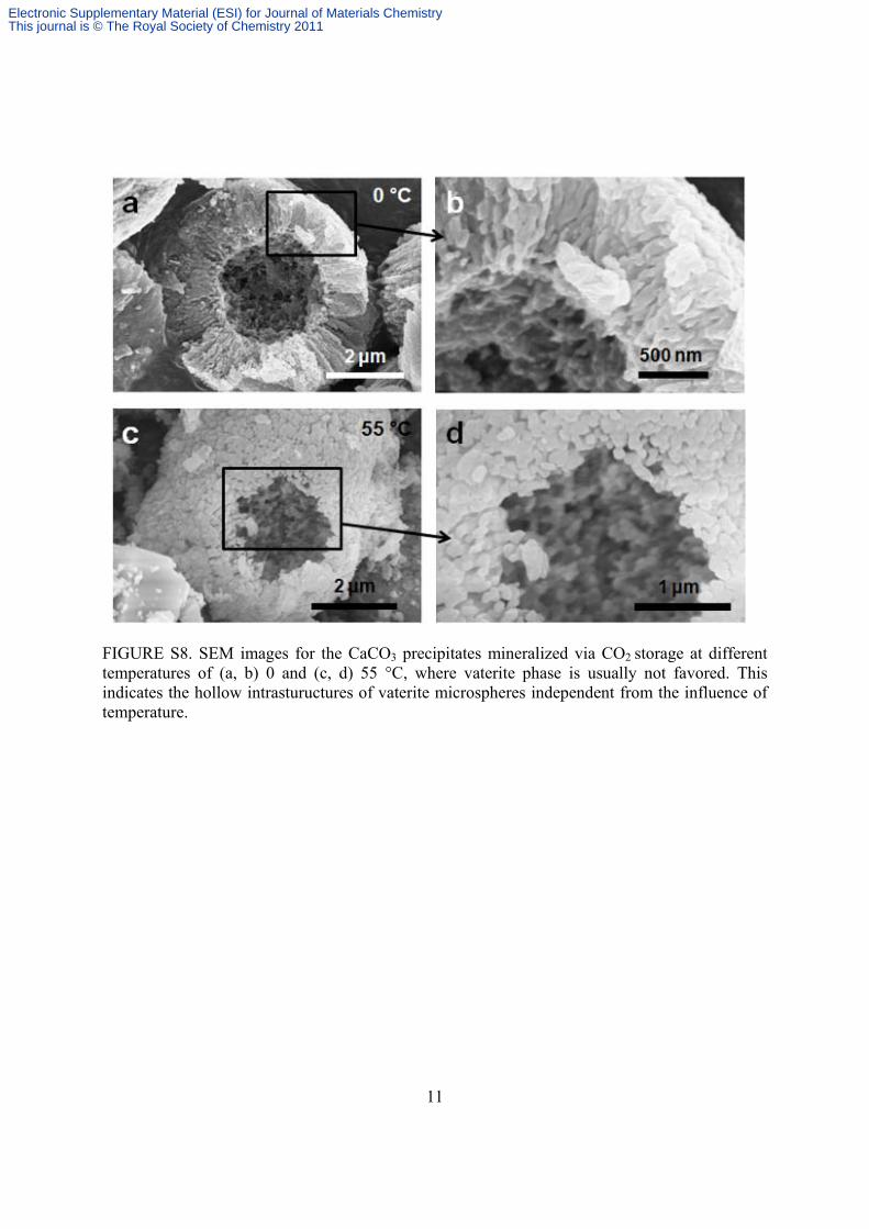
FIGURE S8. SEM images for the CaCO3 precipitates mineralized via CO2 storage at different temperatures of (a, b) 0 and (c, d) 55 °C, where vaterite phase is usually not favored. This indicates the hollow intrastuructures of vaterite microspheres independent from the influence of temperature.
Electronic Supplementary Material (ESI) for Journal of Materials ChemistryThis journal is © The Royal Society of Chemistry 2011

12
FIGURE S9. Synthesis of e-PCL/vaterite composites by CO2 storage. (a) Unmodified e-PCL. (b) Calcite crystals dominantly formed on the unmodified e-PCL after CO2 storage for 1 hour. (c) Vaterite microspheres dominantly formed on the polydopamine-coated e-PCL after CO2 storage for 1 hour. The microspheres consist of CaCO3 nanoparticles and form along the PCL fibers.
Electronic Supplementary Material (ESI) for Journal of Materials ChemistryThis journal is © The Royal Society of Chemistry 2011

13
FIGURE S10. Morphologies of vaterite phase at early stage, which was formed after 15 minutes of CO2 storage. (a, b) Vaterite microspheres show traces of vaterite platelets on their surfaces. (c) Hexagonal vaterite platelets formed on the polydopamine-coated e-PCL fibers. (d) Spherical vaterite with hollow intrastructure is also observable on the polydopamine-coated e-PCL fibers.
Electronic Supplementary Material (ESI) for Journal of Materials ChemistryThis journal is © The Royal Society of Chemistry 2011

14
FIGURE S11. FT-IR spectra for unmodified e-PCL, e-PCL/vaterite composite and e-PCL/hydroxyapatite composite. Absorption band of carbonate group (CO3
2-) at 873 cm-1 and two split bands of vaterite at 1390 and 1470 cm-1 3 increases in the spectra of e-PCL/vaterite composite. Absorption bands of phosphate groups (PO4
3-) at 560 and 1025 cm-1 6,7 increases in the spectra of e-PCL/hydroxyapatite composite. A trace peak for carbonate group found at 873 cm-1 was observed with its intensity significantly decreased, which indicates vaterite crystals were massively transformed to hydroxyapatite on the e-PCL scaffold incubated in SBF. Letter V indicates vaterite. Note that spectrum for polydopamine-coated e-PCL is omitted as it did not show distinct difference from unmodified e-PCL.
References for Electronic Supplementary Information (1) T. Kokubo, H. Takadama, Biomaterials 2006, 27, 2907. (2) C. M. Li, G. D. Botsaris, D. L. Kaplan, Cryst. Growth Des. 2002, 2, 387. (3) L. Addadi, S. Raz, S. Weiner, Adv. Mater. 2003, 15, 959. (4) J. L. Wray, F. Daniels, J. Am. Chem. Soc. 1957, 79, 2031. (5) T. Ogino, T. Suzuki, K. Sawada, Geochim. Cosmochim. Acta 1987, 51, 2757. (6) W. L. Suchanek, P. Shuk, K. Byrappa, R. E. Riman, K. S. TenHuisen, Biomaterials 2002,
23, 699. (7) D. M. Roy, S. K. Linnehan, Nature 1974, 247, 220.
Electronic Supplementary Material (ESI) for Journal of Materials ChemistryThis journal is © The Royal Society of Chemistry 2011