Embed Size (px)
Citation preview

Electronic Supplementary Information (ESI) For
pH-Responsive Mitoxantrone (MX) Delivery Using Mesoporous Silica Nanoparticles (MSN)
5
Yanhang Ma,a†Lin Zhou,b† Haoquan Zheng,a Lei Xing,a Chenguang Li,a Jinghao Cuib* and Shunai Chea*
a School of Chemistry and Chemical Engineering, State Key Laboratory of Metal Matrix Composites, Shanghai Jiao Tong University, 800 Dongchuan Road, Shanghai 200240, P. R. China. Fax: +86 21 5474 1297; Tel: +86
21 5474 2852; E-mail: [email protected] 10
b Department of Pharmaceutics, College of Pharmaceutical Science, Soochow University, 199 Renai Road,
Suzhou 215123,China. Fax: X+86–512–6588–2077; E-mail: [email protected]
1. Material
Cetyltrimethylammonium bromide (CTAB; SCRC, China), tetraethylorthosilicate (TEOS; TCI, 15
Japan), Mitoxantrone (MX). All materials were used as purchased without further purification.
2. Methods
2.1. Synthesis of MCM-41-Type MSN
Typically, 1 g CTAB was first dissolved in the mixture of 480 ml H2O and 7 ml NaOH (1.0 M). The 20
temperature of the solution was adjusted to 80 °C. Then 5.0 ml TEOS was added dropwise to the solution, followed by stirring for 2 h at 80 °C to give rise to white precipitation. The solid product was filtered, washed with deionized water and dried at 100 °C overnight. To remove the surfactant, the as-synthesized materials were extracted in the ethanolic solution of 1 M HCl.
2.2. Synthesis of methylate- and mercapto- functionalized MCM-41 25
The methylate group and mercapto group functionalized MCM-41 nanoparticles were synthesized by post grafting method. Typically, 0.5 g MCM-41 nanoparticles after removal of surfactant were suspended in 20 mL toluene. And then 0.42 mM Methyltriethoxysilane or 3-Mercaptopropyltrimethoxysilane was added dropwise under stirring, followed by refluxing for 12 h.
2.3. Synthesis of carboxyl group functionalized MSN 30
The carboxyl group functionalized mesoporous nanoparticles were synthesized by co-condensation method. 0.14 g C18-3-1 was dissolved in the mixture of 135 mL deionized water and 5.8 g ethanol at 80 °C. And then 0.174 g CES and 0.78 g TEOS were added simultaneously, followed by stirring for 1 h at 80 °C. The obtained solution was aged at 80 °C for 2 days. To removal the surfactant in the nanoparticles, the synthesized materials were extracted with the mixture of 90 mL THF and 10 mL 35% 35
HCl.
2.4. Characterizations
Powder X–ray diffraction (XRD) patterns were recorded on a Rigaku X–ray diffractometer D/MAX–2200/PC equipped with Cu Kα radiation (40 kV, 20 mA) at a rate of 1.0°/min over the range of 1–6° (2θ). The morphology of MCM-41 nanoparticles was observed with scanning electron 40
microscope (SEM, JEOL JSM–7401F) with an accelerating voltage of 1.0 kV. High–resolution transmission electron microscopy (HRTEM) images were taken with a JEOL JEM–3010 microscope operating at 300 kV. The nitrogen adsorption/desorption isotherms were measured at –196 °C with a Quantachrome Nova 4200E porosimeter. The surface area was calculated by the Brunauer–Emmett–Teller (BET). The pore size distribution was calculated by Barrett–Joyner–Halenda (BJH) method 45
according to the adsorption branch of the isotherm. The concentration of anti-cancer drugs (MX) in
Electronic Supplementary Material (ESI) for Journal of Materials ChemistryThis journal is © The Royal Society of Chemistry 2011
solution was measured by a UNICO UV–4802 UV–vis double beam spectrophotometer. The zeta-potential of the nanoparticles was measured in a Malvern ZS90 zeta potential analyzer.
2.5. Loading of MX in the mesopores of the MSN
The mitoxantrone was first prepared as a solution with the concentration of 200 µg/ml in ethanol. In a typical condition, 0.1 g MCM-41 nanoparticles were added into 10 ml MX solution with stirring for 5
24 h. The materials were obtained by centrifugation and then would be washed for three times with PBS of pH 7.4.
2.6. Release of MX from the MSN in PBS solution
In a typical release experiment, about 25 mg of the MCM-41 materials loading with MX was suspended by vibration in 30.0 ml of PBS solution with pH 4.0-7.4 at 37 °C. In the case of sampling, 2 10
ml homogenous solution were withdrew to centrifuged, followed by being measured with UV-vis spectrophotometer.
2.7. In-vitro cell assay
Cells were seeded in a 96-well plate at a seeding density of 5,000 per well in 100µL of RPMI 1640 medium with 10% FBS and 1% penicillin and streptomycin. After the cells were cultured at 37℃ for 15
24h, the growth medium was removed and fresh growth medium containing the predetermined amount of MX-loaded nanoparticles was added. After a 24h incubation, cells were washed three times with 100µL of PBS, and then 100µL of RPMI 1640 medium with 10% FBS was added. Cytotoxicity was assessed using MTT to measure the viability of the cells.10µL of MTT solution (5mg/mL) was added to each well. The plates were incubated for an additional 4h and then 100µL of 10%HCl-SDS were 20
added to dissolve the MTT formazan crystals. After the plates were cultured overnight, the absorbance of each well was measured at 570 nm in a microplate reader.
2.8. Fluorescent experiments
Cells were seeded in a 6 well plate at a seeding density of 2*105 per well in 2 mL of RPMI 1640 medium with 10% FBS and 1% penicillin and streptomycin. After the cells were cultured at 37 °C for 25
24 h with the concentration of CO2 of 5%, the growth medium was removed and fresh growth medium containing the predetermined amount of Rhodamine-loaded nanoparticles was added. After 24 h incubation, cells were washed three times with 2 mL of PBS and then were observed in a confocal laser microscopy.
30
35
40
Electronic Supplementary Material (ESI) for Journal of Materials ChemistryThis journal is © The Royal Society of Chemistry 2011
5
10
15
20
25
I. SEM
The SEM
II. N2 a
Figure nanopa
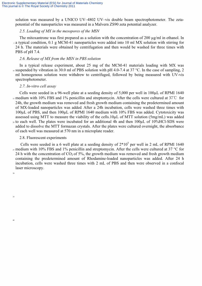
The N2 aThe measu(Barrett-Joy
M images o
M image sho
adsorption/
S2. (a) N2 aarticles after
Table
MCM-41
adsorption/dured BET (Byner-Halend
of MCM-41
Figure
ows that the
/desorption
adsorption/dr calcinations
S1. Porous
Surface a
8
a–c Cal
desorption aBrunauer-Eda) average
1 nanoparti
e S1. SEM im
e MSN are s
n results of
desorption iss shown in F
and compos
areaa (m2/g)
834
culated from
analysis of mmett-Tell pore diame
icles
mages of MC
spherical sh
f MCM-41
sotherm andFigure 1.
sitional prop
Pore volu
0
m N2 adsorp
the extracteler) surface eter was 2.2
CM-41 nano
hape with th
nanopartic
d (b) pore siz
perties of MC
umeb (cm3/g)
0.73
ption/desorpt
ed material area was 8
25 nm and th
oparticles.
e size of 80
cles
ze distributio
CM-41 nano
) Pore s
2
tion data.
revealed ty834 m2/g. he pore volu
0-100 nm.
on of the MC
oparticles.
sizec (nm)
2.25
ypical type The corresume was 0.
CM-41
IV isothermsponding BJ73 cm3/g.
ms. JH
Electronic Supplementary Material (ESI) for Journal of Materials ChemistryThis journal is © The Royal Society of Chemistry 2011

II
Fi
5
Fi2.ch
10
IV15
20
wva
25
II. Mole
igure S3. (a
The two seigure S3a. A07 nm (Fig
harged and m
V. Load
Figure
The loadinwhich could
alue reaches
cular struc
a) Molecular
econdary amAt pH 7.4, og. S3b). Whmolecular s
ding amoun
e S4. Loadin
ng amount obe attribut
s above 8, th
ctures of M
formula of M
mines on thonly aliphathen the pH wsize came to
nt of MX fo
ng amount o
of MX in thted to enhahe loading b
MX and thei
MX and Che
he label chaic amines wwas changedo be about 2
r the drug
f MX in the M
he MSN haansive negabegins to re
ir Chem3D
em3D mode
ain of MX pwere positivd to 4.0, the
2.03 nm (Fig
delivery sy
MCM-41 na
ave increaseative chargeeduce due to
models.
l of MX at (b
put the strucvely chargede aromatic ag. S3c).
ystem at dif
noparticles
ed when thee on silanolo the loss of
b) pH 7.4 an
cture of thed and the moamine grou
fferent pH
under variou
e pH valuesl groups. Hf positive ch
nd (c) pH 4.0
e molecule olecular siz
ups were als
values.
us pH condi
s increase fHowever, wharge of MX
0.
as shown inze was abouso positively
itions.
from 3 to 8when the pHX.
n ut y
8, H
Electronic Supplementary Material (ESI) for Journal of Materials ChemistryThis journal is © The Royal Society of Chemistry 2011

5
10
15
20
25
30
35
40
V. Rel
Figure S5functionalize
Fig. S5a
group functpH 7.4. Fodelivery. Tmolecules. bonding ordeterminedgroups andneeded to fubecause of
ease of MX
. (a) 6% ed MCM-41
shows the tionalized s
or the seconThese result
The pH-resr electrostat
d by CNS eld mercapto further invesa strong ele
X from vari
methyl gro nanoparticl
release prosample, a hnd sample ts reveal thsponsive detic interactilemental chgroups wer
stigate the rectrostatic in
ious organi
up functiones; (c) carbo
files of MXigh release (as shown hat organiclivery of Mions. The qemical analre 6.0 and reason. Fig. nteraction b
ic group fu
nalized MCoxyl group f
X in variouspercentagein Fig. S4b
c groups coMX in Fig. S
quantitativelysis. The re6.3 mmol/gS4c reveals
between carb
nctionalize
CM-41 nanofunctionalize
s organic gre of MX hab), the releould still i4b might be
e determinaesults showg SiO2, ress that MX iboxyl group
ed MCM-41
oparticles; (ed mesoporo
roup functioas been obseease profile nteract wite attributed ation of the
w that the loapectively. Cs hard to beps and MX.
1 nanopart
(b) 6% meous nanopar
onalized MServed in PB shows a pth organic to complica
e functionaading amouCareful woe released ev.
ticles.
ercapto grorticles.
SN. In methBS solution pH-responsiparts of Mated hydrog
al groups wunts of methorks would ven at low p
oup
hyl at
ive MX gen was hyl be
pH
Electronic Supplementary Material (ESI) for Journal of Materials ChemistryThis journal is © The Royal Society of Chemistry 2011

V
FiM4.5
10
afThh Ade15
arreaf
20
25
30
VI. The s
igure S6. XRCM-41nano0) for 48h.
The presenfter loading he surface aand 48 h. H
An obvious emonstratesrea after treaeleased at pHfter treated f
stability of
RD patternsoparticles; (b
MX loTreateTreate
a–c Calcul
nce of peakand release
area, pore vHowever, adecrease o
s that most oated in PBSH 4.0, the sfor 48 h.
MCM-41 M
s, N2 adsorptb) materials
Table S
MC
Materials
oaded MCM-d in 7.4 for 4d in 4.0 for 4lated from N
k (2θ~2.5°e the drug. Nvolume and at pH 4.0, Mof surface aof mesopo
S solution, wsurface area
MSN.
tion/desorptreleased in
2. Porous a
M-41 nanop
Surfac(m
-41 148 h 748 h 4
N2 adsorption
°) indicates N2 analysis pore size al
MX would barea, about res have be
which mighta of sample
tion isothermPBS (pH 7.4
nd composi
particles at v
ce areaa m2/g)
58 77 450 n/desorption
the mainteresults showll decrease be released t 676 m2/g,een occupiet be caused e in PBS so
ms and pore 4) for 48h; (
tional prope
various stage
Pore volum(cm3/g)
0.3 0.2 0.538
n data.
enance of mw that the cafter treatedand mesop
, has been d by MX. Tby the colla
olution at pH
size distribuc) materials
erties of
es.
eb Pore (nm1.1.2.
mesostructurchange of prd in PBS so
pore structurfound afte
There is alsoapse of mesH 4.0 is larg
ution of (a) Ms released in
sizec m) .8 .8 .2
ure of the nroperties ofolution at pHure is mostlyer loading o a decreas
sopores. As rger than tha
MX-loaded n PBS (pH
anoparticlesf mesoporesH 7.4 for 24y preservedMX, which
se of surfaceMX will be
at at pH 7.4
s s. 4 d. h e e 4
Electronic Supplementary Material (ESI) for Journal of Materials ChemistryThis journal is © The Royal Society of Chemistry 2011

5
10
15
20
25
30
35
40
45
VII. Effe
This resu
a favorable VIII. N2
fun
Figure S8functionalize
ect of MCM
Figure
ult showed t drug carrie
adsorptioctionalized
. N2 adsored MSN; (b)
M-41 in inh
e S7. Inhibitio
that the MCer for cancer
on/desorptid MSN.
rption/desor) methyl gro
hibition of c
on ratio of S
CM-41 nanor therapy.
on results
rption isotheup functiona
cancer cell.
SMMC-7721
oparticles sh
s of carbo
erms and alized MCM-
.
cells for MC
how low cyt
oxyl group
pore size -41; (c) mer
CM-41 nano
totoxicity an
p, methyl
distributioncapto group
oparticles.
and could be
and mer
n of (a) cap functionaliz
e employed
capto grou
arboxyl grozed MCM-41
as
up
oup 1.
Electronic Supplementary Material (ESI) for Journal of Materials ChemistryThis journal is © The Royal Society of Chemistry 2011

fu5
Thfusl
10
IX15
20
25
30
in35
Table S2.
All the threunctionalizehe pore size
unctionalizeight reducti
X. Cytot
This resul
ndicating a g
Porous and
Carfunction
Mefunction
Merfunction
ee samples ed materials e of 2.94 nmed samples hion after fun
toxicity of M
Figure S
lt showed tgood biocom
d compositiofunc
Materials
rboxyl groupnalized mateethyl group nalized matercapto groupnalized mate
a–c Calcu
possess a hhave been
m is large enhave been synctionalizati
MCM-41 na
S9. Inhibitio
that the Mmpatibility.
onal propertictionalized m
Surfac(m
p erials
erials p erials ulated from N
high mesoposynthesized
nough to hoynthesized bion.
anoparticles
on ratio of Q
MCM-41 nan
es of carboxmesoporous
ce areaa m2/g)
648
711
845
N2 adsorptio
ore surface ad based on cold the drug
based on po
s to normal
QSF-7701 c
noparticles
xyl group, mnanoparticl
Pore volum(cm3/g)
0.71
0.8
0.86
on/desorptio
and pore voco-condensamolecule. T
ost-grafting
cells.
cells for MC
were of l
methyl group es.
eb Pore (nm2.9
2.0
2.0
on data.
olume. The cation methoThe methyl method. Th
CM-41 nano
ow cytotox
and mercap
sizec m) 94
04
04
carboxyl grod according
and mercaphe pore size
oparticles.
xicity to no
pto group
roup g to ref. 1. pto group es show a
ormal cellss,
Electronic Supplementary Material (ESI) for Journal of Materials ChemistryThis journal is © The Royal Society of Chemistry 2011
5
10
15
20
25
30
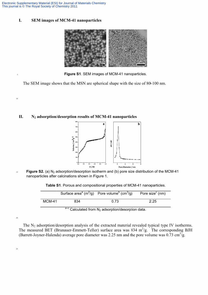
X. Uptand Figureprresp To skindincrcomwhi
take efficied MCF-7.
ure S10. Fluresent diffepectively.
study the ud of commorease of co
mparision ofich also acco
ency of fluo
uorescent imerent conce
uptake efficon fluoresceoncentrationf three groounts for th
orescent M
mages of a) entration of
iencies of Ment agent, han of MCM-ups reveals
he high inhib
MCM-41 in
A549 cells;MCM-41,
MCM-41 naas been use-41, the ins that the cbition in the
three diffe
b) SMMC-7167 mg/m
anoparticlesed as the dyntensity of cellular upte vitro expe
erent cells
7721 cells; cmL, 667 mg
s in three cee. As showfluorescent
take efficienriments.
of A549, S
c) MCF-7 ceg/mL and
ell lines, Rhwn in the rest increases. ncy in A54
SMMC-772
ells. 1, 2 and1000 mg/m
hodamine Bsults, with t
Besides, t49 is highe
21,
d 3 mL
B, a the the est,
Electronic Supplementary Material (ESI) for Journal of Materials ChemistryThis journal is © The Royal Society of Chemistry 2011