Embed Size (px)
Citation preview
Clays and Clay Minerals, 1969, Vol. 17, pp. 31-35. Pergamon Press. Printed in Great Britain
E L E C T R O N MICROSCOPIC IDENTIFICATION SINGLE CRYSTALS OF WAIRAKITE,
A R A R E C O M P O N E N T IN CLAYS
OF
KLARA KISS and HAROLD T. PAGE GAF Corporation, Central Research Laboratory, Easton, Pennsylvania
(Received 17 October 1968)
Abstract--Crystallites of the finest fraction of a clay mineral from Rosamund, California, which account for over 50 per cent of the total weight, are identified as wairakite single crystals of 0.l-1 size. High-magnification electron microscopy revealed flat, almost perfectly square-shaped lamellae, which consist of superimposed layers of approximately < 50 A. thickness. Electron diffraction patterns from a selected single crystal proved that the basal plane of the crystallite aligned perpendicular to the electron beam is the (111) plane. It is suggested on the basis of the present study and the report of previous investigators that the pseudocubic wairakite crystals cleave along their (111) and ( ] l 1) planes. The indices of high order reflections, unpublished or previously reported as uncertain, are determined.
INTRODUCTION
A NEW, unusual zeolite mineral was discovered by A. Steiner in 1953 at Wairakei, New Zealand. This mineral, named wairakite, was characterized as the cMcium analogue of analcime, with the chemical composition: CaO.AI203.4SiO2.2H20. Physical properties and the morphology of macro- scopic crystals have been described. The crystal structure was determined by D. S, Coombs (1955) by means of X-ray diffraction, utilizing the Weiss- enberg technique, on a macroscopic single crystal. The latter was an incomplete octahedron, 15 mm across. The structure was reported as pseudocubic with a = 1 3 - 6 9 A , b = 1 3 - 6 8 A , c = 1 3 . 5 6 A , /3= 90.5 ~ with 8(CaOAI~O3 4SiO~2H20) per unit cell. Debye-Scherrer X-ray diffraction powder patterns were also obtained from wairakite powders.
Since the first report by Steiner (1955), wairakite has been detected at various other locations: at St. Croix and St. Thomas, Virgin Islands (Whetten, 1965; Donnelly, 1962) in Shimane Prefecture, Japan (Umegaki, 1965), in the Geysers of Cali- fornia (Steiner, 1958), and at Mt. Rainier Park, Washington (Wise, 1959) in the U.S.A. Synthetic wairakite was obtained by hydrothermal synthesis (Ames, 1958) and by exchange reaction at high pressure from leucite (Debron, 1965).
The clay investigated in the present study was a commercial product of Paramount Pacific Incor- porated, deposited 10 miles east of Rosamond, California, a few miles from Edwards Air Force base and 90 miles from Los Angeles. This is a high desert area of about 2000 ft altitude with a semi-arid climate. The deposit is at the surface with
31
6"-2' overburden and covers two areas, each 10 miles square. The depth of the clay varies from 20 to 80 ft deep and is generally mined by open pit method.
The major constituent (over 50 per cent) of the sample proved to consist of extremely fine waira- kite crystallites of 0.1-1/~ size. This phenomenon allowed a study on wairakite single crystals by electron microscopy and by electron diffraction from selected, individual crystallites. This com- plementary method to X-ray diffraction has the advantage that structure studies can be carried out on single crystals having dimensions smaller by orders of magnitude than the previously studied crystal and that the indices of high order reflections, not indexed previously or reported as uncertain, can be determined.
A further advantage of the electron diffraction study is that the presence of wairakite can be proved with certainty, even in the presence of large amounts of muscovite and feldspar. The spacings of the high intensity X-ray reflections of the latter minerals are very close to those of wairakite. Their simultaneous presence may pre- vent the recognition of wairakite, when only X-ray technique is applied.
EXPERIMENTAL
Fractionation of the clay. The separation of the silt and the clay fractious was achieved by sedi- mentation under gravity. The grains of the sample were ground lightly in a mortar and the sand was dispersed in cold water for 15 rain utilizing a high speed Hamilton-Beach mixer. Sedimentation was
C.C.M. Vol. 17No. I - C
32 K. KISS and H. T. PAGE
carried out in preweighed beakers and the liquid phase was removed by careful decantation. The fractions were dried at 90-100~ in an oven and exposed to the atmosphere for 24 hr prior to X-ray diffraction analysis to allow equilibration with atmospheric moisture.
Elemental analysis. Fraction 5 (See Table 1) was analyzed by emission spectrophotometry.
X-ray diffraction methods. A Phillips Norelco X-ray diffractometer with Ni filtered CuK~ radi- ation was utilized at 40 kV and 35 mA. Random powders of untreated and dilute-glycerine treated fractions were investigated.
Electron optical methods. The samples for elec- tron optical studies were taken from the highly dispersed, finest Fraction 5, prior to evaporation to dryness. A drop of the highly diluted suspension was deposited on a carbon coated electron micro- scope grid. After evaporation of the water, the samples were shadowed with a mixture of Pt -C in high vacuo, utilizing a 45 ~ shadow angle. An RCA Model EMU-3 electron microscope was used for electron diffraction and high magnification bright field image studies. The spacings of the
experimental reflections were calculated using Pt reflections as internal standards. All distances were measured on the photographic plate. The theoret- ical spacings of the hkl reflections of wairakite were calculated by means of an IBM Model 7044 computer, assuming cubic symmetry and a = 13.7 A lattice constant.
RESULTS AND DISCUSSION Table I summarizes the sedimentation times,
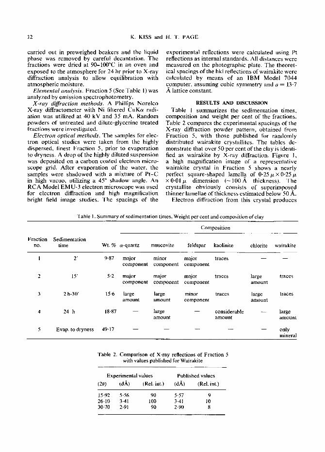
composition and weight per cent of the fractions. Table 2 compares the experimental spacings of the X-ray diffraction powder pattern, obtained from Fraction 5, with those published for randomly distributed wairakite crystallites. The tables de- monstrate that over 50 per cent of the clay is identi- fied as wairakite by X-ray diffraction. Figure 1, a high magnification image of a representative wairakite crystal in Fraction 5 shows a nearly perfect square-shaped lamella of 0-25 p x 0.25/x x0.01/z dimension ( -100 .A thickness). The crystallite obviously consists of superimposed thinner lamellae of thickness estimated below 50 A.
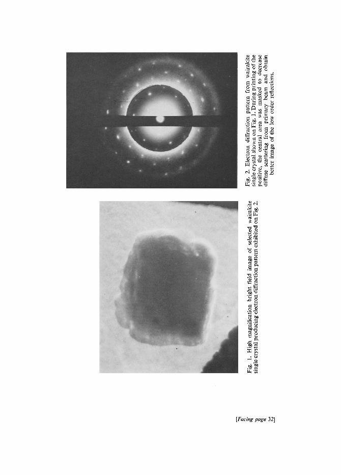
Electron diffraction from this crystal produces
Table 1. Summary of sedimentation times, Weight per cent and composition of clay
Composition
Fraction Sedimentation no. time Wt. % a-quartz muscovite feldspar kaolinite chlorite wairakite
1 2' 9.87 major minor major traces component component component
2 15' 5-2 major major major traces large traces component component component amount
3 2 h-30' 15-6 large large minor traces large traces amount amount component amount
4 24 h 18-87 - - large - - considerable - - large amount amount amount
5 Evap. to dryness 4 9 . 1 7 . . . . . only mineral
Table 2. Comparison of X-ray reflections of Fraction 5 with values published for Wairakite
Experimental values Published values
(20) (d~) (Rel. int.) (d,~) (Rel. int.)
15-92 5-56 90 5-57 9 26-10 3-41 100 3.41 10 30-70 2"91 90 2.90 8
0 0
~ . ~ ~,.~
0
~.=_.
�9 0
[Facing page 32]
ELECTRON MICROSCOPIC I D E N T I F I C A T I O N 33
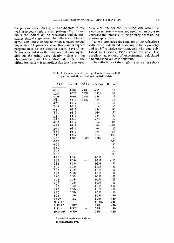
the pa t t e rn s h o w n on Fig. 2. T h e d iagram of this well r e so lved single c rys ta l pa t t e rn (Fig. 3) ex- hibi ts the indices of the ref lect ions and d e m o n - s t ra tes s ixfold s y m m e t r y . T h e ref lect ions ob t a ined agree wi th those expec t ed w h e n a cub ic crys ta l lies on its (111) p lane , i.e. w h e n this p lane is a l igned pe rpend icu l a r to the e l ec t ron beam. Severa l re- f lect ions inc luded in the d iagram, bu t unrecogniz - able on the print , were clear ly vis ible on the pho tog raph ic plate. T h e cent ra l dark s tr ipe on the diffract ion pa t t e rn is an ar t i fac t due to a f rame used
as a subs t i tu te for the b e a m s t o p wi th which the e lec t ron mic roscope was not equ ipped , in o rde r to dec rea se the in tens i ty of the p r imary b e a m on the pho tog raph ic plate.
T a b l e 3 c o m p a r e s the spac ings of the ref lect ions with those ca lcu la ted a s suming cubic s y m m e t r y and a 13.7,& latt ice cons t an t , and wi th da ta pub- l ished by C o o m b s (1955) whe re avai lable . T h e exce l len t a g r e e m e n t of exper imen ta l , ca lcu la ted and pub l i shed values is apparen t .
T h e ref lect ions of the single c rys ta l pa t t e rn show
Table 3. Comparison of spacing of reflections on E.D. pattern with theoretical and published data
h k l d A C a l c d ,~Li t d , ~ E x p Rel. int.t
0 2 2* 4-808 2 2 4* 2-776 4 4 0* 2.404 2 ~ 6 1.817 4 5 6 1-817 2 6 4 1-817 6 2 4 1.817 4 6 2 1.817 6 4 2 1-817 6 4 2 1.817 4 6 2 1.817 6 2 4 1.817 2 6 4 1.817 4 2 6 1.817 2 4 6 1-817 6 6 0 1-603 0 9 6 6 0 6 6 6 0 6 0 6 0 6 g 4 4 8" 1-388 2 6 8 1.334 2 8 6 1-334 8 6 2 1-334 2 8 6 1.334 6 8 2 1'334 6 8 2 1.334 6 2 ~ 1.334 2 6 8 1.334 6 ~ 8 1-334 8 2 6 1.334 8 6 2 1-334 8 2 6 1-334 8 8 0* 1.202 10,4,6" lq03 2, 10, 8" 1.049 8, 12, 4 0-909 10, 0, 10" 0'960
4.84 4-83 10 2.770 2.752 30 2.418 2.38 10 1.822 1-84 30
1 "84 30 1-84 50 1.84 80 1.84 50 1.84 80 1.84 50 1-84 80 1.84 <80 1.84 80 1.84 50 1.84 <50
1.612 1.606 50 100 80 50 5O
100 1.353 1.353 <10 t -353 50 1.353 50 1.353 50 1-353 100 1.353 100 1.353 100 1.353 10 1-353 <10 1.353 <10 1-353 < 10 1.353 < 10 1.205 < 10 1-1086 <10 1-03 < 10 0.94 <10 0.94 <10
* - a n d all equivalent indices. t Estimated by eye.
34 K. KISS and H. T. PAGE
i0~0,I0
~,f0,8 o8~ 26-s ~8 6~s 808 10,8,8
Z,fo,6 ~ 6 096 ~ 6 a~6 606 826 10,4,6
6,~0,4 48~ ~ a oZ~ 2-~ .~o4 6.2a 814 ~o~6,4
880 660 /.40 220 000 220 4Z~O 660 880 �9 �9 �9 @ �9 �9 �9
l"O,g,~ g,Z,-4 g2-Z/:0-4 42Z 04~ 2o6Z 4o8Z 6610,Z 8,12,Z �9 �9 �9 @ �9 �9
To,2,~ ~Sg go~ Z2g ~6 o66 2ag �9 �9 �9 �9 �9 �9 �9
~,ot~o ;,2,~o 4@,ao o,~o,ro
Fig. 3. Diagram of electron diffraction pattern exhibited on Fig. 2.
that the (111) plane and its higher order equivalents are perpendicular, and the [ 111] axis of the re- ciprocal lattice is parallel to the electron beam. The observation that the (111) planes of several single crystals were aligned perpendicular to the electron beam indicates, that the wairakite crystals cleave along their (111) and (111) planes. The data col- lected in the present study are insufficient to allow a statement on a statistical basis and further work is necessary to substantiate this suggestion. A. Steiner (1955) described cleavage-like lines on powdered material obtained by crushing a mega- scopic crystal; he does not suggest, however, the index of the plane along which the cleavage has taken place.
The doubling of many reflections was observed on the electron diffraction pattern. Similar observa- tions reported by D. S. Coombs (1955) were attributed to a measurable departure from cubic symmetry. It is suggested that the twinning of the crystals parallel to (110) planes, a characteristic property of wairakite (Steiner, 1955; Coombs, 1955) furthermore superposition of lamellae, which are rotated with respect to each other, may also contribute to the formation of double reflections.
Acknowledgment s -The authors are indebted to Dr. J. P. G. Beiswanger (GAF Corporation) for providing the clay sample, to Dr. H. J. Stolten (GAF Corporation), to
Professors J. A. Manson and G. Krauss (Lehigh Uni- versity) for their helpful discussions, and to Mr. R. Korastinsky (Lehigh University) for expert assistance.
REFERENCES
Ames, L. L. and Sand, L. B. (1958) Hydothermal synthesis of wairakite and calcium-mordenite: Am. Mineralogist 43,476-80.
Coombs, D. S. (1955) X-ray observations on wairakite and non-cubic analcime: Mineral. Mag. 30, 699-708.
Debron, G. (1965) Investigation of the exchange of alkali metal ions and alkali earth ions in feldspathoids: Bull. Soc. Franc. Mineral. Crist. 88, 69-96.
Donnelly, T. W, (1962) Wairakite in West-Indian spilitic rocks: Am. Mineralogist 47,794-802.
Steiner, A. (1955) Wairakite, the calcium analogue of analcime, a new zeolite mineral: Mineral. Mag. 30, 691-8.
Steiner, A. (1958) Occurrence of wairakite at the Geysers, California: Am. Mineralogist 43, 781.
Whetten, F. T. (1965) Wairakite from low-grade meta- morphic rocks on St. Croix, U.S. Virgin Islands: Am. Mineralogist 50, 752-5.
Wise, W. S. (1959) Occurrence of wairakite in meta- morphic rocks of the Pacific Northwest: Am. Mineral- ogist 44, 1099-1101.
Umegaki, Yoshiharu and Ogawa, Toshihiko (1965) Occurrence of zeolites in the Miocene formation in Shimane Prefecture, Japan: J. Sci. Hiroshima Univ. Ser. C. 4(4), 479-97.
E L E C T R O N M I C R O S C O P I C I D E N T I F I C A T I O N 35
R 6 s u m 6 - L e s crystallites de la fraction la plus fine d 'un min6ral d'argile provenant de Rosamund en Californie, qui repr6sentent plus de 50% du poids total, sont identifi6s comme des crystaux simples wairakite h dimensions de 0 , I - I p,. La microscopie 61ectronique/~ grandissement 61ev6 a montr6 des lamelles plates de forme ~presque parfaitement carr6e, qui comportent des couches surimpos6es 6paisseur d 'environ <50 A. Les modules de diffraction des 61ectrons ~t partir d 'un seul crystal s61ec- tionn6 ont d6montr6 que le plan de base du crystallite align6 de mani~re perpendiculaire au faisceau d'61ectrons est le plan ( 111 ). Sur la base de cette 6tude et selon le rapport ant6rieur d'autres rechercheurs on sugg~re que les crystaux pseudo-cubiques de wairakite se fendent selon leurs plans (11 l) et ( ] l 1). On d6termine les indices des r6flexions d 'ordre 61ev6 qui n 'ont pas 6t6 publi6s ant6rieurement ou qu'on a indiqu6 comme 6tant incertains.
Kurzreferat- Kristallite aus den feinsten Fraktionen eines Tonminerales aus Rosamund, Kalifornien, die fiber 50% des Gesamtgewichtes ausmachen, werden als Wairakit Einzelkristalle von 0,I - 1/x Gr6sse identifiziert. Intensivvergr6sserung dutch Elektronenmikroskopie zeigt flache, beinahe perfekt quadratische Lamellen, die aus iibereinander geschichteten Lagen yon c a . <50 fi~ Dicke bestehen. Elektronenbeugungsbilder eines ausgew~ihlten Einzelkristalles beweisen, dass die Basise- bene des Kristallits, die normal zum Electronenstrahl liegt, die (11 l) Ebene ist. Aufgrund der gegen- w~irtigen Untersuchung und der Arbeiten friiherer Forscher wird angenommen, dass sich die pseudo- kubischen Wairakitkristalle entlang ihrer (1 I l) und (]-1 l) Ebenen spalten. Die bisher unver/Sffentlich- ten bzw. angezweifelten Reflexionsindexe h/3herer Ordnung werden bestimmt.
Pe3mMe--KpucTaa~HTbl Meabqa~mxx ~bpaguH~ raHHacvoro MHHepana ~3 PO3aMyH~la (Kaaagbo- pHHII), KOTOpble COCTaBflglOT IIOqTH ~0% rio aozy, npeacTaa~a~OT MOHOKpHCTa.rlJlb! ua~pa~aTa pa3MepoM 0,1--1 MK. ~neKTpOHHafl MHKpOCKOnnn no3aoaHna o6uapy~fnTb naocgae, nOqTX Hs HO KnailpaTHbte naacrauga, COCTOaLUae n3 Haaox~eunbtx cnoes r o a m a a o ~ npa6aa3nTeabHO<50 A. ,~H~0paKuaonnble KapTHnbl noKa3bwaatOT, ~TO 6aaaabnan H.qOCKOCTB KpHCTaaflHTa, nepne- n~nKyaapHaa K 321r nyqgy, npe~cTaaaaeT HJ]OCKOCTb (I 1 I). Ha OCHOBaHHH HaCToamero accaeaoaaHaa u aaaablx npeJlbzaymux ncc~elloaaan~ eCTh ocnoaanaa noaaraTb, STO nceaao- gy6xnecKae Kpncxaaahl 8afipagHra pacgaJlbma~oTcfl no FU1OCKOeTflM (111)H (I 1 I). OnpeneaeHbt I, IH~eKCb| oTpa)KeHHH BblCOKOFO nopflglKa.