Embed Size (px)
Citation preview

Open AccessResearch Article
Electrochemical Biosensors Based on ZnO Nanostructures to Measure Intracellular Metal Ions and GlucoseMuhammad H. Asif1,2*, Fredrik Elinder3 and Magnus Willander2
1Materials Research, Department of Physics, COMSATS Institute of information Technology, Lahore-54000, Pakistan2Department of Science and Technology, Campus Norrköping, Linköping University, SE-601 74 Norrköping, Sweden3Department of Clinical and Experimental Medicine, Division of Cell Biology, Linköping University, SE- 581 85 Linköping, Sweden
Biosensing
Keywords: ZnO nanorods; Human adipocytes; Frog oocytes; Poten-tiometric; Metal ions; Glucose; Membranes; Enzymes; Functionaliza-tion
IntroductionNanotechnology is attractive in the research community to
create and manipulate materials at the nanoscale to fabricate unique metal-oxide nanostructures with interesting properties. Metal-oxide nanostructures (e.g. nanorods, nanowires, and nanotubes) have been explored for many biochemical and biomedical applications (e.g. biosensing, biological separation, molecular imaging, biomarker, and anticancer therapy) because of their interesting properties and functions. Particularly their high surface/volume ratio, surface tailor ability, biosafety, and biocompatibility open many new possibilities for biomedicine. At present the application of nanostructure in advancement of electrochemical biosensors for the intracellular environment is an active research area. A metal-oxide nanostructure material with exciting surface-charge properties provides a fascinating platform for interfacing biorecognition elements with transducer for signal amplification. These nanostructures display appealing nano-morphological, functional, biocompatible nontoxic and catalytic properties. They show higher electron-transfer kinetics and durable adsorption capability, providing appropriate microenvironments for the immobilization of biomolecules and enhanced biosensing characteristics.
Zinc oxide (ZnO) is one of the best metal-oxide semiconductors, and it is relatively biosafe and biocompatible which is suitable for intracellular sensor/transducer applications [1-5]. The dimensions of ZnO nanostructure can be engineered to the size of the biochemical species being sensed which make the ZnO an excellent primary transducer for producing electrical signals. All these facts make ZnO nanostructures suitable candidates for intracellular measurements [6]. The focus of this review is the fabrication and demonstration of functionalized ZnO nanorod-based potentiometric sensors suitable for intracellular free metal-ions and glucose. Our main effort has
been directed towards the construction of borosilicate glass capillary tips (0.7µm in diameter) selective for metal ions (Ca2+, Mg2+, K+ and Na+) and glucose, capable of penetrating the cell membrane, as well as the optimization of the electrochemical potential properties. Glass tips with grown ZnO nanorods have proven to be a convenient and practical choice as we will demonstrate in this review.
Biological role of intracellular free metal ions
Metal ions play fundamental roles in life processes by acting as cofactors in enzymes, as osmotic regulators, as current carriers and consequently as factors in information processing, and as integrators and stabilizers of proteins and lipids [7]. Na/K-ATPases charge the cellular battery by pumping monovalent Na+ and K+ across the cellular membrane – Na+ to the outside of the cell and K+ to the inside. This generates an electric potential across the resting membrane; opening of passive ion channels release the energy, which can be used to transmit fast electric impulses in the form of action potentials. Divalent cations, such as Ca2+, Mg2+ and Zn2+, normally play a regulatory role in modifying ion channels, receptors and enzymes [8]. The regulation of the metal ion concentration is strictly controlled and failures to do so lead to disease and death.
The intracellular K+ concentration is high, generating a negative
*Corresponding author: Muhammad H. Asif, Materials Research, Department of Physics, COMSATS institute of Information Technology- Lahore-54000, Pakistan, E-mail: [email protected], [email protected]
Received July 01, 2011; Accepted September 26, 2011; Published September 30, 2011
Citation: Asif MH, Elinder F, Willander M (2011) Electrochemical Biosensors Based on ZnO Nanostructures to Measure Intracellular Metal Ions and Glucose. J Anal Bioanal Tech S7:003. doi:10.4172/2155-9872.S7-003
Copyright: © 2011 Asif MH, et al. This is an open-access article distributed under the terms of the Creative Commons Attribution License, which permits unrestricted use, distribution, and reproduction in any medium, provided the original author and source are credited.
AbstractZinc oxide (ZnO) nanostructures have attracted much interest for intracellular electrochemical measurements
because of its large surface area, and its biocompatible properties. To design intracellular biosensors for metal ions and glucose, we grew ZnO nanorods on the tip of borosilicate glass capillaries (0.7µm in diameter) and characterized the nano-scale structure with field-emission scanning electron microscopy and high-resolution transmission electron microscopy. The ZnO nanorods were functionalized accordingly for intracellular free metal ions or glucose measurements. Selectivity was achieved by using a metal-ion selective plastic membrane or glucose oxidase enzyme for glucose measurements. These functionalized ZnO nanorods showed high sensitivity and good biocompatibility for intracellular environments. Human adipocytes and frog oocytes were used for determinations of intracellular free metal ions and glucose concentrations. In this review, we discuss the simple and direct approach for intracellular measurements using ZnO nanostructure-based potentiometric biosensors for clinical and non-clinical applications. The performance of ZnO nanostructure-based intracellular sensor can be improved through engineering of morphology, effective surface area, functionality, and adsorption/desorption capability. This study paves the way to find applications in biomedicine by using this simple and miniaturized biosensing device.
Asif et al. J Anal Bioanal Tech 2011, S7 DOI: 10.4172/2155-9872.S7-003
ISSN:2155-9872 JABT, an open access journal J Anal Bioanal Tech
Journal ofAnalytical & Bioanalytical TechniquesJo
urna
l of A
nalyt
ical & Bioanalytical Techniques
ISSN: 2155-9872

Page 2 of 9
ISSN:2155-9872 JABT, an open access journal J Anal Bioanal Tech
resting membrane potential, and fluctuations in the K+ concentration alters the membrane potential and consequently, affects excitability and other physiological functions of nerve and muscle [9]. The intracellular concentration of Na+ is low and it may act as a second messenger in different cell types. Na+ regulates different transporters and receptors in renal cells and neurons, and it regulates the activation of both Na+ and K+ channels, thereby regulating the reabsorption of salt and water in the kidneys and neuronal excitability [10-15]. Na+ and K+ also play fundamental roles in a number of less explored mechanisms. Intracellular K+ participates in cell growth, maintenance of cell volume, DNA, and protein synthesis, enzymatic activity, acid-base balance, and programmed cell death (apoptosis) [16]. Reduced intracellular K+ and subsequent cell shrinkage are early events in apoptosis. The reduced K+ concentration release caspase inhibition, resulting in DNA degradation and eventually cell death [17-20]. Intracellular Na+ is also important in the early stages of apoptotic processes in cell lines, and may be involved in intracellular signalling during apoptosis [17,21-24].
Mg2+ is the most abundant intracellular divalent cation in living cells, where it plays a major biological role in its involvement with phosphate compounds (e.g. ATP) and phosphate metabolism, binding in chlorophyll pigments, and acting as a critical cofactor in numerous enzymatic reactions [7]. Intracellular Mg2+ also affects ion channels, for instance by gating inward-rectifier K channels [25]. Ca2+ is essential in living cells [26] – it has important roles in regulating enzyme activity, neuronal activity, muscle contraction, vesicle exocytosis, cell death, development, and plastic changes (i.e. LTP, long-term potentiation; [27]) at the synapse. Ca2+ binds to synaptic proteins to trigger synaptic release, it binds to intracellular proteins in muscles to trigger muscle contraction, and it binds to ion channels to regulate their activity [8].
To be able to accurately measure the concentrations and alterations of specific metal ions is therefore of great importance in cell biological research.There are, however, situations when real-time monitoring in single cells with probes inserted in living cells is the preferred configuration. In our studies of (bio) sensors based on probes covered by ZnO nanorods, we have found that it is possible to make sufficiently small probes to measure free metal ions inside large cells [1-4].
Glucose in health and disease
Glucose is one of the main products of photosynthesis and serves as the human body’s primary source of energy. During hypoxic conditions, as in hard working muscles, glucose conversion in glycolysis is the only means of generating cellular adenosine triphosphate (ATP). While most tissues and cells can oxidize fatty acids for their energy supply, the brain is critically dependent on a constant glucose supply. Glucose also serves as a metabolic intermediate in the synthesis of more complex molecules such as fats.In the research community, it has become one of the most targeted analyte in bioanalysis whether it is in pharmacy, medicine or biotechnology. When glucose levels in the bloodstream are not properly regulated, diseases such as diabetes can develop [6].
In fact, there are not many intracellular biosensors reported in literature. Abe et al. (1992) [28] described a glucose microsensor with a tip diameter of 2µm that could provide reliable and accurate measurements of glucose during few hours. As an alternative to direct measurements in cells, metabolite concentrations can be assessed by indirect, non-invasive measurements using various techniques, such as NMR that gives average concentrations on samples containing several cells [29]. Other techniques, like those employing fluorescently labeled glucose molecules, may permit real time measurements in single living cells [30]. A precise and fast measurement of glucose is obviously
significant to diagnosis and handling of the different forms of diabetes, as well as in research to understand the diabetes diseases and to develop new therapies [5].
Electrochemical miniaturization for intracellular measure-ments
In recent years the research community has been working to miniaturize the materials for intracellular measurements in biological and biomedical applications. There have been great progresses in the application of metal-oxide nanomaterials such as ZnO, CuO, Fe2O3 in intracellular biosensors [1-5,31]. Semiconductors, such as ZnO and SnO2 have high resistivity that provides alternative template for gas sensors [32]. Nanomaterial-based biosensors, which represent the integration of material science, molecular engineering, chemistry and biotechnology, can significantly advancement in the sensitivity and selectivity for biomolecule detection, and also hold the capability of detecting atoms and molecules [33-35]. The continuous progress in synthesizing and fabrication of controlled materials on the submicron and nanometer scale results from novel advanced functional materials with tailored properties. When the bulk materials scale down to a nanoscale, most of the materials reveal novel properties that cannot be deduced from their bulk behavior. For the fabrication of an efficient and fast biosensor, the choice of substrate for dispersing the sensing material decides the sensor performance.
The ZnO nanomaterials are applied to biosensors because of their unique physical, chemical, mechanical, and optical properties which markedly enhance the sensitivity and specificity of detection [1-5,36]. The interesting property of ZnO nanostructure may offer excellent sce-narios for interfacing biological recognition events with electronic sig-nal transduction. All these facts described above open up for possible sensitive extra- and intracellular measurements. Metal-oxide nano-structures (nanorods or nanotubes) have been widely used in immobi-lization of polymeric membranes and enzymes for bioanalytical appli-cations. These can play an important role on electrical, optical, electro-chemical and magnetic transducers [37]. ZnO nanostructure can act as excellent material for immobilization of enzymes and rapid electron-transfer agent for the fabrication of efficient biosensors due to the wide direct band gap [38,39].The resulting biosensors could be used to detect the glucose level of serum samples with high sensitivity. Nanostructure metal oxides display appealing nano-morphological, functional, bio-compatible, and catalytic properties. These nanostructures also show enhanced electron-transfer kinetics and strong adsorption capability, providing suitable microenvironments for the immobilization of bio-molecule and improved biosensing characteristics [40]. Miniaturiza-tion of sensors based on nanostructure is very important since a large surface area-to-volume ratio leads to a short diffusion distance of the analyte towards the electrode surface, thereby providing an improved signal-to-noise ratio, faster response time, enhanced analytical perfor-mance and increased sensitivity. Nanostructure-based biosensors are expected to find new manufacture of miniaturized, smart biosensing device. In this review, we focus on new methods for the fabrication of electrochemical sensors with desired properties for health care, intra-cellular environment, and confining different biomolecule onto closely spaced one dimensional nanostructure based on ZnO nanorods [6].
Fundamentals of the ZnO nanostructure
ZnO is a relatively biosafe, biocompatible and highly atomic ionic-ity in the crystal which can be used for biomedical applications (e.g., bio sensing, biological separation, molecular imaging, and/or antican-cer therapy). ZnO nanorods display appealing nano-morphological,
Biosensing
Citation: Asif MH, Elinder F, Willander M (2011) Electrochemical Biosensors Based on ZnO Nanostructures to Measure Intracellular Metal Ions and Glucose. J Anal Bioanal Tech S7:003. doi:10.4172/2155-9872.S7-003

Page 3 of 9
ISSN:2155-9872 JABT, an open access journal J Anal Bioanal Tech
functional, non-toxic and catalytic properties. ZnO is a polar semicon-ductor with two crystallographic planes with opposite polarity and dif-ferent surface relaxation energies, which leads to a higher growth rate along the c-axis. The crystal structures formed by ZnO are wurtzite, zinc blende, and rocksalt. The most stable phase of ZnO is the wurtzite structure in which the sub-lattices of cation (Zn2+) and anions (O2-) are interconnected tetrahedrally. A diverse group of nanostructures has been obtained from ZnO, and this is probably the richest family of nanostructures among the entire family of one dimensional nano-structure [41]. The ZnO nanorods have large surface area-to-volume ratio which makes them attractive for biochemical sensing applica-tions. They have the ability to control their nucleation sites which makes them candidates for high density sensor arrays [42], and they have alternating planes composed of hexagonal with neutral surface to have equal possibility of contribution to the ions with ionicity of around 60%. A literature survey reveals that ZnO nanorods show n-type semiconducting property and their electrical transport is highly dependent on the adsorption/desorption nature of chemical species [43]. The diameters of these nanostructures can be comparable to the size of the biological and chemical species being sensed, which makes them represent excellent primary transducers for producing electrical signals [1-5]. All these facts make them suitable for extra and intracel-lular ion measurements. The sensor in this study was used to detect and monitor real changes in metal ions and glucose using changes in the electrochemical potential at the single cell/ZnO nanorod surface interface. In the past it was demonstrated that the pH inside cells can be monitored without damaging the cell by the use of our grown ZnO nanowires [31,44]. If ZnO nanorods are lost in the body or in a blood vessel, it can be dissolved in the biofluid into ions that can be absorbed by the body and become part of the nutrition; Zn2+ are needed by each one of us every day. Finally the slow solubility and high compatibility of ZnO in biofluids is required for its application in biology [41].
Growth and characterization of ZnO nanostructure
A simple and direct technique was used to grow the nanostructures at desired substrate and positions with suitable arrangements such as nanorod arrays on tip of borosilicate glass capillaries (0.7µm in diameter) for intracellular measurements. The ZnO nanorods grow easily and normally self-organized with a preferential growth direction along the c-axis (0001). A diverse group of ZnO nanostructures, such as nanorods, nanowires, nanobelts, nanocombs, nanosprings, nanorings, nanoflakes, nanoflowers, and nanotubes, can be grown by a variety of different methods. There are many techniques to grow ZnO nanostructure; low-temperature growth methods use temperature below 100oC and high-temperature growth methods use temperatures around 1000oC.
In this article we will briefly review the most commonly used and well developed methods for the growth of ZnO nanorods on the tip of borosilicate glass capillaries (0.7µm in diameter) for intracellular mea-surements. The low-temperature approach used is based on the aque-ous chemical growth (ACG) method [45,46] and is relatively easy and attractive because the nanostructure can be grown on a variety of sub-strates. The ZnO nanorods were grown in 150mL of an aqueous solu-tion of 0.025M Zn(NO3)2 and 0.025M hexamethylenetetramine (HMT, C6H12N4) in a conventional flask. The reaction temperature was kept to 95oC [1-5]. Before the samples were placed inside the solution for growth, they were coated with a ZnO seed layer by using a technique develop by Green et al. [47]. The seed layer plays an important role to achieve vertically aligned ZnO nanostructures. The whole procedure was repeated three times in order to ensure a uniform ZnO seed layer.
Then the substrate was annealed at 110oC to solidify the seed layer. This seed solution was used to facilitate aligned nanorod growth. After the growth the samples were cleaned in de-ionized water and left to dry in air inside a closed beaker [1-5].
Two types of microscopy techniques were used to investigate the morphology, crystal structure, and chemical composition of ZnO nanorods/nanowires: 1) A field-emission scanning electron micro-scope (FESEM), model JEOL JSM-6335F, and 2) a high resolution transmission electron microscopy (HRTEM) using a Tecnai G2 UT instrument operated at 200 kV with 0.19nm point resolution. Figure 1 shows the FESEM images revealing that the diameter of the nano-rods was 100-150nm and that the length was 900 – 1000nm. The ZnO nanorods cover 3mm of the silver-coated film on the glass capillary tip which was used as sensing electrode for intracellular measurement. The appearing nanostructure has a rod like shape with a hexagonal cross section and primarily aligned along the perpendicular direction [1-5]. The morphology of the functionalized ZnO nanorod decorated tip was checked in FESEM after the intracellular measurements at different magnifications as shown in figure 2 which shows that some compo-nents from the cell and the cell membrane adhere to the probe. Possibly this contamination occurs when the probe is pulled out from the cell.
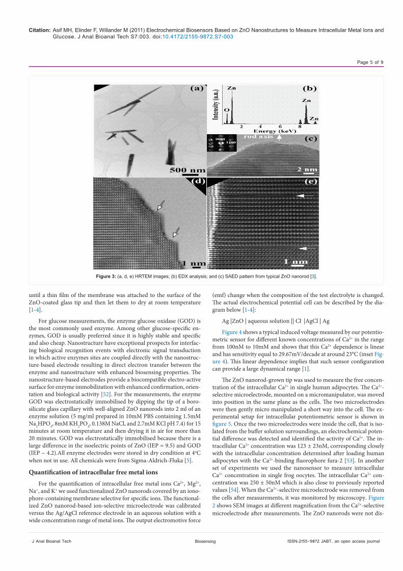
Figure 3 shows the HRTEM images of ZnO nanorods grown by the ACG method on the surface of a glass tip. The specimen was made by scraping the nanorods onto a copper grid with a carbon film. Chemical composition microanalysis using the energy dispersive X-Ray (EDX) demonstrates that the ZnO nanorods consists only of O and Zn with-out other metal impurities as catalysts (see figure 3 (b)). The additional peak at 8keV is the Cu Kα from the copper grid. A HRTEM image and a selected area electron diffraction (SAED) pattern are shown in figure 3 (c) with the inset revealing that the nanorod axis is in the [0001] direc-tion. The rods contain some defects such as surface steps and stacking faults shown by arrows in figures 3 (d, e) [3].
Functionalization of ZnO nanostructure
The nanostructure materials, used in biosensors, are usually cov-ered with a thin layer of biomaterials (i.e. membrane or enzyme) that has many functions, including diffusion control, reduction of interfer-ence and mechanical protection of the sensing probe. There are dif-ferent types of polymers and enzymes commercially available which are commonly used to functionalize the interface due to their suitable physical and chemical properties especially for selectivity. Biosensors with polymer membranes have been successfully applied in many fields such as the monitoring of food production, environmental pollution and pathological specimens. Polymeric membranes are mainly made of polymers, which can selectively transfer certain chemical species. Therefore, membranes are the key components of all potentiometric ion sensors [48-51]. Analogous to biological ion channels, in analyti-cal technology there are the so called ionophores and neutral carriers incorporated into synthetic membranes or biomolecule membranes in order to achieve the desired selectivity or detection of ionic species in complex samples. Solvent polymeric membrane are commercially available and routinely used for the selective detection of several metal ions such as K+, Na+, Ca2+ etc. in complex biological matrices [6].To measure the intracellular free metal ion selectively, the ZnO nanorod layer on the tip of borosilicate glass capillaries were coated with poly-meric membrane incorporated with ionophore by a manual procedure. For optimization, powdered polyvinyl chloride (PVC) was dissolved in tetrahydrofuran (THF) together with the plasticizer dibutylphtalate (DBP) and metal ion-specific ionophores. After preparing the solution, the ZnO-coated glass tip was dipped twice into the resultant solution
Biosensing
Citation: Asif MH, Elinder F, Willander M (2011) Electrochemical Biosensors Based on ZnO Nanostructures to Measure Intracellular Metal Ions and Glucose. J Anal Bioanal Tech S7:003. doi:10.4172/2155-9872.S7-003

Page 4 of 9
ISSN:2155-9872 JABT, an open access journal J Anal Bioanal Tech
Figure 1: SEM images of ZnO nanorods on the surface of glass capillary tip at different magnifications.
Figure 2: FESEM images showing the working electrode after intracellular measurements at different magnifications [5].
Biosensing
Citation: Asif MH, Elinder F, Willander M (2011) Electrochemical Biosensors Based on ZnO Nanostructures to Measure Intracellular Metal Ions and Glucose. J Anal Bioanal Tech S7:003. doi:10.4172/2155-9872.S7-003
Page 5 of 9
ISSN:2155-9872 JABT, an open access journal J Anal Bioanal Tech
until a thin film of the membrane was attached to the surface of the ZnO-coated glass tip and then let them to dry at room temperature [1-4].
For glucose measurements, the enzyme glucose oxidase (GOD) is the most commonly used enzyme. Among other glucose-specific en-zymes, GOD is usually preferred since it is highly stable and specific and also cheap. Nanostructure have exceptional prospects for interfac-ing biological recognition events with electronic signal transduction in which active enzymes sites are coupled directly with the nanostruc-ture-based electrode resulting in direct electron transfer between the enzyme and nanostructure with enhanced biosensing properties. The nanostructure-based electrodes provide a biocompatible electro-active surface for enzyme immobilization with enhanced confirmation, orien-tation and biological activity [52]. For the measurements, the enzyme GOD was electrostatically immobilised by dipping the tip of a boro-silicate glass capillary with well-aligned ZnO nanorods into 2 ml of an enzyme solution (5 mg/ml prepared in 10mM PBS containing 1.5mM Na2HPO4, 8mM KH2PO4, 0.138M NaCl, and 2.7mM KCl pH 7.4) for 15 minutes at room temperature and then drying it in air for more than 20 minutes. GOD was electrostatically immobilised because there is a large difference in the isoelectric points of ZnO (IEP = 9.5) and GOD (IEP ~ 4.2).All enzyme electrodes were stored in dry condition at 4oC when not in use. All chemicals were from Sigma-Aldrich-Fluka [5].
Quantification of intracellular free metal ions
For the quantification of intracellular free metal ions Ca2+, Mg2+, Na+, and K+ we used functionalized ZnO nanorods covered by an iono-phore-containing membrane selective for specific ions. The functional-ized ZnO nanorod-based ion-selective microelectrode was calibrated versus the Ag/AgCl reference electrode in an aqueous solution with a wide concentration range of metal ions. The output electromotive force
(emf) change when the composition of the test electrolyte is changed. The actual electrochemical potential cell can be described by the dia-gram below [1-4]:
Ag |ZnO | aqueous solution || Cl- |AgCl | Ag
Figure 4 shows a typical induced voltage measured by our potentio-metric sensor for different known concentrations of Ca2+ in the range from 100nM to 10mM and shows that this Ca2+ dependence is linear and has sensitivity equal to 29.67mV/decade at around 23°C (inset Fig-ure 4). This linear dependence implies that such sensor configuration can provide a large dynamical range [1].
The ZnO nanorod-grown tip was used to measure the free concen-tration of the intracellular Ca2+ in single human adipocytes. The Ca2+-selective microelectrode, mounted on a micromanipulator, was moved into position in the same plane as the cells. The two microelectrodes were then gently micro manipulated a short way into the cell. The ex-perimental setup for intracellular potentiometric sensor is shown in figure 5. Once the two microelectrodes were inside the cell, that is iso-lated from the buffer solution surroundings, an electrochemical poten-tial difference was detected and identified the activity of Ca2+. The in-tracellular Ca2+ concentration was 123 ± 23nM, corresponding closely with the intracellular concentration determined after loading human adipocytes with the Ca2+-binding fluorophore fura-2 [53]. In another set of experiments we used the nanosensor to measure intracellular Ca2+ concentration in single frog oocytes. The intracellular Ca2+ con-centration was 250 ± 50nM which is also close to previously reported values [54]. When the Ca2+-selective microelectrode was removed from the cells after measurements, it was monitored by microscopy. Figure 2 shows SEM images at different magnification from the Ca2+-selective microelectrode after measurements. The ZnO nanorods were not dis-
Figure 3: (a, d, e) HRTEM images; (b) EDX analysis; and (c) SAED pattern from typical ZnO nanorod [3].
Biosensing
Citation: Asif MH, Elinder F, Willander M (2011) Electrochemical Biosensors Based on ZnO Nanostructures to Measure Intracellular Metal Ions and Glucose. J Anal Bioanal Tech S7:003. doi:10.4172/2155-9872.S7-003

Page 6 of 9
ISSN:2155-9872 JABT, an open access journal J Anal Bioanal Tech
solved. This result was expected because the functionalization provided protection for the surface of the nanorods.
To test the sensitivity of our constructed biosensor we performed measurements while changing the Ca2+ concentration in the buffer solution around the cell. The Ca2+ was varied from 100nM to 10mM. These measurements were performed for two cases; the first was for a configuration where half of the working functionalized ZnO nanorods were inserted inside the cell and the other half was in contact with the surrounding buffer solution. The second configuration was when all the functionalized ZnO nanorods were inserted inside the cell. Signifi-cantly, the functionalized ZnO nanorods exhibited a stepwise decrease
in the induced electrochemical potential only in the first case of the configuration. Once the functionalized ZnO nanorods working elec-trode was totally inserted inside the cell, (i.e. isolated from the buffer solution surrounding the cell), the electrochemical potential difference signal detected was stable (corresponding to the actual intracellular Ca2+ concentration) even when the Ca2+ concentration was varied in the buffer solution. This implies that the constructed electrode has a high sensitivity. In addition, this observation confirms that the values of the potentiometric response when the whole sensing electrode was inserted inside the cell membrane is the signal corresponding to the value of the Ca2+ concentration inside the cell [1].
We have also used functionalized hexagonal ZnO nanorods, grown on silver-coated capillary glass tip, as a selective intracellular sensor for other free metal ions (Na+, K+, Mg2+) in single human adipocytes and frog oocytes. The functionalization was achieved through the use of ion-selective PVC polymeric membrane. The potential difference was found to be linear over a wide concentration range of interest. The measured intracellular metal-ion concentrations using the ZnO nano-rods sensor in human fat cells or in frog egg cells were consistent with values reported in the literature. These results demonstrate the capabil-ity to perform biological relevant measurements inside living cells. The ZnO nanorod-based metal ion microelectrodes thus hold promise for minimally invasive dynamic analyses of single cells [1-4].
Quantification of intracellular glucose
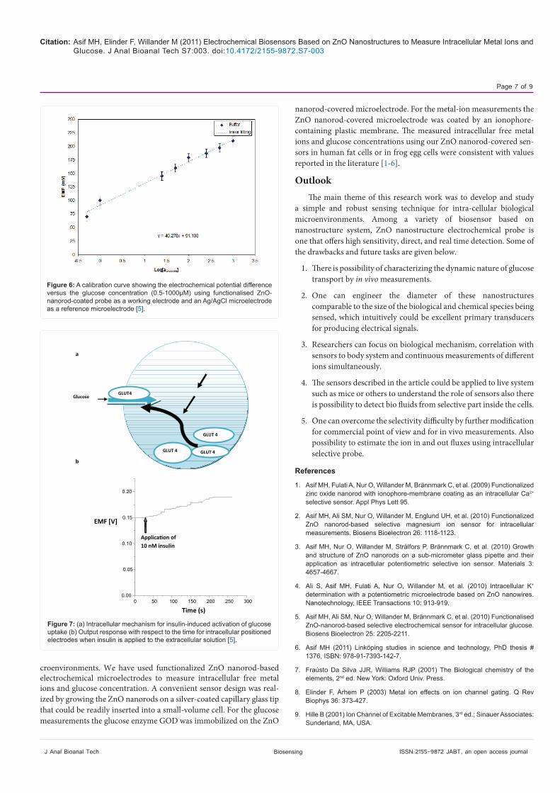
The determination of the intracellular glucose concentration with functionalized ZnO nanorod-coated microelectrodes was performed in two types of cells, human adipocytes and frog oocytes. In the hu-man body, the hormone insulin only stimulates glucose transport into muscle and fat cells. However, insulin has also been found to affect glu-cose uptake in oocytes from the frog Xenopuslaevis [55,56]. The large size of these cells makes it possible to microinject specific reagents that interrupt or activate signal transmission to glucose. The response of the electrochemical potential difference of the ZnO nanorods to the changes in buffer electrolyte glucose was measured for the range of 500nM to 1mM and shows that the glucose dependence is linear and has sensitivity equal to 42.5 mV/decade (Figure 6) The intracellular glucose concentration in the human adipocytes was 50 ± 15µM (n = 5), which can be compared with the 70µM intracellular concentration determined by nuclear magnetic resonance spectroscopy in rat muscle tissue in the presence of a high, 10mM, and extracellular glucose con-centration [29]. For the frog oocytes, it was 125 ± 23µM (n = 5). When we achieved a stable potential for the intracellular measurement, 10nM insulin was added to extracellular solution. After few minutes, the in-sulin increased the glucose concentration in the human adipocyte from 50 ± 15 to 125 ± 15µM and the glucose concentration in the frog oo-cytes increased from 125 ± 23µM to 250 ± 19µM. Figure 7 a,b showed the output response with respect to time for intracellularly positioned electrodes when insulin is applied to the extracellular solution [5]. The reported values of the glucose concentration in human adipocytes and frog oocytes using our functionalized ZnO nanorods sensor was con-sistent with values of glucose concentration reported in the literature survey [5].
ConclusionsWe have developed and studied a simple and robust biosensing
technique, based on ZnO nanorods, for intracellular biological mi-
y = -29.674x + 98.072
-40
-20
0
20
40
60
80
100
120
140
160
-2 -1 0 1 2 3 4 5
Log [aCa2+]
EM
F (
mV
)
Buffer SolutionLinear fitting
Figure 4: Sensitivity diagram showing the potentiometric response versus time as the concentrations of Ca2+ solution is changed in the buffer solution sur-rounding the cell. The Ca2+ concentration was varied from 100nM to 10mM. The inset is a typical calibration curve showing the electrochemical potential difference versus the Ca2+ concentration using the functionalized ZnO nano-rods as working electrode and an Ag/AgCl as reference electrode [1].
Figure 5: Experimental setup for the intracellular potentiometric measure-ments [6].
Biosensing
Citation: Asif MH, Elinder F, Willander M (2011) Electrochemical Biosensors Based on ZnO Nanostructures to Measure Intracellular Metal Ions and Glucose. J Anal Bioanal Tech S7:003. doi:10.4172/2155-9872.S7-003
Page 7 of 9
ISSN:2155-9872 JABT, an open access journal J Anal Bioanal Tech
croenvironments. We have used functionalized ZnO nanorod-based electrochemical microelectrodes to measure intracellular free metal ions and glucose concentration. A convenient sensor design was real-ized by growing the ZnO nanorods on a silver-coated capillary glass tip that could be readily inserted into a small-volume cell. For the glucose measurements the glucose enzyme GOD was immobilized on the ZnO
nanorod-covered microelectrode. For the metal-ion measurements the ZnO nanorod-covered microelectrode was coated by an ionophore-containing plastic membrane. The measured intracellular free metal ions and glucose concentrations using our ZnO nanorod-covered sen-sors in human fat cells or in frog egg cells were consistent with values reported in the literature [1-6].
OutlookThe main theme of this research work was to develop and study
a simple and robust sensing technique for intra-cellular biological microenvironments. Among a variety of biosensor based on nanostructure system, ZnO nanostructure electrochemical probe is one that offers high sensitivity, direct, and real time detection. Some of the drawbacks and future tasks are given below.
1. There is possibility of characterizing the dynamic nature of glucose transport by in vivo measurements.
2. One can engineer the diameter of these nanostructures comparable to the size of the biological and chemical species being sensed, which intuitively could be excellent primary transducers for producing electrical signals.
3. Researchers can focus on biological mechanism, correlation with sensors to body system and continuous measurements of different ions simultaneously.
4. The sensors described in the article could be applied to live system such as mice or others to understand the role of sensors also there is possibility to detect bio fluids from selective part inside the cells.
5. One can overcome the selectivity difficulty by further modification for commercial point of view and for in vivo measurements. Also possibility to estimate the ion in and out fluxes using intracellular selective probe.
References
1. Asif MH, Fulati A, Nur O, Willander M, Brännmark C, et al. (2009) Functionalized zinc oxide nanorod with ionophore-membrane coating as an intracellular Ca2+ selective sensor. Appl Phys Lett 95.
2. Asif MH, Ali SM, Nur O, Willander M, Englund UH, et al. (2010) Functionalized ZnO nanorod-based selective magnesium ion sensor for intracellular measurements. Biosens Bioelectron 26: 1118-1123.
3. Asif MH, Nur O, Willander M, Strålfors P, Brännmark C, et al. (2010) Growth and structure of ZnO nanorods on a sub-micrometer glass pipette and their application as intracellular potentiometric selective ion sensor. Materials 3: 4657-4667.
4. Ali S, Asif MH, Fulati A, Nur O, Willander M, et al. (2010) Intracellular K+ determination with a potentiometric microelectrode based on ZnO nanowires. Nanotechnology, IEEE Transactions 10: 913-919.
5. Asif MH, Ali SM, Nur O, Willander M, Brännmark C, et al. (2010) Functionalised ZnO-nanorod-based selective electrochemical sensor for intracellular glucose. Biosens Bioelectron 25: 2205-2211.
6. Asif MH (2011) Linköping studies in science and technology, PhD thesis # 1376, ISBN: 978-91-7393-142-7.
7. Fraústo Da Silva JJR, Williams RJP (2001) The Biological chemistry of the elements, 2nd ed. New York: Oxford Univ. Press.
8. Elinder F, Århem P (2003) Metal ion effects on ion channel gating. Q Rev Biophys 36: 373-427.
9. Hille B (2001) Ion Channel of Excitable Membranes, 3rd ed.; Sinauer Associates: Sunderland, MA, USA.
Figure 6: A calibration curve showing the electrochemical potential difference versus the glucose concentration (0.5-1000µM) using functionalised ZnO-nanorod-coated probe as a working electrode and an Ag/AgCl microelectrode as a reference microelectrode [5].
0 50 100 150 200 250 300 0.00
0.05
0.10
0.15
0.20
Application of 10 nM insulin
EMF [V]
Time (s)
GLUT4
GLUT 4
GLUT 4 GLUT 4
Glucose
a
b
Figure 7: (a) Intracellular mechanism for insulin-induced activation of glucose uptake (b) Output response with respect to the time for intracellular positioned electrodes when insulin is applied to the extracellular solution [5].
Biosensing
Citation: Asif MH, Elinder F, Willander M (2011) Electrochemical Biosensors Based on ZnO Nanostructures to Measure Intracellular Metal Ions and Glucose. J Anal Bioanal Tech S7:003. doi:10.4172/2155-9872.S7-003

Page 8 of 9
ISSN:2155-9872 JABT, an open access journal J Anal Bioanal Tech
10. Knight KK, Wentzlaff DM, Snyder PM (2008) Intracellular sodium regulates proteolytic activation of the epithelial sodium channel. J Biol. Chem 283: 27477-27482.
11. Ho IH, Murrell-Lagnado RD (1999) Molecular mechanism for sodium-dependent activation of G protein-gated K+ channels. J Physiol 520: 645-651.
12. Yu XM, Salter MW (1998) Gain control of NMDA-receptor currents by intracellular sodium. Nature 396: 469-474.
13. Efendiev R, Bertorello AM, Zandomeni R, Cinelli AR, Pedemonte CH (2002) Agonist-dependent regulation of renal Na+, K+-ATPase activity is modulated by intracellular sodium concentration. J. Biol. Chem 277: 11489-11496.
14. Budelli G, Hage TA, Wei A, Rojas P, Jong YJ, et al. (2009) Na+-activated K+ channels express a large delayed outward current in neurons during normal physiology. Nat Neurosci 12: 745-750.
15. Kameyama M, Kakei M, Sato R, Shibasaki T, Matsuda H, et al. (1984) Intracellular Na+ activates a K+ channel in mammalian cardiac cells. Nature 309: 354-356.
16. Yu SP, Canzoniero LM, Choi D W (2001) Ion homeostasis and apoptosis. Curr Opin Cell Biol 13: 405-411.
17. Thompson GJ, Langlais C, Cain K, Conley EC, Cohen GM (2001) Elevated extracellular [K+] inhibits death-receptor- and chemical-mediated apoptosis prior to caspase activation and cytochrome c release. Biochem J 357: 137-145.
18. Bortner CD, Cidlowski JA (2007) Cell shrinkage and monovalent cation fluxes: role in apoptosis. Arch Biochem Biophys 462: 176-188.
19. Bortner CD, Sifre MI, Cidlowski JA (2008) Cationic gradient reversal and cytoskeleton-independent volume regulatory pathways define an early stage of apoptosis. J Biol Chem 283: 7219-7229.
20. Hughes FM, Bortner CD, Purdy GD, Cidlowski JA (1997) Intracellular K+ suppresses the activation of apoptosis in lymphocytes. J Biol Chem 272: 30567-30576.
21. Akanda N, Tofighi R, Brask J, Tamm C, Elinder F, et al. (2008) Voltage-dependent anion channels (VDAC) in the plasma membrane play a critical role in apoptosis in differentiated hippocampal neurons but not in neural stem cells. Cell Cycle 7: 3225-3234.
22. Banasiak KJ, Burenkova O, Haddad GG (2004) Activation of voltage-sensitive sodium channels during oxygen deprivation leads to apoptotic neuronal death. Neuroscience 126: 31-44.
23. Bortner CD, Gomez-Angelats M, Cidlowski JA (2001) Plasma membrane depolarization without repolarization is an early molecular event in anti-Fas-induced apoptosis. J Biol Chem 276: 4304-4314.
24. Bortner CD, Cidlowski JA (2003) Uncoupling cell shrinkage from apoptosis reveals that Na+ influx is required for volume loss during programmed cell death. J Biol Chem 278: 39176-39184.
25. Matsuda H, Saigusa A, Irisawa H (1987) Ohmic conductance through the inwardly rectifying K channel and blocking by internal Mg2+. Nature 325: 156-159.
26. Berridge MJ, Bootman MD, Roderick HL (2003) Calcium signalling: dynamics, homeostasis and remodeling. Nat Rev Mol Cell Biol 4: 517-529.
27. Bliss TV, Collingridge GL (1993) A synaptic model of memory: long-term potentiation in the hippocampus. Nature 361: 31-39.
28. Abe T, Lau YY, Ewing AG (1992) Characterization of glucose microsensors for intracellular measurements. Anal Chem 64: 2160-2163.
29. Cline GW, Jucker BM, Trajanoski Z, Rennings AJM, Shulman GI (1998) A novel 13C NMR method to assess intracellular glucose concentration in muscle, in vivo. Am J Physiol 274: E381-E389.
30. Yamada K, Nakata M, Horimoto N, Saito M, Matsuoka H, et al. (2000) Measurement of glucosdes uptake and intracellular calcium concentration in single, living pancreatic β-cells. J Biol Chem 275: 22278-22283.
31. Al-Hilli S, Willander M (2007) ZnO nanorods as an intracellular sensor for pH measurements. Methods Mol Bio 544: 187-200.
32. Henrich VE, Cox PA (1994) The surface science of metal oxides, Cambridge University Press, Boston.
33. Zhang Y, Yang M, Portney NG, Cui D, Budak G, et al. (2008) A surface electrical characteristic to probe the interaction of nanoparticles with normal and cancer human breast epithelial cells. Biomed Microdev 10: 321-328.
34. Cui D (2007) Advances and prospects on biomolecules functionalized carbon nanotubes. J. Nanosci Nanotechnol 7: 1298-1314.
35. Pan B, Cui D, Ozkan CS, Ozkan M, Xu P, et al. (2008) Effects of carbon nanotubes on photoluminescence properties of quantum dots. J Phys Chem C 112: 939-944.
36. Zhang X, Guo Q, Cui D (2009) Recent advances in nanotechnology applied to biosensors. Sensors 9: 1033-1053.
37. Liu A (2008) Towards development of chemosensors and biosensors with metal-oxide-based nanowires or nanotubes. Biosens Bioelectron 24: 167-177.
38. Xia C, Wang N, Lidong L, Lin G (2008) Synthesis and characterization of waxberry-like microstructures ZnO for biosensors. Sens Actuat 129: 268-273.
39. Huang ZB, Yan DH, Yang M, Liao XM, Kang YQ, et al. (2008) Preparation and characterization of the biomineralized zinc oxide particles in spider silk peptides. Colloid Interface Sci 325: 356-362.
40. Solanki PR, Kaushik A, Angrawal VV, Malhotra BD (2011) Nanostructured metal oxide-based biosensors. NPG Asia Mater 3: 17-24.
41. Wang ZL, Kong XY, Ding Y, Gao P, Hughes WL, et al. (2004) Semiconducting and piezoelectric oxide nanostructures induced by polar surfaces. Adv Funct Mater 14: 943-956.
42. Willander M, Zhao QX, Hu QH, Klason P, Kuzmin V, et al. (2008) Fundamentals and properties of zinc oxide nanostructures: optical and sensing applications. Supperlattices & Microstructures 43: 352-361.
43. Li QH, Gao T, Wang YG, Wang TH (2005) Adsorption and desorption of oxygen probed from ZnO nanowire films by photocurrent measurements. Appl Phys Lett 86: 123117-123119.
44. Al-Hilli SM, Al-Mofarji RT, Willander M (2006) Zinc oxide nanorod for intracellular pH sensing. Appl Phys Lett 89: 173119-173121.
45. Vayssieres L, Kies K, Lindquist SE, Hagfeldt A (2001) Purpose-built anisotropic metal oxide material: 3D highly oriented microrod array of ZnOJ. Phys Chem B 105: 3350-3352.
46. Vayssieres L (2003) Growth of arrayed nanorods and nanowires of ZnO from aqueous solutions Adv Mater 15: 464-466.
47. Greene L, Law M, Tan D, Montano M, Goldberger J, et al. (2005) General route to vertical ZnO nanowire arrays using textured ZnO seeds. Nano Lett 5: 1231-1236.
48. Bakker E, Buhlmann P, Pretsch ESI (1999) Polymer membrane ion-selective electrodes-what are the limits? Electroanaly 11: 915-933.
49. Bakker E, Buhlmann P, Pretsch ESI (1997) Carrier -based ion selective electrodes and bulk optodes. 1. General characteristics. Chem. Rev 97: 3083-3132.
50. Bakker E, Buhlmann P, Pretsch ESI (1998) Carrier--based ion selective electrodes and bulk optodes. 2. Ionophores for potentiometric and optical sensors. Chem Rev 98: 1593-1687.
51. ZhangS, Wright G, Yang Y (2000) Materials and techniques for electrochemical biosensor design and construction. Biosens Bioelectron 15: 273-282.
52. Rahman MM, Ahammad AJS, Jin J-H, Ahn SJ, Lee J-J (2010) A comprehensive review of glucose biosensors based on nanostructured metal-oxides. Sensors 10: 4855-4886.
53. Draznin B, Sussman KE, Eckel RH, Kao M, Yost T, et al. (1988) Possible role of cytosolic free calcium concentrations in mediating insulin resistance of obesity and hyperinsulinemia. J Clin Invest 82: 1848-1852.
54. Takahashi T, Neher E, Sakmann B (1987) Rat brain serotonin receptors in Xenopus oocytes are coupled by intracellular calcium to endogenous channels. Proc Natl Acad Sci 84: 5063-5067.
Biosensing
Citation: Asif MH, Elinder F, Willander M (2011) Electrochemical Biosensors Based on ZnO Nanostructures to Measure Intracellular Metal Ions and Glucose. J Anal Bioanal Tech S7:003. doi:10.4172/2155-9872.S7-003

Page 9 of 9
ISSN:2155-9872 JABT, an open access journal BiosensingJ Anal Bioanal Tech
55. JanicotM, Lane MD (1989) Activation of glucose uptake by insulin and insulin-like growth factor I in Xenopus oocytes. Proc Natl Acad Sci 86: 2642-2646.
56. Simpson IA, Cushman SW (1986) Hormonal regulation of mammalian glucose transport. Annu Rev Biochem 55: 1059-1089.
This article was originally published in a special issue, Biosensing handled by Editor(s). Dr. Dr. Michael J. Serpe, University of Alberta, Canada
Citation: Asif MH, Elinder F, Willander M (2011) Electrochemical Biosensors Based on ZnO Nanostructures to Measure Intracellular Metal Ions and Glucose. J Anal Bioanal Tech S7:003. doi:10.4172/2155-9872.S7-003