Embed Size (px)
Citation preview

Welcome to Podiatry Management’s CME Instructional program. Our journal has been approved as a sponsor of Contin-uing Medical Education by the Council on Podiatric Medical Education.
You may enroll: 1) on a per issue basis (at $20.00 per topic) or 2) per year, for the special introductory rate of $139 (yousave $61). You may submit the answer sheet, along with the other information requested, via mail, fax, or phone. In the nearfuture, you may be able to submit via the Internet.
If you correctly answer seventy (70%) of the questions correctly, you will receive a certificate attesting to your earned cred-its. You will also receive a record of any incorrectly answered questions. If you score less than 70%, you can retake the test atno additional cost. A list of states currently honoring CPME approved credits is listed on pg. 178. Other than those entities cur-rently accepting CPME-approved credit, Podiatry Management cannot guarantee that these CME credits will be acceptable byany state licensing agency, hospital, managed care organization or other entity. PM will, however, use its best efforts to ensurethe widest acceptance of this program possible.
This instructional CME program is designed to supplement, NOT replace, existing CME seminars. Thegoal of this program is to advance the knowledge of practicing podiatrists. We will endeavor to publish high quality manuscriptsby noted authors and researchers. If you have any questions or comments about this program, you can write or call us at: PodiatryManagement, P.O. Box 490, East Islip, NY 11730, (631) 563-1604 or e-mail us at [email protected].
Following this article, an answer sheet and full set of instructions are provided (p. 178).—Editor
This modality is a useful adjunct in the treatment of non-unions, delayed unions, fresh fractures, and Charcot osteoarthropathy.
JANUARY 2008 • PODIATRY MANAGEMENTwww.podiatrym.com 167
Continuing
Medical Education
Objectives
1) To understandthe different tech-nologies available forbone stimulation.
2) To understandthe risk factors for thedevelopment of anon-union.
3) To understandsome basic scienceconcepts behind thedevelopment of bonestimulation.
the potentials necessary to modifybone. Bone stimulation is a usefuladjunctive therapy for the treat-ment of non-unions, delayedunions, fresh fractures, and Char-cot osteoarthropathy.
There are four distinct tech-niques for the delivery of electric-i ty or mechanical pressure tobone: direct current, capacitivecoupling, inductive coupling, and
Continued on page 168
IntroductionBone stimulation is the use of
energy transmitted through boneto accelerate bone growth. Al-though the concept of stimulat-ing bone growth was discoveredin the mid-twentieth century,technological advances recentlyallowed for the development oftechniques capable of delivering
By Linnie V. Rabjohn, DPM
ElectricalStimulation of Bone: The EvolvingTechnology
There are four distinct
techniques for the
delivery of electricity
or mechanical
pressure to bone.
C L I N I C A L P O D I A T R YC L I N I C A L P O D I A T R Y

later by Bassett and Becker.2 Thefirst modern report of electricmethods being used to heal anon-union was of a medial malle-olus in 1971 by direct currenttechnology. 3 Since that t ime,there has been a large amount ofresearch focused on the use ofpiezoelectric fields associated withbone and the application of bonestimulators based on thisconcept.4 Piezoelectricity is theability of crystals to produce avoltage when subjected to stress.Bone contains calcium phosphatecrystals , making it subject tomodification by the introductionof voltage.
Wolff’s LawWolff’s law encompasses the
idea that bonewill adapt to in-troduced stress,that the form ofbone will followthe function ofthe bone. Stress-generated poten-tials, small elec-tric currents, inbone will changethe activity ofbone, al lowingfor this adapta-
tion by modifying the activity ofosteoblasts.2,5-6 Endogenous bio-electr ic potentials have beenfound in unstressed bone to havean electronegative reaction at thefracture site.3 Bone is most elec-
tronegative at areas of growth,such as fractures and epiphysealplates.7 From this concept camethe idea that electric potentials
sent through bone could acceler-ate the healing process of freshfractures, delayed unions andnon-unions.
The Science Behind BoneStimulation
Bone is subject to electr icstresses because it contains calci-um phosphate crystals. The com-pression side produces a negativepotential, while the tension sidehas a posit ive potential . 4 Theother form of electric potential as-sociated with bone is transmem-brane potential. These potentialsare generated by the metabolismof a cell and are reliant on cell vi-ability. The greater the cellular ac-tivity, the greater the negative po-tential generated. The potentialsplay an important role in earlycallus development and the re-modeling stages of bone healing.
The application of these po-tentials by an externalsource can act to accel-erate osseous healing byinfluencing the bone-healing cascade. The useof electrical stimulationleads to the productionof more bone-formingcells by the osteopro-genitor cells. 8 This inturn accelerates healingby augmenting the de-velopment of extracellu-lar matrix.
The formation of ablood clot at the frac-ture site initiates the be-ginning phases of bonehealing. 4 The clot iscomposed of inflamma-tory cells and platelets.
Continued on page 169
168 www.podiatrym.comPODIATRY MANAGEMENT • JANUARY 2008
Stimulation...
ultrasound. Direct currentstimulation utilizes electrodes
implanted directly on the bone todeliver energy potentials to pro-mote bone growth. Capacitivecoupling uses either percutaneousskin electrodes or charged platesplaced on the skin to deliver anelectric current to the osseousarea of interest. Inductive cou-pling uses electromagnetic fieldsto del iver the necessary bonestimulating potentials . Ultra-sound uses acoustic radiation inthe form of mechanical energy tostimulate healing. There are a va-riety of bone stimulators commer-cially available for adjunctivetherapy which may be categorizedaccording to thetechnique ofs t i m u l a t i o n(Table 1).
Origin of BoneStimulation
The idea ofbone stimulationthrough exoge-nous sources is arelat ively newconcept that hasevolved over thelast sixty years. In 1955, Yasudawas the first to report new boneformation at the s i te of elec-tronegative potential.1 This dis-covery went largely unrecognizeduntil it was confirmed some years
Contin
uing
Medica
l Edu
catio
n
Remodeling of the
bone occurs in
the final stages of
bone healing.
Bone is subject
to electric stresses
because it contains
calcium phosphate
crystals.
TABLE 1Representation of Commercially Available
Bone Stimulators
Direct Current: Osteogen® (EBI, Parsippany, NJ)
Capacitive Coupling: Orthopak® (EBI, Parsippany, NJ)
Inductive Coupling: Pulsed Electromagnetic Fields (PEMF):EBI Bone Healing System® (EBI, Parsippany, NJ)Physio-Stim Lite® (Orthofix, McKinney, TX)
Combined Magnetic Fields (CMF):OL1000® (DJO, Vista, CA)
Ultrasound: Exogen® (Smith & Nephew, Memphis, TN)
JANUARY 2008 • PODIATRY MANAGEMENTwww.podiatrym.com 169
future integrity. Bone that doesnot progress from the bridging tothe calcifying stage of the healingprocess will not unite.10
Non-unionsIndications for the use of bone
stimulators include non-unions,delayed unions, failed arthrodesis,Charcot osteoarthorpathy, and
fresh fractures.11 This article willmainly focus on the use of bonestimulators for non-unions. Thegold standard treatment for non-unions is the removal of necroticbone tissue and stabilization bymeans of internal or external fixa-tion.12 Treatment with a bone
stimulator can be usedas an alternative treat-ment or as an adjunc-tive treatment to thestandard of care.
The most commonlyfractured long bone ofthe lower extremity isthe tibia, which is alsoassociated with thehighest incidence of de-layed union and non-unions.13-15 This is thereason that many stud-ies evaluating the effi-cacy of bone stimula-tion are involving non-unions of the tibia.Arthrodesis of the footand ankle has a non-union rate of 5-10%,making bone stimula-tion a very useful andneeded adjunct to ther-
These cells are responsible for re-leasing factors that are requiredfor bone healing. Primary fracturehealing occurs when there is rigidinternal fixation with minimalstrain.4,9-10 This allows bone to belaid down across the gap directly.Secondary bone healing occursdue to strain, and results in in-tramembranous or endochondralossification bone formation.
Intramembranous ossificationoccurs in the periosteum as theosteoblasts recruit the neededcells to that site. This allows forthe formation of a granulation tis-sue that becomes cartilage. Afterthis occurs, endochondral bonehealing can occur. There are a va-riety of chondrocytes responsiblefor transforming the cartilage intobone. The chondrocytes initiateangiogenesis, which allows forthe formation of vasculaturechannels that can bring os-teoblasts to the site. This tissuemust be calcified at this point be-fore vascular invasion and trabec-ulation can occur.
The osteoblasts are responsiblefor ult imately producing thewoven bone that unites a fracture.Remodeling of the bone occurs inthe final stages of bone healing.The bone produced must be re-modeled to become structuredlike the original bone to allow for
Stimulation... apy for foot and anklesurgeons.16 Risk factors fornon-union include smoking,immunosuppressive drugs forinflammatory arthritides, diabetesmellitus, obesity, alcoholism, pre-vious operation, hormones, use ofillicit drugs, age, nutrition, frac-ture characteristics, location, com-minution, vascular injury, soft tis-
sue damage, infection, prior opentrauma, psychiatric illness, andhistory of Charcot osteoarthropa-thy (Table 2).11,17-18
Having an understanding ofthe contribution of each factor ofincreasing the risk of non-unionwould assist in the decision pro-
cess of utilizing adjunct electricalbone stimulation.11 The existenceof such risks has made adjunctiveprocedures that promote bonehealing desirable in an effort todecrease non-union rate. It iswell-documented throughout lit-erature that smoking can be detri-
Continuing
Medical Education
Continued on page 170
Figure 1: The Orthopak® bone stimulation system byEBI medical products.
TABLE 2Risk Factors for Development
of Non-Union
Smoking Immunosuppressive drugsInflammatory arthritides Diabetes mellitusObesity AlcoholismPrevious surgery HormonesIllicit drug use AgeNutrition Fracture characteristicsFracture location ComminutionVascular injury Soft tissue damageInfection Prior open traumaPsychiatric illness Charcot osteoarthropathy
It is well-documented
throughout literature
that smoking
can be detrimental
to both soft tissue
and bone healing.
Success of bone stimulationon different forms of non-unionsdecreases in the following order:hypertrophic, oligotrophic, andatrophic non-unions.10 A hyper-trophic non-union is a well-vascu-larized callous formation with no
calcification be-tween bone frag-ments. 17 Thisform of non-union is themost capable ofhealing with in-activity and im-m o b i l i z a t i o n ,and adjuncttreatment ofbone st imula-tion. No systemwill work for atrue synovial
pseudoarthrosis.10
To determine the presence of apseudoarthrosis, a technetium 99bone scan may be performed. Ifone is present, a “cold cleft,” orarea of decreased activity sur-rounded by increased radioactivi-ty, will be seen.24
Direct Current BoneStimulation
Direct current bone stimula-tion appears to work by a mecha-nism of stimulating the formationof bone by increasing the amountof intracellular free calcium andhydrogen peroxide generation atthe cathode and the resulting in-crease in pH.25,26 The use of a di-rect current bone stimulation de-vice may be either by an invasive
technique ofsurgically im-planting a bat-tery and leadsor through asemi-invasivet e c h n i q u ewhich usespercutaneouselectrodes con-nected to anexternal powerdevice. 27 Theonly commer-cially availabledirect currentdevice is theOsteogen (EBI,P a r s i p p a n y ,NJ) which is
invasive by technique.There are currently no semi-
invasive direct current devicesavailable. The Osteogen utilizes atitanium cathode wire in the formof a single, double, or mesh wirethat is implanted directly into theosseous site, allowing for maximalsurface area of contact . 27-28 I tshould have no contact withmetal implants. The battery is ananode placed subcutaneously thatis attached to the cathode. Thisbattery will provide power from6-12 months, making the deviceactive for this time period. Thisdevice uses a constant current of20 micro-amps and 1.0 volts perlead.29
The advantages to the use ofdirect current for bone stimula-tion are increased patient compli-ance because the device is im-planted within the body, that thecurrent is directly applied to thesite with maximal intensity, andthe constant stimulation of theosseous area.28 The disadvantagesto the use of direct current in-
clude that the cathode wiringcannot come into contact withany metallic device, which be-comes a concern when you areplacing it within an arthrodesis orreduction site in addition to inter-nal fixation.
Some advocate the removal ofthe battery anode after union hasoccurred or around 9-12 monthsafter implantation, requiring asecondary surgery. The placementof the battery anode subcuta-neously can lead to a prominence
Continued on page 171
170 www.podiatrym.comPODIATRY MANAGEMENT • JANUARY 2008
Stimulation...
mental to both soft tissueand bone healing. Risk of frac-
ture in smokers is two to s ixtimes greater due to reduced bonedensity.19 This increases in womenwho are post-menopausal be-cause of the al-ready presentloss of bone den-sity. 20 Nicotineuse, or smoking,has been shownto cause a signifi-cantly highernumber of non-unions than theoccurrence inn o n - s m o k e r s(18.6% v.7.1%).11 The risk of developing anon-union is 2.7 times greater fora smoker than nonsmoker after arearfoot arthrodesis.21
PseudoarthrosisThe incidence of pseudoarthro-
sis is at least four times greater inpatients who smoke.22 There is alsoan increased risk of pseudoarthro-sis in relation to grafts becausenicotine inhibits the revasculariza-tion of grafts.20 There are a multi-tude of effects of nicotine, carbonmonoxide, and hydrogen cyanideon tissue healing.23 These includetissue anoxia, cellular hypoxia,and an inhibition of the prolifera-tion of cells, vasoconstriction, anda decrease in the oxygen-carryingcapacity of blood.
Contin
uing
Medica
l Edu
catio
n
A hypertrophic non-
union is a well-
vascularized callus
formation with no
calcification between
bone fragments.
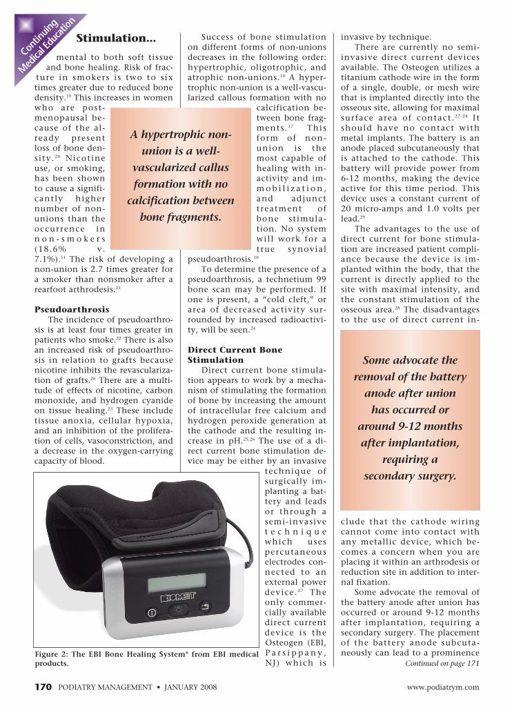
Figure 2: The EBI Bone Healing System® from EBI medicalproducts.
Some advocate the
removal of the battery
anode after union
has occurred or
around 9-12 months
after implantation,
requiring a
secondary surgery.
JANUARY 2008 • PODIATRY MANAGEMENTwww.podiatrym.com 171
A different study also lookedat high-risk patients undergoingfoot and ankle arthrodesis proce-dures with implantation of a di-rect current bone stimulator.11 Pa-tients were considered high riskbased on the presence of one or
more of the fol-lowing: DM,obesity, habitualtobacco and/oralcohol use, im-munosuppressivetherapy, and pre-vious history ofnon-union. Theresults demon-strated thatarthrodesis ofthe foot andankle may be en-
hanced by use of direct currentbone stimulation. The high-risk ofdeveloping a non-union in footand ankle arthrodesis leads to aconsiderable need for revisionarthrodesis.
One study evaluated the use ofdirect current in ten consecutiverevision arthrodeses performed onpatients with aseptic non-unions. 33 Al l obtained a sol idunion at an average of 12.8weeks. The modif ied AOFASscores 70% good to excellent re-sults with use of direct current inrevision arthrodesis. Donley andWard, in the process of using di-rect current st imulation toachieve arthrodesis in a high riskpopulation, also found an im-
which is uncomfortable for thepatient.28 In rare instances, thebattery must be removed prior tounion or inactive state due to aninfection of the area of the device.
Direct CurrentStudies
The first mul-t icenter studyevaluating theuse of direct cur-rent for bonestimulation in178 non-unionsshowed that 83%achieved osseousunion with theuse of a direct current device.30 Astudy performed shortly there-after showed a 86% incidence ofclinical and radiographic osseoushealing after 16 weeks of the useof a direct current device.31 In thepatients who did not achieveunion, the authors suggested thatfailure was due to either the im-proper placement of cathodes orthe premature discontinuation ofcast immobilization.
Brighton and colleagues per-formed a subsequent study look-ing at a case series of 189 non-unions involving the lower ex-tremity, of which 83.7% healedafter direct current implantationof twelve weeks. A retrospectivecase study evaluated the use of di-rect current stimulation inonly patients consideredto be at a high-risk of non-union.32 Results showedthat 78% of patients werefunctionally improved andthat 89% achieved a satis-factory result. The authorsconcluded that the use ofa direct current is reason-able with an acceptablecomplication rate in high-risk groups. Al l had atleast two risk factors fornon-union and 35% wererevision surgeries. In thisgroup of patients, a 15%incidence of deep spaceinfection occurred, mainlyin patients who eithersmoked or had Charcot os-teoarthropathy.
Stimulation... provement in painscores from a mean of 8.5to 1.9 during the process.34
While it is highly apparentthrough numerous clinical trialsthat direct current bone stimula-tion aids in the achievement ofosseous union, some authors be-lieve that there is not sufficientevidence to use an invasive directcurrent device over the non-inva-sive PEMF device.28
Capacitive Coupling BoneStimulation
Capacitance is defined as theability of a device to store electriccharge.4 The technique of capaci-tive coupling for bone stimula-tion has been described as eithersemi-invasive with the use of elec-trodes that are placed percuta-
neously over the area of osseousinterest using a conductive gel ap-plied to the skin, or through anoninvasive technique of usingtwo charged plates to generate aflow of current.27,28 The non-inva-sive form of capacitive coupling isthe only form commercially avail-able as the Orthopak (EBI, Parsip-pany, NJ) (Figure 1).
The mechanism of action ofthis form of bone stimulation isto increase the osteoblastic prolif-eration by inducing an increase inTGF-beta expression through theactivation of the calcium/calmod-ulin pathway.35,36
The advantage to using a ca-pacitive coupling device is theidea that it provides a more directcurrent compared to electromag-netic devices. The disadvantagesto the use of this form centers
Continuing
Medical Education
The advantage of
using an inductive
coupling device
is that it is the
most non-invasive
form of bone
stimulation available.
Capacitance
is defined
as the ability
of a device to store
electric charge.
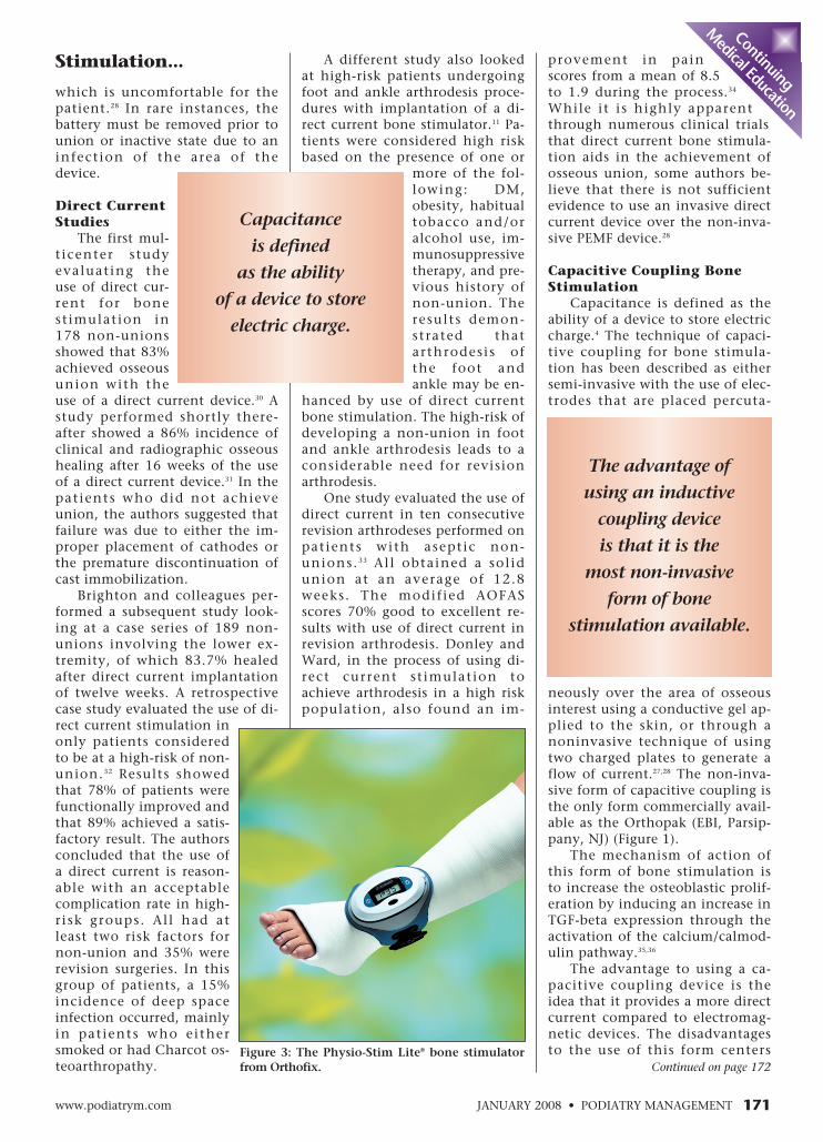
Continued on page 172Figure 3: The Physio-Stim Lite® bone stimulatorfrom Orthofix.
of 21 non-unions of the femoraland tibial shafts showed a signifi-cant difference in the union ratesin patients receiving capacitivebone stimulation.39
In another study, 73% of es-tablished non-unions with at leasta .5 cm. gap wenton to solid unionafter the use ofcapacit ive cou-pling. 40 The re-sults were betterin the non-unions withm e t a p h y s e a lgaps. The averagetime to healingof establ ishednon-unions inone study usingcapacit ive cou-pling was 15weeks. Al l thesubjects with aplate distance of less than 80 mm.healed.41 This suggests that thereis a relationship between the suf-ficient current and healing.
Inductive Coupling BoneStimulation
Inductive coupling bone stim-ulators use a time-varied magnet-ic field using coils to deliver theflow of current to the site of os-seous interest.27 There is an in-verse relationship between thedistance of the coils from thebone of interest and the strengthof the current. Various configura-tions of coils have been devel-oped to produce a more uniformcurrent.42 Inductive coupling ap-pears to stimulate osseous healing
of a non-union by the differentia-t ion of f ibrocarti lage cel ls byTGF-beta expression.43,44 The stim-ulation seems to occur throughosteoblasts by an up-regulation ofBMP-2 and BMP-4 expression andalso by increasing the calcium
uptake by nitricoxide syn-thase.45-47
There are twomain categoriesof inductive cou-pling devicesavai lable com-mercially, com-bined magneticfield (CMF) andpulsed electro-magnetic f ie ld(PEMF). The twocategories differbased on signaltype. PEMF usesa pulsed burst of
power at 15 Hz.27 The two devicesavailable that use the PEMF tech-nique are EBI Bone Healing Sys-tem (EBI, Parsippany, NJ) (Figure2) and Physio-Stim Lite (Orthofix,McKinney, TX) (Figure 3).
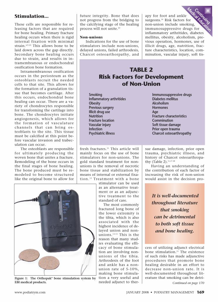
The EBI Bone Healing Systemrequires use of 10 hours a daywhile the Orthofix Physio-Stimrequires three hours a day. CMFprovides magnetic fields througha sinusoidal pattern at 76 Hz thatis superimposed on a constantmagnetic field. The current avail-able model is the OL1000 (DJO,Vista, CA, which requires usage of30 minutes a day.
The use of CMF is based oncalculations that predicted cou-pling to calcium-dependent cellu-
lar signaling processes intissues.48-49 The two differ-ent methods of makingavailable electromagneticwaves at the different Hzfrequencies bring intoquestion which methodis more successful instimulating bone. A studyevaluated the efficacy oflow-frequency electricalf ields on osteogenesisand found that somemethods provide a mag-nitude of power in tissuethat may not be neededto stimulate the desired
Continued on page 173
172 www.podiatrym.comPODIATRY MANAGEMENT • JANUARY 2008
Stimulation...
around patient use. Compli-ance can be an issue since the
device use is recommended for 24hours/day. Also, the electrodesmust be placed on the skin, andcannot be placed over a cast ordressing, and may cause allergicreaction to either the electrode it-self or the conductive gel.
Multiple studies have shownthat the use of capacitive cou-pling for bone stimulation is suc-cessful . Brighton and Pollackfound a 77% rate of solid osseousfusion in 21 long bone non-unions at 22.5 weeks with use of acapacitive coupling device.37 Anevaluation of the use of this de-vice by an independent audit
group showed a successful healingrate of 71% in 534 establishednon-unions that had failed previ-ous treatment, with 65% of thosehealing in less than six months.38
A prospective, double-blind study
Contin
uing
Medica
l Edu
catio
n
Inductive coupling
appears to stimulate
osseous healing
of a non-union by
the differentiation
of fibrocartilage
cells by TGF-beta
expression.
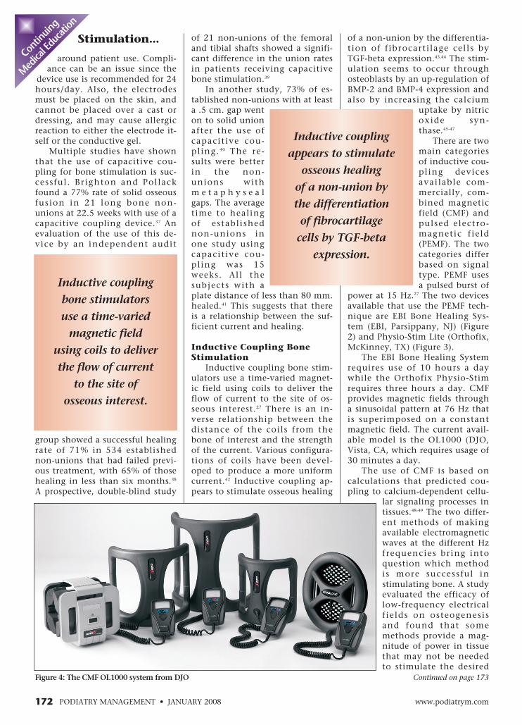
Figure 4: The CMF OL1000 system from DJO
Inductive coupling
bone stimulators
use a time-varied
magnetic field
using coils to deliver
the flow of current
to the site of
osseous interest.

JANUARY 2008 • PODIATRY MANAGEMENTwww.podiatrym.com 173
union reported when com-pared to PEMF used for therecommended time period.52
Prior to FDA approval ofPEMF, it had been shown tohave an efficacy rate in onestudy of 77% fracture non-unions and 82% non-unionsstemming from fai ledarthrodesis.53 Bassett and col-leagues in the same year alsoshowed an overall union rateof 125 tibial delayed unionsor non-unions of 87%.54 A re-view of 44 articles focused onthe effectiveness of PEMF.55
For non-unions of the tibiafractures, 81% healed withPEMF and 82% with surgery.After multiple failed surgeries,the success rate of PEMF is re-ported to be greater than with
surgery; this discrepancy increaseswith additional numbers of priorsurgeries.
In infected non-unions, the re-sults of surgical treatment de-creased by 21% and were lessthan the results utilizing PEMF. Inopen fractures, surgical healingexceeded PEMF whereas in closedinjuries, PEMF cases healed morefrequently. In general, the reviewfound that PEMF treatment of un-united fractures has proved to bemore successful than noninvasivetraditional management, and at
least as effectiveas surgical thera-pies. PEMF treat-ed osteotomiesdemonstrated afaster recovery ofdynamic load-bearing with in-creased loadbearing capacitycompared withthe untreatedgroup. 56 Theb iomechanica lproperties of thehealing osteoto-
my were significantly better inthe PEMF treated group, suggest-ing that not only does it stimulatebone growth, but that the result-ing bone is of high quality andstrength.
In a prospective, randomizedclinical trial evaluating PEMF, 64patients underwent a tr iplearthrodesis or isolated hindfoot
response.50 One study demonstrat-ed that combined magnetic fieldsfor the use of achieving a postero-lateral spine arthrodesis at 30minutes/day was successful.51
The advantage of using an in-ductive coupling device is that itis the most non-invasive form ofbone st imulation avai lable. 28
There is no risk of allergic or sen-sitivity reactionsbecause none ofthe models re-quire the use ofelectrodes orconductive gel.These modelscan be placedover a cast ordressing, whichmakes them easi-er to use anddoes not necessi-tate windowing acast. The disad-vantage to theuse of inductive coupling devicesis patient compliance. Some ofthe devices require a usage perday of up to ten hours. Specifical-ly, the use of a PEMF device forless than the recommended timeperiod has demonstrated a signifi-cant reduction in the efficacy ofbone stimulation in union rates,with approximately 2.3 times less
arthrodesis. There was asignificant reduction inthe t ime required for aunion in the talonavicular andcalcaneocuboid arthrodesis in thePEMF-treated group, and a trendtowards a faster union rate in theSTJ arthrodesis group. This studyexcluded patients at high-risk,specif ical ly those who hadrheumatoid arthritis, diabetesmellitus, or use of corticosteroids,which decreases the strength ofthe study.57
In a retrospective case serieslooking at non-unions after footand ankle arthrodesis, the authorsdo not recommend the use of
PEMF and immobilization as aprotocol for treating delayedunion in foot and ankle arthrode-sis. They hypothesized that themechanical difficulties in orient-ing the coils around the foot andankle may partially explain thelower success compared with longbones. The rate of success wassubstantially lower than that ofuse in long bone delayed unions.16
A study looking at time usageand efficacy of PEMF for fracturenon-unions showed that patientswho used the device less than anaverage of three hours a day had asuccess rate of 35.7%. Patientswho used it more than threehours daily had an 80% successrate.58 This study also showed thatthere was no statistical signifi-cance beyond three hours.
A fairly consistent radiograph-ic progression to union from non-unions treated with PEMF hasbeen described.10,59 The non-uniongap is predicted to widen duringthe first six to eight weeks oftreatment. The theory behind thewidening is that an increase invascular activity occurs along
Continued on page 174
Continuing
Medical Education
Patients who
used it more than
three hours daily
had an 80%
success rate.
In a prospective,
randomized clinical
trial evaluating
PEMF, 64 patients
underwent a triple
arthrodesis or isolated
hindfoot arthrodesis.
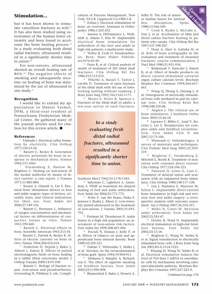
Figure 5: The Exogen® bone stimulatorfrom Smith & Nephew products.
Stimulation...

covery, and to protect articularcartilage.62
PEMF has the ability to perme-ate the articular cartilage and theunderlying subchondral bone.The use of PEMF can be aimed atcontrolling inflammation, stimu-lating the anabolic activity of thechondrocytes, and preventing car-tilage degeneration resulting in achondroprotective effect. The au-thors do not advocate the use ofPEMF on joint inflammation asso-ciated with systemic disease likerheumatoid arthritis. In a differ-ent study, patients who were sta-tus post-arthroscopic chon-droablasion or microfracturetreatment of chondral lesionswere instructed to use PEMF forsix hours a day. 63 The resultsshowed a lower use of non-
steroidal anti-inflammatories anda faster functional recovery.
Ultrasound Bone Stimulation
Ultrasound is the use of acous-tic radiation above the limit ofhuman hearing.64 It is a form ofmechanical energy that can betransmitted into the bodythrough pressure waves.17 Thiscauses a biochemical event at thecellular level. Ultrasound for bonestimulation uses low-intensity,high frequency waves to stimulategrowth.65 The ultrasound modelavailable for bone stimulation isthe Exogen (Smith & Nephew,Memphis, TN) (Figure 5).
The recommended daily use ofExogen is 20 minutes. The pres-ence of metallic implants seemsto have no effect on the efficacyof electromagnetic stimulators.With ultrasound, however, only
one study has shown that tibiafractures fixated with an intra-medullary nail showed no acceler-ation of bone healing both clini-cally and radiographically.66
The mechanism of action forbone growth using ultrasound isnot based on thermal effects.65 Ona micro-level, ultrasound appearsto stimulate the production ofprostaglandin E2. 67 Also, in astudy evaluating rat models, ultra-sound increased the mechanicalproperties of the callus and stimu-lated the production of aggrecanmRNA and procollagen mRNA.68 Acalcium flux also occurs withinseconds of starting the ultrasoundtreatment.64
The effect of ultrasound onbone at a larger level appears tobe on the hypertrophic cartilagecells that produce calcified matrixas well as the osteoblasts that pro-duce the bony collar.65 Ultrasoundused in this way has been foundto not only increase the chondro-cyte population and the soft-cal-lus formation, but also shows ac-celeration of the endochondralossification, and increases thestrength of the fracture site.
In animal studies, the stimula-tion of callus tissue and bonehealing has been apparent withthe use of ultrasound.68-70 Duarte’sstudy showed both radiographicand histologic acceleration ofheal ing in rabbit f ibular os-teotomies made by drill hole.69 Onin vitro mouse metatarsals, low-intensity ultrasound applied for20 minutes daily stimulated thegrowth lengthwise of the calcifieddiaphysis within days. Both thebony collar and the calcified hy-pertrophic cart i lage were in-creased with treatment. This is ei-ther due to an increased numberof osteoblasts or increased activityof the osteoblasts present.65 Scinti-graphic control of the healingprocess was faster in ultrasound-treated animals when comparedto untreated control animals.71
In humans, ultrasound hasbeen shown to accelerate the con-solidation of acute tibia diaphy-seal fractures by 40% both radio-graphically and clinically.72 Notonly does ultrasound appear tostimulate cortical bone growth,
Continued on page 175
174 www.podiatrym.comPODIATRY MANAGEMENT • JANUARY 2008
Stimulation...
with a debridement of thenon-union gap. During the sec-
ond and third months, “gapclouding”, which is the stippledcalcification in the central por-tion of the non-union fibrocarti-lage, occurs.10 Sclerosis appears inthe third and fourth months asthe sharp sclerotic margins of thenon-union become more diffusein appearance due to the vascularinvasion and process of creepingsubstitution. Between the fourthand sixth month, trabecularbridging should be present. Thefinal radiographic healing stage isthe restitution of the intramedu-lary canal.
PEMF will not work if a syn-ovial pseudoarthrosis is present, ifthe fracture exceeds 5 mm., orwhen the fracture is not adequate-ly immobilized.60 Presence of fi-brocartilage is correlated with re-sponsiveness to PEMF leading tobony union. Presence of a fracturegap of dense fibrous tissue lackingthese osteogenic biochemicalmarkers is correlated with unre-sponsiveness to PEMF.61
PEMF and Articular Cartilage
The physiology of articularcartilage is relatively unknown,but it is generally accepted thatthe cartilage has little to no re-generative potential and degradesover time.62 Development of tech-nology to slow the degradation ofcartilage overtime is lacking be-cause of the lack of information.Articular cartilage is composed ofhypocellular, avascular, and alym-phatic tissue. The cartilage con-tains a dense collagen and proteo-glycan matrix which provides alow-friction surface that is resis-tant from wear for both shear andcompressive forces. PEMF hasbeen considered for adjunctivetherapy to treat the inflammationassociated with degenerative jointdisease.
In a study evaluating the effec-tiveness of PEMF in such a situa-tion, the authors concluded thatPEMF can be used following mini-mally-invasive surgery, such asarthroscopy, to control inflamma-tion and enhance functional re-
Contin
uing
Medica
l Edu
catio
n
PEMF has
the ability to
permeate the
articular cartilage
and the underlying
subchondral
bone.
JANUARY 2008 • PODIATRY MANAGEMENTwww.podiatrym.com 175
cations of Fracture Management. NewYork, NY:J.B. Lippincott Co;1984:1-8.
10 Zoltan J. Electrical stimulation ofbone: an overview. Seminars in Ortho-pedics 1986;1:242-252.
11 Saxena A, DiDomenico L, Widt-feldt A, Adams T, Kim W. Implantableelectrical bone stimulation forarthrodesis of the foot and ankle inhigh risk patients: a multicenter study.
12 Weber B, Cezh O. Pseudoarthro-sis. Bern: Hans Huber Publish-ers;1976:45-50.
13 Puno R, et al. Critical analysis ofresults of treatment of 201 tibial shaftfractures. Clin Orthop Rel Res1986;212:113-213.
14 Whittle A, Russel T, Taylor J,Lavelle D. Treatment of open fracturesof the tibial shaft with the use of inter-locking nailing without reaming. JBone Joint Surg 1992;74A:1162-1171.
15 Moore S, Storts R, Spencer J.Fractures of the tibial shaft in adults: aten-year survey of such fractures.
Southern Med J 1962;55:1178-1183.16 Saltzman C, Lightfoot A, Amen-
dola A. PEMF as treatment for delayedhealing of foot and ankle arthrodesis.Foot Ankle Int 2004;25:771-773.
17 Nolte P, van der Krans, Patka P,Janssen I, Ryaby J, Albers G. Low-inten-sity pulsed ultrasound in the treatmentof non-unions. J Trauma 2001;51:693-703.
18 Perlman M, Thordarson D. Anklefusion in a high risk population: an as-sessment of non-union risk factors.Foot Ankle Int 1999;208:491-496.
19 Pocock N, Eisman J, Kelly P, etal. Effects of tobacco on axial and ap-pendicular bone mineral density. Bone1989;10:329-331.
20 Daftari T, Whitesides T, Heller J,et al. Nicotine on the revascularizationof bone graft. Spine 1994;19:904-911.
21 Ishikawa S, Murphy A, Richard-son E. The effect of cigarette smokingon hindfoot fusions. Foot Ankle2002;2311:996-998.
22 Blumenthal S, Baker J, Dossett A,
but it has been shown to stimu-late cancellous fractures as well.73
It has also been studied using os-teotomies of the human lower ex-tremity and been found to accel-erate the bone healing process.74
In a study evaluating fresh distalradial fractures, ultrasound result-ed in a significantly shorter timeto union.64
For non-unions, ultrasoundshowed an overall healing rate of86%.65 The negative effects ofsmoking and subsequently nico-tine on healing of bone was mini-mized by the use of ultrasound inone study.75
RecognitionI would like to extend my ap-
preciat ion to Daniel Yarmel,DPM, a third-year resident atPennsylvania Presbyterian Medi-cal Center. He gathered many ofthe journal articles used for cita-tion for this review article. ■
References1 Yashuda I. Electrical callus forma-
tion by electricity. Clin Orthop1977;124:53-56.
2 Bassett C, Becker R. Generationof electric potentials by bone in re-sponse to mechanical stress. Science1962;137:1063.
3 Friedenberg Z, Harlow M,Brighton C. Healing on non-union ofthe medial malleolus by means of di-rect current: a case report. J Trauma1971;11:883-884.
4 Kesani A, Ghandi A, Lin S. Elec-trical bone stimulation devices in footand ankle surgery: types of devices, sci-entific basis, and clinical indicationsfor their use. Foot Ankle Int2006;27:149-156.
5 Bassett C, Hermann L. Influenceof oxygen concentration and mechani-cal factors on differentiation of con-nective tissues in vitro. Nature1961;190:460.
6 Bassett C. Electrical effects inbone. Scientific American 1965;213:18.
7 Bassett C, Pawluk R, Becker R. Ef-fects of electric currents on bone invivo. Nature 1964;204:652-654.
8 Fredericks D, Nepola J, Baker J,Abbott J, Simon B. Effects of pulsedelectromagnetic fields on bone healingin a rabbit tibial osteotomy model. JOrthop Trauma 2000;14:93-100.
9 Bassett C. Biology of fracture re-pair, non-union and pseudoarthrosis.In:Gossling H, Pillsbury S, eds. Compli-
Stimulation... Selby D. The role of anteri-or lumbar fusion for internaldisc disruption. Spine1988;13:566-569.
23 Cook S, Ryaby J, McCabe J,Frey J, et al. Acceleration of tibia anddistal radius fracture healing in pa-tients who smoke. Clin Orthop Rel Res1997;337:198-207.
24 Desai A, Alavi A, Galinka M, etal. Role of bone scintigraphy in theevaluation and treatment of ununitedfractures: concise communication. JNucl Med 1980;21:931-934.
25 Bodamyali T, Kanczler J, SimonB, et al. Effect of faradic products ondirect current-stimulated calvarialorgan culture calcium levels. BiochemBiophys Res Commun 1999;264:657-661.
26 Wang Q, Zhong S, Ouyang J, etal. Osteogenesis of electrically stimulat-ed bone cells mediated in part by calci-um ions. Clin Orthop Relat Res1998;348:259-68.
27 Anglen J. The clinical use ofbone stimulators. J Southern OrthoAssoc 2003;12:46-54.
28 Liporace F, Bibbo C, Azad V, Ko-erner J, Lin S. Bioadjuvants for com-plex ankle and hindfoot reconstruc-tion. Foot Ankle Clin N Am2007;12:75-106.
29 Weinraub G. Orthobiologics: Asurvey of materials and techniques.Clin Podiatr Med Surg 2005;22:509-519.
30 Brighton C, Friedenberg Z,Mitchell E, Booth R. Treatment of non-union with constant direct current.Clin Orthop 1977;124:106-123.
31 Paterson D, Lewis G, Cass C.Treatment of delayed union and non-union with an implanted bone stimu-lator. Clin Orthop 1980;148:117-128.
32 Lau J, Stamatos E, Myerson M,Schon L. Implantable direct-currentbone stimulators in high risk and revi-sion foot and ankle surgery: a retro-spective analysis with outcome assess-ment. Am J Orthop 2007;36:354-357.
33 Midis N, Conti SF. Revisionankle arthrodesis. Foot Ankle Int2002;23:243-47.
34 Donley B, Ward D. Implantableelectrical stimulation in high-risk hind-foot fusions. Foot Ankle Int2002;23:13-18.
35 Brighton C, Wang W, Seldes R,et al. Signal transduction in electricallystimulated bone cells. J Bone Joint SurgAm 2001;83-A:1514-1523.
36 Khuang H, Wang W, Seldes R, etal. Electrical stimulation induces thelevel of TGF-beta 1 mRNA in osteoblas-tic cells by mechanism involving calci-um/calmodulin pathway. Biochem Bio-phys Res Commun 1997;237:225-9.
Continued on page 176
Continuing
Medical Education
In a study
evaluating fresh
distal radial
fractures, ultrasound
resulted in a
significantly shorter
time to union.

York: Plenum Press, 1987:97-108.50 McLeod K, Rubin C, Brook S.
The effect of low-frequency electricalfields on osteogenesis. J Bone JointSurg 1992;74A:920-929.
51 Linovitz R, Pathria M, BernhardtM, et al. Combined magnetic fields ac-celerate and increase spine fusion. Adouble-blind, randomized, placebo-controlled study. Spine 2002;27:1383-1388.
52 Garland DE, Moses B, Salyer W.Long-term follow-up of fracture non-unions treated with PEMF. ContempOrthop 1991;22:295-302.
53 Bassett C, Mitchell S, Gaston S.Pulsing electromagnetic field treatmentin ununited fractures and failedarthrodesis. JAMA 1982;247:623-628.
54 Bassett C, Mitchell S, Schink M.Treatment of therapeutically resistantnon-unions with bone grafts and puls-ing electromagnetic fields. J Bone JointSurg 1982;64-A:1214-1220.
55 Gossling H, Berstein R, Abbott J.Treatment of ununited tibial fractures:a comparison of surgery and pulsedelectromagnetic fields (PEMF). Ortho-pedics 1992;15:711-719.
56 Inoue N, Ohnishi I, Chen D, etal. Effect of pulsed electromagneticfields (PEMF) on late phase osteotomygap healing in a canine tibial model. JOrthop Res 2002;20:1106-1114.
57 Dhawan S, Conti S, Towers J, etal. The effect of pulsed electromagneticfields on hindfoot arthrodesis: a pro-spective study. J Foot Ankle Surg2004;43:93-6.
58 Garland D, Moses B, Salyer W.Long-term follow-up of fracture non-unions treated with PEMFs. ContemOrthop 1991;22:295-302.
59 Giurini J. Healing fractures withpulsed magnetic fields. Cont Pod Phy-sician 1991:41-45.
60 Bassett C. The electrical manage-ment of ununited fractures. In:Gossling H, Pillsbury S, eds. Complica-tions of fracture management. NewYork, NY: J.B.Lippincott Co; 1984:9-39.
61 Gossling H, Kromplinger W,Broom M. Fracture gap biopsy as a pre-dictor of response to PEMF. In: Currentstatus of electricity in the clinical sci-ences. April 1985:18-19.
62 Massari L, Benazzo F, De MatteiM, Setti S, Fini M. Effects of electricalphysical stimuli on articular cartilage. JBone Joint Surg 2007;89(suppl 3):152-161.
63 Zorzi C, Dall’oca C, Cadossi R,Setti S. Effects of pulsed electromagnet-ic fields on patients’ recovery afterarthroscopic surgery: prospective, ran-domized and double-blind study. KneeSurg Sports Traumatol Arthrosc 2007.
64 Kristiansen T, Ryaby J, McCabe J,Frey J, Roe L. Accelerated healing of
distal radial fractures with the use ofspecific, low-intensity ultrasound. Amulticenter, prospective, randomized,double-blind, placebo-controlled study.J Bone Joint Surg 1997:79A:961-973.
65 Nolte P, Klein-Nulend G, AlbersR, Semeins C, Goie S, Burger E. Low-in-tensity ultrasound stimulates endo-chondral ossification in vitro. J OrthopRes 2001;19:301-307.
66 Emani A, Petren-Mallmin M,Larsson S. No effect of low intensity ul-trasound on healing time of in-tramedullary fixed tibia fractures. J Or-thop Trauma 1999;13:252-257.
67 Tsai C, Chang W, Liu T, Song G.Ultrasonic effect on fracture repair andprostaglandin E2 production. Chin JPhysiol 1992;35:27-34.
68 Yang K, Parvizi J , Wang S,Lewallen D, et al. Exposure to low-in-tensity ultrasound increases aggrecangene expression in a rat femur fracturemodel. J Orthop Res 1996;14:802-809.
69 Duarte L. The stimulation ofbone growth by ultrasound. Arch Or-thop Trauma Surg 1983;101:153-159.
70 Pilla A, Mont M, Nasser P, KahnS, et al. Non-invasive low-intensitypulsed ultrasound accelerates bonehealing in the rabbit. J Orthop Trauma1990;4:246-253.
71 Klug W, Franke W, Knoch H.Scintigraphic control of bone-fracturehealing under ultrasonic stimulation:an animal experimental study. Eur JNucl Med 1986;11:494-497.
72 Heckman J, Ryaby J, McCabe J,Frey J, Kilcoyne R. Acceleration of tib-ial fracture healing by non-invasive,low-intensity pulsed ultrasound. J BoneJoint Surg 1994;76-A:26-34.
73 Kristiansen T. The effect of lowpower specifically programmed ultra-sound on the healing time of freshfractures using a Colles’ model. J Or-thop Trauma 1990;4:227-228
74 Nolte P, Maas M, Van Dijk C, Al-bers G. The effect of ultrasound on thebone healing time in osteotomies.SICOT 1999:0081.
75 Cook S, Ryaby J, McCabe J, FreyJ, Heckman J, et al. Acceleration oftibia and distal radius fracture healingin patients who smoke. Clin Orthop1997;337:198-207.
176 www.podiatrym.comPODIATRY MANAGEMENT • JANUARY 2008
Stimulation...37 Brighton C, Pollack S. Treat-
ment of recalcitrant non-unionwith a capacitively coupled electrical
field. A preliminary report. J Bone JointSurg 1985;67-A:577-585.
38 Phipps Associates, an Indepen-dent Audit Control Group. An efficacysurvey of bone growth stimulation uti-lizing capacitive coupling technology.Bioelectron Inc, Hackensack, NJ 1992.
39 Scott G, King J. A prospective,double-blind trial of electrical capacita-tive coupling in the treatment of non-union of long bones. J Bone Joint Surg1994;76-A:820-826.
40 Zamora-Navas P, Verdera B,Lorenzo A, et al. Electrical stimulationof bone non-union with the presenceof a gap. Acta Orthop 1995;61:169-176.
41 Abeed R, Naseer M, Abel E. Ca-pacitively coupled electrical stimula-tion treatment: results from patientswith failed long bone fracture unions. JOrthop Trauma 1998;12:510-513.
42 Giurini J. Healing fractures withpulsed magnetic fields. Cont Pod Phy-sician 1991:41-45.
43 Guerkov H, Lohmann C, Liu Y,et al. Pulsed electromagnetic fields in-crease growth factor release by non-union cells. Clin Orthop Relat Res2001;384:265-279.
44 Aaron R, Wang S, Ciombor D.Upregulation of basal TGFbeta1levelsby EMF coincident with chondrogene-sis-implications for skeletal repair andtissue engineering. J Orthop Res2002;20:233-240.
45 Spadero J, Bergstrom W. In vivoand in vitro effects of a pulsed electro-magnetic field on net calcium flux inrat calvarial bone. Calcif Tissue Int2002;70:496-502.
46 Bodamyali T, Bhatt B, Hughes F,et al. Pulsed electromagnetic fields si-multaneously induce osteogenesis andupregulate transcription of bone mor-phogenetic proteins 2 and 4 in rat os-teoblasts in vitro. Biochem Biophys ResCommun 1998;250:458-61.
47 Diniz P, Shomura K, Soejima K,et al. Effects of pulsed electromagneticfield (PEMF) stimulation on bone tis-sue-like formation are dependent onthe maturation stages of the os-teoblasts. Bioeletromagnetics2002;23:398-405.
48 Fitzsimmons R, Ryaby J, MageeF, et al. Combined magnetic fields in-creased net calcium flux in bone cells.Calcif Tissue Int 1994;55:376-380.
49 McLeod B, Liboff A. Cyclotronresonance in cell membranes: the theo-ry of the mechanism. In: Blank M,Findl E, eds. Mechanistic approaches tointeractions of electric and electromag-netic fields with living systems. New
Contin
uing
Medica
l Edu
catio
n
Dr. Rabjohn is an associate at the Ar-lington/Mans-field Foot andAnkle Center inA r l i n g t o n ,Texas. She com-pleted her resi-dency at TheGraduate Hospi-tal of Philadel-phia, Pennsylva-nia.
JANUARY 2008 • PODIATRY MANAGEMENTwww.podiatrym.com 177
B) Established non-unionsC) Epiphyseal platesD) All of the above
7) Which type of non-union ismost likely to obtain a beneficialresult with the use of a bonestimulator?
A) OligotrophicB) PseudoarthrosisC) AtrophicD) Hypertrophic
8) Which type of non-union canbe defined as having a well-vas-cularized callus with no calcifica-tion between bone fragments?
A) HypertrophicB) OligotrophicC) AtrophicD) Pseudoarthrosis
9) Smoking causes which of thefollowing effects on tissue healing?
A) Tissue anoxiaB) VasodilationC) Both a and bD) Neither a or b
10) Which form of bone stimula-tion will be successful in a truesynovial pseudoarthrosis?
A) Direct currentB) Inductive couplingC) UltrasoundD) None of the above
11) A technetium 99 bone scancan be used to detect a “coldcleft” which is indicative of thepresence of a pseudoarthrosis.Which of the following best de-scribes the “cold cleft?”
A) Increased activity at thesite of non-union surroundedby increased radioactivityB) Decreased activity at the siteof non-union surrounded by
1) Which form of bone stimula-tion uses electromagnetic fieldsto deliver electricity to bone?
A) Capacitive couplingB) Inductive couplingC) Direct currentD) Ultrasound
2) Which form of bone stimula-tion uses electrodes that are di-rectly implanted on bone?
A) Inductive couplingB) Capacitive couplingC) Direct currentD) Ultrasound
3) Which form of bone stimula-tion uses acoustic radiation totransmit mechanical energy inthe form of pressure waves tobone?
A) Inductive couplingB) Capacitive couplingC) Direct currentD) Ultrasound
4) Which form of bone stimula-tion uses charged plates toconduct electricity acrossbone?
A) Inductive couplingB) Capacitive couplingC) Direct currentD) Ultrasound
5) Which portion of the directcurrent technology is placed indirect contact with the bone?
A) the titanium cathodeB) the anode batteryC) bothD) neither
6) In which of the followingconditions would one expectnot to find electronegativecharges on bone?
A) Fractures
increased radioactivityC) Decreased activity at thesite of non-union surroundedby increased radioactivityD) Increased activity at thesite of non-union surroundedby decreased radioactivity
12) Which form of bone stimula-tion requires not only a primarysurgical implantation but also apossible secondary surgery forremoval?
A) Direct currentB) Inductive couplingC) Capacitive couplingD) Ultrasound
13) Which type of inductive cou-pling uses a sinusoidal pattern ofmagnetic energy superimposedon a constant magnetic field?
A) Direct currentB) PEMF (pulsed electromag-netic fields)C) CMF (constant magneticfields)D) Both b and c
14) Which form of bone stimu-lation, by the instructions givenby the manufacturer, has a rec-ommended usage of less thanone hour?
A) UltrasoundB) PEMFC) CMFD) Both a and c
15) Which bone is the most com-monly fractured long bone ofthe lower extremity and also hasa high risk of non-union?
A) FemurB) FibulaC) TibiaD) Metatarsal
Continuing
Medical Education
E X A M I N A T I O N
See answer sheet on page 179.
Continued on page 178
178 PODIATRY MANAGEMENT
16) Which of the following bone stimulatorsneeds direct contact with skin to beeffective?
A) PEMFB) CMFC) UltrasoundD) All of the above
17) Which of the following is the bestdescription of Wolff’s Law?
A) When possible, primary bone healingshould be attempted on fracturesB) Form follows function and bone willadapt to stresses placed on itC) All fractures should be immobilizedD) Both a and c
18) Which of the following is the definitionof primary fracture healing?
A) Providing minimal to moderate strainsto accelerate the rate of healingB) The bone healing process that includesintramembranous and endochondralossificationC) Providing minimal strain and rigidinternal fixation across fracture sitesD) All of the above
19) The “gold standard” of treatment for anon-union includes all the following except:
A) Removal of all necrotic boneB) Immediate placement of a directcurrent bone stimulatorC) Stabilization of the fracture withinternal or external fixationD) All of the above are part of the “goldstandard” treatment.
20) The risk factors for the development of anon-union include all of the following except:
A) Soft tissue damageB) InfectionC) Psychiatric illnessD) Rigid fixation
E X A M I N A T I O N
(cont’d)
See answer sheet on page 179.
Contin
uing
Medica
l Edu
catio
n
PM’sCPME Program
Welcome to the innovative Continuing EducationProgram brought to you by Podiatry ManagementMagazine. Our journal has been approved as asponsor of Continuing Medical Education by theCouncil on Podiatric Medical Education.
Now it’s even easier and more convenientto enroll in PM’s CE program!
You can now enroll at any time during the yearand submit eligible exams at any time during yourenrollment period.
PM enrollees are entitled to submit ten examspublished during their consecutive, twelve–monthenrollment period. Your enrollment period beginswith the month payment is received. For example,if your payment is received on September 1, 2006,your enrollment is valid through August 31, 2007.
If you’re not enrolled, you may also submit anyexam(s) published in PM magazine within the pasttwelve months. CME articles and examinationquestions from past issues of Podiatry Man-agement can be found on the Internet athttp://www.podiatrym.com/cme. Each lessonis approved for 1.5 hours continuing education con-tact hours. Please read the testing, grading and pay-ment instructions to decide which method of partici-pation is best for you.
Please call (631) 563-1604 if you have any ques-tions. A personal operator will be happy to assist you.
Each of the 10 lessons will count as 1.5 credits;thus a maximum of 15 CME credits may beearned during any 12-month period. You may se-lect any 10 in a 24-month period.
The Podiatry Management Magazine CMEprogram is approved by the Council on PodiatricEducation in all states where credits in instruction-al media are accepted. This article is approved for1.5 Continuing Education Contact Hours (or 0.15CEU’s) for each examination successfully completed.
www.podiatrym.com
Home Study CME credits nowaccepted in Pennsylvania
Over, please
Please print clearly...Certificate will be issued from information below.
Name _______________________________________________________________________Soc. Sec. #______________________________Please Print: FIRST MI LAST
Address_____________________________________________________________________________________________________________
City__________________________________________________State_______________________Zip________________________________
Charge to: _____Visa _____ MasterCard _____ American Express
Card #________________________________________________Exp. Date____________________
Note: Credit card is the only method of payment. Checks are no longer accepted.
Signature__________________________________Soc. Sec.#______________________Daytime Phone_____________________________
State License(s)___________________________Is this a new address? Yes________ No________
Check one: ______ I am currently enrolled. (If faxing or phoning in your answer form please note that $2.50 will be charged to your credit card.)
______ I am not enrolled. Enclosed is my credit card information. Please charge my credit card $20.00 for each exam submitted. (plus $2.50 for each exam if submitting by fax or phone).
______ I am not enrolled and I wish to enroll for 10 courses at $139.00 (thus saving me $61 over the cost of 10 individual exam fees). I understand there will be an additional fee of $2.50 for any exam I wish to submit via fax or phone.
Note: If you are mailing your answer sheet, you must completeall info. on the front and back of this page and mail with yourcredit card information to: Podiatry Management, P.O. Box490, East Islip, NY 11730.
TESTING, GRADING AND PAYMENT INSTRUCTIONS(1) Each participant achieving a passing grade of 70% or
higher on any examination will receive an official computer formstating the number of CE credits earned. This form should be safe-guarded and may be used as documentation of credits earned.
(2) Participants receiving a failing grade on any exam will benotified and permitted to take one re-examination at no extra cost.
(3) All answers should be recorded on the answer formbelow. For each question, decide which choice is the best an-swer, and circle the letter representing your choice.
(4) Complete all other information on the front and back ofthis page.
(5) Choose one out of the 3 options for testgrading: mail-in,fax, or phone. To select the type of service that best suits yourneeds, please read the following section, “Test Grading Options”.
TEST GRADING OPTIONSMail-In GradingTo receive your CME certificate, complete all information
and mail with your credit card information to:Podiatry Management
P.O. Box 490, East Islip, NY 11730There is no charge for the mail-in service if you have already
enrolled in the annual exam CPME program, and we receive this
E N R O L L M E N T F O R M & A N S W E R S H E E T
✄
179
Continuing
Medical Education
exam during your current enrollment period. If you are not en-rolled, please send $20.00 per exam, or $139 to cover all 10 exams(thus saving $61* over the cost of 10 individual exam fees).
Facsimile GradingTo receive your CPME certificate, complete all information and
fax 24 hours a day to 1-631-563-1907. Your CPME certificate willbe dated and mailed within 48 hours. This service is available for$2.50 per exam if you are currently enrolled in the annual 10-examCPME program (and this exam falls within your enrollment period),and can be charged to your Visa, MasterCard, or American Express.
If you are not enrolled in the annual 10-exam CPME pro-gram, the fee is $20 per exam.
Phone-In GradingYou may also complete your exam by using the toll-free ser-
vice. Call 1-800-232-4422 from 10 a.m. to 5 p.m. EST, Mondaythrough Friday. Your CPME certificate will be dated the same dayyou call and mailed within 48 hours. There is a $2.50 charge forthis service if you are currently enrolled in the annual 10-examCPME program (and this exam falls within your enrollment peri-od), and this fee can be charged to your Visa, Mastercard, Ameri-can Express, or Discover. If you are not currently enrolled, the feeis $20 per exam. When you call, please have ready:
1. Program number (Month and Year)2. The answers to the test3. Your social security number4. Credit card information
In the event you require additional CPME information,please contact PMS, Inc., at 1-631-563-1604.
Enrollment/Testing Informationand Answer Sheet
✄
180 www.podiatrym.comPODIATRY MANAGEMENT • JANUARY 2008
E N R O L L M E N T F O R M & A N S W E R S H E E T (cont’d)Con
tinuin
g
Medica
l Edu
catio
n
LESSON EVALUATION
Please indicate the date you completed this exam
_____________________________
How much time did it take you to complete the lesson?
______ hours ______minutes
How well did this lesson achieve its educational objectives?
_______Very well _________Well
________Somewhat __________Not at all
What overall grade would you assign this lesson?
A B C D
Degree____________________________
Additional comments and suggestions for future exams:
__________________________________________________
__________________________________________________
__________________________________________________
__________________________________________________
__________________________________________________
__________________________________________________
EXAM #1/08Electrical Stimulation of Bone:
The Evolving Technology(Rabjohn)
1. A B C D
2. A B C D
3. A B C D
4. A B C D
5. A B C D
6. A B C D
7. A B C D
8. A B C D
9. A B C D
10. A B C D
11. A B C D
12. A B C D
13. A B C D
14. A B C D
15. A B C D
16. A B C D
17. A B C D
18. A B C D
19. A B C D
20. A B C D
Circle: