Embed Size (px)
Citation preview
Jarrett and Dawood
for polymorphonuclear leukocytes. Prostaglandins 1978; 16:239.
15. Castor CW. Connective tissue activation. VII. Evidence supporting a role for prostaglandins and cyclic nucleotides. J Lab Clin Med 1975;85:392.
December 1986 Am J Obstet Gynecol
16. Hunt TK, Van Winkle W Jr. Fundamentals of wound management in surgery. Wound healing: normal repair. South Plainfield, New Jersey: Chirurgecom, 1976.
Electrical and mechanical uterine activity and gap junctions in
estrogen-treated oophorectomized sheep
A. Verhoef£, M.D., Ph.D., R. E. Garfield, Ph.D.,J. Ramondt, M.D., and H. C. S. Wallenburg, M.D., Ph.D.
Rotterdam, The Netherlands, and Hamilton, Ontario, Canada
The effects of a single dose (0.1 mg) of 17~-estradiol on myometrial electrical activity, intrauterine pressure, and myometrial gap junctions were studied in six chronically instrumented oophorectomized
ewes. The intrauterine pressure cycles were analyzed for the maximum rate of rise, peak pressure, and active pressure area, together with the frequency of the cycles and the burst frequency, and related to the
gap junction area. 17~-estradiol temporarily depressed uterine activity. The period of quiescence was followed by a pronounced increase in all variables of electrical and mechanical activity and also by a rise in gap junction area. The greatest increase in the maximum rate of rise of the intrauterine pressure cycles occurred 24 hours after estrogen and was associated with the maximum increase in gap junction area.
These results indicate that a single dose of 17~-estradiol induces formation of myometrial gap junctions, which may facilitate the spread of electrical activity over the myometrium and improve coordination of
uterine contractility. (AM J 0BSTET GYNECOL1986;155:1192-6.)
Key words: Uterine activity, gap junctions, estrogen treatment, oophorectomy
Gap junction formation during labor has been suggested to be a necessary step in facilitating coordination of contractile forces in the myometrium. 1 In a previous study in sheep we showed that the rise in the number of gap junctions during parturition is related to increased coordination of myometrial activity! The formation of gap junctions is thought to be regulated by steroid hormones and prostaglandins. 3 In sheep the number of gap junctions is low during pregnancy; during parturition it shows a rise, which appears to be related to an increase in the estrogen/progesterone ratio in plasma.2 A recent study in rats suggests that these
From the Department of Obstetrics and Gynecology, Erasmus University Medical School, Rotterdam, and the Departments of Neurosciences and Obstetrics and Gynecology, McMaster University Health Sciences Centre, Hamilton.
This work was supported by a grant from theM edical Research Council of Canada and from the Netherlands Organization for the Advancement of Pure Research-ZWO.
Presented in part at the Thirty-first Annual Meeting of the Society for Gynecologic Investigation, San Francisco, California. March 21-24, 1984.
Reprint requests: H. C. S. Wallenburg, M.D., Ph.D., Department of Obstetrics and Gynecology, Erasmus University Medical School EE 2283, P. 0. Box 1738, 3000 DR Rotterdam, The Netherlands.
1192
changes in circulating steroid hormone concentrations may lead to an increase in the level of estradiol nuclear receptors and to gap junction formation by nuclear receptor-mediated transcription and protein synthesis!
In nonpregnant oophorectomized ewes administration of 17[3-estradiol induces an initial decrease in myometrial activity, followed by an increase.5
• 6 Together
with the increase in frequency of intrauterine pressure cycles, the shape of the cycles is also changed after administration of 17[3-estradiol, as shown by an increased rate of rise of the intrauterine pressure cycles.7
The aim of the present study was to investigate whether the increase in myometrial activity after the administration of 17[3-estradiol is related to gap junction formation. The chronically instrumented nonpregnant ewe is a useful model to investigate the regulation of myometrial gap junction formation and the influence of gap junctions on myometrial contractility, as it allows a much easier manipulation of the factors that seem to control gap junction formation than does the pregnant animal. In this study we investigated electrical and mechanical myometrial activity in oophorectomized ewes before and after administration of a sin-
Volume 155 Number 6
1mV[ o•
100 [ mmH:
2.5mV[ t H
... •• Ill
~ n ~ ' r
~ tl lfil"' Ill• IH ~·
0 hrs
IHtl 11111111!1
100 [ 24 h<S mmHg . . . 0 -----~------~--·
2.5mV[--frtJ ..... j ~~ ·~ II -~ II II tl ~
1min
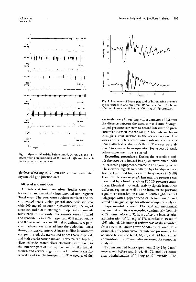
Fig. I. Myometrial activity before and 6, 24, 48, 72, and 144 hours after administration of 0.1 mg of 17[3-estradiol at 0 hours, recorded in one ewe.
gle dose of O.I mg of I713-estradiol and we quantitated myometrial gap junction area.
Material and methods
Animals and instrumentation. Studies were performed in six chronically instrumented nonpregnant Texel ewes. The ewes were oophorectomized and instrumented while under general anesthesia induced with 500 mg of ketamine hydrochloride, 0.5 mg of atropine, and 300 to 500 mg of thiopental sodium administered intravenously. The animals were intubated and ventilated with 40% oxygen and 60% nitrous oxide and 0.5 to 4 volumes per 100 ml of enflurane. A polyvinyl catheter was inserted into the abdominal aorta through a femoral artery. A lower midline laparotomy was. performed, the uterus and adnexa were exposed, and both ovaries were removed. Three pairs of bipolar, silver chloride-coated silver electrodes were fixed to the anterior part of the myometrium in the fundal, medial, and cervical regions of both uterine horns for recording of the electromyogram. The needles of the
Uterine activity and gap junctions in sheep 1193
120
0 ·e 0 90 M
1 >. u 0 60 ~ ~ C]'
! 30
0 90 ·e 0 M
>- 60 u 0 ~ ~
30 C]'
! -12 12 24 36 48 60
hours
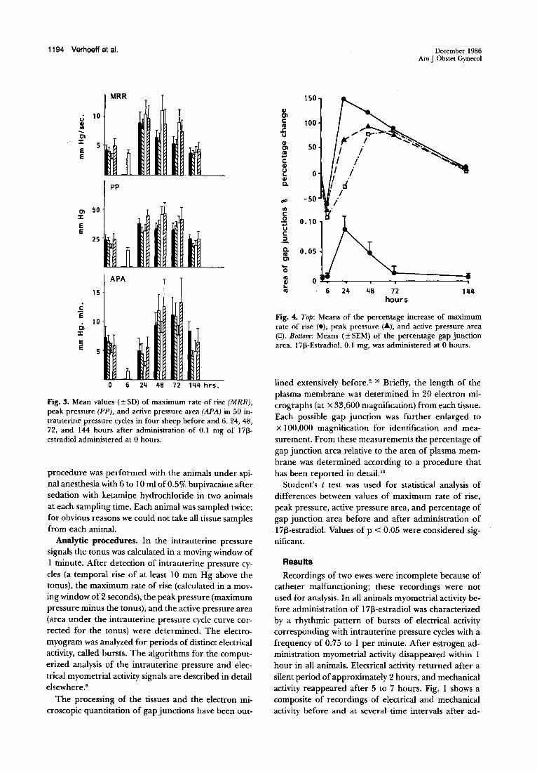
Fig. 2. Frequency of bursts (top) and of intrauterine pressure cycles (bottom) in one ewe from 12 hours before to 72 hours after administration (0 hours) of 0.1 mg of 17[3-estradiol.
electrodes were 3 mm long with a diameter of 0.2 mm; the distance between the needles was 2 mm. Spongetipped pressure catheters to record intrauterine pressure were inserted into the cavity of both uterine horns through a small incision in the cervical region. The wires and catheters were passed subcutaneously to a pouch attached to the ewe's flank. The ewes were allowed to recover from operation for at least I week before experiments were started.
Recording procedures. During the recording periods the ewes were housed in a quiet environment, with the recording equipment situated in an adjoining room. The electrical signals were filtered by a band-pass filter. For the lower and higher cutoff frequencies (- 3 dB) I and 30 Hz were selected. Intrauterine pressure was measured by a Gould Statham P23 ID pressure transducer. Electrical myometrial activity signals from three different regions as well as one intrauterine pressure signal were recorded on a Gould Brush eight-channel polygraph with a paper speed of 25 mm · min- 1 and stored on magnetic tape for off-line computer analysis.
Experimental protocol. Electrical and mechanical myometrial activity was recorded continuously from I6 to 24 hours before to 72 hours after the intra-arterial administration of O.I mg of I713-estradiol in I 0 ml of 10% ethanol. Myometrial activity was again recorded from I40 to I 50 hours after the administration of I713-estradiol. Fifty consecutive intrauterine pressure cycles obtained before and 6, 24, 48, 72, and I44 hours after administration of I713-estradiol were used for computer analysis.
Two myometrial biopsy specimens (5 by 2 by I mm) were taken before and 6, 24, 48, 72, and I44 hours after administration of 0.1 mg of 1713-estradiol. The
1194 Verhoeff et al.
MRR
u 10 8! -01
::1: 5 E
] E
PP
01 50 ::1:
E E
25
~ APA
15
c: ·e 10
01 ::1:
e e 5
0 6 24 48 72 144 hrs.
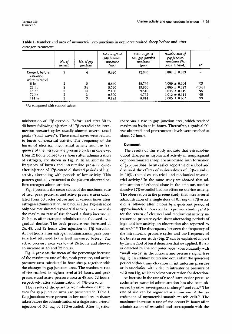
Fig. 3. Mean values (±SD) of maximum rate of rise (MRR), peak pressure (PP), and active pressure area (APA) in 50 intrauterine pressure cycles in four sheep before and 6, 24, 48, 72, and 144 hours after administration of 0.1 mg of 1713-estradiol administered at 0 hours.
procedure was performed with the animals under spinal anesthesia with 6 to 10 ml of0.5% bupivacaine after sedation with ket;amine hydrochloride in two animals at each sampling time. Each animal was sampled twice; for obvious reasons we could not take all tissue samples from each animal.
Analytic procedures. In the intrauterine pressure signals the tonus was calculated in a moving window of 1 minute. After detection of intrauterine pressure cycles (a temporal rise of at least 10 mm Hg above the tonus), the maximum rate of rise (calculated in a moving window of 2 seconds), the peak pressure (maximum pressure minus the tonus), and the active pressure area (area under the intrauterine pressure cycle curve corrected for the tonus) were determined. The electromyogram was analyzed for periods of distinct electrical activity, called bursts. The algorithms for the computerized analysis of the intrauterine pressure and electrical myometrial activity signals are described in detail elsewhere.8
The processing of the tissues and the electron microscopic quantitation of gap junctions have been out-
cu en r::: Ia J: u cu en Ia ... r::: cu u ... cu 0.
dP
Ill r::: .2 ... u
r::: .2.. 0. Ia en ... 0 Ia cu ... Ia
150
100
50
0
-50
0.10
0.05
0 6 24 48 72
hours
December 1986 Am J Obstet Gynecol
144
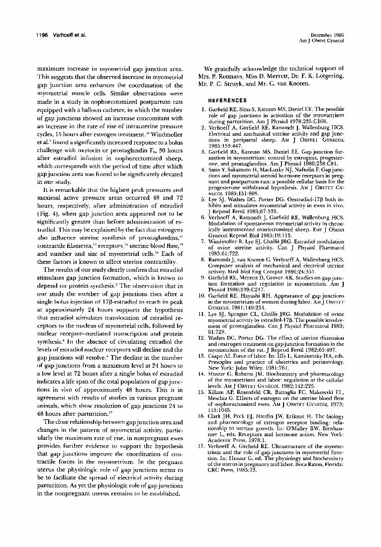
Fig. 4. Top: Means of the percentage increase of maximum rate of rise (e), peak pressure (A); and active pressure area (D). Bottom: Means ( ± SEM) of the percentage gap junction area. 1713-Estradiol, 0.1 mg, was administered at 0 hours.
lined extensively before.9•
10 Briefly, the length of the plasma membrane was determined in 20 electron micrographs (at X 33,600 magnification) from each tissue. Each possible gap junction was further enlarged to x 100,000 magnification for identification and measurement. From these measurements the percentage of gap junction area relative to the area of plasma membrane was determined according to a procedure that has been reported in detail. 10
Student's t test was used for statistical analysis of differences between values of maximum rate of rise, peak pressure, active pressure area, and percentage of gap junction area before and after administration of 17~-estradiol. Values of p < 0.05 were considered significant.
Results
Recordings of two ewes were incomplete because of catheter malfunctioning; these recordings were not used for analysis. In all animals myometrial activity before administration of 17~-estradiol was characterized by a rhythmic pattern of bursts of electrical activity corresponding with intrauterine pressure cycles with a frequency of 0.75 to 1 per minute. After estrogen administration myometrial activity disappeared within 1 hour in all animals. Electrical activity returned after a silent period of approximately 2 hours, and mechanical activity reappeared after 5 to 7 hours. Fig. 1 shows a composite of recordings of electrical and mechanical activity before and at several time intervals after ad-
Volume 155 Number 6
Uterine activity and gap junctions in sheep 1195
Table I. Number and area of myometrial gap junctions in oophorectomized sheep before and after estrogen treatment
Total length of Total length of Relative area of gap junction non-gap junction gap junction
membrane (%, No. of No. of gap membrane animals junctions (mm)
Control, before 2 4 0.420 estradiol
After estradiol 6 hr 2 8 0.840 24 hr 2 34 3.720 48 hr 2 14 2.100 72 hr 2 3 0.300 144 hr 2 3 0.195
*As compared with control values.
ministration of 1713-estradiol. Before and after 30 to 40 hours following injection of 1713-estradiol the intrauterine pressure cycles usually showed several small peaks ("small waves"). These small waves were related to bursts of electrical activity. The frequency of the bursts of electrical myometrial activity and the frequency of the intrauterine pressure cycles in one ewe, from 12 hours before to 72 hours after administration of estrogen, are shown in Fig. 2. In all animals the frequency of bursts and intrauterine pressure cycles after injection of 1713-estradiol showed pe.riods of high activity alternating with periods of low activity. This pattern gradually returned to the pattern observed before estrogen administration.
Fig. 3 presents the mean values of the maximum rate of rise, peak pressure, and active pressure area calculated from 50 cycles before and at various times after estrogen administration. At 6 hours after 1713-estradiol only one ewe showed myometrial activity. In all animals the maximum rate of rise showed a sharp increase at 24 hours after estrogen administration followed by a gradual decline. The peak pressure was increased at 24, 48, and 72 hours after injection of 1713-estradiol. At 144 hours after estrogen administration peak pressure had returned to the level measured before. The active pressure area was low at 24 hours and showed an increase at 48 and 72 hours.
Fig. 4 presents the mean of the percentage increase of the maximum rate of rise, peak pressure, and active pressure area calculated in four sheep, together with the changes in gap junction area. The maximum rate of rise reached its highest level at 24 hours, and peak pressure and active pressure area at 48 and 72 hours, respectively, after administration of 1713-estradiol.
The results of the quantitative evaluation of the tissues for gap junction area are presented in Table I. Gap junctions were present in low numbers in tissues taken before the administration of a single intra-arterial injection of 0.1 mg of 1713-estradiol. After injection
membrane (f.Lm) mean± SEM) p*
12.330 0.007 ± 0.003
18.766 0.009 ± 0.004 NS 13.370 0.086 ± 0.023 <0.01 9.510 0.045 ± 0.019 NS 4.752 0.012 ± O.Oll NS 8.814 0.005 ± 0.003 NS
there was a rise in gap junction area, which reached maximum levels at 24 hours. Thereafter, a gradual fall was observed, and pretreatment levels were reached at about 72 hours.
Comment The results of this study indicate that estradiol-in
duced changes in myometrial activity in nonpregnant oophorectomized sheep are associated with formation of gap junctions. In an earlier report we described and discussed the effects of various doses of 1713-estradiol in 10% ethanol on electrical and mechanical myometrial activity.6 In the same study we showed that administration of ethanol alone in the amounts used to dissolve 1713-estradiol had no effect on uterine activity. The observation in the present study that intra-arterial administration of a single dose of 0.1 mg of 1713-estradiol is followed after 1 hour by a quiescent period of approximately 2 hours confirms previous findings. 6 After the return of electrical and mechanical activity intrauterine pressure cycles show alternating periods of high and low activity, an observation also reported by others. 5•
7• " The discrepancy between the frequency of
the intrauterine pressure cycles and the frequency of the bursts in our study (Fig. 2) can be explained in part by the method of burst detection that we applied. Bursts as detected by the computer occur concomitantly with "small waves" in the intrauterine pressure signal (see Fig. 1). In addition bursts also occur after the quiescent period without any elevation in intrauterine pressure or in association with a rise in intrauterine pressure of < 10 mm Hg, which is below our criterion for detection.
An increase in the rate of rise of intrauterine pressure cycles after estradiol administration has also been observed by other investigators in sheep6
• 7 and rats. 12 The
rate of rise can be regarded as a function of the recruitment of myometrial smooth muscle cells. 13 The maximum increase in rate of rise occurs 24 hours after administration of estradiol and corresponds with the
1196 Verhoeff et al.
maximum increase in myometrial gap junction area. This suggests that the observed increase in myometrial gap junction area enhances the coordination of the myometrial muscle cells. Similar observations were
made in a study in oophorectomized postpartum rats equipped with a balloon catheter, in which the number of gap junctions showed an increase concomitant with an increase in the rate of rise of intrauterine pressure cycles, 15 hours after estrogen treatment. 12 Windmoller et aU found a significantly increased response to a bolus challenge with oxytocin or prostaglandin F2a 30 hours after estradiol infusion in oophorectomized sheep, which corresponds with the period of time after which gap junction area was found to be significantly elevated in our study.
It is remarkable that the highest peak pressures and maximal active pressure areas occurred 48 and 72 hours, respectively, after administration of estradiol (Fig. 4), when gap junction area appeared not to be significantly greater than before administration of estradiol. This may be explained by the fact that estrogens also influence uterine synthesis of prostaglandins," contractile filaments, 13 receptors, 14 uterine blood flow, 15
and number and size of myometrial cells. 16 Each of these factors is known to affect uterine contractility.
The results of our study clearly confirm that estradiol stimulates gap junction formation, which is known to depend on protein synthesis.9 The observation that in our study the number of gap junctions rises after a single bolus injection of 17j3-estradiol to reach its peak at approximately 24 hours supports the hypothesis that estradiol stimulates translocation of estradiol receptors to the nucleus of myometrial cells, followed by nuclear receptor-mediated transcription and protein synthesis.< In the absence of circulating estradiol the levels of estradiol nuclear receptors will decline and the gap junctions will resolve. 4 The decline in the number of gap junctions from a maximum level at 24 hours to a low level at 72 hours after a single bolus of estradiol indicates a life span of the total population of gap junctions in vivo of approximately 48 hours. This is in agreement with results of studies in various pregnant animals, which show resolution of gap junctions 24 to 48 hours after parturitionY
The close relationship between gap junction area and changes in the pattern of myometrial activity, particularly the maximum rate of rise, in nonpregnant ewes provides further evidence to support the hypothesis that gap junctions improve the coordination of contractile forces in the myometrium. In the pregnant uterus the physiologic role of gap junctions seems to be to facilitate the spread of electrical activity during parturition. As yet the physiologic role of gap junctions in the nonpregnant uterus remains to be established.
December 1986 Am J Obstet Gynecol
We gratefully acknowledge the technical support of Mrs. P. Rotmans, Miss D. Merrett, Dr. F. K. Lotgering, Mr. P. C. Struyk, and Mr. C. van Kooten.
REFERENCES 1. Garfield RE, Sims S, Kannan MS, Daniel EE. The possible
role of gap junctions in activation of the myometrium during parturition. Am J Physiol 1978;235:C168.
2. Verhoeff A, Garfield RE, Ramondt J, Wallenburg HCS. Electrical and mechanical uterine activity and gap junctions in peripartal sheep. AM j 0BSTET GYNECOL 1985;153:447.
3. Garfield RE, Kannan MS, Daniel EE. Gap junction formation in myometrium: control by estrogens, progesterone, and prostaglandins. AmJ Physiol1980;238:C81.
4. Saito Y, Sakamoto H, MacLusky NJ, Naftolin F. Gap junctions and myometrial steroid hormone receptors in pregnant and postpartum rats: a possible cellular basis for the progesterone withdrawal hypothesis. AM J OBSTET GvNECOL 1985; 151:805.
5. Lye SJ, Wathes DC, Porter DG. Oestradiol-17[3 both inhibits and stimulates myometrial activity in ewes in vivo. J Reprod Fertil 1983;67:335.
6. Verhoeff A, Ramondt J, Garfield RE, Wallenburg HCS. Modulation of spontaneous myometrial activity in chronically instrumented ovariectomized sheep. Eur J Obstet Gynecol Reprod Bioi 1985;19:113.
7. Windmoller R, Lye SJ, ChallisJRG. Estradiol modulation of ovine uterine activity. Can J Physiol Pharmacol 1983;61:722.
8. Ramondt], van Kooten C, Verhoeff A, Wallenburg HCS. Computer analysis of mechanical and electrical uterine activity. Med Bioi Eng Comput 1986;24:351.
9. Garfield RE, Merrett D, Grover AK. Studies on gap junction formation and regulation in myometrium. Am J Physiol1980;239:C217.
10. Garfield RE. Hayashi RH. Appearance of gap junctions in the myometrium of women during labor. AM J 0BSTET GYNECOL 1981;140:254.
11. Lye SJ, Sprague CL, Challis JRG. Modulation of ovine myometrial activity by estradiol-17[3. The possible involvement of prostaglandins. Can J Physiol Pharmacol 1983; 61:729.
12. Wathes DC, Porter DG. The effect of uterine distension and oestrogen treatment on gap junction formation in the myometrium of the rat. J Reprod Fertil 1982;65:497.
13. Csapo AI. Force oflabor. In: Iffy L, Kaminetzky HA, eds. Principles and practice of obstetrics and perinatology. New York: John Wiley, 1981:761.
14. Huszar G, Roberts JM. Biochemistry and pharmacology of the myometrium and labor: regulation at the cellular levels. AM j 0BSTET GYNECOL 1982; 142:225.
15. Killam AP, Rosenfeld CR, Battaglia FC, Makowski EL, Meschia G. Effects of estrogen on the uterine blood flow of oophorectomized ewes. AM J OBSTET GYNECOL 1973; 115:1045.
16. Clark JH, Peck EJ, Hardin JW, Erikson H. The biology and pharmacology of estrogen receptor binding: relationship to uterine growth. In: O'Malley BW, Birnbaumer L, eds. Receptors and hormone action. New York: Academic Press, 1978: 1.
17. Verhoeff A, Garfield RE. Ultrastructure of the myometrium and the role of gap junctions in myometrial function. In: Huszar G, ed. The physiology and biochemistry of the uterus in pregnancy and labor. Boca Raton, Florida: CRC Press, 1985:73.