Embed Size (px)
Citation preview
ELAINE N. MARIEB
EIGHTH EDITION
12
Copyright © 2006 Pearson Education, Inc., publishing as Benjamin Cummings
PowerPoint® Lecture Slide Presentation by Jerry L. Cook, Sam Houston University
ESSENTIALSOF HUMANANATOMY
& PHYSIOLOGY
PART A
The Lymphatic System and Body Defenses
Copyright © 2006 Pearson Education, Inc., publishing as Benjamin Cummings
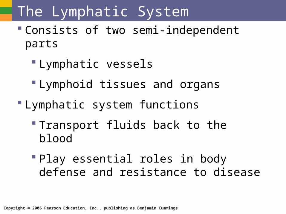
The Lymphatic System Consists of two semi-independent parts
Lymphatic vessels
Lymphoid tissues and organs
Lymphatic system functions
Transport fluids back to the blood
Play essential roles in body defense and resistance to disease
Copyright © 2006 Pearson Education, Inc., publishing as Benjamin Cummings
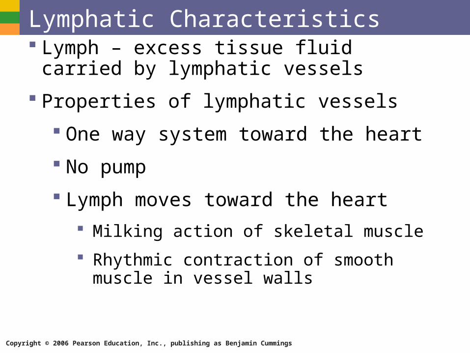
Lymphatic Characteristics Lymph – excess tissue fluid carried by
lymphatic vessels
Properties of lymphatic vessels
One way system toward the heart
No pump
Lymph moves toward the heart
Milking action of skeletal muscle
Rhythmic contraction of smooth muscle in vessel walls
Copyright © 2006 Pearson Education, Inc., publishing as Benjamin Cummings
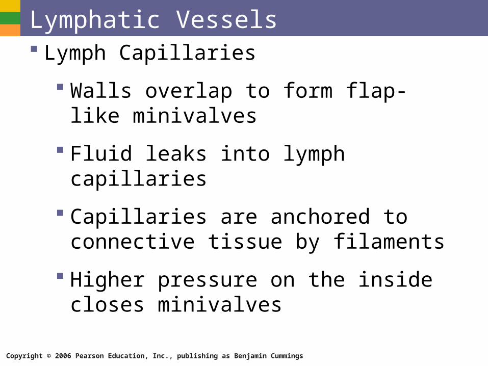
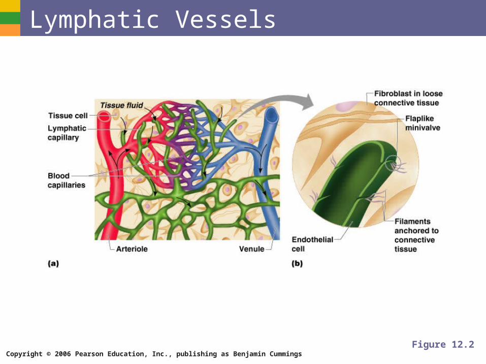
Lymphatic Vessels Lymph Capillaries
Walls overlap to form flap-like minivalves
Fluid leaks into lymph capillaries
Capillaries are anchored to connective tissue by filaments
Higher pressure on the inside closes minivalves
Copyright © 2006 Pearson Education, Inc., publishing as Benjamin Cummings
Lymphatic Vessels
Figure 12.2
Copyright © 2006 Pearson Education, Inc., publishing as Benjamin Cummings
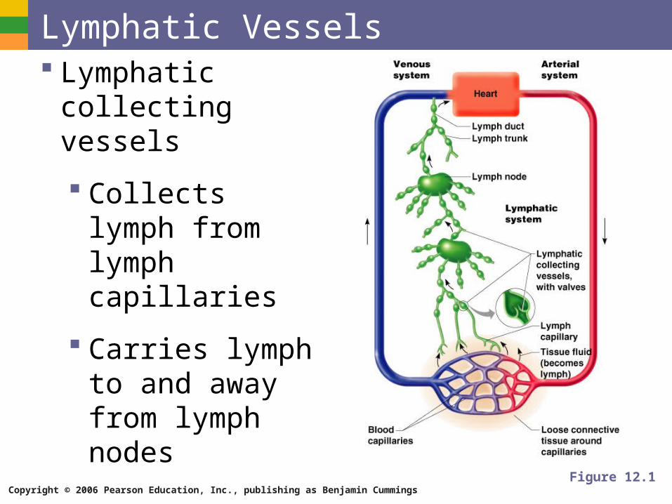
Lymphatic Vessels Lymphatic collecting
vessels
Collects lymph from lymph capillaries
Carries lymph to and away from lymph nodes
Figure 12.1
Copyright © 2006 Pearson Education, Inc., publishing as Benjamin Cummings
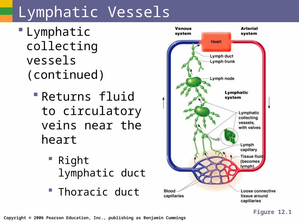
Lymphatic Vessels Lymphatic collecting
vessels (continued)
Returns fluid to circulatory veins near the heart
Right lymphatic duct
Thoracic duct
Figure 12.1
Copyright © 2006 Pearson Education, Inc., publishing as Benjamin Cummings
Lymph Materials returned to the blood
Water
Blood cells
Proteins
Copyright © 2006 Pearson Education, Inc., publishing as Benjamin Cummings
Lymph Harmful materials that enter lymph vessels
Bacteria
Viruses
Cancer cells
Cell debris
Copyright © 2006 Pearson Education, Inc., publishing as Benjamin Cummings
Lymph Nodes Filter lymph before it is returned to the blood
Defense cells within lymph nodes
Macrophages – engulf and destroy foreign substances
Lymphocytes – provide immune response to antigens
Copyright © 2006 Pearson Education, Inc., publishing as Benjamin Cummings
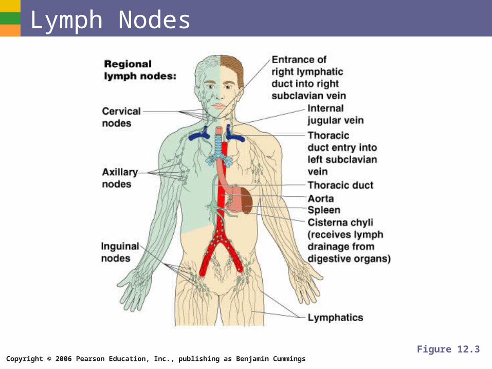
Lymph Nodes
Figure 12.3
Copyright © 2006 Pearson Education, Inc., publishing as Benjamin Cummings
Lymph Node Structure Most are kidney-shaped, less than 1 inch long
Cortex
Outer part
Contains follicles – collections of lymphocytes
Medulla
Inner part
Contains phagocytic macrophages
Copyright © 2006 Pearson Education, Inc., publishing as Benjamin Cummings
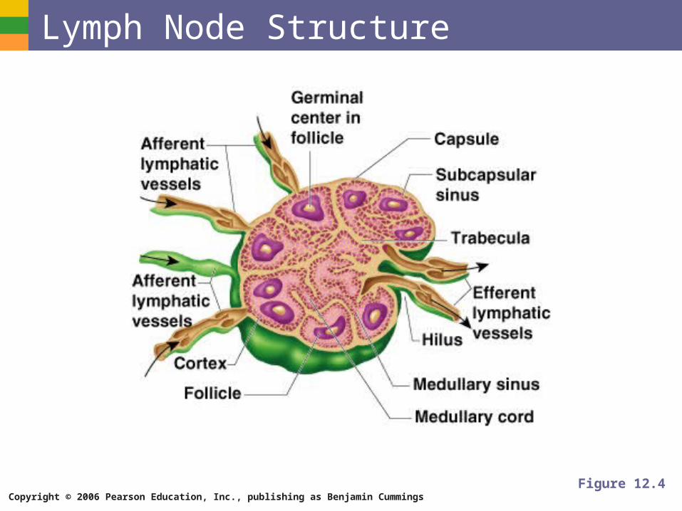
Lymph Node Structure
Figure 12.4
Copyright © 2006 Pearson Education, Inc., publishing as Benjamin Cummings
Flow of Lymph Through Nodes Lymph enters the convex side through
afferent lymphatic vessels
Lymph flows through a number of sinuses inside the node
Lymph exits through efferent lymphatic vessels
Fewer efferent than afferent vessels causes flow to be slowed
Copyright © 2006 Pearson Education, Inc., publishing as Benjamin Cummings
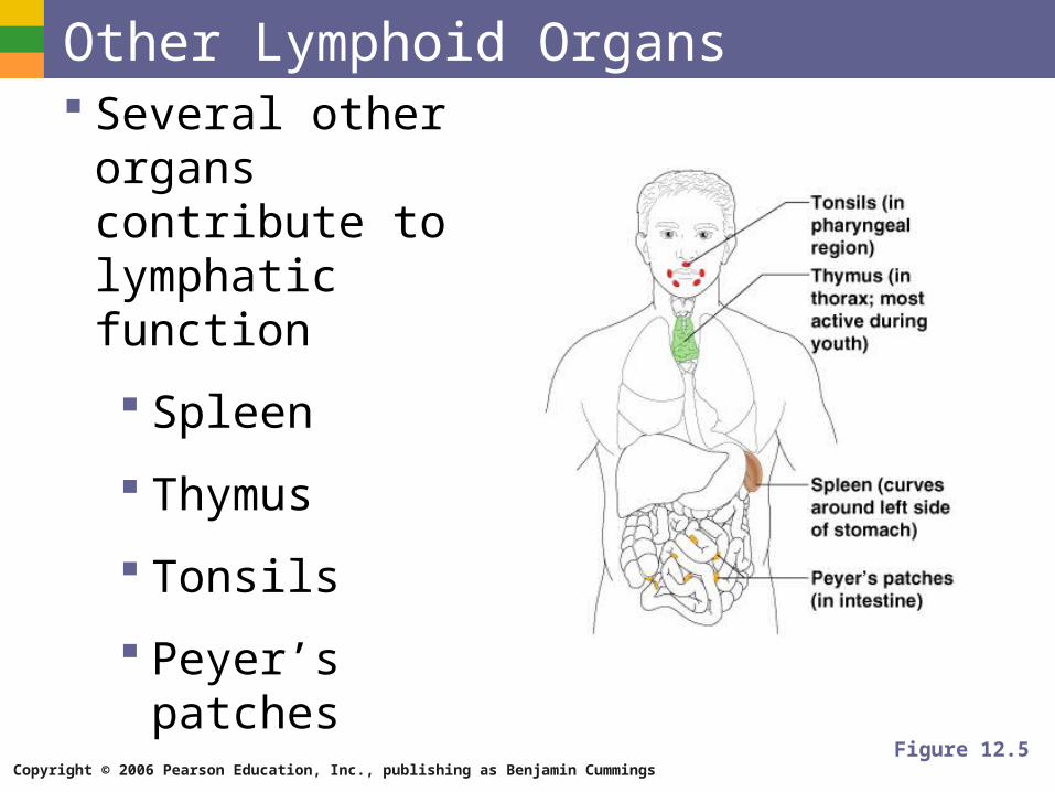
Other Lymphoid Organs Several other organs
contribute to lymphatic function
Spleen
Thymus
Tonsils
Peyer’s patches
Figure 12.5
Copyright © 2006 Pearson Education, Inc., publishing as Benjamin Cummings
The Spleen Located on the left side of the abdomen
Filters blood
Destroys worn out blood cells
Forms blood cells in the fetus
Acts as a blood reservoir
Copyright © 2006 Pearson Education, Inc., publishing as Benjamin Cummings
The Thymus Located low in the throat, overlying the heart
Functions at peak levels only during childhood
Produces hormones (like thymosin) to program lymphocytes
Copyright © 2006 Pearson Education, Inc., publishing as Benjamin Cummings
Tonsils Small masses of lymphoid tissue around the
pharynx
Trap and remove bacteria and other foreign materials
Tonsillitis is caused by congestion with bacteria
Copyright © 2006 Pearson Education, Inc., publishing as Benjamin Cummings
Peyer’s Patches Found in the wall of the small intestine
Resemble tonsils in structure
Capture and destroy bacteria in the intestine
Copyright © 2006 Pearson Education, Inc., publishing as Benjamin Cummings
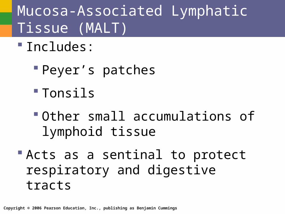
Mucosa-Associated Lymphatic Tissue (MALT) Includes:
Peyer’s patches
Tonsils
Other small accumulations of lymphoid tissue
Acts as a sentinal to protect respiratory and digestive tracts
Copyright © 2006 Pearson Education, Inc., publishing as Benjamin Cummings
Body Defenses The body is constantly in contact with
bacteria, fungi, and viruses
The body has two defense systems for foreign materials
Nonspecific defense system
Specific defense system
Copyright © 2006 Pearson Education, Inc., publishing as Benjamin Cummings
Body Defenses Nonspecific defense system
Mechanisms protect against a variety of invaders
Responds immediately to protect body from foreign materials
Specific defense system
Specific defense is required for each type of invader
Also known as the immune system
Copyright © 2006 Pearson Education, Inc., publishing as Benjamin Cummings
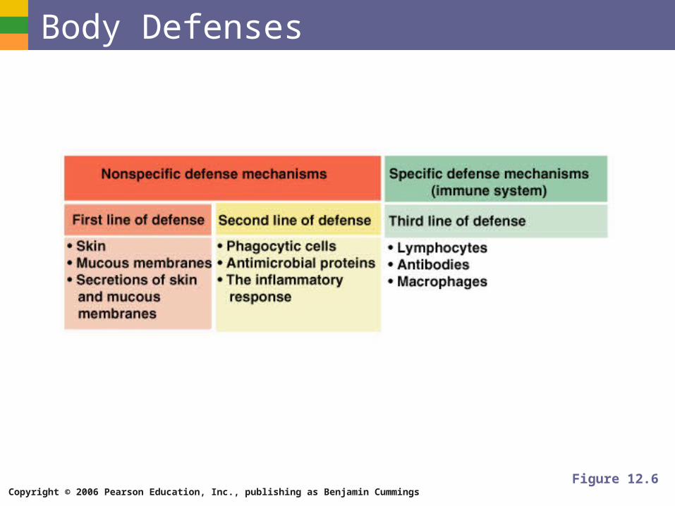
Body Defenses
Figure 12.6
Copyright © 2006 Pearson Education, Inc., publishing as Benjamin Cummings
Nonspecific Body Defenses Body surface coverings
Intact skin
Mucous membranes
Specialized human cells
Chemicals produced by the body
Copyright © 2006 Pearson Education, Inc., publishing as Benjamin Cummings
Surface Membrane Barriers – First Line of Defense The skin
Physical barrier to foreign materials
pH of the skin is acidic to inhibit bacterial growth
Sebum is toxic to bacteria
Vaginal secretions are very acidic
Copyright © 2006 Pearson Education, Inc., publishing as Benjamin Cummings
Surface Membrane Barriers – First Line of Defense Stomach mucosa
Secretes hydrochloric acid
Has protein-digesting enzymes
Saliva and lacrimal fluid contain lysozyme
Mucus traps microogranisms in digestive and respiratory pathways
Copyright © 2006 Pearson Education, Inc., publishing as Benjamin Cummings
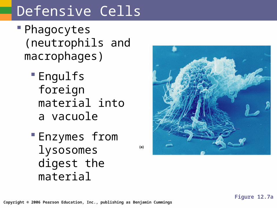
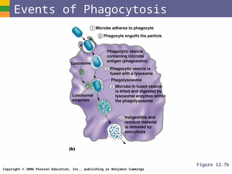
Defensive Cells Phagocytes
(neutrophils and macrophages)
Engulfs foreign material into a vacuole
Enzymes from lysosomes digest the material
Figure 12.7a
Copyright © 2006 Pearson Education, Inc., publishing as Benjamin Cummings
Events of Phagocytosis
Figure 12.7b
Copyright © 2006 Pearson Education, Inc., publishing as Benjamin Cummings
Defensive Cells Natural killer cells
Can lyse and kill cancer cells
Can destroy virus- infected cells
Copyright © 2006 Pearson Education, Inc., publishing as Benjamin Cummings
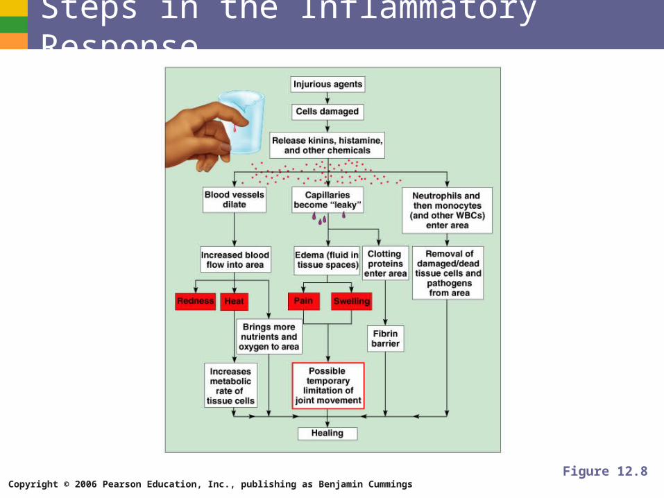
Inflammatory Response - Second Line of Defense Triggered when body tissues are injured
Produces four cardinal signs
Redness
Heat
Swelling
Pain
Results in a chain of events leading to protection and healing
Copyright © 2006 Pearson Education, Inc., publishing as Benjamin Cummings
Functions of the Inflammatory Response Prevents spread of damaging agents
Disposes of cell debris and pathogens
Sets the stage for repair
Copyright © 2006 Pearson Education, Inc., publishing as Benjamin Cummings
Steps in the Inflammatory Response
Figure 12.8
Copyright © 2006 Pearson Education, Inc., publishing as Benjamin Cummings
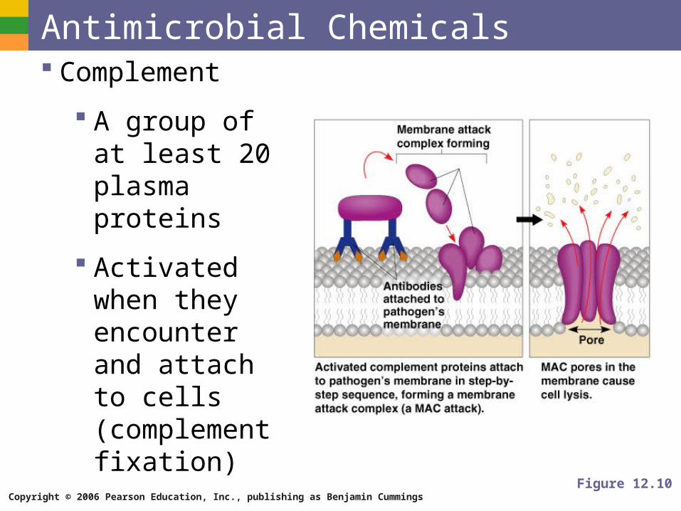
Antimicrobial Chemicals Complement
A group of at least 20 plasma proteins
Activated when they encounter and attach to cells (complement fixation)
Figure 12.10
Copyright © 2006 Pearson Education, Inc., publishing as Benjamin Cummings
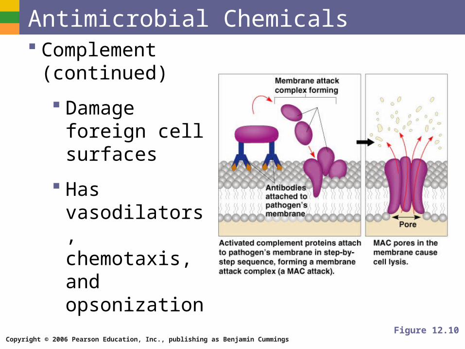
Antimicrobial Chemicals Complement
(continued)
Damage foreign cell surfaces
Has vasodilators, chemotaxis, and opsonization
Figure 12.10
Copyright © 2006 Pearson Education, Inc., publishing as Benjamin Cummings
Antimicrobial Chemicals Interferon
Secreted proteins of virus-infected cells
Bind to healthy cell surfaces to inhibit viruses binding