Embed Size (px)
Citation preview

ORIGINAL ARTICLE
The Myocardial Support
El soporte del miocardio
1 Research Department, Hospital Presidente Perón, Buenos Aires, Argentina.2 Department of Cardiac Surgery, Clínica Güemes, Luján, Buenos Aires, Argentina.3 Department of Pathology, Clínica Güemes, Luján, Buenos Aires, Argentina.4 Department of Cardiac Surgery, Hospital Presidente Perón, Buenos Aires, Argentina5 Department of Cardiology, Investigaciones Médicas, Buenos Aires, Argentina.6 Department of Computed Tomography, and Magnetic Resonance Imaging, Clínica Güemes, Luján, Buenos Aires, Argentina.
JORGE TRAININI1, , MARIO BERAUDO2, MARIO WERNICKE3, ALEJANDRO TRAININI2,4, , DIEGO HABER LOWENSTEIN5, MARÍA ELENA BASTARRICA2, , DARÍO CARLOS MARTINO6, JORGE LOWENSTEIN5,
ABSTRACT
Background: The cardiac muscle cannot be anatomically free in the thorax and without a support to fulfill its hemodynamic func-tion. Therefore, we analyzed the possibility of a supporting point, acting as a lever.Material and Methods: Cardiac dissection was performed in ten young (two-year-old) bovine hearts (800-1000 g); and in eight hu-man hearts: one belonging to a 23-week gestation fetus, one to a 10-year-old child, weighing 250 g, and six to adult patients, with mean weight of 300 g. The myocardial band was totally unfolded. The different pieces were anatomically and histologically analyzed and the study was completed using magnetic resonance imaging, computed tomography and simple radiology.Results: In the anatomical investigation of all the human and bovine hearts studied we found a nucleus underlying the right trigone, of osseous, chondroid or tendinous histological structure. The microscopic analysis revealed in bovine hearts a trabecular osteochon-dral matrix (fulcrum) and in human hearts the fulcrum was formed by chondroid tissue. The origin and end of the myocardial fibers were inserted into this structure, not previously described by other authors. Imaging studies confirmed the existence of the cardiac fulcrum.Conclusions. The cardiac fulcrum found in the anatomical investigation of bovine and human hearts would clarify the supporting point of the myocardial muscle to complete its torsion motion.
Key words: Heart – Cardiac anatomy – Myocardium- Cardiac support
RESUMEN
Objetivo: El músculo cardíaco no puede estar anatómicamente libre en el tórax y sin un soporte para cumplir con su función hemo-dinámica. Por tanto, se analizó la posibilidad de la existencia de un punto de apoyo que actuara a modo de palanca. Material y métodos: Se utilizaron: 1) disección cardíaca en diez corazones bovinos jóvenes (dos años) (800-1000 g); 2) disección car-díaca en ocho corazones humanos: un embrión de 23 semanas de gestación; uno de 10 años, 250 g; y seis adultos, peso medio 300 g. La banda miocárdica se desenrolló en su totalidad. Las piezas extraídas fueron analizadas por anatomía e histología. Se completó la investigación con estudios de imágenes radiográficas simples, resonancia nuclear magnética y tomografia computada. Resultados: En investigaciones anatómicas hemos encontrado en todos los corazones humanos y bovinos estudiados un núcleo sub-yacente al trígono derecho de estructura histológica ósea-condroide-tendinosa. El análisis microscópico reveló en corazones bovinos una matriz osteocondral trabecular (fulcro). En todos los corazones humanos se encontró que el fulcro se halla formado por tejido condroide. En esta estructura, no descrita por otros autores, tienen inserción muscular el origen y el final de las fibras miocárdicas. Las técnicas con imágenes confirmaron su existencia. Conclusiones: El fulcro cardíaco encontrado en la investigación anatómica de corazones humanos y bovinos aclararía sobre el nece-sario punto de apoyo del músculo miocárdico para completar sus movimientos de torsión.
Palabras clave: Corazón – Anatomía cardiaca – Miocardio- Apoyo miocárdico
REV ARGENT CARDIOL 2021;89:217-223. http://dx.doi.org/10.7775/rac.v89.i3.20407
Address for reprints: Jorge Carlos Trainini - Hospital Presidente Perón - Avellaneda, Provincia de Buenos Aires, Argentina – Tel: 54 11 15 40817028 -
Email: [email protected]
Financing: The present work did not receive any scolarship or grant.
Received: 03/14/2021 – Accepted: 05/08/2021

ARGENTINE JOURNAL OF CARDIOLOGY / VOL 89 Nº 3 / JUNE 2021218
INTRODUCTIONThe anatomy of the heart was traditionally consid-ered to be formed by muscles arranged in spiraling bundles, but these were never described in relation to their physiology. Richard Lower in 1669 considered that the myocardium was subjected to a torsion mo-tion related to the helical fibers that formed it. He expressed that the heart exerted a motion similar to "wringing a towel" and not as it was considered since Harvey who claimed it was due to ventricular radial compression comparable to "closing a fist". (1)
Andreas Vesalius in his work “De Humani Corpo-ris Fabrica” (1543) referred to the difficulty in identi-fying the myocardial layers. He expressed verbatim: “No matter how you perform the dissection of the meat of the heart, whether raw or cooked…, you can scarcely remove a portion of only one type of fiber, because they have multiple and different directions, especially transversal”. (2)
Three centuries later, this situation was also re-marked by JB Pettigrew (1864) “Of the complexity of the arrangement I don´t need to say more than Vesalius, Haller and De Blainville; they all confessed their inability to decipher it”. (3) RF Shaner in 1923 states that “the myocardium is formed by two flat-tened muscles with the shape of an 8. These muscles twist in opposite directions in systole, emptying their content”. (4)
Lack of an adequate anatomical dissection of the myocardium prevented seeing its real functional structure. Recent interpretations pose controversial opinions, mainly between band and mesh models. Torrent Guasp (5, 6) in 1973 considered the myocar-dium as a cardiac muscle band, showing in numerous dissections that it is formed by a set of muscle fibers coiled unto themselves similar to a rope, flattened lat-erally, which by giving two spiraling turns define a he-lix limiting the two ventricles. Maclvear (7, 8) explains that the ventricular walls are shaped by an intricate cardiomyocyte three-dimensional mesh, implying that cardiomyocytes are arranged with radial and longitu-dinal angulations.
The inevitable reflection that arises is that to ful-fill torsion, the myocardium must perform it on a sup-porting point, same as the skeletal muscle does it on a firm insertion. Does that structure exist in the heart? If this supporting point is real, how are the cardiac muscle fibers inserted into this structure? Can it be differentiated from the trigones? This premise cor-relates with a cardiac structure presenting remark-able characteristics: that of being a suction-impelling pump, equivalent to a human fist, weighing an aver-age of 270 grams, which ejects 4 to 5 liters/minute of blood at a speed of 300 cm/s with an efficacy that al-lows pumping 70% of the left ventricular volume with only 12% shortening of its contractile unit, the sar-comere.
The aim of this work was to demonstrate through macroscopic dissection and histological studies that
the myocardium is a continuous, single muscle with helical arrangement. All these anatomo-functional considerations may help both to quantify the severity of morbid processes as in therapeutic strategies. (9)
METHODS1) Myocardial dissection of 18 hearts: a) 10 two-year-old
bovine hearts with an average weight of 800-1000 g; b) 8 human hearts: one from a 23-week gestation fetus; one from a 10-year-old child weighing 250 g and 6 adult hearts with an average weight of 300 gr.
2) Histological and histochemical analysis of anatomical samples. The histological study was performed using 10% formalin as buffer, 4-micron sections and hematox-ylin-eosin and Masson’s trichrome staining. All samples underwent histological and histochemical analysis using Alcian blue staining as a reliable marker to identify the presence of hyaluronic acid as an antifriction mecha-nism. and even provide a semiquantitative assessment.
3) Imaging studies. Bovine hearts were studied with com-puted tomography, nuclear magnetic resonance imaging and simple X-rays. One patient could be studied with a computed tomography scan.The hearts examined were obtained from the morgue
and slaughterhouse. In order to dissect it, the heart must be boiled in water during two hours with the convenient addi-tion of acetic acid (15 ml per liter). This step removes the fat attached to the myocardium, making the dissection easier and neater. The aorta and the pulmonary artery are then cut at three centimeters from their origin, separating the at-tachment between them, followed by a longitudinal incision at the level of the interventricular sulcus of the superficial fibers extending transversally along the anterior wall of the ventricles. As there is only connective tissue between the atria and the ventricles, the denaturation process produced by heat allows the easy separation of these chambers.
The key maneuver to unfold the myocardium consists in entering the anterior interventricular sulcus with a blunt instrument, leaving on the left side of the operator the muscle end corresponding to the pulmonary artery and its contiguity with the right ventricular free wall (right seg-ment). Then, traction is applied towards the same left side, a maneuver which completely releases the pulmonary artery from the rest of the myocardium. This myocardial dissection exposes the fulcrum below and in front of the aorta, in a plane inferior to the right trigone and the origin of the right coronary artery, without continuity with the aortic valve and inserted as a complementary element between the aorta and the myocardium. This structure, the supporting site of the origin and end of the cardiac muscle, is more rigid than the trajectory between the trigones.
It should be understood that as the myocardium is un-folded, separating the pulmonary artery and the pulmo-tricuspid cord (anterior) from the ascending segment (pos-terior), the vision of the homogeneous anatomical integrity is lost. This conjunction between the origin and end of the cardiac muscle in the cardiac fulcrum constitutes a meet-ing point between the right segment and the ascending seg-ment, origin and end of the myocardium. Thus, both ends are situated in the same point, with the origin of the myocar-dial fibers placed anteriorly to those of its end.
The progress of the myocardial muscle dissection implies finding the whole length of the right segment, the origin of the left segment and, at the posterior margin of the right ventricular chamber, the dihedral angle formed by the inter-

219MYOCARDIAL SUPPORT / Jorge Trainini et al.
ventricular septum and the free wall of the right ventricle (right segment). The next step (the most delicate one) con-sists in approaching the dihedral angle between the right ventricular and intraseptal fibers. This separation from the right ventricle allows access to the ventral part of the sep-tum. Then, the dorsal part of the septum is dissected to de-tach and separate the aorta.
Finally, to the right of the operator, the muscle plane of the descending segment is separated in blunt fashion from the one corresponding to the ascending segment leading to the cardiac fulcrum next to the aorta, allowing it to be ex-tended in its full length. Being able to unfold the myocar-dium with a similar thickness in all its length shows that it is a single and continuous muscle and not a heuristic con-struction.
RESULTSIn all the human and bovine hearts studied we have found a nucleus underlying the right trigone, whose osseus, chondroid or tendinous histological structure depends on the specimens analyzed (Figures 1 to 3). The microscopic analysis of the hearts revealed an osterochondral trabecular matrix (fulcrum), with seg-mental lines in bovines (Figures 1 and 2). A central zone, formed by chondroid tissue was found in the ful-crum of the 10-year-old human heart (Figure 4A) and prechondroid areas in myxoid stroma in the fetus ful-crum (Figures 4 B), while in adult human hearts, the histological analysis revealed a matrix similar to that of a tendon. All the hearts presented myocardial inser-tion into the rigid structure of the fulcrum (Figures 2 and 5) No cardiomyocytes were found either in the left or right trigone or at the base of the cardiac valves.
This fixation point implies, as in any skeletal mus-cle, its ability to achieve the necessary support and
also to act as a bearing or pad preventing the ventricu-lar rotation force, either by torque or torsion, from spreading to the great vessels, thus dissipating the en-ergy produced by the helical motion of the muscle and avoiding aortic constriction or flexion during systolic ejection.
Radiological images evidenced the osteochondral nucleus found in dissections, with the same morpholo-gy and analogous size (Figure 1). In computed tomog-raphy scans, we saw that the analysis of the region described as cardiac fulcrum in the dissections per-formed had an intensity above 110 Hounsfeld units (HU), while the adjacent muscle had values below 80 HU. In one patient, the area described as fulcrum had a mean value of 132±4.5 HU, and in the adjacent ar-eas this value was 47.96±12.5 and 77.59±21.64 HU.
DISCUSSIONIn all the bovine and human hearts used in this re-search, we found an osseous, chondroid or tendinous nucleus, which we have termed cardiac fulcrum. Fib-ers from the right segment and the ascending seg-ment, i.e., the origin and end of the cardiac muscle, are oriented and inserted into this fulcrum.
The existence of the “os cordis”, a formation found in bovine, sheep and chimpanzee hearts is a fact men-tioned in veterinary medicine, without any physiologi-cal relationship. It is located in the same position in which we have investigated this structure, both in bovines and humans. Beyond its mere mention in bo-vines, it was never assigned any function or signifi-cance of its presence, and it also lacks description in humans.
In the human hearts analyzed, the findings are
Fig. 1. Cardiac fulcrum (bo-vine heart). A: Mature osse-ous trabecula forming the cardiac fulcrum. Hematoxy-lin-eosin technique. (10x); B: In the area shown with the arrow, an image adjacent to the aortic root is observed on the interventricular septum (computed tomography); C: Another view of the fulcrum.
Fulcrum
Aorta
Fulcrum
ARGENTINE JOURNAL OF CARDIOLOGY / VOL 89 Nº 3 / JUNE 2021220
different. And this difference in the intimate analysis of the cardiac fulcrum is undoubtedly associated with the resistance it must oppose to the cardiac muscle action in hearts of different sizes.
A fact that satisfies the structural and mechani-cal logic of the myocardium is finding in the human heart this point of attachment of myocardial fibers of unequivocal characteristics to simple observation and palpation, in the same location and with similar
remarkable from an interpretative point of view, as-suming that it is logical to consider its presence in all the evolutionary history of mammals. This structure, when analyzed in the different specimens, has the common function of providing support for the myo-cardium to generate the strength needed by any mus-cle, which varies in different mammals. Therefore, its presence is constant in all the hearts analyzed, both bovine and human, but its structural characteristic is
Fig. 2. Note the insertion line of the myocardial fibers in the fulcrum of a bovine heart. Histological image of the insertion. A: 1) Myocar-dial fibers and myxoid stro-ma. 2) Myocardial bands in a chondroid stroma (insertion). 3) Osseous cortical tissue of the fulcrum. Hematoxylin-eosin technique (15x). B: re-sected piece.
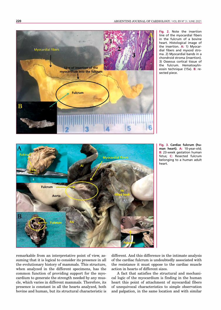
Fig. 3. Cardiac fulcrum (hu-man heart). A: 10-year-old; B: 23-week gestation human fetus; C: Resected fulcrum belonging to a human adult heart.
Fulcrum
Line of insertion of the myocardium into the fulcrum
Myocardial fibers
Fulcrum
Fulcrum
Fulcrum
Aorta
PA tilted back
Myocardial fibers
RA
Fulcrum
AI

221
triangular morphology as that mentioned in different species. However, the histological analysis in the adult human heart revealed a matrix similar to that of a tendon. At this point, several questions arise: Why does the human fulcrum have characteristics similar to a tendon, despite it fulfills the same function of at-taching the myocardium to a support as in other spe-cies? Why does it not have the same structure as in the human fetal or childhood heart?
MYOCARDIAL SUPPORT / Jorge Trainini et al.
The interpretation we have is that perhaps the osseus fulcrum in bovine, chimpanzee, sheep and hu-man fetus is a vestigial organ inherent to mammalian evolution. A vestigial structure must be understood as the preservation during the evolutionary process of genetically determined traits which have partly or totally lost their ancestral function in a certain spe-cies. As a result, we find it in the initial process of human development, but then its osseous nature dis-
Fig. 4. A: 10-year-old human heart. Central zone of the fulcrum consisting of chon-droid tissue. Hematoxylin-eosin technique (15x). B: Prechondrial bluish areas in a myxoid stroma in a 23-week gestation fetus. Masson’s tri-chrome technique (15x).
Fig. 5. Scalloped cardiomyo-cytes penetrating a fibrocol-lagenous matrix (adult hu-man heart). 1: Cardiomyocytes 2: Cardio-myocyte fraying; 3: Atrophied cardiomyocytes, 4: Fibrocol-lagenous matrix. Hematoxy-lin-eosin technique (15x).
Cardiac fulcrum

ARGENTINE JOURNAL OF CARDIOLOGY / VOL 89 Nº 3 / JUNE 2021222
1. Henson RE, Song SK, Pastorek JS, Ackerman JH, Lorenz CH. Left ventricular torsion is equal mice and humans. Am J Physiol Heart Circ Physiol 2000;278:H1117-23. https://doi.org/10.1152/ajpheart.2000.278.4.H11172. Trainini J, Lowenstein J, Beraudo M, Trainini A, Mora Llabata V, Wernicke M. “Myocardial Torsion”. Ed Biblos Buenos Aires; Ar-gentina, 2019.3. Pettigrew JB. On the arrangement of the muscular fibres in the ventricles of the vertebrate heart with phisiological remarks. Philos Trans 1864;154:445-500. https://doi.org/10.1098/rstl.1864.00144. Shaner R.F. On the muscular architecture of the vertebrate ven-tricle. J Anat 1923;58:59-70.5. Torrent-Guasp F, Kocica MJ, Corno AF, Komeda M, Carreras-Costa F, Flotats A, et al.Towards new understanding of the heart structure and function. Eur J Cardiothorac Surg 2005;27:191-201. https://doi.org/10.1016/j.ejcts.2004.11.0266. Torrent Guasp F, Buckberg G, Carmine C, Cox J, Coghlan H, Gharib M. The structure and function of the helical heart and its buttress wrapping. I. The normal macroscopic structure of the heart. Semin Thorac Cardiovasc Surg 2001;13:301-19. https://doi.org/10.1053/stcs.2001.299537. Mac Iver DH, Stephenson RS, Jensen B, Agger P, Sanchez-Quin-
REFERENCES
appears, remaining as a tendinous structure sufficient to achieve the myocardial insertion that generates a muscle strength inferior to that of larger mammals. Let us recall that in our research the nature of the bovine fulcrum is osseous.
The results of the dissection showed that the myo-cardium is a continuous, helical muscle. Cardiac func-tion cannot be explained by a mesh configuration. (25) In this regard, Maclver's work states: “None of the histological studies of the myocardium that we are aware, in contrast have provided any evidence for an origin and insertion as described for the alleged unique myocardial band” and “None of these inves-tigations have provided any evidence of an alignment of the cardiomyocytes that follows the course of the unique myocardial band.” (3) First of all, the cardiac fulcrum that we have investigated in human and ani-mal hearts describes the cardiac support that would give rise to that single, continuous, helical muscu-lar myocardial conformation. Regarding the second conclusion of this author, the sequential histological analysis of the unfolded myocardium shows the lon-gitudinal orientation of fibers in agreement with the continuity of the segments resulting from its spatial arrangement, an orientation that is parallel both in the internal and external surfaces of each segment (Figure 2).
No segment of the histological sequence corre-sponding to the longitudinal continuity of the myocar-dium presents a mesh configuration. In the external surface of the distal end of the descending segment, when it turns at the apex and becomes the ascending segment, the orientation of the cardiomyocytes gener-ates, in planimetric sections, a dissimilar architecture in their orientation to that of the internal surface, only place where this situation occurs. The rest of the orientation is always parallel. In the apex, the spiral-ing course of the myocardial fibers which shift from the periphery to the center determines a torsion in which the subepicardial fibers become subendocar-dial, overlapping like the tiles of a roof, as depicted in the aforementioned figure. This resembles the Moe-bius band due to the progressive change in fiber angu-lations, turning them from epicardial to endocardial.
It can be seen that the myocardial structure is not a mesh but a continuous muscle. (12) The mesh concept was developed by segment overlap due to the folding of the myocardial helix.
A histological analysis of the trigones has also been performed, trying to find cardiomyocytes in them, as a possibility of myocardial insertion into these struc-tures. Only collagenous tissue without cardiomyocytes was observed in our investigation of the trigones, con-firming that the fulcrum is the support of the cardiac muscle, both at its origin and its end.
The myocardium cannot be anatomically suspend-ed and free in the thoracic cavity because it would be impossible for the heart to eject blood at a speed of 300 cm/s. There must be a point of attachment, which
we found and called cardiac fulcrum. In this support-ing point, the muscle fibers are inevitably obliged to intertwine with the fulcrum. In our anatomical and histological research, the connective, chondroid or os-seus fulcrum showed this insertion, attaching the ori-gin and end of the cardiac muscle.
In the fulcrum, the heart finds the fixed point ena-bling the mechanics of muscular torsion. The opposite rotation of the left ventricle from base to apex (13, 14) allows the development of elevated pressure which reduces tension, exactly as “wringing a towel”. This mechanic, found in mice and humans, (15-18) helps the ejection of the blood content in a limited time span with the necessary force to circulate throughout the whole organism.
LimitationsThere were few human specimens studied because it is difficult to have access to intact, well-preserved hearts for a careful dissection. We think that this work should be expanded with a greater number of human adult and specially children’s hearts. Our investigation was limited to eight human and ten bovine hearts.
CONCLUSIONSThe cardiac fulcrum found in this anatomical research would help to clarify the supporting point of the myo-cardial band to complete its torsion function. Without its presence, the heart would not achieve its hemody-namic efficiency of pumping blood at a speed of 300 cm/s.
Conflicts of interestNone declared.
(See authors’ conflicts of interest forms on the website/Supplementary material)
Note: Part of this article has been previously published in Morphologie, 2021;105:15-23.

223MYOCARDIAL SUPPORT / Jorge Trainini et al.
tana D, Jarvis JC, et al. The end of the unique myocardial band: Part I. Anatomical considerations. Eur J Cardiothorac Surg 2018;53:112-9. https://doi.org/10.1093/ejcts/ezx2908. Mac Iver DH, John B. Partridge JB, Agger P, Stephenson RS , Boukens BJD, Omann C, Jarvis JC, Zhang H. The end of the unique myocardial band: Part II. Clinical and functional considerations. Eur J Cardio-Thoracic Surg 2018;53:120-8. https://doi.org/10.1093/ejcts/ezx3359. Elencwajg B, López-Cabanillas N, Cardinali EL, Barisani JL, Trainini J, Fischer A, et al. The Jurdham procedure endocardial left ventricular lead insertion via a femoral transseptal sheath for cardiac resynchronization therapy pectoral device implanta-tion. Heart Rhythm 2012;9:1798-804. https://doi.org/10.1016/j.hrthm.2012.07.01010. Trainini JC, Herreros J, Elencwajg B, Trainini A, Lago N, López Cabanillas N, et al. Disección del miocardio. Rev Argent Cardiol 2017;85:44-50. https://doi.org/10.7775/rac.es.v85.i1.1019811. Moittié S, Baiker K, Strong V, Cousins E, White K, Liptovszky M, et al.. Discovery of os cordis in the cardiac skeleton of chimpan-zees (Pan troglodytes). Scientific Reports 2020;10:9417 https://doi.org/10.1038/s41598-020-66345-7. 12. Anderson R, Ho S, Redman K, Sanchez-Quintana D, Punken-heimer P. The anatomical arrangement of the myocardial cells mak-
ing up the ventricular mass. Eur J Cardiothoracic Surg 2005;28:517-25. https://doi.org/10.1016/j.ejcts.2005.06.04313. Trainini JC, Trainini A, Valle Cabezas J, Cabo J. Left Ventricu-lar Suction in Right Ventricular Dysfunction. EC Cardiology 2019; 6:572-7. 14. Mora V, Roldán I. Romero E, Saurí A, Romero D, Perez-Gozabo J, et al. Myocardial contraction during the diastolic isovolumetric period: analysis of longitudinal strain by means of speckle track-ing echocardiography. J Cardiovasc Dev Dis 2018;5:4. https://doi.org/10.3390/jcdd503004115. Trainini JC, Elencwajg B, Herreros J. New Physiological Concept of the Heart. Ann Transplant Res 2017;1:1001. 16. Carreras F, Ballester M, Pujadas S, Leta R, Pons-Lladó G. Mor-phological and functional evidences of the helical heart from non-invasive cardiac imaging. Eur J Cardiothoracic Surg 2006; 29(Suppl 1):S50-5. https://doi.org/10.1016/j.ejcts.2006.02.06117. Ballester M, Ferreira A, Carreras F. The myocardial band. Heart Fail Clin 2008;4:261-72. https://doi.org/10.1016/j.hfc.2008.02.01118. Kocica MJ, Corno AF, Carreras-Costa F, Ballester-Rodes M, Moghbel MC, Cueva CN, et al. The helical ventricular myocardial band: global, three-dimensional, functional architecture of the ven-tricular myocardium. Eur J Cardiothorac Surg 2006; 29:Suppl-I:S21-40. https://doi.org/10.1016/j.ejcts.2006.03.011


![Ticagrelor - CADIME · inestable, infarto de miocardio sin elevación del segmento ST [IMSEST] o infarto de miocardio con elevación del segmento ST [IMCEST]), incluidos los pacientes](https://img.pdfslide.us/doc/110x75/5ba4661709d3f2c0278d5ec2/ticagrelor-inestable-infarto-de-miocardio-sin-elevacion-del-segmento-st.jpg)