Embed Size (px)
Citation preview

ISSN 0355-1180
UNIVERSITY OF HELSINKI
Department of Food and Environmental Sciences
EKT Series 1493
CHROMATOGRAPHIC SEPARATION, FRACTIONATION AND OXIDATION OF
SELECTED BETA-LACTOGLOBULIN PEPTIDES
Göker Gürbüz
Helsinki 2010

HELSINGIN YLIOPISTO HELSINGFORS UNIVERSITET UNIVERSITY OF HELSINKI
Tiedekunta/Osasto Fakultet/Sektion Faculty
Faculty of Agriculture and Forestry
Laitos Institution Department
Department of Food and Environmental Sciences
Tekijä Författare Author
Göker Gürbüz Työn nimi Arbetets titel Title
Chromatographic separation, fractionation and oxidation of selected beta-lactoglobulin peptides Oppiaine Läroämne Subject
Food Sciences (Food Safety) Työn laji Arbetets art Level
M. Sc. Thesis
Aika Datum Month and year
November 2010
Sivumäärä Sidoantal Number of pages
59
Tiivistelmä Referat Abstract
The literature review elucidates the mechanism of oxidation in proteins and amino acids and gives an overview of the detection and analysis of protein oxidation products as well as information about -lactoglobulin and studies carried out on modifications of this protein under certain conditions. The experimental research included the fractionation of the tryptic peptides of -lactoglobulin using preparative-HPLC-MS and monitoring the oxidation process of these peptides via reverse phase-HPLC-UV. Peptides chosen to be oxidized were selected with respect to their amino acid content which were susceptible to oxidation and fractionated according to their m/z values. These peptides were: IPAVFK (m/z 674), ALPMHIR (m/z 838), LIVTQTMK (m/z 934) and VLVLDTDYK (m/z 1066). Even though it was not possible to solely isolate the target peptides due to co-elution of various fractions, the percentages of target peptides in the samples were satisfactory to carry out the oxidation procedure. IPAVFK and VLVLDTDYK fractions were found to yield the oxidation products reviewed in literature, however, unoxidized peptides were still present in high amounts after 21 days of oxidation. The UV data at 260 and 280 nm enabled to monitor both the main peptides and the oxidation products due to the absorbance of aromatic side-chains these peptides possess. ALPMHIR and LIVTQTMK fractions were oxidatively consumed rapidly and oxidation products of these peptides were observed even on day 0. High rates of depletion of these peptides were acredited to the presence of His (H) and sulfur-containing side-chains of Met (M). In conclusion, selected peptides hold the potential to be utilized as marker peptides in
-lactoglobulin oxidation. Avainsanat Nyckelord Keywords
-Lactoglobulin, protein oxidation, peptides, HPLC Säilytyspaikka Förvaringsställe Where deposited
Viikki Campus Library Muita tietoja Övriga uppgifter Further information
EKT Series 1493

PREFACE
This study was carried out at the Department of Food and Environmental Sciences, Food
Chemistry Division.
Firstly, I would like to express my sincere thanks to Professor Marina Heinonen for all her
support and advice throughout the program and accepting me into her skilled group. My
warm thanks and deep gratitude go to my supervisor Marjo Poutanen for her guidance and
Tuuli Koivumäki for her patience and understanding all along the way, exploring the
“combos” with me. I also thank everyone at Food Chemistry Division for helping me
around the lab and making me feel welcome as a colleague.
I would like to thank my fellow students Flora Agalga, Bhawani Chamlagain, Xiaoyan Liu,
Marta Nogueroles-Moya and Qiao Shi, for going through this journey together as the first
students of the MScFood program.
Finally, I would like to express my heartfelt gratitude to my family, Mehmet, Gül and
Güney Gürbüz for their encouragement and support. Last, but not least I thank Ruta
Kazlauskaite for simply being there for me, even at times I am grumpy and intolerable.
Helsinki, November 2010
Göker Gürbüz

LIST OF ABBREVIATIONS
-Lg Beta-lactoglobulin A, Ala Alanine AAS Alpha-aminoadipic semialdehyde C, Cys Cysteine D, Asp Aspartic acid DNPH Dinitrophenylhydrazine DOPA 3,4-dihydroxyphenylalanine E, Glu Glutamic acid ESI Electrospray ionization F, Phe Phenylalanine FLD Fluorescence detector G, Gly Glycine GC Gas chromatography GGS Gamma-glutamic semialdehyde H, His Histidine HACA 6-hydroxy-2-aminocaproic acid HAVA 5-hydroxy-2-aminovaleric acid HPLC High performance liquid chromatography I, Ile Isoleucine K, Lys Lysine L, Leu Leucine M, Met Methionine MALDI Matrix-assisted laser desorption-ionization MCO Metal-catalyzed oxidation MS Mass spectrometry N, Asn Asparagine P, Pro Proline PAD Photodiode array detector Q, Gln Glutamine R, Arg Arginine ROS Reactive oxygen species S, Ser Serine SDS-PAGE Sodium dodecyl sulfate - polyacrylamide gel electrophoresis T, Thr Threonine TOF Time-of-flight (mass spectrometer) W, Trp Tryptophan V, Val Valine Y, Tyr Tyrosine

TABLE OF CONTENTS ABSTRACT PREFACE LIST OF ABBREVIATIONS 1 INTRODUCTION 6
2 LITERATURE REVIEW 8
2.1 Protein oxidation 8
2.1.1. Reactive oxygen species 8
2.1.2 Metal-catalyzed oxidation 9
2.1.3 Peptide bond cleavage 10 2.1.4 Side-chain oxidation 11
2.1.5 Cross-link formation 12
2.2 Amino acid oxidation 12
2.2.1 Sulfur-containing amino acids 12
2.2.2 Aromatic side-chains 14
2.2.3 Aliphatic amino acids and other heteroatom-containing side-chains 17
2.3 Detection and analysis of protein oxidation in foods 19
2.4 -Lactoglobulin 21
2.4.1 Structure, amino acid composition and allergenicity 21
2.4.2 Effect of thermal treatment 23
2.4.3 Antioxidant activity and photo-oxidation 23
3 EXPERIMENTAL RESEARCH 26
3.1 Aims 26
3.2 Materials and methods 26
3.2.1 Materials 26
3.2.2 Digestion of -lactoglobulin with modified trypsin 27
3.2.3 Fractionation of -lactoglobulin peptides with preparative-HPLC-MS 27
3.2.4 Preparation of peptide samples 30
3.2.5 Checking the concentration of H2O2 solution 30
3.2.6 Monitoring the oxidation process 31
3.3 Results 33
3.3.1 Peptide fractions obtained via preparative-HPLC 33
3.3.2 Monitoring the oxidation process in peptide fractions 35
3.4 Discussion 46
3.4.1 Digestion of -lactoglobulin and fractionation of peptides 46
3.4.2 Interpretation of the oxidative changes in -lactoglobulin peptides 46
4 CONCLUSIONS 54
REFERENCES 56

6
1 INTRODUCTION
Oxidative deterioration of proteins in foods leads to many undesirable alterations in
physical, chemical and functional features. Even though studies on oxidation mechanisms
in food are generally dominated by other oxidation mechanisms, mainly of lipids in foods,
this trend tends to change. The complex mechanisms and products formed as a result of
protein oxidation in food are better understood with respect to progressing studies on the
topic. On the other hand, oxidation reactions in foods are often studied and referred with
respect to loss in quality and nutritional value. However, studies that are aimed in the
direction of possible toxic products and adverse health effects of protein oxidation have
been carried out (Matsukura et al. 1981; Stadtman and Berlett 1997; Stadtman and Levine
2003). It is fair to admit that the mechanisms and interactions of protein oxidation that take
place during processing and storage in food systems are yet to be understood adequately.
Thus, further studies are required in order to identify, explain and finally prevent the
undesirable deterioration of proteins in food systems.
Proteins and amino acids in food systems are susceptible to oxidation, for instance, via
reactive oxygen species (ROS) that are present in the atmosphere as pollutants, or formed
as by-products of normal metabolic processes as well as a result of exposure to irradiation.
Reactive oxygen-mediated modification of proteins can lead to several pathways of
oxidation of the protein backbone such as cleavage of peptide bonds, oxidation in side
chains of amino acid residues and several cross-linking reactions (Stadtman and Berlett
1997). Additionally, certain side chains of amino acids and amino acid residues are readily
oxidized by metal-ion catalyzed oxidation systems (Stadtman and Levine 2003).
Determination of protein oxidation is performed by methods that were generally
developed in biomedical research. However, a number of these can be applied in food
systems. The main understanding in the assessment of protein oxidation involves the
detection of oxidative modifications and analysis of the specific products generated with
the proposed oxidation mechanism. The analysis of oxidized proteins has been performed
traditionally by quantification of protein carbonyls via dinitrophenylhydrazine (DNPH)
method which even though considered accurate, lacks the information on specific
generation of protein carbonyls (Requena et al. 2003). Quantification of carbonyls by other
late and advanced methods includes liquid chromatography-electrospray ionization-mass
spectrometry (LC-ESI-MS) of -aminoadipic and -glutamic semialdehydes (Estevez et al.

7
2009). However, since other mechanisms that yield formation of carbonyls are present,
analysis of carbonyls as sole indicators of oxidation products and mechanisms would not
give enough information on the field. In addition to carbonyl assays, certain biomarkers are
subject to recent methods to analyze ROS-damaged proteins such as, dityrosine, 3-nitro-
tyrosine, chlorine derivatives and certain mono- or di-hydroxy phenylalanine derivatives
(Stadtman and Levine 2003). The methods developed and being used so far clearly should
be subjected to further advancements since current studies in the field of protein oxidation
cover only a small area among the unknowns of oxidized protein mechanisms and
interactions in food systems. Thus more methods should be set up to enlighten other
protein oxidation mechanisms.
-Lactoglobulin is a major whey protein that is found in several mammals’ milk but
humans. It constitutes up to 50-55% of total whey proteins in bovine milk. -Lactoglobulin
has been widely studied due to its nutritional value as a whey protein, its structural
similarity to retinol binding protein in human serum, thermal denaturation in milk
processing and the questions arising with respect to its unknown biological functions
(Hambling et al. 1992). There are 6 genetic variants known among which A and B are the
most significant ones in terms of human diet. The amino acid sequence of bovine -
lactoglobulin A and B contain 162 amino acids. An important feature of -lactoglobulin is
its antigenicity manifesting itself mainly in infants as intolerance and allergic reactions
(Wal 1998). Thus, many studies have been conducted to reduce its antigenicity via
enzymatic and physical treatments. In addition, -lactoglobulin has been studied with
respect to the relatively mild antioxidant properties of its certain peptides and amino acid
residues with high radical-scavenging activity (Elias et al. 2005).
The modifications in chosen -lactoglobulin peptides that were imposed by oxidation
reactions were investigated and discussed in this study in the light of literature reviewed on
protein and amino acid oxidation. The peptides chosen were obtained as a result of tryptic
digestion of the protein and fractionation process via preparative high performance liquid
chromatography coupled with mass spectrometer. These fractions were later oxidized
under laboratory conditions and the changes were monitored via reverse phase high
performance liquid chromatography. The resulting data of this process provided
information on the oxidatively vulnerable peptide fractions that may be used as markers of
oxidation in -lactoglobulin.

8
2 LITERATURE REVIEW
2.1 Protein oxidation
2.1.1 Reactive oxygen species
Oxidation reactions of proteins are generally initiated by the reactive oxygen species
(ROS). These species can result from pollutants present in the atmosphere, exposure to UV,
X-ray or -irradiation, or metabolic processes such as lipid peroxidation, metal-catalyzed
systems, glycation/glycoxidation and so on (Stadtman and Berlett 1997). Major sources of
ROS that affect the scheme of oxidation are depicted in Figure 1. Most common ROS that
set off oxidation reactions are hydroxyl (HO•), superoxide anion (O2-•) and hydroperoxyl
(HO2•) radicals. Among these radicals, initial sites of oxidation in proteins and peptides
are vulnerable to HO• attack. HO• can arise due to the before-mentioned sources of ROS
or the breakdown of H2O2 by iron or copper in metal-catalyzed oxidation systems. There
are several pathways and modifications observed of the oxidation reactions in proteins and
peptides. These include the hydrogen atom abstraction from the -carbon of the
polypeptide backbone, cleavage of peptide bonds, side-chain modifications (including
addition reactions), cross-linking of proteins, glycation/glycoxidation, chlorination and
modification by reactive nitrogen species (Garrison 1987; Fu et al. 1998; Stadtman and
Levine 2003; Hawkins et al. 2003).
Figure 1. Major sources of reactive oxygen species (ROS) and other reactive species (RS) that lead to protein oxidation (Stadtman and Berlett 1997). (NOS: NO synthase, MCO: Metal-catalyzed oxidation).

9
2.1.2 Metal-catalyzed oxidation
Many studies have confirmed that metal-catalyzed oxidation (MCO) systems are a
significant source of oxidative modification in proteins (Amici et al. 1989; Stadtman and
Oliver 1991; Huggins et al. 1993). The activity of Fe(II) or Cu(I) in the initiation of
oxidation reactions lie in their ability to convert relatively unreactive H2O2 into HO•
radicals that lead to yield carbon-centered radicals. Formation of HO• may either involve
direct reduction of O2 into H2O2 while Fe(III) is reduced to Fe(II) or a sequential reduction
of O2 through an intermediate O2-• and then to HO• (Stadtman and Oliver 1991).
Following these pathways, Fe(II) acts in the generation of alkyl peroxides from peroxyl
protein radicals which later again by the action of Fe(II) are converted into highly reactive
alkoxyl radicals. These radicals finally undergo a hydrogen atom addition through another
Fe(II) catalyzed-system to yield the hydroxy derivative of the protein. It is known that in
MCO systems Fe(II) achieves its function via the metal-binding site on the protein which
reacts with the oxygen radicals. This site specific binding is favored on the side-chains of
amino acid residues on proteins. Oxidation of side-chains of several amino acids such as
Lys, Arg, Pro and Thr result in carbonyl compounds whereas His ends up as 2-oxo-
histidine (Stadtman and Berlett 1997). Figure 2 presents the general scheme of a site
specific metal-catalyzed oxidation including the mechanisms mentioned above on a lysyl
residue.
Figure 2. Site-specific binding of metal ions that lead to oxidative modification on a lysyl residue (Stadtman and Oliver 1991). (P: Protein/ Peptide)

10
2.1.3 Peptide bond cleavage
Irrespective of their source, HO• radicals are highly reactive towards proteins, peptides
and amino acids. Most of the times, these radicals start the free radical-mediated reactions
by abstracting a H atom from the -carbon on the polypeptide backbone to generate
carbon-centered radicals (alkyl radicals). In the presence of O2, these alkyl radicals are
converted into peroxyl protein radicals. Peroxyl radicals can react readily with another
molecule or HO2• radicals in the medium to yield alkyl peroxides via the abstraction of a
H atom. Further reactions of alkylperoxides with HO2• radicals lead to the formation of
alkoxyl radicals and hydroxy derivatives of the proteins (Garrison 1987; Stadtman and
Berlett 1997). The scheme of the radical-mediated oxidation up to this point including the
involvement of Fe(III) and Fe(II) ions is presented below in Figure 3.
Figure 3. Pathway of free radical-mediated protein oxidation in the presence of O2 (Stadtman and Levine 2003).
Garrison (1987) along with Stadtman and Berlett (1997) have explained that the alkyl
peroxide and alkoxyl derivatives of proteins can be involved in a backbone cleavage
process via two pathways, namely -amidation and diamide. Cleavage of the polypeptide
chain via these pathways is illustrated in Figure 4. As a result of the peptide bond scission
in the -amidation pathway, fragments of amide and -keto-acyl derivatives are formed.
On the other hand, the cleavage reaction occurring via diamide pathway yields fragments

11
of diamide and isocyanate derivatives (Davies 1996; Stadtman and Berlett 1997). Cleavage
of peptide bond is also observed as a result of side-chain oxidation of some amino acid
residues. The examples to this incident include oxidation of prolyl residues (Uchida 1990),
aspartyl and glutamyl residues (Garrison 1987).
Figure 4. Oxidative backbone cleavage of polypeptides via -amidation and diamide pathways (Stadtman and Levine 2003).
2.1.4 Side-chain oxidation
Side-chains of amino acid residues in proteins and peptides are among the most susceptible
sites to oxidative modifications by various means such as radiolysis and MCO systems.
However, the extent of the changes caused by different systems varies according to the
side-chain characteristics. For instance, Stadtman and Berlett (1991) have noted that while
ionizing radiation causes modifications in almost all amino acids (especially Cys, Met, Tyr,
Trp and His), in MCO systems Trp, Met and Tyr are not targets of preference. Moreover,
studies have showed that some amino acids are much more susceptible to oxidative
modifications than others. These include sulfur-containing, aromatic side chains and
several aliphatic residues such as Pro, Lys and Arg. Amino acid oxidation including their
side-chain alterations and major oxidation products will be presented in Section 2.2.

12
2.1.5 Cross-link formation
There are several mechanisms that have been reported to yield inter and intra-protein
cross-linking. One of these involves direct dimerization of two carbon-centered radicals
that have been formed in MCO systems or other ROS sources. This process is known to
occur in the absence of O2 (Stadtman 1993). Another mechanism involves the side-chain
interaction of two amino acid residues, namely the sulfur-bridge formation between the
two sulfhydryl groups of Cys and the formation of links between the aromatic side-chains
of Tyr. The rest of the cross-linking occurs via several reactions of Lys and Arg residues
with carbonyl compounds arising from oxidation reactions of other proteins (Davies 2005).
2.2 Amino acid oxidation
Free amino acids or amino acid residues in proteins are prone to attack of reactive oxygen
species and undergo several modifications. These changes are commonly observed on the
side-chains as a result of reactions with oxygen free radicals. Some amino acids react with
these radicals more readily than others due to characteristics of their side-chains. Davies
(2005) tabulates the rate constants for the reactions of HO• with amino acids as highest in
the case of Cys, followed by His, Trp, Tyr, Met and Phe. It is now widely known that the
sulfur-containing amino acids and amino acids with aromatic side-chains are main targets
of oxidative damage. Main oxidative modifications are discussed below in three categories
as sulfur-containing amino acids, aromatic side-chains and aliphatic and other heteroatom-
containing side-chains.
2.2.1 Sulfur-containing amino acids
Met and Cys are the two sulfur-containing amino acids which have been shown to be
significant sites of oxidation in proteins. Met, a non-polar amino acid, owes its rapid
oxidative depletion to its thioether side-chain while polar Cys holds a thiol side-chain. Due
to the weak bonds at the sulfur-binding site, these amino acids are highly susceptible to
oxidation.
Oxidation of cysteine occurs by metal ions or by other oxidants generating thiyl radicals
through hydrogen abstraction from a free thiol group (Hawkins and Davies 2001). These
radical species may react reversibly and an intermediate species, sulfenic acid (-SOH) is

13
observed. Sulfenic acid later may lead to the formation of a dimer via the disulfide linkage
formed between thiol groups (cystine). Cystine can also react further with O2 to form
species such as mono-sulfoxide or go through radical oxidation that results in disulfide
bond cleavage (Rehder and Borges 2010; Davies 2005). On the other hand, sulfenic acid
may also react with O2 to produce peroxyl radicals which later end up generating oxyacids
such as sulfinic acid (-SOOH) and sulfonic acid (-SO2OH) (Davies 2005; Elias et al. 2008).
Figure 5 schemes simply the oxidation pathway of Cys into oxyacids and disulfide
structure.
Figure 5. Structures and oxidation pathway of cysteine and its products (Griffiths 2002).
The other sulfur-containing amino acid Met has been the subject of several studies in terms
of oxidative modification. Many oxidants can rapidly react with the thioether side-chain of
Met such as HO•, H• species as well as HOCl and HOBr. A table of rate constants for the
reactions with Met has been presented by Davies 2005. The high tendency of this amino
acid to scavenge radicals and the fact that Met sulfoxide can be reduced back to Met have
even been investigated as whether surface-exposed Met residues could act as an
antioxidant in proteins against oxidative damages due to the reversible oxidation reaction
(Levine et al. 1996; Levine et al. 1999). Unlike Cys, Met is unable to form disulfide
bridges and most of the reactions with the oxidants result in the production of methionine
sulfoxide as the major product. Further oxidation is also known to lead to the formation of

14
methionine sulfone but in a lesser extent (Vogt 1995; Davies 2005). Figure 6 shows the
structures of Met and its oxidation products.
Figure 6. Structure of methionine and its oxidation products (Vogt 1995)
2.2.2 Aromatic side-chains
Amino acids with aromatic side-chains are another group that is highly susceptible to
oxidation. These include Trp, Tyr, Phe and His. The reaction of the aromatic ring with the
reactive oxygen species generally takes place as an addition to the side-chain. Although
hydrogen abstraction from the aromatic ring occurs initially that results in several short-
lived adducts radical formation (Hawkins and Davies 2001).
Oxidation of Phe by various reactive species takes place as a hydroxylation process and
yields in o-Tyr, m-Tyr and p-Tyr. The latter hydroxylated compound is Tyr itself and thus
is not considered to be a credible oxidation marker; whereas o-Tyr and m-Tyr are stable
markers in Phe oxidation even in acid hydrolysis of proteins (Huggins et al. 1993; Davies
et al. 1999). Figure 7 shows Phe and the oxidation products already mentioned. In addition,
further dimerization of these hydroxylated amino acids is also observed as a result of
phenoxyl radical formation due to one electron oxidation (Hawkins and Davies 2001; Elias
et al. 2008).

15
Figure 7. Phe and its oxidation products (Stadtman and Levine 2003).
The attack of oxidant species on Tyr in metal-catalyzed H2O2 systems ends up in the
formation of 3,4-dihydroxyphenylalanine (DOPA) and dityrosine. However, these products
can also be observed in other systems both in the absence and presence of O2 (Davies et al.
1999). Dityrosine, formed as dimer of Tyr, can arise as a result of other systems as well
such as peroxidase-catalyzed and reactions with reactive nitrogen species (Malencik and
Anderson 2003). DOPA can be formed via the disproportionation of radicals resulting
from the reaction of Tyr with HO• in the absence of O2 or through a peroxyl radical
pathway in the presence of O2 where the latter series of reactions give way to higher yields
of DOPA (Hawkins and Davies 2001). In addition, even though DOPA is known to be a
reductant which is susceptible to further oxidation to form quinones and even result in
damage to other biomolecules, this compound was also investigated for its use as a
potential marker for oxidative damage (Gieseg et al. 1993; Morin et al. 1998; Davies et al.
1999). The structures of Tyr and its oxidation products, DOPA and dityrosine are
displayed in Figure 8.
Figure 8. Structures of Tyr and its major oxidation products (Stadtman and Levine 2003).
Oxidation of Trp can be observed as modifications either on the benzene ring or the
pyrrole unit (Figure 9). The benzene moiety is hydroxylated by HO• attacks to yield 2-, 4-,

16
5-, 6- or 7-hydroxytryptophan. On the other hand, HO• can also be added to the pyrrole
ring to form neutral radicals. Furthermore, these pyrrole radicals react with O2 to form
peroxyl radicals which later on cause the cleavage of the heterocyclic ring to produce N-
formylkynurenine and further kynurenine, kynurenic acid and 3-hydroxy-kynurenine
(Hawkins and Davies 2001). Among the oxidation products of Trp, hydroxylated materials
are considered as stable markers of oxidation more than N-formylkynurenine and
kynurenine due to the susceptibility of the latter compounds for additional and complex
reactions (Davies et al. 1999).
Figure 9. Oxidation pathways and products of Trp (Salminen et al. 2008).
His has been known to be vulnerable to oxidation most in metal-catalyzed systems and the
reactions that take place to yield the end-products are not fully identified (Uchida and
Kawakashi 1993; Schöneich 2000; Hawkins and Davies 2001). However, several studies
reported that the oxidation of His resulted in formation of asparagines, aspartic acid and 2-
oxo-histidine (Figure 10), the last one being used as a marker by a method revealed by
Uchida and Kawakishi (1993). The addition of HO• on the imidazole ring forms radical
species that can react with O2 to form peroxyl radicals. These peroxyl radicals through
further reactions yield 2-oxo-histidine (Hawkins and Davies 2001; Uchida 2003).

17
Figure 10. Formation of 2-oxo-histidine from His (Hawkins and Davies 2001).
2.2.3 Aliphatic amino acids and other heteroatom-containing side-chains
The majority of oxidation reactions taking place with the aliphatic amino acids and other
heteroatom-containing side-chains are initiated by a hydrogen atom abstraction from the -
carbon via the attack of HO• and other radicals, resulting in the generation of carbon-
centered radicals (Stadtman 1993). In the absence of O2, these radicals are found to
dimerize by forming bonds with the other carbon-centered radicals (Davies 2005).
However, in the presence of O2, these radicals react with O2 to generate peroxyl radicals.
Peroxyl radicals can further be involved in hydrogen abstraction, fragmentation and
dimerization reactions that result in the formation of hydroperoxides, alcohol and carbonyl
compounds. Due to their unstable nature, hydroperoxides rapidly decompose with a wide
range of oxidizing systems to yield further radicals such as alkoxyl and superoxides as well
as more alcohols and carbonyl compounds (Davies et al. 1995; Davies 2005; Stadtman
1993). In addition to carbon-centered oxidation pathway, Ser, Thr, Pro, Lys and Arg yield
carbonyl compounds also from their side-chain oxidation (Garrison 1987).
Some of the alcohols produced as a result of these reactions are considered as stable
markers of oxidation such as 5-hydroxyleucine and 3-hydroxyvaline (Davies et al. 1999).
On the other hand, the carbonyl compounds arisen have been the subject of many studies
and employed as markers of oxidation through development of several methods for the
quantification of these materials (Amici et al. 1989; Requena et al. 2001; Akagawa et al.
2006; Estevez et al. 2009). Table 1 presents the known major oxidation products of the
amino acids with aliphatic and heteroatom-containing side chains from selected references.

18
Table 1. Amino acid oxidation products via various oxidation mechanisms
Amino Acid Products Reference
Glycine aminomalonic acid Davies et al. (1999) Glutamic acid 4-hydroxyglutamic acid Davies et al. (1999) Valine 3- and 4-hydroxyvaline Garrison (1987); Davies et al. (1999) Leucine 3- and 4-hydroxyleucine Garrison (1987)
Lysine 3-, 4- and 5-hydroxylysine
-aminoadipic semialdehyde 6-hydroxy-2-aminocaproic acid (HACA)
Morin et al. (1998); Davies et al. (1999); Requena et al. (2001)
Arginine -glutamic semialdehyde 5-hydroxy-2-aminovaleric acid (HAVA) Requena et al. (2001)
Proline -glutamic semialdehyde
5-hydroxy-2-aminovaleric acid (HAVA) 3- and 4-hydroxyproline
Requena et al. (2001); Davies et al. (1999)
Threonine 2-amino-3-keto butyric acid Taborsky (1973)
2.3 Detection and analysis of protein oxidation in foods
Majority of the methods that are employed in the detection of oxidative modifications of
proteins and determination of oxidation products are developed for biomedical research.
Nevertheless, a number of these methods are also applied to food systems and produce
informative results. The common approach in the evaluation of protein oxidation includes
the detection of modifications caused by oxidation and analysis of the specific products
that are formed as a result of these modifications.
Detection of aggregation, polymerization and fragmentation in proteins has been used by
many researchers investigating the structural changes occurring in protein during oxidation.
A traditional method utilized in determination of these modifications involves the sodium
dodecyl sulfate and polyacrylamide gel electrophoresis (SDS-PAGE) where an increase or
decrease in the molecular size can be quantitated by a densitometer according to the
staining density of protein bands on polyacrylamide gels (Davies and Delsignore 1987).
Additionally, changes in the overall electrical charges detected by using isoelectric
focusing gel assayss can provide information about the alterations in primary structure
(Davies et al. 1987). SDS-PAGE method is still applied also for qualitative purposes in
protein oxidation studies to obtain useful information besides other advanced methods. A
report by Salvi et al. (1999) informs the development of a capillary zone electrophoresis
method where the changes in peak heights are observed as during radical-induced protein
fragmentation.

19
Another notable factor of polymerization of proteins is the cross-linking via disulfide
bonds. The method developed by Ellman (1959) considers the loss of free sulfhydryl (thiol)
groups and detects the loss by reacting the sample with 5,5’-dithiobis(2-nitrobenzoic acid)
measuring the absorbance at 412 nm. In an example of the application of the technique,
Liu and Xiong (2000) estimated the disulfide content in oxidized myosin, -lactoglobulin
-Lg) and 7S globulin proteins by subtracting free sulfhydryl content from total sulfhydryl
and disulphide groups. The calculations included the absorbance measurement at 412 nm
after reacting the protein samples with 5,5’-dithiobis(2-nitrobenzoic acid) and disodium 2-
nitro-5-thiosulfobenzoate to find the free sulfhydryls and total disulphide content,
respectively.
Due to the presence of aromatic amino acids, Trp, Tyr and Phe in proteins and peptides,
detections of oxidative modifications in these amino acid residues provide a useful tool in
assessment of protein oxidation. Among these amino acids, Trp is considered as the most
susceptible one to oxidative deterioration (Estevez et al. 2008). Trp fluorescence is known
to be observed at 280 nm excitation and 340-350 nm emission wavelengths. As a result of
the formation of Trp oxidation products such as N-formylkynurenine a loss in fluorescence
of Trp takes place. Thus, this change in Trp residues has been monitored to determine the
oxidation by several studies (Davies et al. 1987; Finley et al. 1998). Tyr and its oxidation
products have also been investigated in traditional methods such as fluorometric detection
with high performance liquid chromatography (HPLC) monitored at excitation at 280 nm
(320 nm emission) and coupling gas chromatography (GC) with mass spectrometry (MS)
(Davies et al. 1999; Miyahara et al. 2000). However, newer studies also focused on
employing electron spin resonance (ESR) and nuclear magnetic resonance (NMR)
spectroscopy and electrospray ionization tandem-mass spectrometry (ESI-MS/MS) with
respect to oxidation products of Tyr and Trp (Saeed et al. 2006; Gracanin et al. 2009;
Grosvenor et al. 2010).
In addition to the methods mentioned above, analysis of protein oxidation has been
dominated by the analysis of carbonyl products formed during several pathways (including
metal-catalyzed oxidation of specific amino acid side chains). Formation of carbonyl
compounds is related to a number of amino acid side chains such as Ala, Val, Leu, Asp,
Lys, Pro and Arg (Headlam and Davies 2004) and has been used widely as markers of
protein oxidation. As a result, quantification of protein oxidation has been traditionally and
extensively performed by carbonyl assay, that is, spectrophotometric detection of carbonyl
groups after reacting with 2,4-dinitrophenylhydrazine, also known as DNPH method

20
(Levine et al. 1994; Requena et al. 2003). Recently, two main carbonyl products
considered as biomarkers of metal-catalyzed oxidative protein damage are in focus of
protein oxidation analysis studies. These are -glutamic semialdehyde (GGS), oxidation
product of Arg and Pro and -aminoadipic semialdehyde (AAS), of Lys. Daneshvar et al.
(1997) studied the identification of biomarkers in metal-catalyzed oxidation of bovine
serum albumin, employing high-performance liquid chromatography (HPLC) with diode
array detector (DAD) and mass spectrometer (MS). The study concludes with the GGS and
AAS being important markers of oxidation in the serum proteins. In another study,
Requena et al. (2001) developed a method for quantification of GGS and AAS after their
reduction to 5-hydroxy-2-aminovaleric acid (HAVA) and 6-hydroxy-2-aminocaproic acid
(HACA) via gas chromatography – selected ion monitoring mass spectrometry (GC-SIM-
MS). The method was applied to several model proteins and biological samples,
concluding that GGS and AAS make up of majority of carbonyl products of oxidation.
Akagawa et al. (2006) employed a different method for quantification of AAS and GGS
where the semialdehydes in proteins are derivatized by a fluorescent reagent followed by
acid hydrolysis. The hydrolysates were analyzed by HPLC and fluorescence detector
(FLD). A most recent study by Estevez et al. (2009) used liquid chromatography –
electrospray ionization – multistage tandem mass spectrometry (LC-ESI-MS/MS) to
determine GGS and AAS formation in several food proteins (myofibrillar proteins, soy
proteins and -lactalbumin) and bovine serum albumin and achieved accurate detection of
both semialdehydes. Even though, formation of carbonyl compounds is the most focused
protein oxidation detection, it should not be forgotten that a number of amino acid residues
are present which undergo other oxidative modification mechanisms other than carbonyl
production.
As it can be observed through the progress of protein oxidation analysis methods, mass
spectrometry is more frequently employed with the development of soft ionization
techniques such as ESI and matrix-assisted laser desorption-ionization (MALDI) combined
with multi stage and hybrid mass analyzers, time-of-flight (TOF), Fourier-transform ion
cyclotron resonance (FTICR), Q-q-Q, Q/TOF and TOF/TOF. Additionally, establishment
of protein databases increased the sensitivity of peptide characterization and mapping
(Mamone et al. 2009). However, it should be noted that a specific MS technique should be
considered for the individual objective of analysis, thus choice of the particular
instrumentation and fragmentation method becomes significant. An example of a recent
study on metal-catalyzed oxidation of bovine serum albumin in vitro system includes

21
application of MALDI-TOF/TOF MS/MS and a protein database for identification of
oxidatively modified tryptic peptides (Guedes et al. 2009).
2.4 -Lactoglobulin
2.4.1 Structure, amino acid composition and allergenicity
-Lactoglobulin ( -Lg) is a major whey protein found in bovine milk. The protein is also
present in several other mammals such as reindeer, horse, pig, sheep and cat, but not in
humans (Hambling et al. 1992). It is known to make up of 10 to 15% of total milk proteins
and 50 to 55% of total whey proteins (Liu et al. 2007). At neutral pH values, -Lg is
present as a dimer, while under acidic (pH<3.5) or basic (pH>7.5) conditions, it is
dissociated into its monomers (Hambling et al. 1992). As a monomer, bovine -Lg has a
molecular weight of 18350 Da and its structure is composed of nine -strands and one -
helix together to form a hydrophobic pocket as shown in Figure 11. (Shimoyamada et
al.1996).
Figure 11. Three-dimensional view of -lactoglobulin monomer.
-Lg is secreted within the epithelial cells of mammary glands, where mRNA coding is
transferred to produce a 180 amino acid pre- -Lg. This pre- -Lg contains a sequence of
18 highly conserved amino acids which are later cleaved from the C terminal of Ala by a
protease enzyme inside the membrane to yield the generally known bovine -Lg sequence

22
consisting of 162 amino acids (Hambling et al. 1992). Bovine -Lg is found to have 6
genetic variants among which -Lg A and -Lg B are the most important and frequently
encountered in commercial preparations. -Lg A and B differ in their amino acid sequence
where A variant includes Asp at position 64 and Val at 118, whereas B variant hosts Gly
and Ala at the same positions, respectively (Figure 12) (Jankowski and Sichel 2003). -Lg
monomer contains five Cys residues at positions 66, 106, 119, 121 and 160. The thiol
groups of Cys residues form disulphide bonds located at Cys66-Cys160 and Cys106-Cys119
leaving the thiol group free at Cys121. Moreover, Tyr and Trp residues that bovine -Lg
monomer includes, contribute to the spectroscopic properties of the protein (Hambling et
al. 1992).
Figure 12. The amino acid sequence of bovine -lactoglobulin A (www.rcsb.org, code: 1BEB).
-Lg has been extensively studied due to its nutritional value as a whey protein, its
structural similarity to retinol binding protein in human serum, and the questions arising
with respect to its unknown biological functions (Hambling et al. 1992). In addition, -Lg
belongs to lipocalin protein family that transports hydrophobic ligands and is known to be
allergenic (Wal 1998). Cow milk allergy is one of the significant food allergies especially
affecting infants due to intolerance developed against milk proteins. Intact -Lg protein in
cow milk is known to be responsible for this allergy phenomenon, and therefore,
proteolytic treatment of -Lg has been of research interest (Jakobsson et al. 1983; Madsen
et al. 1997). Several studies aimed and reported to reduce the antigenicity of -Lg with
proteolytic procedures that are combined with physical treatments under various conditions
(Iametti et al. 2002; Bonomi et al. 2003; Kim et al. 2007). However, the existence of the
cases that residual antigens are found in hydrolyzed milk formulas shows that the
allergenic nature of -Lg will continue to be an interest for researchers in terms of food
safety (Mamone et al. 2009).

23
2.4.2 Effect of thermal treatment
Thermal modifications of -Lg and the importance of their consequences in milk
processing have been widely investigated. Bertrand-Harb et al. (2002) have observed the
aggregation of -Lg and the resistance to hydrolysis by trypsin and pepsin enzymes after
thermal treatment as well as its co-denaturation with -lactalbumin. On the other hand,
Chen et al. (2005) have discussed the use of -Lg as thermal marker in processed milk as a
result of its denaturation at temperature values of 80ºC and above. The lack of the same
behavior in other whey proteins has put -Lg in a significant position in terms of
determining processing conditions of milk.
A number of the studies concerning -Lg modifications under heat treatment include the
mechanism and products of Maillard and oxidation reactions in the presence of lactose in
milk and dairy products. Meltretter et al. (2007) analyzed the site-specific formation of
lactosylation products arising from -Lg after incubation with lactose, H2O2 and partially
hydrolyzed by endoproteinase AspN. In the same study, several oxidative modifications
were also reported in glycated -Lg such as formation of aminoadipic semialdehyde from
Lys which was explained to be promoted by the dicarbonyl compounds yielded from
sugars during Maillard reaction. In addition, a mass shift of 16 Da and its multiples were
contributed to the oxidation of Cys and Trp residues of glycated protein. Another article by
Meltretter et al. (2008) reports the oxidation of Met residues to Met-sulfoxide in
lactosylated -Lg.
2.4.3 Antioxidant activity and photo-oxidation
Antioxidant activity of -Lg has been investigated by several studies. Elias et al. (2005)
examined the antioxidant activity of Cys, Trp and Met residues in continuous phase -Lg
in oil-in-water emulsions and found that Cys and Trp residues underwent oxidative
modification before lipid oxidation. Oxidation of Met residues, however, was not observed
in this study and this was attributed to its position in the tertiary structure of intact -Lg at
pH 7.0. In another study, Elias et al. (2006) studied antioxidant mechanisms of
enzymatically hydrolyzed -Lg by chymotrypsin in food lipid dispersions. Chymotryptic
hydrolyzates were found to be effective peroxyl radical scavengers. In particular, peptide
fragments of DIQKVAGTWY (positions of 11-20, oxidized at Tyr residue) and
SLAMAASDISLL (positions 21-32, oxidized at Met residue) displayed significant

24
oxidation levels. The comparison of these two studies carried out shows that the oxidative
stability of -Lg is higher when it is in its intact form rather than its enzyme-hydrolyzed
peptide fragments. Another study about antioxidant nature of enzymatically digested -Lg
peptides was carried out by Hernandez-Ledesma et al. (2005). They have found that after
hydrolysis of -Lg A by Corolase PP enzyme, several peptides display high radical
scavenging activity. These peptides include MHIRL (fragments 145-149), YVEEL
(fragments 42-46) and WYSLAMAASDI (fragments 19-29). As it was reported in
previous articles, this oxidative tendency was credited to the presence of Trp, Try, Met and
His due to their high capacity of serving as hydrogen donors. A later article by Elias et al.
(2007) focused on the effect of thermal processing on the antioxidant activity of -Lg in
oil-in-water emulsions. The best inhibition against lipid oxidation by denatured -Lg was
achieved at 95ºC (for 30 min) in continuous phase. This is hypothesized to result from the
enhanced solvent accessible area of radical scavenging amino acid residues which are
normally buried in the tertiary structure of the intact protein. In addition to these
experiments, Liu et al. (2007) investigated and compared the antioxidant influence of -Lg
with other known antioxidants. In this study, inhibition of LDL oxidation was chosen to be
the subject. It was reported that -Lg is a mild antioxidant with its effectiveness being
lower than vitamin E and probucol. This mild potency was credited to the partially buried
position of Cys121, which bears the only free sulfhyrdyl group in the protein that acts as a
hydrogen donor. Meltretter and Pischetsrieder (2008) analyzed -Lg modification both as
intact protein and in partially hydrolyzed form in a heated milk model (with lactose) via
MALDI-TOF-MS. As a result, a number of oxidative modifications on certain amino acid
residues were observed. These included Lys8 modification to lysine aldehyde, methionine
sulfoxide formation at positions Met7, Met24, Met145 and a mass shift of 32 Da at a peptide
containing Trp and Met. Thus, it was concluded that MALDI-TOF-MS proved to be a
useful method for identification of glycation and oxidation systems in heated whey
proteins.
Structural modifications as a result of UV irradiation and photo-oxidation of -Lg under
various conditions and treatments have also been of interest to many researchers in order to
reveal ways to enhance protein functionality. Faergemand et al. (1998) investigated the
utilization of enzymes to increase the cross-linking of whey proteins by enzyme-induced
polymerization. They observed that microbial peroxidase and monoamine oxidase have a
clear effect on -Lg, enhancing the oligomer and polymer formation. Another example to
the cause of structural changes in -Lg is brought upon UV irradiation of the protein

25
(Kehoe et al. 2008). The role of UV radiation was examined in denaturation of -Lg.
According to this study, the decrease in the intensity of the emitted light in fluorescence
measurement was due to the oxidation of Trp into N-formylkynurenine. Furthermore,
irradiation-induced formation of aggregates was observed and attributed to the generation
of covalent links constructed by sulfhydryl groups. Dalsgaard et al. (2007) studied changes
in structures of milk proteins, among which -Lg was present, upon photo-oxidation.
Protein oxidation was determined by changes occurring in amino acid residues as
formation of carbonyl groups and dityrosine upon incubation at 10°C illuminated with
fluorescence light. Carbonyl groups upon acid hydrolysis of the protein solutions were
detected via RP-HPLC with diode array detector and MS detection with an electrospray
ionization source. UV detections were also accounted for the quantification of carbonyl
groups. Dityrosine amount was determined by acid hydrolysis of the proteins and detection
by HPLC-FLD. The results demonstrated that medium levels of carbonyl product of Trp
kynurenine were formed in -Lg similar to carbonyls introduced by the oxidation of Met.
On the other hand, dityrosine, a photo-oxidation product resulted from dimerization of Tyr
residues were recorded as low compared to other milk proteins. Another oxidation product
of Trp, N-Formylkynurenine, content was found the lowest in -Lg.

26
3. EXPERIMENTAL RESEARCH
3.1 Aims
Enzymatic hydrolyzates of -lactoglobulin have been the interest of numerous studies
investigating various aspects of this protein and its behavior under certain conditions and
modifications including oxidation. The purposes of this experimental research included
performing tryptic digestion of -lactoglobulin, observing and fractioning the cleaved
peptides via preparative high performance liquid chromatography coupled with a mass
analyzer, oxidizing the selected peptides under laboratory conditions and monitoring the
changes that occur during oxidation of the peptides through reversed phase-high
performance liquid chromatography. In addition, oxidatively susceptible peptides were
determined and possible modifications imposed by the oxidation mechanism were
discussed.
3.2 Materials and methods
3.2.1 Materials
-Lactoglobulin ( -Lg) used in the method was obtained as a chromatographically
purified, lyophilized preparation of both variants A and B from bovine milk (Sigma-
Aldrich, Inc., USA). Sequencing grade modified trypsin (Promega Corp., Madison, WI,
USA) was employed in enzymatic digestion of -Lg. Buffers, reagents and other chemicals
used during the experimental work were:
- Piperazine-1,4-bis(2-ethanesulfonic acid) buffer (Sigma-Aldrich, Fluka Biochemika,
Switzerland)
- L(+)-Ascorbic acid (Merck, Darmstadt, Germany)
- Iron(III) chloride (FeCl3), reagent grade 97% (Sigma-Aldrich, Steinheim, Germany)
- Ammonium bicarbonate (NH4HCO3) (Sigma-Aldrich, Steinheim, Germany)
- Hydrogen peroxide (H2O2), 30% wt. solution in water (Sigma-Aldrich, Steinheim,
Germany)
- Sodium hydroxide (NaOH), solid pellets (Merck, Darmstadt, Germany)
- Acetonitrile (CH3CN), HPLC grade (Rathburn Chemicals, Walkerburn, Scotland)

27
- Formic acid (HCOOH), reagent grade, 98-100%, (Sigma-Aldrich, Riedel-de Haën,
Steinheim, Germany)
- Trifluoroacetic acid (CF3COOH), reagent grade, 98% (Sigma-Aldrich, Steinheim,
Germany)
Water used throughout the work was purified through the Milli-Q equipment (Millipore
Corp., Bedford, MA, USA).
3.2.2 Digestion of -Lg with modified trypsin
Site-specific enzymatic digestion of -Lg was performed to achieve the cleavage of
peptides which were later to be separated and collected. 10 mg of -Lg was weighed using
Sartorius ME5 microbalance and transferred into 50 mM ammonium bicarbonate buffer
(pH 7.5 - 8.0) to obtain the desired pH range (pH 7.0 - 9.0) for trypsin activity. Modified
trypsin solution was prepared by resuspending the enzyme (20 µg in each vial) in the
buffer of 50 mM acetic acid provided with the product to achieve a final enzyme
concentration of 1 µg/ml. Two vials of trypsin were used to digest the 10 mg of -Lg
sample to attain the protease:protein ratio of 1:250. Then, samples in the glass vials were
placed in the oven at 37°C overnight to allow the enzymatic digestion. During this
experimental research 15 digestions were prepared on different days.
3.2.3 Fractionation of -Lg peptides with preparative-high performance liquid
chromatography-mass spectrometry
The fractionation of enzymatically digested -Lg peptides was carried out by preparative-
high performance liquid chromatography-mass spectrometry (preparative-HPLC-MS). The
set of the equipment (Waters Corp., Milford, MA, USA) included a sample manager for
injection of the sample and collection of the fractions, a 2545 binary pump, a system
fluidics organizer, an active flow splitter to divide and direct part of the flow to the
detectors. The splitter was set to divide the flow by 1:100 and divert to the detectors with
the help of an assisting 515 HPLC pump. The system was connected to a 2996 Photodiode
Array Detector and a single quadrupole (ZQ) 3100 mass detector. The retention of the
peptides was monitored at 214 and 280 nm (UV data in a range of 205-800 nm were
recorded) via UV detector as well as the total ion current via the mass spectrometer. A

28
semi-preparative column (Waters XBridgeTM Prep BEH130, C18, particle size: 5 µm, 10 x
250 mm) was chosen for the separation of the peptides. Sample injection volume was 950
µl and the flow rate was 5 ml/min. Eluents employed in the method were 0.1% formic acid
in acetonitrile and 0.1% formic acid in water with a gradient programmed to achieve better
separation. The acetonitrile flow was kept at 5% in the gradient method for the first 15
minutes which was followed by a constant rate of increase up to 35% over a time of 120
minutes. After this point, acetonitrile content of the eluent mixture was raised rapidly to
75% to be kept at this level for 5 minutes and finally returned back to 5% at the 145th
minute of the whole run. The scheme of the gradient method chosen in terms of acetonitrile
percentage is presented in Figure 13.
Figure 13. Acetonitrile gradient set for the chromatographic separation of digested protein via preparative-HPLC.
The selection of peptides was determined by examining the peptide cleavage sites of
trypsin enzyme on -Lg. The amino acid sequence and m/z values of these peptides were
found on ExPASy proteomics server (www.expasy.org) and via PeptideMass tool. For this
experimental study, amino acid sequences of selected peptides were IPAVFK (m/z 674),
ALPMHIR (m/z 838), LIVTQTMK (m/z 934) and VLVLDTDYK (m/z 1066). These
peptides were chosen because of their content of amino acids that are highly susceptible to
oxidation which are of interest to this study, such as Phe (F), Met (M), His (H) and Tyr
(Y). The software used was MassLynx V4.1 (Waters Laboratory Informatics, Waters, Inc.)
to monitor the separation and collection process. The peptides to be selected were
programmed into the software according to their mass-to-charge ratio (m/z). After setting
up the program, the collection of the peptide fractions into glass vials was performed

29
automatically by the sample manager as fractionated peptides were separated and
recognized with respect to their m/z values. Upon the completion of one run, the peptide
fractions were transferred into plastic tubes to be kept in storage at -20ºC to avoid any
uncontrolled oxidation. In total, 15 runs were performed in order to increase the amount of
selected peptides in the oxidation samples.
The digested protein samples were also run with HPLC (Waters Alliance, Waters Corp.,
Milford, MA, USA) connected to 996 Photodiode Array Detector (Waters Corp., Milford,
MA, USA). This part was included in the procedure to obtain a chromatogram of the
digested protein that would be useful in the later stage of the experiment where individual
gradient methods were going to be determined for each oxidized peptide. Digested -Lg
with an injection volume of 5 µl was monitored via UV detector at wavelengths 214 and
280 nm. The column used was Waters XBridgeTM BEH130 (C18, particle size: 3,5 µm,
2,1x150 mm) and the eluents consisted of 0.1% formic acid in acetonitrile and 0.1% formic
acid in water. The gradient method for the eluent 0.1% formic acid in acetonitrile included
a constant increasing level from 5 to 35% over a time period of 80 minutes. Afterwards,
the level was raised to 75% to be kept for 5 minutes as a washing step, followed by a fall
back to 5% (Figure 14).
Figure 14. Gradient method set for the eluent 0.1% formic acid in acetonitrile for monitoring the digested protein via HPLC.

30
3.2.4 Preparation of peptide samples
After the completion of 15 runs of peptide fractionation, chosen peptides were taken to
evaporate the eluent portion. In this stage, 15 fractions of a certain peptide were combined
in a round-bottom glass flask and evaporation process was carried out via Rotavapor®-R
rotary evaporator (Büchi Labortechnik AG, Flawil, Switzerland) with a water bath
temperature of 30ºC.
The aqueous solution that the peptides were to be oxidized in included preparation of
piperazine-1,4-bis(2-ethanesulfonic acid) buffer (final concentration of 15 mM) with the
addition of ascorbic acid (final concentration of 0.1 mM). The pH of the buffer was
adjusted to 6.0 by addition of 10 % NaOH solution using PHM220 Lab pH Meter
(Radiometer Analytical SAS, Lyon, France). Then, FeCl3 solution (final concentration of
10 µM) was added into piperazine-1,4-bis(2-ethanesulfonic acid) and ascorbic acid buffer
and the 100 ml volumetric flask was filled with water up to the mark. Later, 16 ml buffer
solution was added into the round-bottom flask to solute the peptide. Afterwards, the
peptide solution was divided into three replicates in volumetric flasks of 5 ml where H2O2
working solution (0.3% wt.) was added to achieve a final H2O2 concentration of 1 mM.
H2O2 stock solution was checked beforehand spectrophotometrically to make sure the
concentration was as required (see the section below). In order to maintain a homogeneous
aqueous sample throughout the oxidation process, glass covered stirrer magnets were
added in the sample vials at the last stage. Finally, the samples were placed in an oven at
37ºC on a magnetic tray.
3.2.5 Checking the concentration of H2O2 solution
The H2O2 concentration of the stock solution (30% wt.) was determined as stated by Noble
and Gibson (1970) at 240 nm using a molar extinction coefficient of 43.6 M-1cm-1.
According to National Diagnostics’ Hydrogen Peroxide Assay Kit, a standard 3% H2O2
solution is 0.88 M and at a dilution of 1:100 has the absorbance value of 0.388 at 240 nm.
Therefore, the necessary dilutions of our 30% stock solution were prepared and
absorbances were measured in three replicates via PerkinElmer Lambda 25 UV/VIS
spectrometer (PerkinElmer, Inc., USA) and UV WinLab v5.1.4 software (PerkinElmer,
Inc., USA). The resulting concentration values were determined according to Beer-
Lambert Law (A = ·c).

31
3.2.6 Monitoring the oxidation process
While peptide samples were kept at 37ºC, oxidation samples were taken on days 0, 1, 4, 7,
10, 14 and 21. The reversed-phase HPLC equipment used in monitoring the changes in the
samples included Waters Alliance Separation Module (Waters Corp., Milford, MA, USA)
connected to Photodiode Array Detector (UV) (Waters Corp., Milford, MA, USA). The
software used to monitor and handle the data was Empower 2 (2005-2008, Waters Corp.).
The UV data were recorded at a range from 205 nm to 800 nm with 1.2 nm intervals and
were examined at wavelengths of 214 and 280 nm. For the separation purpose the column
employed in the process was Waters XBridgeTM BEH130 (C18, particle size: 3.5 µm,
2.1x150 mm). 0.1% Trifluoroacetic acid in acetonitrile and 0.1% trifluoroacetic acid in
water were chosen as eluents for the process. Additionally, other common operation
parameters consisted of an injection volume of 20 µl, sample temperature of 10ºC, column
temperature of 30ºC, and a flow rate of 0.2 ml/min.
Furthermore, different gradient methods were adjusted to each selected peptide sample to
achieve better separation and peak resolution. For the peptide fraction IPAVFK (m/z 674),
acetonitrile percentage stayed at 5% for 2 minutes followed by a constant increase up to
13% in 15 minutes. After that, the percentage was risen up to 18.5% in 18 minutes that
lead to an isocratic step for 8 minutes. Then, the acetonitrile level was increased to 22%
constantly in 5 minutes after which a washing step containing 80% acetonitrile was
included (Figure 15 a). For the peptide fractions ALPMHIR (m/z 838) and LIVTQTMK
(m/z 934), same gradient method was employed which begun with an isocratic step of 5%
acetonitrile level for 2 minutes and then displayed a constant increase up to 22%
acetonitrile in 48 minutes, finally ending up with a 80% acetonitrile washing step (Figure
15 b). For the last peptide fraction VLVLDTDYK (m/z 1066), two steps were included: a
constant 5% acetonitrile level for 2 minutes and a following constant increase to 25% over
58 minutes. The method ended with a washing step of 80% acetonitrile (Figure 15 c).

32
Figure 15. Gradient methods employed in terms of 0.1% trifluoroacetic acid in acetonitrile percentage for the oxidation samples of peptide fractions (a) IPAVFK, (b) ALPMHIR and LIVTQTMK and (c) VLVLDTDYK.
33
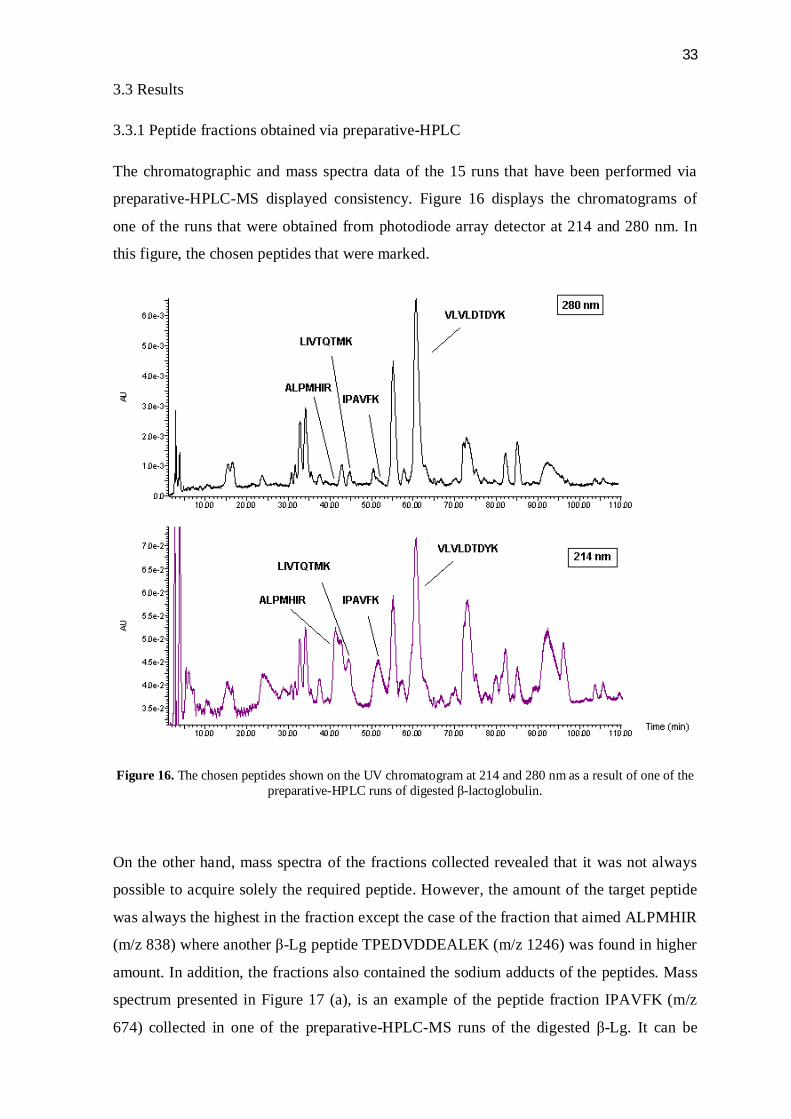
3.3 Results
3.3.1 Peptide fractions obtained via preparative-HPLC
The chromatographic and mass spectra data of the 15 runs that have been performed via
preparative-HPLC-MS displayed consistency. Figure 16 displays the chromatograms of
one of the runs that were obtained from photodiode array detector at 214 and 280 nm. In
this figure, the chosen peptides that were marked.
Figure 16. The chosen peptides shown on the UV chromatogram at 214 and 280 nm as a result of one of the preparative-HPLC runs of digested -lactoglobulin.
On the other hand, mass spectra of the fractions collected revealed that it was not always
possible to acquire solely the required peptide. However, the amount of the target peptide
was always the highest in the fraction except the case of the fraction that aimed ALPMHIR
(m/z 838) where another -Lg peptide TPEDVDDEALEK (m/z 1246) was found in higher
amount. In addition, the fractions also contained the sodium adducts of the peptides. Mass
spectrum presented in Figure 17 (a), is an example of the peptide fraction IPAVFK (m/z
674) collected in one of the preparative-HPLC-MS runs of the digested -Lg. It can be

34
observed here that sodium adducts of the peptide (+22, +44) were present. In addition, this
fraction had another -Lg peptide that has the amino sequence of VLVLDTDYKK (m/z
1194). The presence of this peptide resulted from the activity of trypsin enzyme missing
one cleavage site at Lys. Fractions of LIVTQTMK (m/z 934) included another peptide
resulting from -Lg digestion which had the amino acid sequence of TPEDVDDEALEK
(m/z 1246). The intensity of this peptide in mass spectra was around 1:5 compared to the
target peptide (Figure 17 b). Similar to previous peptide fractions, the sodium adduct of the
target peptide was observed (+22). In another peptide fraction that belonged to
VLVLDTDYK (m/z 1066), several undefined materials were observed besides the target
peptide and its sodium adduct (Figure 17 c). Finally, as mentioned before, fractions of the
peptide ALPMHIR (m/z 838) contained another peptide of TPEDVDDEALEK (m/z 1246)
with an average ratio of 2:5 as seen in MS data in terms of intensity (Figure 17 d). Thus,
the amount of the target peptide was less than half in the fractions collected.
Figure 17. Examples of mass spectra obtained from the fractionation of chosen -lactoglobulin peptides (a) IPAVFK (m/z 674), (b) LIVTQTMK (m/z 934), (c) VLVLDTDYK (m/z 1066) and (d) ALPMHIR (m/z
838).
(d)
(a) (b)
(c)

35
3.3.2 Monitoring the oxidation process in peptide fractions
The data acquired through HPLC was monitored at 214 and 280 nm via photodiode array
detector. In addition, identification of the peaks of interest was confirmed through LC-ESI-
QIR-MS (performed by Tuuli Koivumäki, outside the scope of this thesis).
Peptide fraction VLVLDTDYK (m/z 1066)
This fraction was found to contain several other compounds other than the target peptide in
relatively small amounts as it was viewed from the MS data during the fractionation
process via preparative-HPLC (Figure 17 c). Thus, various peaks were observed on day 0
of the oxidation process (Figure 18). The target peptide VLVLDTDYK was found to elute
after 47.2 minutes with the highest peak area both in 214 and 280 nm. Additionally,
another noticeable peak was present eluting at minute 51.7 with a relatively less peak area
than the target peptide both at 214 and 280 nm. High absorbance values of this compound
at 280 nm as well as 214 nm could be attributed to its hosting the aromatic amino acid Tyr
(Y) and therefore could be the oxidation product of the Tyr-containing target peptide. This
product could have been formed as a result of the biphenyl linkage formed between the
aromatic side-chains of two Tyr residues among peptides and yield a dipeptide as in the
formation of dityrosine.
Figure 18. Presence of the peaks in the peptide fraction VLVLDTDYK on day 0 at 214 nm and 280 nm.

36
On the 21st day of oxidation, UV data at 214 and 280 nm displayed several oxidation
products of both the peptide VLVLDTDYK and other compounds that were initially
present in the fraction. Some of these compounds absorbed UV light considerably at 280
nm. This could be credited to the presence of Tyr in their amino acid sequence, thus these
compounds could be the oxidation products of the main peptide. On the other hand, the
ones that lack this absorbance ability could be the undesired compounds that were included
during the fractionation process. In the day 21 chromatogram presented below in Figure
19, the oxidation products are marked as “P” whereas, the compounds that are considered
to be included during the fractionation are marked as “C”.
Figure 19. Day 21 chromatogram of the peptide fraction VLVLDTDYK where the peaks of interest are marked. (P: oxidation product, C: compounds that were initially present in the fraction other than the target
peptide)
The plot of the main peaks of interest against oxidation time can be observed in Figure 20
in terms of individual percentage values of the peak areas. The target peptide
VLVLDTDYK showed a steep decline during the first day of oxidation and started to

37
increase in peak area during the following days according to the UV data. The peak
interpreted as the dipeptide that was already present on day 0 also decreased in peak area
on the first day of oxidation but later on continued with slow rate of climbing up.
Similarly, the compounds present in the fraction at the beginning had risen during the
course of oxidation. One of the oxidation products which covered a relatively small in peak
area at 214 nm displayed a high absorbance at 280 nm which continued to increase
throughout the monitoring (Figure 19, P 1). Additionally, another product of interest
(Figure 19, P 2) which was observed noticeably both at 214 and 280 nm followed a
continuous level of growth in terms of peak area.
Figure 20. The changes in peak areas of the compounds of interest in terms of percentage versus oxidation period. (P: oxidation product, C: compound initially present in the peptide fraction other than the target
peptide).
Peptide fraction IPAVFK (m/z 674)
Among the several peaks observed in chromatograms of oxidation samples on the different
days, it was identified that the peptide IPAVFK was retained very closely with another
-Lg peptide VLVLDTDYKK (m/z 1194). Due to the presence of Phe (F) in the target
peptide IPAVFK and Tyr (Y) in VLVLDTDYKK, it was possible to identify the peaks of
these different peptides by observing the UV data in 260 nm besides 214 and 280 nm. Day
0 chromatogram at 214 nm that can be found in Figure 21 shows these fractions.

38
According to this, the target peptide for oxidation had a retention time of around 41.1
minutes while the peptide with lower intensity was retained around 40.5th minute.
Figure 21. The presence of peptide fractions of IPAVFK and VLVLVDTDYKK on Day 0 chromatogram
at 214 nm.
Over the course of oxidation period, a slight decrease in the intensity of both present
peptides was observed while several new peaks have emerged that had relatively high
absorbance values in 260 and 280 nm. The comparison of day 0 and day 21 in the three
wavelengths examined are presented in Figure 22.

39
Figure 22. Comparison of oxidation days at 214, 260 and 280 nm on (a) day 0 and (b) day 21.

40
Moreover, changes observed of the target peptide IPAVFK in terms of peak area at 214 nm
indicated a slight decrease while a product formed (retention time = 20 min) at later stages
of the oxidation process has arisen. A graph was prepared where the changes in
compounds of main interest were plotted against oxidation days in terms of percentage that
was calculated from the peak area (Figure 23).
Figure 23. Changes of the main compounds over time in terms of peak area percentage obtained at 214 nm.
Peptide fraction LIVTQTMK (m/z 934)
At day 0, this peptide fraction contained 4 peaks of interest at 214 nm (Figure 24) and none
at 280 nm. These were, in the order of elution: (1) the target peptide with an addition of 16
in m/z value, (2) target peptide with a shift of +32 in m/z value, (3) another -Lg peptide
that was detected in preparative-HPLC-MS data with a m/z value of 1246 (see Figure 17 b)
and (4) the target peptide LIVTQTMK.

41
Figure 24. The UV data obtained in oxidation samples of peptide fraction LIVTQTMK at 214 nm.
Several peaks that occured as a result of oxidative changes in the fraction were observed
that display a continuous increase throughout the oxidation period. These are presented in
Figure 25 on oxidation days 0, 7, 14 and 21.
The target peptide LIVTQTMK was considerably low in day 0 at 214 nm and declined in
amount until day. The peptide with m/z value of 950 displayed slight changes in peak area
at 214 nm during the course of oxidation and reached a higher peak area value at the end of
day 21 than day 0. Another compound of interest with m/z value of 966, showed an
increase between day 0 and 1 in peak area after which a slight decline was observed on day
4. Later on, a continuous increase was detected during the rest of the course. Peptide with a
m/z value of 1246 (TPEVDDEALEK) displayed a route of depletion throughout the
oxidation process. This decline first followed a rapid course until day 4 and then slowed
down during the rest of the oxidation course. Meanwhile, presence of a compound retained
around 22.5th minute was observed at day 7 which continued to increase its peak area on
later stages of oxidation period both at 214 and 280 nm. The changes in the above-
mentioned peaks at 214 nm were plotted against time in Figure 26 in terms of percentage
of individual peak areas.

42
Figure 25. The UV chromatograms obtained during oxidation of the peptide fraction LIVTQTMK at 214 nm on (a) day 0, (b) day 7, (c) day 14 and (d) day 21.
43
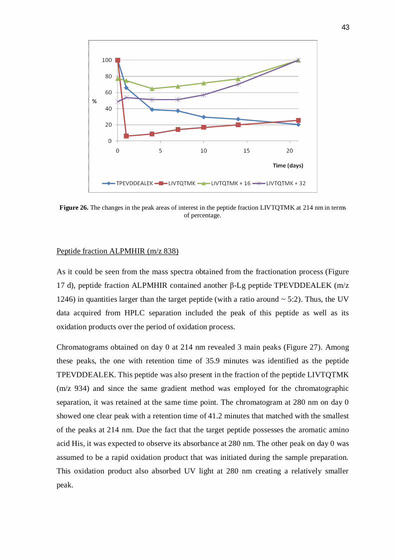
Figure 26. The changes in the peak areas of interest in the peptide fraction LIVTQTMK at 214 nm in terms of percentage.
Peptide fraction ALPMHIR (m/z 838)
As it could be seen from the mass spectra obtained from the fractionation process (Figure
17 d), peptide fraction ALPMHIR contained another -Lg peptide TPEVDDEALEK (m/z
1246) in quantities larger than the target peptide (with a ratio around ~ 5:2). Thus, the UV
data acquired from HPLC separation included the peak of this peptide as well as its
oxidation products over the period of oxidation process.
Chromatograms obtained on day 0 at 214 nm revealed 3 main peaks (Figure 27). Among
these peaks, the one with retention time of 35.9 minutes was identified as the peptide
TPEVDDEALEK. This peptide was also present in the fraction of the peptide LIVTQTMK
(m/z 934) and since the same gradient method was employed for the chromatographic
separation, it was retained at the same time point. The chromatogram at 280 nm on day 0
showed one clear peak with a retention time of 41.2 minutes that matched with the smallest
of the peaks at 214 nm. Due the fact that the target peptide possesses the aromatic amino
acid His, it was expected to observe its absorbance at 280 nm. The other peak on day 0 was
assumed to be a rapid oxidation product that was initiated during the sample preparation.
This oxidation product also absorbed UV light at 280 nm creating a relatively smaller
peak.

44
Figure 27. Day 0 chromatograms of peptide fraction ALPMHIR and the peaks identified at 214 and 280 nm.
The target peptide ALPMHIR which had already undergone rapid oxidation could not be
detected after day 0. The other -Lg peptide TPEVDDEALEK displayed a fast depletion
during the first 4 days and later on continued with a slight reduction in peak area.
Similarly, the compound assumed to be an initial oxidation product on day 0 declined in
peak area over the first 4 days of oxidation process and then on displayed a decrease on a
lower rate at both wavelenghts. On the other hand, starting from day 1, oxidation products
started to appear and continued to rise at 214 nm. One of those products with a retention
time of 22.1 minutes considerably grew over oxidation days both at 214 and 280 nm.
Additionally, another peak that developed at 280 nm during the oxidation process was
retained at 18.1th minute even though it covered a relatively small area at 214 nm. The
oxidative changes between days 1 and 21 with peaks of interest can be observed in Figure
28 both at 214 and 280 nm where oxidation products are marked as “P”.

45
Figure 28. The comparison of oxidation products formed between (a) day 1 and (b) day 21 at 214 nm and their absorbance at 280 nm.
Furthermore, the rate of change in the depletion of initial compounds present in the
fraction and the formation of new oxidation products are plotted in terms of individual
peak area percentage versus oxidation days in Figure 29.
Figure 29. Changes observed in the areas of main peaks over oxidation period.

46
3.4 Discussion
3.4.1 Digestion of -lactglobulin and fractionation of peptides
Majority of the theoretical peptides to be cleaved from the protein as a result of trypsin
activity were observed in the data acquired from preparative high performance liquid
chromatography coupled with mass spectrometry (preparative-HPLC-MS). This enzyme
was chosen due its wide field of use in preparation of -Lg hydrolyzates in several studies
(Iametti et al. 2002; Bonomi et al. 2003; Cheison et al. 2010). In addition the theoretical
peptides that could be achieved from the activity of trypsin on proteins are present in
databases and proteomics software tools. In the case of this study, it was known that
trypsin cleaves -Lg at the carboxyl side of amino acids Arg and Lys unless they are
preceded by Pro.
The chromatograms and the mass spectra obtained from the 15 runs performed with this
equipment showed high similarity with each other and thus proved the digestion of the
protein was successful. Collection of the chosen peptide fractions did not always provide
the single presence of the target peptides due to the co-elution of other -Lg peptides in the
separation process. However, the amounts of the peptides in the fractions were satisfactory
to proceed with the preparation of the oxidation sample. In this part of the experimental
study, chosen peptides were fractionated by programming the auto-collection according to
the m/z values of the peptides. Another technique that could be used was to perform the
fractionation according to the retention time of the peptides to be selected. In conclusion,
the fractionation procedure via preparative-HPLC-MS provided a useful tool to obtain
individual peptides from the enzymatically digested protein.
3.4.2 Interpretation of the oxidative changes in chosen -lactoglobulin peptides
Oxidation of peptide fraction Val108 – Lys116 (VLVLDTDYK)
The amino acid sequence of the target peptide for oxidation in this fraction was Val-Leu-
Val-Leu-Asp-Thr-Asp-Tyr-Lys with a m/z value of 1066. Among these amino acids, Tyr is
the notable one to undergo oxidation with highest rates due to its aromatic side-chain. In
this fraction, day 0 chromatograms showed the presence of another compound with similar
absorbance attributes as the main peptide VLVLDTDYK. It could be assumed that this was
one of the main oxidation products that occurred rapidly due to the high susceptibility of

47
Tyr side-chain. This main oxidation product resulting from Tyr oxidation was considered
to be a dipeptide formation. As it was found from literature, dityrosine is a major end
product of Tyr. Dityrosine is formed via hydrogen atom abstraction from the hydroxyl
group of aromatic side-chain that goes through radical recombination and isomerization as
it was presented in Figure 30 (Giulivi et al. 2003). Similarly, in the main peptide, Tyr side-
chains might have formed biphenyl linkages to undergo a dipeptide formation.
Furthermore, retention of the dipeptide formed through intermolecular Tyr side-chain
linkages were expected to take place after the main peptide in the reverse-phase HPLC
system as it was noted from literature (Gilulivi et al. 2003; Winterbourn et al. 2004;
Roeser et al. 2010). In addition to this major product, there are several other compounds
that can yield through Tyr oxidation that involves hydroxylation and hydrogen abstraction
processes. These mechanisms are able to form several products that have m/z values of -2,
+16 and +14 compared to the main peptide (Roeser et al. 2010). Nevertheless, presence of
these oxidation products were also confirmed through LC-MS data with m/z values of
1064, 1082 and 1080. These compounds in oxidation samples were observed to absorb UV
light at 280 nm, meaning that they contain aromatic side-chain modifications of Tyr. In
conclusion, formation of intermolecular links between Tyr side-chains could be utilized as
a marker for peptides containing Tyr as it is detected easily.
Apart from oxidation products of Tyr in this peptide, Lys was also capable of undergoing
oxidation during the monitored time period. As reported by Requena et al. 2001, Lys
residues in peptides and proteins are known to yield -aminoadipic semialdehyde (AAS) in
metal-catalyzed oxidation systems that is initiated by the abstraction of a H atom from
carbon at position 6 and through losing the NH2 moiety. AAS is also known to be involved
in further reactions to result in 6-hydroxy-2-aminocaproic acid (HACA). However, this
process requires reduction of AAS by NaBH4 and later hydrolysis by HCl which are not
present in our oxidation system and thus HACA was not expected to form in the oxidation
samples. In addition, 3-hydroxylysine was reported to be another oxidation product arising
from Lys (Morin et al. 1998). Thus, some of the products observed during oxidation could
be attributed to the reactions of Lys side-chains combined with Tyr side-chain oxidation.
Moreover, due to the presence of several other compounds than the target peptide in the
fraction that was found to be included after fractionation process, various peaks were
observed on the HPLC chromatograms. These peaks were believed to be identified as those
undesired compounds in the fraction since they do not display absorbance at 280 nm,
considering that they were not related to the peptide VLVLDTDYK.

48
The initial rapid depletion of the target peptide on the first day of oxidation was accredited
to vulnerability of Tyr residues to radical attack. However, during the rest of the
monitoring, the peak of this peptide displayed a route of growth which was assumed to be
due to the co-elution of some of the oxidation products with the main peptide peak.
Additionally, at the end of the monitoring process of oxidation period, the resulting peak
area of the target peptide may suggest that the further oxidation of the target peptide after
the initial stages were hindered or the peptide displayed some resistance to oxidation.
Figure 30. Mechanism of dityrosine formation (Giulivi et al. 2003).
Oxidation of peptide fraction Ile94 – Lys99 (IPAVFK)
This peptide had the amino acid sequence of Ile-Pro-Ala-Val-Phe-Lys (m/z 674). Among
these amino acids, Phe (F) is known to be the most vulnerable one to oxidation which
contains an aromatic side-chain that could be observed in UV chromatograms at 280 nm.
On the other hand, this peptide sample also contained another -Lg peptide that resulted
from one missed cleavage by trypsin. The amino acid sequence of this peptide was found
to be Val-Leu-Val-Leu-Asp-Thr-Asp-Tyr-Lys-Lys with a m/z value of 1194. Thus, it is
fair to assume that the samples would also include the changes that were manifested by the
oxidation of this peptide. Retention times of these two peptides appeared to be close to
each other at during chromatographic separation. Therefore, due to the high absorbance of

49
UV light of Phe in the peptide IPAVFK at 260 nm, chromatograms at this wavelength
enabled us to identify the target peptide retention.
Oxidation products of Phe side-chains arise due to the hydroxylation of the aromatic ring.
These result in the formation of m-Tyr, o-Tyr and Tyr side-chains. Further oxidative
reactions of these side-chains may end up forming links between the side chains and lead
to dimerization Huggins et al. 1993; Davies et al. 1999). In our oxidation samples, several
oxidation samples appeared and followed a growing route that displayed noticeable
absorbance at 280 nm. The fact that these peaks were noted at 280 nm was accredited to
the conversion of Phe side-chains into hydroxylated ones, since Phe is not expected to
absorb UV light at 280 nm at this level. Moreover, it was also possible that one of these
oxidation products could be the resulting from the dimerization of the target peptide due to
the links formed between hydroxylated Phe side-chains. On the other hand, the slight
changes and the relatively low amount of depletion in the peak area of the target peptide
during the oxidation period may point out that the extent of oxidation did not reach very far
even though several oxidation products were observed.
Oxidative behavior of the other -Lg peptide that was present in the fraction,
VLVLDTDYKK (m/z 1194) was expected to be similar to the main peptide in the previous
fraction VLVLDTDYK (m/z 1066) due the similarity in amino acid sequence. Thus, Tyr
(Y) side-chains were presumed to play a significant role in the oxidation of this peptide.
However, the anticipated oxidation product of this peptide, the dipeptide was not observed
clearly due to the difference in the gradient method applied for the chromatographic
separation between this fraction and the previous one that harbors the similar peptide. It
was possible that the dipeptide formation of VLVLDTDYKK due to Tyr side-chain
oxidation was retained at the same time of the unoxidized peptide and thus creating an
overlap in peaks. This could also be the reason that the peak area of this peptide showed
fluctuations.
Oxidation of peptide fraction Leu17 – Lys24 (LIVTQTMK)
The main peptide in this fraction had the following amino acid sequence: Leu-Ile-Val-Thr-
Gln-Thr-Met-Lys. Met (M), here represents an amino acid that is highly susceptible to
oxidation. Initial oxidation product of Met residues is Met sulfoxide which results from the
addition of an oxygen atom to the sulfur-containing side-chain. This reaction, however, is

50
reversible and Met sulfoxide can be reduced back to Met residues and thus not considered
to be dependable marker for oxidation especially in quantitative studies (Davies et al.
1999). Further oxidation of Met sulfoxide leads to the formation of Met sulfone via
addition of another oxygen atom to sulfur. This oxidation reaction is known to be
irreversible, however is not encountered in high rates (Vogt 1995).
In our fraction, the target peptide LIVTQTMK (m/z 934) was readily oxidized to a great
extent and oxidation products could be observed on day 0. Met sulfoxide (m/z 950) was
highest in amount, demonstrating the ease of the yield of this product. In addition, Met
sulfoxide was also oxidized to Met sulfone (m/z 966) to a lesser extent. The fast decline of
the absorbance peak of the peptide LIVTQTMK on the first day showed that the oxidation
of this peptide continued. However, the slight increase of the peak area of this peptide
during the following days could be a sign of the reduction of Met sulfoxide residues to Met.
However, after day 4, a growth pattern was observed both in the major oxidation products
and the target peptide itself. Thus, it can be concluded that Met sulfone was produced
continuously. The slight growth in peptide peak could be attributed to Met sulfoxide
reduction, however, it was also found out that amount of Met sulfoxide was on the rise
during the later stages of oxidation process. This could be caused by various oxidation
products formed through other mechanisms being retained the same time as Met sulfoxide
peak.
Another -Lg peptide was also present in this fraction in relatively lesser quantities. The
amino sequence of this peptide was found to be Thr-Pro-Glu-Val-Asp-Asp-Glu-Ala-Leu-
Glu-Lys (m/z 1246). It would be fair to assume that the presence of this peptide also
contributed to the outcome of oxidation products observed in the chromatograms. In fact, it
was noticeable that the peak area of this peptide reduced continuously. The main oxidation
products were assumed to be resulting from the oxidation of Lys and Pro. As mentioned
before, oxidation of Lys leads to formation of AAS and 3-hydroxylysine. On the other
hand, Requena et al. 2001 notes that oxidation of Pro residues yield in -glutamic
semialdehyde (GGS). Similar to AAS, GGS is susceptible to further reduction reactions by
NaBH4 and hydrolysis by HCl to form 5-hydroxy-2-aminovaleric acid (HAVA) which was
not anticipated to appear in our samples. Formation mechanism of AAS from Lys residues
and GGS from Pro residues is presented below in Figure 31.
Furthermore, formation of two other notable oxidation products was observed in this
peptide fraction with a retention time of 18.3 and 22.2 minutes. These products both

51
displayed high absorbance at 280 nm. This could be credited to the presence of aromatic
side-chains that featured from modification of AAS and GGS into a nitrogen-containing
ring derived side-chains as could be seen in Figure 31.
On the whole, oxidation of peptide fraction LIVTQTMK provided a clear oxidation
monitoring, especially due to the products formed through oxidation of Met side-chain and
Met sulfoxide in particular could be considered as a marker in detection of oxidation of
-Lg peptides.
Figure 31. Formation mechanism for -aminoadipic semialdehyde (AAS) and -glutamic semialdehyde (GGS) from Lys and Pro residues in proteins, respectively (Akagawa et al. 2006).
Oxidation of peptide fraction Ala158 – Arg164 (ALPMHIR)
The target peptide in this fraction had a m/z value of 838 and the amino acid sequence of
Ala-Leu-Pro-Met-His-Ile-Arg where His and Met were the vulnerable amino acids to
oxidative damage. Presence of these two amino acids that are highly reactive with radical
species made this peptide a readily oxidized one. Thus, it was not possible to detect it after
day 0 at either 214 or 280 nm.

52
Modifications likely to occur due to the sulfur-containing side-chain of Met were discussed
before in the previous peptide fraction as Met sulfoxide and Met sulfone being the major
oxidation products. The other highly reactive amino acid His is easily oxidized in metal-
catalyzed systems to yield 2-oxo-histidine as the main product and several others such as
asparagine and aspartic acid that are formed through ring-opening reactions (Uchida 2003).
Thus, the absence of absorbance peaks at 280 nm of several oxidation products observed at
214 nm could be attributed to the fact that these ring-opening reactions took place at the
side-chain of His. Moreover, various products arising as a result of both Met and His side-
chains in the target peptide were observed during the monitoring of oxidation process in
this fraction. These products were also confirmed by LC-MS with respect to their m/z
values which displayed shifts of +16, +32, +48 and +64 due to the addition of oxygen
atoms to the side-chains of these amino acids.
In addition, presence of the amino acids Pro and Arg in the target peptide was also likely to
contribute to the oxidation of this peptide. Pro was known to form GGS as oxidation
product as discussed before. Similarly, Arg also causes the formation of GGS in case of
oxidation. Thus, similar peaks were to be expected and observed as to the previous peptide
fraction LIVTQTMK (m/z 934). The reaction scheme of Arg oxidation into GGS can be
followed in Figure 32.
Figure 32. Reaction scheme of Arg oxidation into -glutamic semialdehyde, GGS (Akagawa et al. 2006).
It was also revealed during the fractionation process that another -Lg peptide was present
in this fraction. This peptide, which was also seen in the previous peptide fraction of
LIVTQTMK, had the m/z value of 1246 and amino acid sequence of TPEVDDEALEK.
Therefore, a comparison with the other peptide fraction helped identify the peak of this

53
peptide retained in the day 0 chromatogram. Again, in similar to the oxidation of the
peptide fraction LIVTQTMK, existence of this peptide was likely to cause formation of its
oxidation products. Consequently, same peaks that eluted at minutes 18.3 and 22.2 during
the oxidation of the previous fraction appeared to rise in this one as well. These peaks were
assumed to be the result of the ring-forming modifications occurring at the side chains of
AAS and GGS residues that were formed in the peptide TPEVDDEALEK due to oxidation
Lys (K) and Pro (P) side-chains.

54
4 CONCLUSIONS
This study investigated the oxidative modifications certain -lactoglobulin peptides
underwent. In order to obtain these peptides, the protein was enzymatically digested with
trypsin and the resulting amino acid sequences of the peptides were found in a database via
a proteomics tool. Fractionation of peptides to be oxidized was performed with respect to
their m/z values via preparative-HPLC-MS followed by the preparation of oxidation
samples of each peptide fraction. Oxidation was monitored for 21 days using reverse
phase-HPLC connected to a photodiode array detector. The chromatographic separations
of the samples were viewed at 214 and 280 nm based on the literature data.
Preparative-HPLC-MS provided to be a useful tool in the fractionation process where the
peptides chosen were selected according to their m/z value and chromatographic separation
was achieved with an optimized gradient method. However, it was not possible to collect
the target peptides exclusively in the fractions due to the co-elution of several compounds.
Considering the high amount of cleavage possibilities caused by the enzyme activity, it
was anticipated to find such yield of compounds. Nonetheless, the amounts of the target
peptides in the fractions were satisfactory to carry out the oxidation and monitor the
changes.
The oxidation system employed for the peptides functioned satisfactorily since evident
oxidative alterations in the samples were observed. Gradient methods for clear separation
for each peptide sample were adjusted during the analysis with reverse phase-HPLC runs.
These methods were proved to be optimal applications in most cases to observe the
changes in peaks of main peptides and oxidation products.
The peptide fraction of Val108 – Lys116 with an amino acid sequence of Val-Leu-Val-Leu-
Asp-Thr-Asp-Tyr-Lys (m/z 1066) had an oxidation product that could be observed on day
0. This product was assumed to be resulting from the intermolecular linkages formed via
Tyr side-chains during oxidation. In later stages of oxidation, products arising from
hydroxylated and hydrogen abstracted forms of Tyr side-chains appeared as well. However,
no considerable change of this peptide was displayed after the initial oxidative depletion.
In conclusion, the oxidation of this peptide could be marked by the formation of the
products that could be viewed at 280 nm due to the aromatic side-chain of Tyr.

55
The peptide cleaved at Ile94 – Lys99 from the protein had the amino acid sequence of Ile-
Pro-Ala-Val-Phe-Lys with a m/z value of 674. This peptide could easily be marked for its
possession of Phe which exhibits high absorbance of UV light at 260 nm. However, this
was not the case for the products arising from the oxidative modification of Phe side-
chains since they were not especially favoring this wavelength. On the other hand,
according to the UV data, a considerable amount of this peptide stayed unoxidized even
though several oxidation products formed.
The peptide with the sequence of Leu-Ile-Val-Thr-Gln-Thr-Met-Lys (m/z 934) cleaved at
Leu17 – Lys24 from -lactoglobulin showed an initial high-rate oxidation and started to
increase due to the reduction of an oxidation product of Met side-chains. This formation
that involves the addition of an oxygen atom to the sulfur on the side-chain proved to be
the major oxidation product and easily detectable even though the oxidation reaction was
not irreversible. Furthermore, addition of another oxygen atom to this product provided
another marker for the oxidative changes of the main peptide.
The last peptide sample including Ala158 – Arg164 fraction as the target peptide with the
amino acid sequence of Ala-Leu-Pro-Met-His-Ile-Arg (m/z 838) supplied several products
upon oxidation. This was because of the presence of highly oxidative amino acids Met and
His. The modifications observed during the HPLC analysis included the formation of
oxygenated and hydroxylated side chains of these amino acids and rapid consumption of
the target peptide already on the first day.
In conclusion, the method employed in this study provided a useful means to monitor the
oxidative alterations manifested in the chosen -lactoglobulin peptides. In addition, further
characterizations of these modifications can be performed through recent advance methods
such as mass spectrometry. Being a significant protein in milk and widely studied allergen,
oxidation of -lactoglobulin and the utilization of its peptides in protein oxidation studies
may offer a better understanding to this relatively uncovered field.

56
REFERENCES
Akagawa M, Sasaki D, Ishii Y, Kurota Y, Yotsu-Yamashita M, Uchida K, Suyama K. 2006. New method for the quantitative determination of major protein carbonyls, alpha-aminoadipic and gamma-glutamic semialdehydes: Investigation of the formation mechanism and chemical nature in vitro and in vivo. Chem Res Toxicol 19: 1059-1065.
Amici A, Levine RL, Tsai L, Stadtman ER. 1989. Conversion of amino acid residues in proteins and amino acid homopolymers to carbonyl derivatives by metal-catalyzed oxidation reactions. J Biol Chem 264(6): 3341-3346.
Bertrand-Harb C, Baday A, Dalgalarrondo M, Chobert J-, Haertle T. 2002. Thermal modifications of structure and co-denaturation of alpha-lactalbumin and beta-lactoglobulin induce changes of solubility and susceptiblity to proteases. Nahrung 46(4): 283-289.
Bonomi F, Fiocchi A, Frokiaer H, Gaiaschi A, Iametti S, Poiesi C, Rasmussen P, Restani P. 2003. Reduction of immunoreactivity by bovine beta-lactoglobulin upon combined physical and proteolytic treatment. J Dairy Res 70: 51-59.
Chen WL, Hwang MT, Liau CY, Ho JC, Hong KC, Mao SJT. 2005. Beta-lactoglobulin is a thermal marker in processed milk as studied by elechtophoresis and circular dichroic spectra. J Dairy Sci 88: 1618-1630.
Dalsgaard TK, Otzen D, Nielsen JH, Larsen LB. 2007. Changes in structures of milk proteins upon photo-oxidation. J Agric Food Chem 55: 10968-10976.
Daneshvar B, Frandsen H, Autrup H, Dragsted LO. 1997. Gamma-glutamyl semialdehyde and 2-amino-adipic semialdehyde: Biomarkers of oxidative damage to proteins. Biomarkers 2: 117-123.
Davies KJA, Delsignore ME. 1987. Protein damage and degradation by oxygen radicals III. Modification of secondary and tertiary structure. J Biol Chem 262(20): 9908-9913.
Davies KJA, Delsignore ME, Lin SW. 1987. Protein damage and degradation by oxygen radicals II. Modification of amino acids. J Biol Chem 262(20): 9902-9907.
Davies MJ. 2005. The oxidative environment and protein damage. Biochim Biophys Acta 1703: 93-109.
Davies MJ. 1996. Protein and peptide alkoxyl radicals can give rise to C-terminal decarboxylation and backbone cleavage. Arch Biochem Biophys 336(1): 163-172.
Davies MJ, Fu S, Dean RT. 1995. Protein hydroperoxides can give rise to free radicals. Biochem J 305: 643-649.
Davies MJ, Fu S, Wang H, Dean RT. 1999. Stable markers of oxidative damage to proteins and their application in the study of human disease. Free Radical Bio Med 27(11/12): 1151-1163.
Elias RJ, Bridgewater JD, Vachet RW, Waraho T, McClements DJ, Decker EA. 2006. Antioxidant mechanisms of enzymatic hydrolysates of beta-lactoglobulin in food lipid dispersions. J Agric Food Chem 54: 9565-9572.
Elias RJ, Kellerby SS, Decker EA. 2008. Antioxidant activity of proteins and peptides. Crit Rev Food Sci 48: 430-441.
Elias RJ, McClements DJ, Decker EA. 2007. Impact of thermal processing on the antioxidant mechanisms of continuous phase beta-lactoglobulin in oli-in-water emulsions. Food Chem 104: 1402-1409.
Elias RJ, McClements DJ, Decker EA. 2005. Antioxidant activity of cysteine, tryptophan and methionine residues in continuous phase beta-lactoglobulin in oil-in-water emulsions. J Agric Food Chem 53: 10248-10253.
Ellman GL. 1959. Tissue sulfhydryl groups. Arch Biochem Biophys 82(1): 70-77.
Estevez M, Morcuende D, Ventanas S. 2008. Determination of Oxidation. In: L. M. L. Nollet, F. Toldra, Editors. Handbook of Processed Meats and Poultry Analysis. USA: CRC Press. p141-162.

57
Estevez M, Ollilainen V, Heinonen M. 2009. Analysis of protein oxidation markers alpha-aminoadipic and gamma-glutamic semialdehydes in food proteins using liquid chromatography (LC) - electrospray ionization (ESI) - multistage tandem mass spectrometry (MS). J Agric Food Chem 57: 3901-3910.
Faergemand M, Otte J, Qvist KB. 1998. Cross-linking of whey proteins by enzymatic oxidation. J Agric Food Chem 46: 1326-1333.
Finley EL, Dillon J, Crouch RK, Schey K. 1998. Identification of tryptophan oxidation products in bovine alpha-crystallin. Protein Sci 7: 2391-2397.
Garrison WM. 19897. Reaction mechanisms on the radiolysis of peptides, polypeptides, and proteins. Chem Rev 87: 381-398.
Gieseg SP, Simpson JA, Charlton TS, Duncan MW, Dean RT. 1993. Protein-bound 3,4-dihydroxyphenylalanine is a major reductant formed during hydroxyl radical damage to proteins. Biochemistry 32: 4780-4786.
Giulivi C, Traaseth NJ, Davies KJA. 2003. Tyrosine oxidation products: analysis and biological relevance. Amino Acids 25: 227-232.
Gracanin M, Hawkins CL, Pattison DI, Davies MJ. 2009. Singlet-oxygen-mediated amino acid and protein oxidation: Formation of tryptophan peroxides and decomposition products. Free Radical Biol Med 47: 92-102.
Griffiths SW. 2002. Oxidation of the Sulfur-Containing Amino Acids in Recombinant Human Alpha1-Antitrypsin. [dissertation]. MA, USA: Massachusetts Institute of Technology.
Grosvenor AJ, Morton JD, Dyer JM. 2010. Profiling of residue-level photo-oxidative damage in peptides. Amino Acids 39: 286-296.
Guedes S, Vitorino R, Domingues R, Amado F, Domingues P. 2009. Oxidation of bovine serum albumin: Identification of oxidation products and structural modifications. Rapid Commun Mass Spectrom 23: 2307-2315.
Hambling SG, McAlpine AS, Sawyer L. 1992. Beta-Lactoglobulin. In: P. F. Fox, Editor. Advanced Dairy Chemistry - Volume 1: Proteins. Cambridge, UK: Elsevier Science Publishers, Ltd. p141.
Hawkins CL, Davies MJ. 2001. Generation and propagation of radical reactions on proteins. Biochim Biophys Acta 1504: 196-219.
Hawkins CL, Pattison DI, Davies MJ. 2003. Hypochlorite-induced oxidation of amino acids, peptides and proteins. Amino Acids 25: 259-274.
Headlam HA, Davies MJ. 2004. Markers of protein oxidation: Different oxidants give rise to variable yields of bound and released carbonyl products. Free Radical Bio Med 36(9): 1175-1184.
Hernandez-Ledesma B, Davalos A, Bartolome B, Amigo L. 2005. Preparation of antioxidant enzymatic hydrolysates from alpha-lactalbumin and from beta-lactoglobulin. Identification of active peptides by HPLC-MS/MS. J Agric Food Chem 53: 588-593.
Huggins TG, Wells-Knecht MC, Detorie NA, Baynes JW, Thorpe SR. 1993. Formation of o-tyrosine and dityrosine in proteins during radiolytic and metal-catalyzed oxidation. J Biol Chem 268(17): 12341-12347.
Iametti S, Rasmussen P, Frokiaer H, Ferranti P, Addeo F, Bonomi F. 2002. Proteolysis of bovine beta-lactoglobulin during thermal treatment in subdenaturating conditions highlights some structural features of the temperature-modified protein and yields fragments with low immunoreactivity. Eur J Biochem 269: 1362-1372.
Jakobsson I, Borulf S, Lindberg T, Benediktsson B. 1983. Partial hydrloysis of cow's milk proteins by human trypsins and elastases in vitro. J Pediatr Gastr Nutr 2(4): 613-616.
Jankowski CK, Sichel DI. 2003. Modelling of beta-lactoglobulin (BLG) oxidative folding. J Mol Struc-Theochem 629: 185-196.

58
Kehoe JJ, Remondetto GE, Subirade M, Morris ER, Brodkorb A. 2008. Tryptophan-mediated denaturation of beta-lactoglobulin A by UV radiation. J Agric Food Chem 56: 4720-4725.
Kim SB, Ki KS, Khan MA, Lee WS, Lee HJ, Ahn BS, Kim HS. 2007. Peptic and tryptic hydrolysis of native and heated whey protein to reduce its antigenicity. J Dairy Sci 90: 4043-4050.
Levine RL, Berlett BS, Moskovitz J, Mosoni L, Stadtman ER. 1999. Methonine residues may protect proteins from critical oxidative damage. Mech Ageing Dev 107: 323-332.
Levine RL, Mosoni L, Berlett BS, Stadtman ER. 1996. Methionine residues as endogenous antioxidants in proteins. Proc Natl Acad Sci USA 93: 15036-15040.
Levine RL, Williams JA, Stadtman ER. 1994. Carbonyl assays for determination of oxidatively modified proteins. Methods Enzymol 233: 346-357.
Liu G, Xiong YL. 2000. Oxidatively induced chemical changes and interactions of mixed myosin, beta-lactoglobulin and soy 7S globulin. J Sci Food Agric 80: 1601-1607.
Liu HC, Chen WL, Mao SJT. 2007. Antioxidant nature of bovine milk beta-lactoglobulin. J Dairy Sci 90: 547-555.
Madsen JS, Ahmt TO, Otte J, Halkier T, Qvist KB. 1997. Hydrolysis of beta-lactoglobulin by four different proteinases monitored by capillary electrophoresis and high performance liquid chromatography. Int Dairy J 7: 399-409.
Malencik DA, Anderson SR. 2003. Dityrosine as a product of oxidative stress and fluoroscent probe. Amino Acids 25: 233-247.
Mamone G, Picariello G, Caira S, Addeo F, Ferranti P. 2009. Analysis of food proteins and peptides by mass spectrometry-based techniques. J Chromatogr A 1216: 7130-7142.
Matsukura N, Kawachi K, Morino K, Ohgaki H, Sugimura T, Takayama S. 1981. Carcinogenicity in mice of mutagenic compounds from a tryptophan pyrolyzate. Science 213: 346-347.
Meltretter J, Becker C-, Pischetsrieder M. 2008. Identification and site-specific relative quantification of beta-lactoglobulin modifications in heated milk and dairy products. J Agric Food Chem 56: 5165-5171.
Meltretter J, Pischetsrieder M. 2008. Application of mass spectrometry for the detection of glycation and oxidation products in milk proteins. Ann N Y Acad Sci 1126: 134-140.
Meltretter J, Seeber S, Humeny A, Becker C-, Pischetsrieder M. 2007. Site-specific formation of Maillard, oxidation and condensation products from whey proteins during reaction with lactose. J Agric Food Chem 55: 6096-6103.
Miyahara M, Ito H, Nagasawa T, Kamimura T, Saito A, Kariya M, Izumi K, Kitamura M, Toyoda M, Saito Y. 2000. Determination of o-tyrosine production in aqueous solutions of phenylalanine irradiated with gamma ray, using high performance liquid chromatography with automated pre-column derivatization and LASER fluorometric detection. J Health Sci 46(3): 192-199.
Morin B, Bubb WA, Davies MJ, Dean RT, Fu S. 1998. 3-Hydroxylysine, a potential marker for studying radical-induced protein oxidation. Chem Res Toxicol 11: 1265-1273.
Morin B, Davies MJ, Dean RT. 1998. The protein oxidation product 3,4-dihydroxyphenylalanine (DOPA) mediates oxidative DNA damage. Biochem J 30: 1059-1067
Noble RW, Gibson QH. 1970. The reaction of ferrous horseradish peroxidase with hydrogen peroxide. J Biol Chem 245(9): 2409-2413.
Rehder DS, Borges CR. 2010. Cysteine sulfenic acid as an intermediate in disulfide bond formation and nonenzymatic protein folding. Biochemistry 49: 7748-7755.
Requena JR, Chao C-, Levine RL, Stadtman ER. 2001. Glutamic and aminoadipic semialdehydes are the main carbonyl products of metal-catalyzed oxidation of proteins. Biochemistry 98(1): 69-74.

59
Requena JR, Levine RL, Stadtman ER. 2003. Recent advances in the analysis of oxidized proteins. Amino Acids 25: 221-226.
Roeser J, Permentier HP, Bruins AP, Bischoff R. 2010. Electrochemical oxidation and cleavage of tyrosine- and tryptphan-containing tripeptides. Anal Chem 82: 7556-7565.
Saeed S, Gillies D, Wagner G, Howell NK. 2006. ESR and NMR spectroscopy studies on protein oxidation and formation of dityrosine in emulsions containing oxidized methyl linoleate. Food Chem Toxicol 44: 1385-1392.
Salminen H, Jaakkola H, Heinonen M. 2008. Modifications of tryptophan oxidation by phenolic-rich materials. J Agric Food Chem 56: 11178-11186.
Salvi A, Carrupt P-, Testa B. 1999. Development of a capillary electrophoresis method to monitor protein oxidation and antioxidant protection. Helv Chim Acta 82: 870-878.
Schöneich C. 2000. Mechanisms of metal-catalyzed oxidation of histidine to 2-oxo-histidine in peptides and proteins. J Pharm Biomed Anal 21: 1093-1097.
Shimoyamada M, Yoshimura H, Tomida K, Watanabe K. 1996. Stabilities of bovine beta-lactoglobulin/retinol or retinoic acid complexes against tryptic hydrolysis, heating and light-induced oxidation. Lebensm Wiss u Technol 29: 763-766.
Stadtman ER. 1993. Oxidation of free amino acids and amino acid residues in proteins by radiolysis and by metal-catalyzed reactions. Annu Rev Biochem 62: 797-821.
Stadtman ER, Berlett BS. 1997. Reactive oxygen-mediated protein oxidation in aging and disease. Chem Res Toxicol 10: 485-494.
Stadtman ER, Oliver CN. 1991. Metal-catalyzed oxidation of proteins: Physiological consequences. J Biol Chem 266(4): 2005-2008.
Taborsky G. 1973. Oxidative modification of proteins in the presence of ferrous ion and air. Effect of ionic constituents of the reaction medium on the nature of oxidation products. Biochemistry 12(7): 1341-1348.
Uchida K. 2003. Histidine and lysine as targets of oxidative modification. Amino acids 25: 249-257.
Uchida K, Kato Y, Kawakishi S. 1990. A novel mechanism for oxidative cleavage of prolyl peptides induced by the hydroxyl radical. Biochem Biophys Res Commun 169(1): 265-271.
Uchida K, Kawakishi S. 1993. 2-Oxo-histidine as a novel biological marker for the oxidatively modified proteins. FEBS Lett 332(3): 208-210.
Vogt W. 1995. Oxidation of methionyl residues in proteins: Tools, targets, and reversal. Free Radical Biol Med 18(1): 93-105.
Wal JM. 1998. Cow's milk allergens. Allergy 53:1013-1022.
Winterbourn CC, Parsons-Mair HN, Gebicki S, Gebicki JM, Davies MJ. 2004. Requirements for superoxide-dependent tyrosine hydroperoxide formation in peptides. Biochem J 381: 241-248.