Embed Size (px)
Citation preview

Citation: Egypt. Acad. J. Biolog. Sci. (E-Medical Entom. & Parasitology Vol.12(1) pp 77-84(2020)
Egypt. Acad. J. Biolog. Sci., 12 (1):77 – 84 (2020)
Egyptian Academic Journal of Biological Sciences
E. Medical Entom. & Parasitology
ISSN: 2090 – 0783
www.eajbse.journals.ekb.eg
Morphological Characterization of Monezia expansa Rudolphi, 1810 (F:
Anoplocephalidae) isolated from the intestine of the domestic sheep, Ovis aries (Bovidae)
by light microscopy
and1,3 , Kareem Morsy1, Salma Yehia2, Manal Ahmed2Ganainy-, Sahar El1Mona Fol
* 4Asmaa Adel
1-Zoology Department, Faculty of Science, Cairo University, Cairo, Egypt
2- Zoology Department, Faculty of Science, Minia University, Minya, Egypt
3- Biology Department, Faculty of Science, King Khalid University, Saudi Arabia
4- Zoology Department, Faculty of Science, South Valley University, Qena, Egypt
E-mail: [email protected]
ARTICLE INFO ABSTRACT Article History
Received:16/4/2020
Accepted:26/6/2020
_________________
Keywords: Cestoda, Ovis
aries,
Anoplocephalidae,
morphology.
In the present study, A cestode parasite was recovered from the
intestine of the domestic sheep, Ovis aries (Bovidae) from the main
slaughterhouse of Cairo, Egypt during the year of 2015. The parasite was
observed attached to the wall of the host intestine by unarmed scolex with
suckers only. Five out of 10 gastrointestinal tracts (50%) were found to be
infected. Light microscopic examination showed that the adult worm of this
species was whitish in color measured 421-492 (470±0.4) cm in length and
4.2-6.99 (5.6±0.7) mm in width. The scolex was with prominent four suckers
measured 0.38-0.42mm in diameter. The scolex led into a long neck followed
by segments. The mature segment was broader than longer, each proglottid
measured 3.23-5.29 (4.6± 0.2) mm in width and 1.0-1.64 (1.4±0.02) mm in
width and showed a two set of genital organs. The ovaries and the vitelline
glands formed a ring on either side, median to the longitudinal excretory
canals, while the testes were distributed throughout the central field or they
may be concentrated toward the sides. Each ovary was of ovoid shape,
measured about 0.16-0.19 (0.14±0.01) × 0.12-0.15 (0.13±0.02) mm and was
located 0.8± 0.1 mm apart from the lateral side. The testes were concentrated
on both sides. At the posterior border of each proglottid, a row of
interproglotidal glands was arranged around small pits. Gravid segments
measured 0.022-0.076 (0.048±0.2) mm in length and 2.10-5.02 (3.05±0.02)
mm in width. The present parasite was compared with the previous species of
the same host which showed that the parasite isolated is Monezia expansa.
INTRODUCTION
Moniezia is a genus of tapeworms parasitic in mammals, comprises four known
species such named M. expansa, M. benedeni, M. autumnalis and M. baeri. M. expansa is the
most well-known species within the genus because of its high prevalence. Members of the
genus are among the largest cestodes reaching up to 10 m in length. They inhabit the small
intestine of the mammalian host. Their life cycle is indirect requiring intermediate hosts,
which are the oribatid mites. They are characterized by the presence of interproglottid glands
(Mehlhorn, 2008).

Mona Fol et al. 78
Moniezia expansa is commonly
known as sheep tapeworm or double-pored
ruminant tapeworm. It could be considered
as the most important cestode parasite
infecting sheep causing monieziasis which
constitutes a problem in sheep breeding
(Becker et al., 1981, Polec 1990 and Maziad
and El-Nemr, 2002). It is a large tapeworm
inhabiting the small intestines of ruminants
such as sheep, goats, and cattle (Gómez-
Puerta et al., 2008). There is an unusual
report of human infection in Egypt (El-
Shazly et al., 2004). It is characterized by the
presence of an unarmed scolex (i.e., hooks
and rostellum are absent), two sets
of reproductive systems in each proglottid,
and each proglottid being short but very
broad. M. expansa has a typical cestode
body, consisting of the anterior scolex,
followed by the neck and a highly extended
body proper, the strobilus. It is an extremely
long tapeworm and can reach an enormous
length of up to 6–10 m. The scolex bears
four large suckers, which are the holdfast
organs to the host (Bashtar et al., 2011).
There are no rostellum and rostellar hooks,
and the suckers are devoid of spines. This
tapeworm, being monecious, contains both
male and female reproductive organs in an
individual. Thus, each proglottid is a
complete reproductive unit. Moreover, one
defining feature of the genus is that there are
two sets of reproductive organs situated at
lateral sides with the associated cirrus
pouches and genital pores in each proglottid.
The testes are numerous (Bashtar et al.,
2011). M. expansa infections are generally
harmless and asymptomatic, even when the
tapeworms are present in large numbers in
young lambs. However heavy infection may
cause intestinal obstruction, diarrhea, and
weight loss (Elliott, 1986). The complete life
cycle requires two hosts, ruminants
as definitive hosts, and oribatid mites
as intermediate hosts (Sinitsin, 1931 and
Denegri et al., 1998). Eggs are passed out
from the intestine of the ruminant host along
the gravid proglottids in the feces into the
soil. The eggs are eaten by soil mites. Eggs
must reach the gut of mite hosts within 1 day
of release otherwise they are desiccated.
However, chances of development are very
good as soil mites can be so numerous on
a pasture that even if only 3% are infected
(with 4-13 cysticercoids each), a grazing
ruminant may ingest over 2,000
cysticercoids per kilogram of grass. Once
inside the intestine of mites, the eggs hatch,
and the oncospheres penetrate into
the haemocoel and develops into the
cysticercoid stage. This stage may take up to
4 months. When the infected mite is eaten by
the grazing ruminants, mature cysticercoids
are digested out of the mite and develop into
mature tapeworms in the small intestine
within 5–6 weeks (Denegri et al., 1998). In
the present study morphological
characterization of M. expansa, a cestode
parasite in the intestine of the domestic
sheep, Ovis aries is carried out on the basis
of light microscopy with a complete
description of its different body parts
including scolex, immature, and mature
segments.
MATERIALS AND METHODS
The present study was conducted on ten
gastrointestinal tracts collected from the
domestic sheep, Ovis aries (Bovidae), from
the main slaughterhouse of Cairo, Egypt, the
work was approved by the institute of animal
care and ethics committee; Faculty of
veterinary medicine. After dissection and
isolation of the gastrointestinal tracts, they
were transported to the Parasitology
laboratory. The various organs were
separated from each other, placed
individually in shallow plastic jars containing
normal saline (0.85%), and were examined
for helminth parasites followed by standard
methods of Boomker et al. (1989). The
contents of the abomasa, intestine, and
stomach for each tract were put into separate
plastic containers and each was made up of
1000ml with water. The contents were
thoroughly mixed using a glass pipette and
the digest of abomasa and small intestine
were sieved through a sieve with 25 μm mesh
size. The various aliquots of the ingesta and
Morphological Characterization of Monezia expansa 79
the entire digests were taken into large Petri
dishes and were examined under the
microscope for parasitic worms. The parasites
after their recovery from the hosts were
washed in normal saline to free them from
mucus. Relaxation is the first important step
during the examination of cestodes, worms
were placed in 4% formalin, 2 - 4 hours.
After fixation, samples were washed in
distilled water for 15 minutes to remove the
excess fixative and then processed to staining
which is carried out by using acetic acid alum
carmine for 5-10 minutes according to
(Carlton, 1967). After staining, a
differentiation step must be carried out to
remove the excess stain by placing the stained
worms to a dilute solution of acid alcohol (0.5
ml in 1000 ml alcohol), it is better to carry
out this process under a binocular dissecting
microscope to detect the end point of
differentiation. This is followed by
dehydration in an ascending series of ethyl
alcohol, 30%, 50%, 70%, 90%, 95% and
absolute alcohol, leaving parasites for 2-5
minutes in each grade. The specimens were
then cleared in xylene, then mounted in
Canada balsam, covered with cover glass, and
left to dry in an oven at 40C.
Photomicrographs were taken by the use of
Olympus BX53 microscope (Olympus
Corporation, Tokyo, Japan). Drawings were
made by camera Lucida.
RESULTS
Worms were recovered from the
intestine of the examined host, where they
were morphologically described and
identified as Moniezia expansa (F:
Anoplocephalidae).
Moniezia expansa Rudolphi (1810)
Figures.1-10
Description: Light microscopic
examination showed that the adult worm of
this species was whitish in color measured
421-492 (470±0.4) cm in length and 4.2-6.99
(5.6±0.7) mm in width. The scolex was with
prominent four suckers measured 0.38-
0.42mm in diameter. The scolex led into a
long neck followed by segments. The mature
segment was broader than longer, each
proglottid measured 3.23-5.29 (4.6± 0.2) mm
in width and 1.0-1.64 (1.4±0.02) mm in width
and showed a two set of genital organs. The
ovaries and the vitelline glands formed a ring
on either side, median to the longitudinal
excretory canals, while the testes were
distributed throughout the central field or
they may be concentrated toward the sides.
Each ovary was of ovoid shape, measured
about 0.16-0.19 (0.14±0.01) × 0.12-0.15
(0.13±0.02) mm and was located 0.8± 0.1
mm apart from the lateral side. The testes
were concentrated on both sides. At the
posterior border of each proglottid, a row of
interproglotidal glands was arranged around
small pits. Gravid segments measured 0.022-
0.076 (0.048±0.2) mm in length and 2.10-
5.02 (3.05±0.02) mm in width. A line
diagram illustrated M. expansa scolex and
mature segments were shown in Fig. (10).
Taxonomic Summary:
Taxonomy: Animalia, Platyhelminthes,
Cestoda, Cyclophyllidea, Anoplocephalidae.
Type species: Moniezia expansa Rudolphi
(1810).
Type host: the domestic sheep Ovis aries (F:
Bovidae)
Type habitat and infection site: the adult
worms were isolated from the intestine of the
infected sheep
Type locality: Egypt.
Prevalence: 5(50%) out of the ten examined
gastrointestinal tracts were found to be
naturally infected by this parasite.

Mona Fol et al. 80
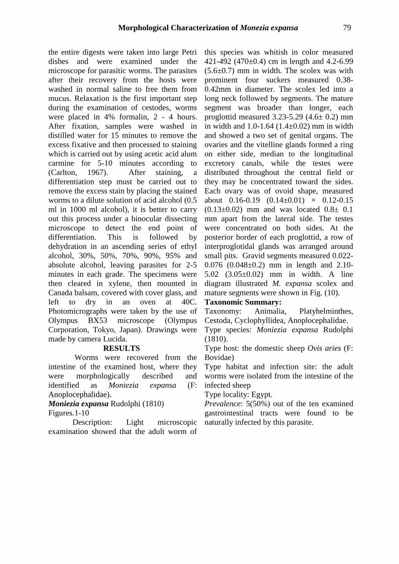
Figs.1-9: Photomicrographs of Moniezia expansa (F: Cyclophylidea) infecting intestine of the domestic sheep Ovis aries
showing high magnifications of: (1) Scolex (SC) with four prominent suckers (SU) followed by a long neck (N), ×100. (2)
Immature proglottids (IS), ×120. (3, 4) Mature broad proglottids (MS) with more developed genital structures, ×120, ×200.
(5-7) Mature segments (MS) with two sets of genital organs per segment. The two horse shoe shaped ovaries (OV)
surrounding a small mass of vitellaria (VT) forming a ring in the lateral sides of each segment, testes (TE) were distributed
throughout the central field or they may be concentrated towards the sides. At the posterior border of each proglottid, a row of
interproglotidal glands (IP) arranged as small pits. (C) cirrus, (CS) cirrus sac, (GA) genital atrium, (V) vagina, (fig. 5, 6 ×300,
fig. 7 ×500). (8) Ovary (OV) and vitellaria (VT), ×800. (9) Testes (TE) and the interproglotidal glands (IP), ×800.

Morphological Characterization of Monezia expansa 81
Fig. 10: A line diagram of M. expansa, (A) scolex, scale bar 0.1mm; (B) mature segments,
scale bar 0.5mm.
DISCUSSION
Sheep and goats cover more than 30%
of all domestic meat consumption and
generate cash income through the export of
meat and edible organs (Fletcher and
Zelalem, 1991). Even though the livestock
sub-sector contributes much to the national
economy, its development is hampered by
different constraints which include rampant
animal diseases, poor nutrition, poor
husbandry, poor infrastructure, shortage of
trained manpower, and lack of government
policies (Fletcher and Zelalem, 1991).
Tapeworms have been noticed to be the most
abundant helminth parasites infecting sheep
through postmortem examinations. These
parasites play an important role in the large
economic losses of farm animals (Maziad and
El-Nemr, 2002). Moniezia expansa (Family:
Anoplocephalidae) and other tapeworms
infect sheep, goats, cattle (Khan et al., 1989)
and constituted a big problem in sheep raising
countries (Tinar et al., 1993). The present
study has indicated a natural infection rate of
50% (5/10) which is in agreement with many
investigators (Khan et al., 1989, Kaur et al.,
1995, Umur and Gicik, 1995) whereas this
value is higher than that reported by
Hassanien (1978) and lower than those
recorded by Ndarathi et al. (1989), Tinar et
al. (1993) and Swarnkar et al. (1996)
(16.21%). The present study reported that the

Mona Fol et al. 82
highest peak of infection was recorded in the
winter season which is similar to some
authors (Swarnkar et al., 1996 and Tilahun,
1996). Other reports observed higher
infection rates during spring (Hassanien,
1978 and Arvinder, 1995) and during summer
(Arvinder et al., 1993). These differences
may be attributed to many environmental
factors, vectors, parasites, and host habits in
different countries (Arvinder et al., 1993).
The morphological studies of the present
specimens confirmed their identification as
Moniezia expansa as described by (Bambroo,
1969, Bali, 1970 and Soulsby, 1982) on the
basis of various morphological characters as
body length, maximum width, and size of
suckers, scolex, presence of two sets of
reproductive organs per mature segment, and
presence of a continuous band of
interproglotids. However, some intraspecific
variations in the size ratio of the body and
various other body organs were recorded in
the present specimens. M. expansa was the
most dominant helminth species identified
during the course of the study. These results
agreed with many records of previous authors
(Tinar et al., 1993, Arvinder et al., 1993,
Kaur et al., 1995, Umur and Gicik). Moniezia
is a unique member among Cyclophyllidea in
possessing groups of interproglottidal glands
in the parenchyma along the posterior edge of
each proglottid. These gland cells are
arranged in a follicular form containing a
central sac. The secretions of these glands
contain acetylcholin-esterase and alkaline
phosphatase (Gunn and Probert, 1983). REFERENCES
Arvinder, K. (1995): Epizootiology and
biology of common anoploce-phaline
cestodes in sheep along with
histopathology and haematology of
the definitive host. Journal of
Veterinary parasitology, 9:153–154.
Arvinder, M.; Bali, H.S. and Gill, J.S.
(1993): Epizootiology of
anoplocepha-line cestodes in sheep in
Punjab. Indian Journal of Ecology,
20:147–151.
Bali, H.S. (1976): A survey of helminth
parasites of sheep (Ovis aris) in
Jammu and Kashmir. Journal of
Animal and Health Production, 4: 25-
32.
Bambroo, N. (1970): On two species of
Gastrothylax Poirier, 1883 from
sheep in Kashmir. Kashmir Science,
7(1-2): 147-155.
Bashter, A.R.; Hassanain, M.; Abdel-
Ghaffar, F.; Al-Rasheid, K.; Hassan,
S. and Mehlhorn, H. (2011): Studies
on monieziasis of sheep I. Prevalence
and anthelmintic effects of
some plant extracts, a light and
electron microscope study.
Parasitology Research, 108: 177-186.
Becker, B.; Mehlhorn, H.; Andrews, P. and
Thomas, H. (1981): Ultrastructural in
vestigations on the effect of
Praziquantel on the tegument of five
species of cestodes. Zeitschrift fur
Parasitenkunde, 64(3):257–269.
Brooker, S.; Clements, A.C.A. and Bundy,
D.A.P. (2006): Global epidemiology,
ecology and control of soil-
transmitted helminth infections.
Global mapping of infectious
diseases: Methods, Examples and
emerging applications book series.
Advances in Parasitology, 62:221–
261.
Carlton, B.C. (1967): Transformation
mapping of the genes controlling
tryptophan biosynthesis in Bacillus
subtilis. Journal of Bacteriology. 94:
660-665.
Denegri, G.; Bernadina, W.; Perez-Serrano, J
and Rodriguez-Caabeiro, F. (1998):
Anoplocephalid cestodes of
veterinary and medical significance: a
review. Folia Parasitologica, 45 (1):
1–8.
Elliott, D.C. (1986): Tapeworm (Moniezia
expansa) and its effect on sheep
production: the evidence
reviewed. New Zealand Veterinary
Journal, 34 (5): 61–5.
El-Shazly, A.M.; Morsy, T.A. and Dawoud,
H.A. (2004): Human Monieziasis M.
expansa: the first Egyptian parastic

Morphological Characterization of Monezia expansa 83
zoonosis. Journal of Egyptian society
of Parasitology, 34 (2): 380–381.
Fletcher, I. and Zelalem, A. (1991): Small
ruminant productivity in the central
Ethiopia mixed farming system.
Instituteof Agricultural research
proceeds. 4 thed. National livestock
improvement conference, Addis
Ababa, Ethiopia. Pp. 15-45.
Gómez-Puerta, L.A.; Lopez-Urbina, M.T.
and González, A.E. (2008):
Occurrence of Moniezia
expansa (Rud, 1810) Blanchard, 1891
(Cestoda: Anoplocephalidae) in
domestic pig (Sus scrofa
domestica Linnaeus, 1758) in Perú.
Veterinary parasitology, 158 (4):
380–381
Gunn, A. and Probert, A.J. (1983): Moniezia
expansa: the interproglottidal glnds
and their secretions. Journal of
Helminthology, 57:51–55.
Hassanien, M.A. (1978): Morphological and
biological studies on tapeworms
infesting sheep under the Egyptian
environment condition. M.Sc. Thesis
Faculty of Veterinary Medicine,
Cairo University, Egypt.
Kaur, A.; Bali, H.S. and Duggal, C.L.
(1995): Seasonal variation of
anoplocephaline cestodes infection in
sheep of Punjab. Indian Journal of
Animal Sciences, 65:38–40.
Khan, M.N.; Hayat, C.S.; Chaudhry, A.H.;
Iqbal, Z. and Hayat, B. (1989):
Prevalence of gastrointestinal
helminths in sheep and goats at
Faisalabad abattoir. Pakistan.
Veterinary Journal, 9:159–161.
Maziad, S.A. and El-Nemr, H.I. (2002): The
endoparasites of sheep and goats,and
shepherd in North Sinai Governorate,
Egypt. Journal of the Egyptian
society of Parasitology, 32(1):119–
126.
Mehlhorn, H. (2008): Encyclopedia of
Parasitology, 3rd edn. Springer
Verlag, Heidelberg.
Ndarathi, C.M.; Waghela, S. and Semenye,
P.P. (1989): Helminthiansis in Massai
ranches in Kenya. Bulletin of Animal
Health and Production in Africa,
37:205–208.
Polec, W. (1990): Immunological studies on
lambs experimentally infected with
Moniezia expansa. Acta
Parasitolgica Polonica, 35(4):333–
339.
Rudolphi, G.A. (1802): Fortesetzing der
Beobachtingen uber die
Eingeweldewurmer. Archiv für
Zoologie und Zootomie, 2:23–25.
Sinitsin, D.F. (1931): A glimpse into the life
history of the tapeworm of
sheep, Moniezia expansa. Journal of
Parasitology, 17 (4): 223–227.
Soulsby, E.J.L. (1982): Helminths,
arthropods and protozoa of
domesticated animals. Bailliere,
Tindal and Cassel, London, 809 pp
Swarnkar, C.P.; Singh, D.; Srivastava, C.P.;
Bhagwan, P.S.K. and Dimri, U.
(1996): A retrospective study on
ovine gastrointestinal helminthoses
under semi-arid conditions. Journal
of Veterinary parasitology, 10:15–21.
Tilahun, G. (1996): Epidemiology of
helminth parasites of small ruminants
in mid-low land Ethiopia. In:
Dioulasso B, Faso B (eds)
parasitology research in Africa.
Proceeding of an IFSworkshop.
International Foundation for Science,
6–10: 255–269.
Tinar, R.; Coskun, S.Z.; Demir, S.; Akyol,
C.V.; Dogan, H. and Aydin, L.
(1993): Cestode species
(Anoplocephalidae) and their
prevalence inruminants slaughtered at
the Bursa meat and fish plant.
Veteriner Fakultesi DergUludag
Unioversitesi, 12:32–40.
Umur, S. and Gicik, Y. (1995): Incidence of
Anoplocephalidae species in
ruminants in kars district. Turk
Parazitoloii Dergist, 19:272–28.

Mona Fol et al. 84
ARABIC SUMMARY
بإستخدام أوفيس إيريس )الديدان الشريطية( الذي تم عزله من أمعاء الخروف مونيزيا إكسبانساالوصف الشكلي لطفيل
الميكروسكوب الضوئي
4اء عادل، أسم1،1، كريم مرسي1، سلمي يحي2، منال أحمد2، سحر الجنايني1فل منى
صرجامعة القاهرة، القاهرة، م ،قسم علم الحيوان، كلية العلوم -1
جامعة المنيا، المنيا، مصر ،قسم علم الحيوان، كلية العلوم -2
قسم الأحياء، كلية العلوم، جامعة الملك خالد، المملكة العربية السعودية -3
قسم علم الحيوان، كلية العلوم جامعة جنوب الوادي، قنا، مصر -4
ريطية التى تصيب الخراف والتى تم ذبحها بالمسلخ خلال الدراسة الحالية تم عزل نوع من انواع الديدان الش
. تم ملاحظة وجود الطفيل متشبثا بجدار الامعاء بواسطة منطقة الرأس وعن طريق ممصات 2112الرئيسي بالقاهرة، مصر عام
تقنية التى تم فحص القناة الهضمية لهم. فحص الطفيل باستخدام فقط. تم عزل الطفيل من خمسة خراف فقط من العشر خراف
الميكروسكوب الضوئي أوضحت أن الطفيل الذي تم عزله هو طفيل بالغ أبيض اللون. تحتوي منطقة الرأس على أربعة
ية الناضجة. وتم وصف ممصات. تلي منطقة الرأس منطقة العنق الغير مقسمة يتبعها القطع اللسانية الغير ناضجة ثم القطع اللسان
ية للقطع اللسانية الناضجة مثل المبيض، والخصي والرحم. وبعد مقارنة الطفيل بمثيله من الجهاز التناسلي والاعضاء التكاثر
.مونيزيا إكسبانسا الانواع السابقه من الديدان الشريطية تم تصنيف الطفيل على أنه طفيل ال