Embed Size (px)
Citation preview

Title EGFR Tyrosine kinase regulates small conductance Ca2+-activated K+ (hSKCa1) channels expressed in HEK 293 cells
Author(s) Wu, W; Sun, H; Deng, XL; Li, GR
Citation Biochemical Journal, 2013, v. 452 n. 1, p. 121-129
Issued Date 2013
URL http://hdl.handle.net/10722/183747
Rights The final version of record is available athttp://www.biochemj.org/bj/default.htm

To whom correspondence should be addressed (email [email protected]).
Accepted by Biochemical Journal for publication EGFR tyrosine kinase regulates small conductance Ca2+-activated K+ (hSKCa1) channels expressed in HEK 293 cells Wei Wu,1 Hai-Ying Sun,1 Xiu-Ling Deng,2 Gui-Rong Li1,3 1Department of Medicine, and Research Centre of Heart, Brain, Hormone and Healthy Aging, Li Ka Shing Faculty of Medicine, The University of Hong Kong, Pokfulam, Hong Kong, China; 2Department of Physiology and Pathophysiology, Medical School of Xi’an Jiaotong University, Xi’an, China;3Department of Physiology, Li Ka Shing Faculty of Medicine, The University of Hong Kong, Pokfulam, Hong Kong, China Running title: hSKCa1 channels and tyrosine kinase regulation Small conductance Ca2+-activated K+ (SKCa) channels are widely distributed in different tissues including the brain, pancreatic islets, myocardium, etc., and play an important role in controlling electrical activity and cellular functions. However, intracellular signal modulation of SKCa channels is not fully understood. The present study was designed to investigate the potential regulation of hSKCa1 channels by protein tyrosine kinases (PTKs) in HEK 293 cells expressing hSKCa1 (KCNN1) gene using approaches of whole-cell patch voltage-clamp, immunoprecipitation, Western blot, and mutagenesis. We found that hSKCa1 current was inhibited by the broad spectrum PTK inhibitor genistein, the selective EGFR (epidermal growth factor receptor) kinase inhibitors T25, and AG556, but not by the Src-family kinases inhibitor PP2. The inhibitory effect of these PTK inhibitors was significantly antagonized by the protein tyrosine phosphatases (PTPs) inhibitor orthovanadate. Tyrosine phosphorylation level of hSKCa1 channels was reduced by genistein, T25, or AG556. The reduced tyrosine phosphorylation was countered by orthovanadate. Interestingly, the mutant hSKCa1 channel with Y109F lost the inhibitory response to T25 or AG556, and showed a dramatic reduction of tyrosine phosphorylation level and a reduced current density. These results demonstrate the novel information that hSKCa1 channels are inhibited by genistein, T25, and AG556 via EGFR tyrosine kinase inhibition, which is related to the phosphorylation of Tyr109 in the N-terminus. This effect may affect electrical activity and cellular functions in brain, pancreatic islets, and myocardium.
Key words: Small conductance Ca2+-activated K+ channels; ion channel modulation; protein tyrosine phosphorylation; EGFR tyrosine kinase; mutagenesis Abbreviations: SKCa channels, small conductance Ca2+-activated potassium channels; PTKs, protein tyrosine kinases; PTPs, protein tyrosine phosphatases; HEK 293 cells, human embryonic kidney 293 cells; EGFR, epidermal growth factor receptor; T63, tyrphostin 63; T25, tyrphostin 25; AG556, tyrphostin AG 556
Introduction
Small conductance Ca2+-activated potassium (SKCa or SK) channels, like
intermediate-conductance Ca2+-activated potassium (IKCa) channels, are voltage-independent, activated by intracellular free Ca2+, and play an

important role in regulating many processes involving Ca2+-dependent signaling in both electrically excitable and nonexcitable cells [1, 2]. There are three types of SKCa channels, including SKCa1 (SK1), SKCa2 (SK2) and SKCa3 (SK3) encoded by KCNN1, KCNN2, and KCNN3, respectively [3]. SKCa channels are expressed in different types of tissues/cells including various regions of the brain [4, 5], pancreatic islets [6], cardiac myocytes [7-9], and endothelial cells (for SKCa3) [10], etc. Like BKCa channels (large conductance Ca2+- and voltage-activated K+ channels) [11] and IKCa channels [1], SKCa channels are sensitive to intracellular free Ca2+ ions [2, 12, 13]. BKCa channels are found to be enhanced by protein kinase C (PKC) and/or cAMP/protein kinase A (PKA) via activating cyclic guanosine monophosphate- (cGMP) dependent kinase [14-17]. Nonetheless, information for the regulation of SKCa channels by intracellular signal transduction is not fully understood. Recent studies reported that Ca2+-sensitivity of SKCa channels was regulated by interaction of protein kinase CK2 and protein phosphatase 2A [18, 19], and the plasma membrane surface expression of SKCa2 channels was regulated by PKA via phosphorylation of serine 568-570 in the C terminus of the channel [20]. Activation of PKA reduced membrane expression of SKCa channels in hippocampal and amygdale neurons [21].
Protein tyrosine kinases (PTKs), including receptor PTKs (e.g. EGFR kinase, epidermal growth factor receptor kinase) and non-receptor PTKs (e.g. Src-family kinases), are important intracellular signals [22]. In addition to the modulation of cell growth, differentiation, embryonic development, metabolism, immune system function, oncogenesis etc. [22], PTKs also
modulate different types of ion channels [23], including L-type Ca2+ (ICa.L) channels [24], the voltage-gated Na+ (INa) channels [25, 26], volume sensitive Cl− (ICl.vol) channels [27], as well as voltage-gated K+ channels [28-35], and also BKCa channels [36]. Currently, the PTK modulation of SKCa1 channels is unknown. The present study was therefore designed to determine whether/how PTKs regulate hSKCa1 channels stably expressed in HEK 293 cells using approaches of electrophysiology, immunoprecipitation, Western blotting, and mutagenesis. We found that PTK inhibitors genistein, T25, and AG556 decreased hSKCa1 current by inhibiting EGFR kinase.
Material and Methods
Cell culture, mutagenesis, and gene transfection
The pCDNA3/hSKCa1 (KCNN1) plasmid kindly provided by Dr. Nicole Schmitt (University of Copenhagen, Denmark) was transfected into HEK 293 cells (ATCC, Manassas, VA) using Lipofectamine 2000TM. The HEK 293 cell line stably expressing hSKCa1 gene was established as previously described [31, 32]. The hSKCa1 cell line was maintained in Dulbecco’s modified eagle’s medium (DMEM, Invitrogen, Hong Kong, China) supplemented with 10% fetal bovine serum and 400 µg/ml G418 (Invitrogen). Cells used for electrophysiology were cultured on a glass cover slip.
The predicted potential tyrosine phosphorylation sites of hSKCa1 channels were examined using the software NetPhos 2.0 (www.cbs.dtu.dk/cgi-bin). Single amino acid substitutions of hSKCa1 were generated using a QuikChange II XL

Site-directed mutagenesis kit (Stratagene, La Jolla, CA) following the manufacturer’s instruction, and confirmed by DNA sequencing. Polymerase chain reaction (PCR) was made with hSKCa1 as a template and a pair of complementary mutagenesis primers. The PCR products were digested with Dpn I and then transformed into competent cells DH5α for plasmids amplification. The three mutants Y109F, Y138F and Y406F were generated and confirmed by full DNA sequencing. The mutants were transiently expressed in HEK 293 cells with a 35 mm culture dish using 10 µl of Lipofectamine 2000TM with 4 µg of pCDNA3.1/hKv4.3-mutant vector. Cells used for electrophysiology were seeded on a glass coverslip.
Solutions and reagents
Tyrode’s solution contained (mM) NaCl 140, KCl 5.4, MgCl2 1.0, CaCl2 1.8, 4-(2-hydroxyethyl)-1-piperazineethanesulfonic acid (HEPES) 10.0 and glucose 10 (pH adjusted to 7.3 with NaOH). For whole-cell recordings, the pipette solution contained (mM) KCl 20, K-aspartate 110, MgCl2 1.0, HEPES 10, ethyleneglycoltetraacetic acid (EGTA) 5, GTP 0.1, Na-phosphocreatine 5, and Mg-ATP 5 (pH adjusted to 7.2 with KOH) with including 300 nM free Ca2+ (calculated using the Cabuf software provided by Dr. G. Droogmans in the Department of Physiology, KU Leuven, Leuven, Belgium).
3-(4-Chlorophenyl)1-(1,1-dimethyl- ethyl)-1H-pyrazolo[3,4-d]pyrimidin-4-amine (PP2) was purchased from Tocris (Bristol, UK). All other reagents were obtained from Sigma-Aldrich (St Louis, MO). Stock solutions were made with dimethylsulfoxide (DMSO) for genistein (100 mM), tyrphostin AG 556 (AG556, 100 mM), tyrphostin 25 (T25, 100 mM), tyrphostin 63 (T63, 100
mM), and PP2 (30 mM). The stocks were divided into aliquots and stored at −20°C. Sodium orthovanadate stock solution (200 mM) was made with distilled water, and pH value was adjusted to 9.0.
Electrophysiology
Cells on a coverslip were transferred to a cell chamber (0.5 ml) mounted on the stage of an inverted microscope (Diaphot, Nikon, Japan) and superfused at ~2 ml/min with Tyrode solution. Whole-cell currents were recorded as described previously [31, 32, 34]. Borosilicate glass electrodes (1.2-mm OD) were pulled with a Brown-Flaming puller (model P-97, Sutter Instrument Co, Novato, CA) and had a resistance of 2–3 MΩ when filled with the pipette solution. A 3-M KCl agar bridge was used as the reference electrode. The tip potential was zeroed before the patch pipette contacted the cell. After a gigaohm seal was obtained, the cell membrane was ruptured by applying a gentle negative pressure to establish the whole-cell configuration. Series resistance (Rs) was 4-6 MΩ and was compensated by 50-80% to minimize voltage errors. The liquid junction potential (~12 mV) was not corrected in the experiment. Membrane currents were measured using an EPC-10 amplifier and Pulse software (Heka Elektronik, Lambrecht, Germany). Command pulses were generated by a 12-bit digital-to-analog converter controlled by Pulse software. Current signals were low-pass filtered at 5 kHz and stored in the hard disk of an IBM compatible computer. All experiments were conducted at room temperature (22–23 ºC).
Immunoprecipitation and Western blotting
The immunoprecipitation and Western blotting were performed following the procedure described previously [32, 34].

HEK 293 cells (~80% confluence) stably expressing hSKCa1 channels were treated respectively with different compounds, e.g. genistein, T25, AG556, PP2, orthovanadate, epidermal growth factor (EGF), etc. for 30 min at room temperature, and centrifuged at 4oC. The cell pellet was then lysed with the lysis buffer containing (mM) 25 Tris, 150 NaCl, 1.0 NaF, 1.0 EDTA, 1.0 orthovanadate, 1.0 phenylmethylsulfonyl fluoride, and 1% Na deoxycholate, 0.1% SDS, 1% Triton X-100, 1 µg/ml leupeptin, and 1 µg/ml aprotinin. Protein quantification of lysates was made using a protein assay reader (Bio-Rad Laboratories, Hercules, CA), and diluted to equal concentrations. Proteins were immunoprecipitated overnight at 4°C using 2 µg of goat anti-hSKCa1 (sc-17991) antibody (Santa Cruz Biotechnology, Santa Cruz, CA) and 100 µl of protein A/G beads (DAM 1460243, Millipore, Billerica, MA). The immunoprecipitated proteins bound to pelleted protein A/G beads were washed thoroughly in PBS, denatured in Laemmli sample buffer, separated using SDS-PAGE, and electroblotted onto nitrocellulose membranes. The immunoblots were probed with anti-phosphotyrosine antibody (1:1000, Cell Signaling Tech., Danvers, MA) overnight at 4°C in a blocking solution containing 5% BSA in TBS and Tween 20, and subsequently treated with goat anti-mouse IgG-HRP antibody (1:5000, Santa Cruz Biotechnology) for 2 h at room temperature. Blots were developed with enhanced chemiluminescence (ECL, Amersham Biosciences) and exposed on X-ray film (Fuji Photo Film GmbH). The blots were then stripped and reprobed with the anti-hSKCa1 channel antibody to determine total hSKCa1 channel protein. The film was scanned, imaged by a Bio-Imaging System (Syngene, Cambridge,
UK), and the intensity of the bands was analyzed via GeneTools software (Syngene).
Statistical analysis
Experimental group data are expressed as means ± SEM. Paired and/or unpaired Student’s t-test were used as appropriate to evaluate the statistical significance of differences between two group means, and ANOVA was used for multiple groups. Values of P<0.05 were considered to be statistically significant.
Results
SKCa1 current inhibition by apamin in HEK 293 cell line
Figure 1 illustrates the membrane currents HEK 293 cells without/with transfecting hSKCa1 (KCNN1) gene. Only a small background current (Fig. 1A) was recorded using a pipette solution containing 300 nM free Ca2+ with the voltage steps as shown in the inset in a HEK 293 cell without hSKCa1 gene transfection (n=4), and showed a linear I-V relationship when recorded using a 3-s ramp from −100 to +80 mV (left panel). However, a lager membrane current was recorded under the same conditions (as in A) in a HEK 293 cell stably expressing hSKCa1 gene, and the current was inhibited by the SKCa channel blocker apamin [37]. I-V relationship curve of the current exhibited a weak inward rectification was inhibited by 300 nM apamin (Fig. 1C, n=4), suggesting a typical of SKCa current.
Inhibition of hSKCa1 current by genistein
Our previous studies have demonstrated that genistein decreases several potassium currents via inhibiting PTKs [28, 32, 33]. To investigate whether it is the case for

hSKCa1 channels, genistein (30 µM) was tested in HEK 293 cells stably expressing hSKCa1 gene. The membrane current elicited by 200-ms voltage steps to between −70 to +80 mV from a holding potential of −80 mV or a 3-s ramp from −100 to +80 mV exhibited an inward rectifier property, a typical of hSKCa1 current (Fig. 2A and 2B). The current was significantly inhibited by application of genistein in bath solution (6 min), and the effect was reversed on washout (8 min). Genistin, a glycoside derivative (PTK-inactive) compound of genistein had no inhibitory effect on hSKCa1 current at 30 µM, while 30 µM genistein significantly inhibited the current in the same cell (Fig. 2B), suggesting that PTK inhibition is involved in the current reduction by genistein.
If the reduction of hSKCa1 current by genistein is related to the inhibition of PTKs, its effect should be antagonized by the PTP inhibitor orthovanadate. Figure 2C shows that the hSKCa1 current inhibition by genistein was partially reversed by co-application of genistein and orthovanadate (1 mM). As the observations in Kv4.3 [35] and hERG [32] and IKs [28] channels, orthovanadate (1 mM) did not show any effect on hSKCa1 current (Fig. 2D). It however, significantly prevented the current reduction by genistein. Figure 2E illustrates the mean percentage values of hSKCa1 current (at +40 mV) in cells treated with 30 µM genistein, 30 µM genistin, 1 mM orthovanadate, genistein plus orthovanadate, 1 mM orthovanadate, or orthovanadate plus 30 µM genistein. Genistein decreased the current to 59.8±4.6% of control (n=12, P<0.01), and the current inhibition was reversed by orthovanadate to 74.9±3.4% of control (n=5, P<0.05 vs. genistein alone). Genistin and orthovanadate had no effect on hSKCa1
current. Pretreatment with orthovanadate significantly prevented the current inhibition by genistein (current reduction: by 11.5±5.5% vs. 25.1±3.4% with genistein plus orthovanadate, n=6, P<0.05). The results suggest that genistein-induced hSKCa1 current reduction is likely related at least in part to inhibition of PTKs.
To further investigate which specific PTK is involved in the regulation of hSKCa1 channels, the selective Src-family kinases inhibitor PP2 [38], and the highly selective EGFR kinase inhibitors T25 and AG556 [39-41] were tested in the following experiments.
Effect of PP2 on hSKCa1 current
Figure 3A shows the time course of hSKCa1 current recorded in a typical experiment with a 200-ms voltage step to +40 mV from a holding potential of −80 mV in the absence and presence of PP2. PP2 (10 µM, 2000 times higher than IC50 for inhibiting Src kinases) [38] had no inhibitory effect on hSKCa1 current during 15 min exposure. Similarly, step hSKCa1 current and the I-V relationships were not affected by the application of 10 µM PP2 (Fig. 3B and 3C). Mean percentage value of hSKCa1 current at +40 mV was not altered by PP2 (10 µM, Fig. 3D). These results suggest that hSKCa1 channels are not regulated by Src-family kinases.
Inhibition of hSKCa1 current by T25 and AG556
Figure 4 shows the effect of the EGFR kinase inhibitor T25 on hSKCa1 current. Voltage step-activated hSKCa1 current was significantly reduced by T25 application in bath solution, and the reduction partially recovered on washout (Fig. 4A). Figure 4B shows the time course of hSKCa1 current recorded with a ramp protocol in a

representative cell. T63 (a PTK-inactive analog of tyrphostin) at 30 µM had a slight inhibitory effect on hSKCa1 current, while T25 at 10 µM induced a significant current reduction in the same cell. The current inhibition by T25 (10 µM) was partially reversed by co-application of T25 and 1 mM orthovanadate (Fig. 4C). Pretreatment of the cell with orthovanadate significantly prevented the current reduction by T25 (Fig. 4D). The mean percentage values of hSKCa1 current ( +40 mV) are illustrated in Fig. 4E in cells treated with 10 µM T25, 30 µM T63, T25 plus 1 mM orthovanadate, 1 mM orthovanadate, or orthovanadate plus 10 µM T25. T25 decreased the current to 60.1±2.9% of control (n=11, P<0.01 vs. control) and the inhibition recovered on washout to 84.1±5.9% of control (P<0.01 vs. T25 alone). The current inhibition was antagonized to 82.7±3.9% of control by orthovanadate (P<0.01 vs. T25 alone). The PTK-inactive analog T63 (30 µM) and orthovanadate had no significant inhibition of hSKCa1 current (n=6, P=NS vs. control). In cells pretreated with orthovanadate, the current inhibition by T25 was significantly prevented (current inhibition: by 8.0±4.5%, n=6, P<0.05 vs. 17.3±3.9% with T25 plus orthovanadate). These results indicate that T25-induced hSKCa1 current reduction is related to the inhibition of EGFR kinase.
We have previously demonstrated that the highly selective EGFR kinase inhibitor AG556 inhibits several K+ channels (e.g. hERG, hEAG1, IKs, Kir2.1, and Kir2.3) [28, 31-34] in a PTK-dependent manner. To determine whether it is the case for hSKCa1 channels, AG556 was tested. Figure 5A shows the hSKCa1 current recorded in a representative cell during control, AG556 application, and washout. AG556 (10 µM, 5 min) inhibited hSKCa1 current and the current reduction was partially recovered by
washout. Figure 5B shows the time-course of the step current in the absence and presence of 10 µM AG556, and co-application of 1 mM orthovanadate. The current inhibition by AG556 was partially reversed by co-application of AG556 and orthovanadate. The pretreatment of the cell with orthovanadate also significantly prevented the current reduction by AG556 (Fig. 5C). The mean percentage values of hSKCa1 current at +40 mV are illustrated in Fig. 5D. The current at +40 mV was reduced by AG556 to 62.4±7.8% of control (n=10, P<0.01 vs. control), the reduction significantly recovered on washout (P<0.01 vs. AG556 alone). The current inhibition was reversed to 78.5±5.7% of control with co-application of genistein and orthovanadate (P<0.01 vs. AG556 alone). The current reduction by AG556, as in genistein and T25, significantly prevented in cells pretreated with orthovanadate (current inhibition: by 9.9±4.5%, n=6, P=0.05 vs. 21.5±5.5% with AG556 plus orthovanadate). These results further support the notion that hSKCa1 current is regulated by EGFR kinase.
However, hSKCa1 channels were not stimulated by application of the EGFR kinase activator EGF (100 ng/ml) in the bath solution (n=5, data not shown). This suggests that tyrosine phosphorylation of hSKCa1 channels, like hERG, IKs, and Kir2.1 channels [28, 32, 33], is at a saturated state under control conditions.
Tyrosine phosphorylation level of hSKCa1 channels
To investigate how tyrosine phosphorylation of hSKCa1 channels is affected by PTK inhibitors, immunoprecipitation and Western blot analysis were performed in hSKCa1 HEK 293 cells. Figure 6A displays the images of tyrosine phosphorylation level of

hSKCa1 channels with different treatments (30 min). Tyrosine phosphorylation was reduced by the broad spectrum PTK inhibitor genistein (30 µM) and the EGFR kinase inhibitors AG556 (10 µM) and T25 (10 µM), but not by the Src-family kinase inhibitor PP2 (10 µM), orthovanadate (1 mM) or EGF (100 ng/ml). The reduced phosphorylation level by PTK inhibitors was significantly antagonized in cells treated with co-application of orthovanadate (1 mM). Figure 6B illustrates the mean percentage values of tyrosine phosphorylation in cells with different treatments. PP2, orthovanadate, and EGF had no effect on the phosphorylation level. Tyrosine phosphorylation was reduced to 59.8±6.9% by genistein (P<0.01 vs. vehicle), to 46.2±6.7% by AG556 (P<0.01 vs. vehicle), and to 53.7±5.7% by T25 (P<0.01 vs. vehicle). The reduced phosphorylation was reversed to 84.7 ± 10.1% by orthovanadate plus genistein (P<0.05 vs genistein alone), 86.7±8.5% by orthovanadate plus AG556 (P<0.05 vs. AG556 alone), and to 85.7±4.5% by orthovanadate plus T25 (P<0.05 vs. T25 alone). These results demonstrate that the tyrosine phosphorylation level is affected by PTK inhibitors genistein, AG556 and tyrphostin 25 (but not PP2), and their interactions can be countered with the PTP inhibitor orthovanadate. No alteration of tyrosine phosphorylation of hSKCa1 channel protein with application of EGF or orthovanadate implicates a saturated phosphorylation state of the channel.
Tyrosine phosphorylation sites of hSKCa1 channels
To investigate the potential tyrosine phosphorylation sites of hSKCa1 channels, three mutant channels (Y109F, Y138F and Y406F) were generated using phenylalanine to substitute for tyrosine. The inhibitory
response of these mutants to PTK inhibitors was determined with the selective EGFR kinase inhibitors T25 and AG556. Figure 7A shows the hSKCa1 mutant currents in the absence and presence of 10 µM T25. T25 significantly inhibited Y138F and Y406F currents, but exhibited only a slight inhibition of Y109F current. Interestingly, AG556 also only slightly reduced Y109F current, while suppressed Y138F and Y406F currents (Fig. 7B). The mean percentage values of WT and mutant hSKCa1 channels inhibition by T25 or AG556 are illustrated in Fig. 7C. T25 (10 µM) and AG556 (10 µM) inhibited hSKCa1 Y109F current (at +40 mV) by 9.1±2.9% (n=5, P<0.05 vs. control) and 10.3±1.5% (n=6, P<0.05 vs. control). The inhibitory effect of these two EGFR kinase inhibitors on WT, Y138F, and Y406F was stronger than that on Y109F current (P<0.01 vs. WT, Y138F or Y406F). These results suggest that tyrosine phosphorylation site of hSKCa1 channels is likely located at Y109 residue of the N-terminus.
If the Y109 of hSKCa1 channels is responsible for tyrosine phosphorylation, the phosphorylation should be reduced in the mutant channels with Y109F. To determine whether it is the case, immunoprecipation and Western blot analysis were employed in HEK 293 cells expressing Y109F mutant or WT hSKCa1 channels. Figure 8A shows the tyrosine phosphorylation levels in HEK 293 cells expressing WT or Y109F hSKCa1 channels with or without 10 µM AG556 treatment. Tyrosine phosphorylation level was evident in WT hSKCa1 channels, which was significantly reduced by AG556. However, the phosphorylation was dramatically reduced in Y109F hSKCa1 channels. The results obtained in the present study demonstrated that the reduction of hSKCa1 current by the PTK inhibitors genistein, T25, and AG556 were related to

the inhibition of EGFR kinase and the decrease of tyrosine phosphorylation at Y109 of hSKCa1 channels. Orthovanadate inhibits PTPs thereby antagonizes the PTK inhibitors-induced reduction of the current amplitude and tyrosine phosphorylation of hSKCa1 channels (Fig. 8B). The current density was significantly reduced in cells expressing the mutant Y109F hSKCa1 channels (Fig. 8C and 8D), suggesting that tyrosine phosphorylation of hSKCa1 channels at Y109 plays an important role in regulation of the channel activity.
Discussion
In the present study, we have shown that inhibition of EGFR kinase by genistein, T25, and AG556 reduces hSKCa1 channels stably expressed in HEK 293 cells. Tyrosine phosphorylation level of hSKCa1 channels is decreased by genistein, T25, or AG556. Their inhibitory effects on hSKCa1 current and tyrosine phosphorylation are antagonized by the PTP inhibitor orthovanadate. Site-directed mutagenesis reveals that Y109 residue is the phosphorylation site of the channel by EGFR kinase.
Previous studies have demonstrated that PTKs regulate different ion channels, including ICa.L channels [24], INa
channels [25, 26], and ICl.vol channels [20, 27] in mammalian heart myocytes, as well as several types of K+ channels in different types of cells [23, 28, 30-35], and also TRPC6 channels [42]. Both EGFR kinase and Src-family kinases participate in regulating cardiac ICl.vol [20, 27], INa channels [25, 26], Kv4.3 channels [35], and hERG channels [32], whereas Src-family kinases are not involved in modulation of cardiac slowly-delayed rectifier K+ channel
current (IKs) [28], the inwardly-rectifier K+ channels (Kir2.1 and Kir2.3) [33, 34], and hEAG1 channels [31]. The present study provided the novel information that hSKCa1 channels stably expressed in HEK 293 cells are regulated by EGFR kinase, but not by Src-family kinases.
Using multiple experimental techniques including electrophysiology, immunoprecipitation, Western blot analysis, and mutagenesis, we found that hSKCa1 current was reduced by the broad spectrum PTK inhibitor genistein and the selective EGFR kinase inhibitors T25 and AG556 via inhibiting PTKs. Four lines of evidence support the conclusion that EGFR kinase regulates hSKCa1 channels. First, T63, an inactive analog of tyrphostins, respectively, did not have any meaningful effect on hSKCa1 channels. Second, as expected for phosphorylation-dependent processes, inhibition of PTPs by orthovanadate antagonized the current reduction by PTK inhibitors. Third, tyrosine phosphorylation level of hSKCa1 channels was reduced by genistein, T25, and AG556, and the reduction was reversed by orthovanadate. Finally, the mutant Y109F hSKCa1 channels exhibited a weak inhibitory response to the EGFR kinase inhibitors T25 and AG556, a reduced tyrosine phosphorylation. Also the mutant channels showed a decreased current density. Moreover, the the Src-family kinase inhibitor PP2 did not alter hSKCa1 channel current or the tyrosine phosphorylation level, indicating that Src-family kinases are not involved in PTK regulation of hSKCa1 channels. This is consistent with IKs, hEAG1, Kir2.1 and Kir2.3 channels [28, 30, 31, 33, 34], which are regulated by EGFR kinase, but not by Src-family kinases.
Earlier studies have demonstrated that SKCa channels (SKCa1, 2, 3) are expressed

in the brain [43]. In addition controlling dendritic integration, synaptic transmission, and synaptic plasticity, SKCa channels play a role in regulating learning and memory [43-45]. SKCa channels are also present in pancreatic islets and regulate glucose response of β-cells [6]. It has been reported in rat insulinoma cell line (INS-1) that SKCa1 channels are responsible for a more negative resting membrane potential, regulate the duration of action potentials, and therefore control insulin secretion. Silencing SKCa1 channels with KCNN1 siRNA results in a more depolarized resting membrane potential and increases insulin secretion [46]. Moreover, recent studies have reported that SKCa channels are expressed predominantly in atrial myocytes of the heart [7-9]. Therefore, SKCa channels are believed to be a potential target for the study of anti-atrial fibrillation [47, 48].
In the present study, we demonstrated that hSKCa1 channel activity was decreased by EGFR kinase inhibition. We therefore speculate that tyrosine phosphorylation of SKCa channels by EGFR kinase may be important in controlling electrical activity in neuronal cells, islet β-cells, and cardiac atrial myocytes. If it is the case, inhibition of EGFR kinase by genistein, T25, and AG556 would affect the dentritic integration and excitability, synaptic transmission and synaptic plasticity in the brain, insulin release in the β-cells, and would also show some anti-atrial fibrillation effects. However, these remain to be demonstrated in future experimental study in different systems.
In addition to the requirement of additional series of experiments to demonstrate potential physiological roles of EGFR kinase regulation on hSKCa1 channels, another limitation of the present study is that we are unable to demonstrate the stimulatory effect on hSKCa1 channels
with orthovanadate or EGF. We have previously shown that the inhibition of PTPs with orthovanadate or EGFR activation with EGF increased current amplitude and tyrosine phosphorylation levels in Kir2.3 channels [34] and cardiac INa [25]. However, no effect was observed on hSKCa1 current or tyrosine phosphorylation with orthovanadate or EGF in the present study. One possible interpretation is that tyrosine phosphorylation is already saturated, therefore no additional increase of current amplitude and phosphorylation level can be observed by inhibiting PTPs or activating EGFR kinase. Saturated tyrosine phosphorylation was also observed in hERG [32], hEAG1 [31], IKs [28], Kir2.1 channels [33], and hKv4.3 channels [35], and therefore stimulation of EGFR kinase with EGF or inhibition of dephosphorylation with orthovanadate does not increase the current amplitude or tyrosine phosphorylation level of these channels.
In summary, the present study has provided the first evidence that the PTK inhibitors genistein, T25, and AG556 decrease hSKCa1 channels stably expressed in HEK 293 cells via inhibiting EGFR kinase that is related to phosphorylation of Y109 residue of the channel in the N-terminus. This effect may affect cellular electrical activity and thus modulate cellular functions in mammalian related organs/tissues including brain, islets and hearts.
Acknowledgement
The study was supported in part by a grant from Sun Chieh Yeh Heart Foundation of Hong Kong. Wei Wu was supported by a postgraduate studentship from the University of Hong Kong. The authors thank Dr. Nicole Schmitt, University of Copenhagen, Denmark, for generously

providing PCDNA3/hSKCa1 plasmid.
Conflict interest. None
References
1 Wei, A. D., Gutman, G. A., Aldrich, R., Chandy, K. G., Grissmer, S. and Wulff, H. (2005) International Union of Pharmacology. LII. Nomenclature and molecular relationships of calcium-activated potassium channels. Pharmacol. Rev. 57, 463-472
2 Berkefeld, H., Fakler, B. and Schulte, U. (2010) Ca2+-activated K+ channels: from protein complexes to function. Physiol. Rev. 90, 1437-1459
3 Kohler, M., Hirschberg, B., Bond, C. T., Kinzie, J. M., Marrion, N. V., Maylie, J. and Adelman, J. P. (1996) Small-conductance, calcium-activated potassium channels from mammalian brain. Science. 273, 1709-1714
4 Rimini, R., Rimland, J. M. and Terstappen, G. C. (2000) Quantitative expression analysis of the small conductance calcium-activated potassium channels, SK1, SK2 and SK3, in human brain. Brain Res. Mol. Brain Res. 85, 218-220
5 Stocker, M. and Pedarzani, P. (2000) Differential distribution of three Ca(2+)-activated K(+) channel subunits, SK1, SK2, and SK3, in the adult rat central nervous system. Mol. Cell. Neurosci. 15, 476-493
6 Tamarina, N. A., Wang, Y., Mariotto, L., Kuznetsov, A., Bond, C., Adelman, J. and Philipson, L. H. (2003) Small-conductance calcium-activated K+ channels are expressed in pancreatic islets and regulate glucose responses. Diabetes. 52, 2000-2006
7 Tuteja, D., Rafizadeh, S., Timofeyev, V., Wang, S., Zhang, Z., Li, N., Mateo, R. K., Singapuri, A.,
Young, J. N., Knowlton, A. A. et al. (2010) Cardiac small conductance Ca2+-activated K+ channel subunits form heteromultimers via the coiled-coil domains in the C termini of the channels. Circ. Res. 107, 851-859
8 Tuteja, D., Xu, D., Timofeyev, V., Lu, L., Sharma, D., Zhang, Z., Xu, Y., Nie, L., Vazquez, A. E., Young, J. N. et al. (2005) Differential expression of small-conductance Ca2+-activated K+ channels SK1, SK2, and SK3 in mouse atrial and ventricular myocytes. Am. J. Physiol. Heart Circ. Physiol. 289, H2714-2723
9 Xu, Y., Tuteja, D., Zhang, Z., Xu, D., Zhang, Y., Rodriguez, J., Nie, L., Tuxson, H. R., Young, J. N., Glatter, K. A. et al. (2003) Molecular identification and functional roles of a Ca(2+)-activated K+ channel in human and mouse hearts. J. Biol. Chem. 278, 49085-49094
10 Burnham, M. P., Bychkov, R., Feletou, M., Richards, G. R., Vanhoutte, P. M., Weston, A. H. and Edwards, G. (2002) Characterization of an apamin-sensitive small-conductance Ca(2+)-activated K(+) channel in porcine coronary artery endothelium: relevance to EDHF. Br. J. Pharmacol. 135, 1133-1143
11 Sansom, S. C., Ma, R., Carmines, P. K. and Hall, D. A. (2000) Regulation of Ca(2+)-activated K(+) channels by multifunctional Ca(2+)/calmodulin-dependent protein kinase. Am. J. Physiol. Renal Physiol. 279, F283-288
12 Xia, X. M., Fakler, B., Rivard, A., Wayman, G., Johnson-Pais, T., Keen, J. E., Ishii, T., Hirschberg, B., Bond, C. T., Lutsenko, S. et al. (1998) Mechanism of calcium gating in small-conductance calcium-activated potassium channels. Nature. 395, 503-507
13 Hirschberg, B., Maylie, J., Adelman, J. P. and Marrion, N. V. (1998) Gating of recombinant small-conductance Ca-activated K+ channels by calcium. J. Gen. Physiol. 111, 565-581
14 Barman, S. A., Zhu, S., Han, G. and White, R. E. (2003) cAMP activates BKCa channels in pulmonary arterial smooth muscle via cGMP-dependent protein kinase. Am. J. Physiol. Lung Cell. Mol. Physiol. 284, L1004-1011
15 Barman, S. A., Zhu, S. and White, R. E. (2004) PKC activates BKCa channels in rat pulmonary arterial smooth muscle via cGMP-dependent protein kinase. Am. J. Physiol. Lung Cell. Mol. Physiol. 286, L1275-1281
16 Wischmeyer, E., Doring, F. and Karschin, A. (1998) Acute suppression of inwardly rectifying Kir2.1 channels by direct tyrosine kinase phosphorylation. J. Biol. Chem. 273, 34063-34068
17 Zhou, X. B., Arntz, C., Kamm, S., Motejlek, K., Sausbier, U., Wang, G. X., Ruth, P. and Korth, M. (2001) A molecular switch for specific stimulation of the BKCa channel by cGMP and cAMP kinase. J. Biol. Chem. 276, 43239-43245
18 Bildl, W., Strassmaier, T., Thurm, H., Andersen, J., Eble, S., Oliver, D., Knipper, M., Mann, M., Schulte, U., Adelman, J. P. et al. (2004) Protein kinase CK2 is coassembled with small conductance Ca(2+)-activated K+ channels and regulates channel gating. Neuron. 43, 847-858
19 Allen, D., Fakler, B., Maylie, J. and Adelman, J. P. (2007) Organization and regulation of small conductance Ca2+-activated K+ channel multiprotein complexes. J. Neurosci. 27, 2369-2376
20 Ren, Y. J., Barnwell, L. F., Alexander, J. C., Lubin, F. D., Adelman, J. P., Pfaffinger, P. J., Schrader, L. A. and Anderson, A. E. (2006) Regulation of surface localization of the small conductance
Ca2+-activated potassium channel, Sk2, through direct phosphorylation by cAMP-dependent protein kinase. J. Biol. Chem. 281, 11769-11779
21 Faber, E. S., Delaney, A. J., Power, J. M., Sedlak, P. L., Crane, J. W. and Sah, P. (2008) Modulation of SK channel trafficking by beta adrenoceptors enhances excitatory synaptic transmission and plasticity in the amygdala. J. Neurosci. 28, 10803-10813
22 Hunter, T. (2000) Signaling--2000 and beyond. Cell. 100, 113-127
23 Davis, M. J., Wu, X., Nurkiewicz, T. R., Kawasaki, J., Gui, P., Hill, M. A. and Wilson, E. (2001) Regulation of ion channels by protein tyrosine phosphorylation. Am. J. Physiol. Heart Circ. Physiol. 281, H1835-1862
24 Ogura, T., Shuba, L. M. and McDonald, T. F. (1999) L-type Ca2+ current in guinea pig ventricular myocytes treated with modulators of tyrosine phosphorylation. Am. J. Physiol. Heart Circ. Physiol. 276, H1724-1733
25 Liu, H., Sun, H. Y., Lau, C. P. and Li, G. R. (2007) Regulation of voltage-gated cardiac sodium current by epidermal growth factor receptor kinase in guinea pig ventricular myocytes. J. Mol. Cell. Cardiol. 42, 760-768
26 Ahern, C. A., Zhang, J. F., Wookalis, M. J. and Horn, R. (2005) Modulation of the cardiac sodium channel Na(V)1.5 by Fyn, a Src family tyrosine kinase. Circ. Res. 96, 991-998
27 Du, X. L., Gao, Z., Lau, C. P., Chiu, S. W., Tse, H. F., Baumgarten, C. M. and Li, G. R. (2004) Differential effects of tyrosine kinase inhibitors on volume-sensitive chloride current in human atrial myocytes: evidence for dual regulation by Src and EGFR kinases. J. Gen. Physiol. 123, 427-439
28 Dong, M. Q., Sun, H. Y., Tang, Q., Tse, H. F., Lau,

C. P. and Li, G. R. (2010) Regulation of human cardiac KCNQ1/KCNE1 channel by epidermal growth factor receptor kinase. Biochim Biophys Acta. 1798, 995-1001
29 Gao, Z., Lau, C. P., Wong, T. M. and Li, G. R. (2004) Protein tyrosine kinase-dependent modulation of voltage-dependent potassium channels by genistein in rat cardiac ventricular myocytes. Cell Signal. 16, 333-341
30 Missan, S., Linsdell, P. and McDonald, T. F. (2008) Involvement of tyrosine kinase in the hyposmotic stimulation of I Ks in guinea-pig ventricular myocytes. Pflugers Arch. 456, 489-500
31 Wu, W., Dong, M. Q., Wu, X. G., Sun, H. Y., Tse, H. F., Lau, C. P. and Li, G. R. (2012) Human ether-a-go-go gene potassium channels are regulated by EGFR tyrosine kinase. Biochim Biophys Acta. 1823, 282-289.
32 Zhang, D. Y., Wang, Y., Lau, C. P., Tse, H. F. and Li, G. R. (2008) Both EGFR kinase and Src-related tyrosine kinases regulate human ether-a-go-go-related gene potassium channels. Cell Signal. 20, 1815-1821
33 Zhang, D. Y., Wu, W., Deng, X. L., Lau, C. P. and Li, G. R. (2011) Genistein and tyrphostin AG556 inhibit inwardly-rectifying Kir2.1 channels expressed in HEK 293 cells via protein tyrosine kinase inhibition. Biochim Biophys Acta. 1808, 1993-1999
34 Zhang, D. Y., Zhang, Y. H., Sun, H. Y., Lau, C. P. and Li, G. R. (2011) Epidermal growth factor receptor tyrosine kinase regulates the human inward rectifier potassium channel Kir2.3 stably expressed in HEK 293 cells. Br. J. Pharmacol. 164, 10
35 Zhang, Y. H., Wu, W., Sun, H. Y., Deng, X. L., Cheng, L. C., Li, X., Tse, H. F., Lau, C. P. and Li, G. R. (2012) Modulation of human cardiac
transient outward potassium current by EGFR tyrosine kinase and Src-family kinases. Cardiovasc. Res. 93, 424-433
36 Prevarskaya, N. B., Skryma, R. N., Vacher, P., Daniel, N., Djiane, J. and Dufy, B. (1995) Role of tyrosine phosphorylation in potassium channel activation. Functional association with prolactin receptor and JAK2 tyrosine kinase. J. Biol. Chem. 270, 24292-24299
37 Blatz, A. L. and Magleby, K. L. (1985) Single chloride-selective channels active at resting membrane potentials in cultured rat skeletal muscle. Biophys. J. 47, 119-123
38 Hanke, J. H., Gardner, J. P., Dow, R. L., Changelian, P. S., Brissette, W. H., Weringer, E. J., Pollok, B. A. and Connelly, P. A. (1996) Discovery of a novel, potent, and Src family-selective tyrosine kinase inhibitor. Study of Lck- and FynT-dependent T cell activation. J. Biol. Chem. 271, 695-701
39 Brenner, T., Poradosu, E., Soffer, D., Sicsic, C., Gazit, A. and Levitzki, A. (1998) Suppression of experimental autoimmune encephalomyelitis by tyrphostin AG-556. Exp. Neurol. 154, 489-498
40 Gazit, A., Osherov, N., Posner, I., Yaish, P., Poradosu, E., Gilon, C. and Levitzki, A. (1991) Tyrphostins. 2. Heterocyclic and alpha-substituted benzylidenemalononitrile tyrphostins as potent inhibitors of EGF receptor and ErbB2/neu tyrosine kinases. J. Med. Chem. 34, 1896-1907
41 Gazit, A., Yaish, P., Gilon, C. and Levitzki, A. (1989) Tyrphostins I: synthesis and biological activity of protein tyrosine kinase inhibitors. J. Med. Chem. 32, 2344-2352
42 Hisatsune, C., Kuroda, Y., Nakamura, K., Inoue, T., Nakamura, T., Michikawa, T., Mizutani, A. and Mikoshiba, K. (2004) Regulation of TRPC6 channel activity by tyrosine phosphorylation. J.

Biol. Chem. 279, 18887-18894
43 Faber, E. S. (2009) Functions and modulation of neuronal SK channels. Cell Biochem. Biophys. 55, 127-139
44 Hammond, R. S., Bond, C. T., Strassmaier, T., Ngo-Anh, T. J., Adelman, J. P., Maylie, J. and Stackman, R. W. (2006) Small-conductance Ca2+-activated K+ channel type 2 (SK2) modulates hippocampal learning, memory, and synaptic plasticity. J. Neurosci. 26, 1844-1853
45 Vick, K. A. t., Guidi, M. and Stackman, R. W., Jr. (2010) In vivo pharmacological manipulation of small conductance Ca(2+)-activated K(+) channels influences motor behavior, object memory and fear conditioning. Neuropharmacology. 58, 650-659
46 Andres, M. A., Baptista, N. C., Efird, J. T., Ogata, K. K., Bellinger, F. P. and Zeyda, T. (2009) Depletion of SK1 channel subunits leads to constitutive insulin secretion. FEBS Lett. 583, 369-376
47 Diness, J. G., Sorensen, U. S., Nissen, J. D., Al-Shahib, B., Jespersen, T., Grunnet, M. and Hansen, R. S. (2010) Inhibition of small-conductance Ca2+-activated K+ channels terminates and protects against atrial fibrillation. Circ. Arrhythm. Electrophysiol. 3, 380-390
48 Diness, J. G., Skibsbye, L., Jespersen, T., Bartels, E. D., Sorensen, U. S., Hansen, R. S. and Grunnet, M. (2011) Effects on atrial fibrillation in aged hypertensive rats by Ca(2+)-activated K(+) channel inhibition. Hypertension. 57, 1129-1135

Figure 1. Apamin inhibits the membrane current in HEK 293 cells expressing hSKCa1 channels. A. Only a small background current was recorded with the voltage steps as shown in the inset in a HEK 293 cell without hSKCa1 gene transfection with a pipette solution containing 300 nM free Ca2+ (n=3), showing a linear I-V relationship of a 3-s ramp voltage protocol from −100 to +80 mV (left panel). B. Representative current was recorded with same the protocol as in panel A in a HEK 293 cell stably expressing hSKCa1 gene in the absence and presence of 300 nM apamin. C. I-V relationship curves of the ramp current recorded in the same cell as in B in the absence and presence of apamin (n=4). Arrows indicate zero current level.

Figure 2. Effect of genistein on hSKCa1 current. A. hSKCa1 current recorded in a representative cell with 200-ms voltage steps between −70 and +80 mV from a holding potential of −80 mV (inset) in the absence (control) and presence of 30 µM genistein, and upon washout. B. Time-course of hSKCa current recorded in a typical experiment with a 3-s ramp (-100 to +80 mV) from a holding potential -80 mV in the absence and presence of 30 µM genistin or 30 µM genistein. The current was measured at -100 and +40 mV. The original ramp I-V current traces at corresponding time points are shown in right of the panel. C. hSKCa1 current recorded in a representative cell during control, in the presence of 30 µM genistein, and genistein plus 1 mM orthovanadate (OV). D. hSKCa1 current recorded in a representative cell during control, in the presence of 1 mM orthovanadate, and orthovanadate plus 30 µM genistein. E. Mean percentage values of hSKCa1 current at +40 mV during control, 30 µM genistein, washout, 30 µM genistin, genistein plus 1 mM orthovanadate. 1 mM orthovanadate, or orthovanadate plus 30 µM genistein (n=5-8, *P<0.05, **P<0.01 vs. control; #P<0.05; ##P<0.01 vs. genistein alone; $P<0.05 vs. genistein plus orthovanadate). The arrows indicate the zero level.

Figure 3. Effect of PP2 on hSKCa1 current. A. Time-course of hSKCa1 current recorded in a representative cell with a 200-ms voltage step to +40 mV from a holding potential of -80 mV (inset) in the absence and presence of 10 µM PP2. Original current traces at corresponding time points are shown in right of the panel. B. hSKCa1 current recorded in a typical experiment in the absence and presence of 10 µM PP2. The arrow indicates the zero level. C. I-V relationship traces recorded in another cell with a ramp voltage protocol as described in Fig. 1B in the absence and presence of 10 µM PP2. D. Mean percentage values of hSKCa1 current in the absence (control) and presence of 10 µM PP2 (n=5, P=NS vs. control).
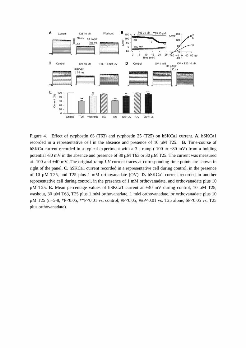
Figure 4. Effect of tyrphostin 63 (T63) and tyrphostin 25 (T25) on hSKCa1 current. A. hSKCa1 recorded in a representative cell in the absence and presence of 10 µM T25. B. Time-course of hSKCa current recorded in a typical experiment with a 3-s ramp (-100 to +80 mV) from a holding potential -80 mV in the absence and presence of 30 µM T63 or 30 µM T25. The current was measured at -100 and +40 mV. The original ramp I-V current traces at corresponding time points are shown in right of the panel. C. hSKCa1 current recorded in a representative cell during control, in the presence of 10 µM T25, and T25 plus 1 mM orthovanadate (OV). D. hSKCa1 current recorded in another representative cell during control, in the presence of 1 mM orthovanadate, and orthovanadate plus 10 µM T25. E. Mean percentage values of hSKCa1 current at +40 mV during control, 10 µM T25, washout, 30 µM T63, T25 plus 1 mM orthovanadate, 1 mM orthovanadate, or orthovanadate plus 10 µM T25 (n=5-8, *P<0.05, **P<0.01 vs. control; #P<0.05; ##P<0.01 vs. T25 alone; $P<0.05 vs. T25 plus orthovanadate).
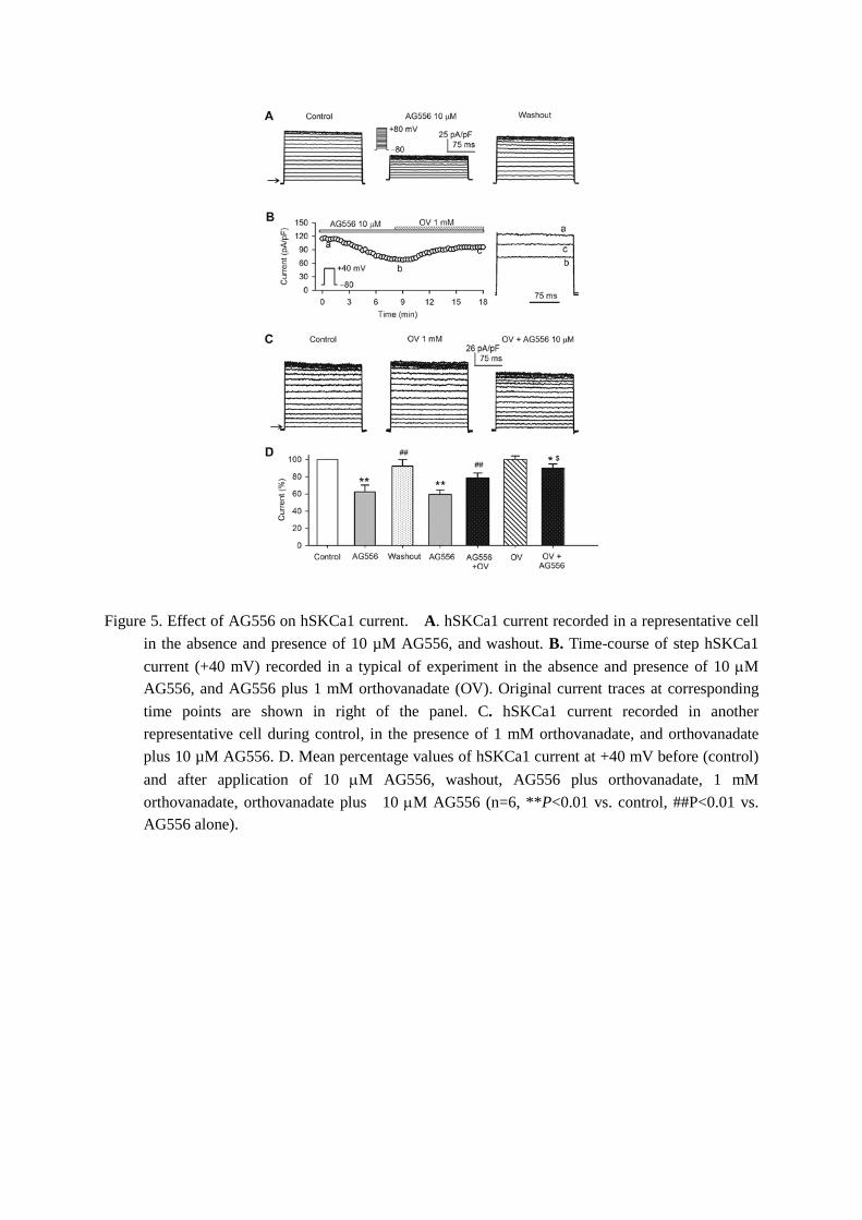
Figure 5. Effect of AG556 on hSKCa1 current. A. hSKCa1 current recorded in a representative cell in the absence and presence of 10 µM AG556, and washout. B. Time-course of step hSKCa1 current (+40 mV) recorded in a typical of experiment in the absence and presence of 10 µM AG556, and AG556 plus 1 mM orthovanadate (OV). Original current traces at corresponding time points are shown in right of the panel. C. hSKCa1 current recorded in another representative cell during control, in the presence of 1 mM orthovanadate, and orthovanadate plus 10 µM AG556. D. Mean percentage values of hSKCa1 current at +40 mV before (control) and after application of 10 µM AG556, washout, AG556 plus orthovanadate, 1 mM orthovanadate, orthovanadate plus 10 µM AG556 (n=6, **P<0.01 vs. control, ##P<0.01 vs. AG556 alone).

Figure 6. Tyrosine phosphorylation levels of hSKCa1 channels. A. Images of immunoprecipitation and Western blotting in cells treated with vehicle, 1 mM orthovanadate (OV), 30 µM genistein, 10 µM AG556, 10 µM T25, or genistein, AG556, T25 plus 1 mM orthovanadate, or 100 ng/ml EGF. B. Mean percentage values of relative tyrosine-phosphorylated hSKCa1 channel protein. The amount of protein from the immunoprecipitation (as shown in A) was normalized to those from the Western blots. Relative phosphorylated protein level (n=4-7 experiments) is expressed as a percentage of vehicle control. **P<0.01 vs. control, #P<0.05 vs. genistein, AG556 or T25 alone.

Figure 7. Effects of T25 and AG556 on mutant hSKCa1 channels. A. hSKCa1 currents recorded in representative cells expressing Y109F, Y138F or Y406F mutant in the absence (control) and presence of 10 µM T25. B. hSKCa1 currents recorded in cells expressing Y109F, Y138F and Y406F mutants in the absence and presence of 10 µM AG556. C. Left panel: mean percentage values of hSKCa1 current at +40 mV in cells expressing WT (n=12), Y109F (n=5), Y138F (n=5) and Y406F (n=5) mutants in the absence (control) and presence of 10 µM T25 (*P<0.05, **P<0.01 vs control; #P<0.05 vs. WT, Y138F or Y406F). Right panel: mean percentage values of hSKCa1 current at +40 mV in cells expressing WT (n=11), Y109F (n=6), Y138F (n=6) and Y406F (n=6) mutants in the absence (control) and presence of 10 µM AG556 (*P<0.05, **P<0.01 vs. control; #P<0.05 vs. WT, Y138F or Y406F).
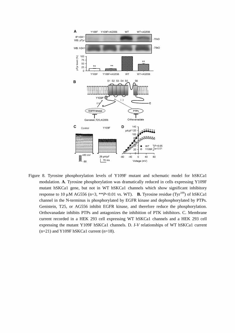
Figure 8. Tyrosine phosphorylation levels of Y109F mutant and schematic model for hSKCa1 modulation. A. Tyrosine phosphorylation was dramatically reduced in cells expressing Y109F mutant hSKCa1 gene, but not in WT hSKCa1 channels which show significant inhibitory response to 10 µM AG556 (n=3, **P<0.01 vs. WT). B. Tyrosine residue (Tyr109) of hSKCa1 channel in the N-terminus is phosphorylated by EGFR kinase and dephosphorylated by PTPs. Genistein, T25, or AG556 inhibit EGFR kinase, and therefore reduce the phosphorylation. Orthovanadate inhibits PTPs and antagonizes the inhibition of PTK inhibitors. C. Membrane current recorded in a HEK 293 cell expressing WT hSKCa1 channels and a HEK 293 cell expressing the mutant Y109F hSKCa1 channels. D. I-V relationships of WT hSKCa1 current (n=21) and Y109F hSKCa1 current (n=18).