Embed Size (px)
Citation preview
EFNS/MDS-ES GUIDELINES/CME ARTICLE
EFNS/MDS-ES recommendations for the diagnosis ofParkinson’s disease
A. Berardellia*, G. K. Wenningb, A. Antoninic, D. Bergd, B. R. Bloeme, V. Bonifatif, D. Brooksg,
D. J. Burnh, C. Colosimoi, A. Fanciullib, J. Ferreiraj, T. Gasserd, F. Grandask, P. Kanovskyl,
V. Kosticm, J. Kulisevskyn, W. Oertelo, W. Poeweb, J.-P. Reesep, M. Reljaq, E. Ruzickar,
A. Schrags, K. Seppib, P. Tabat and M. VidailhetuaDipartimento di Neurologia e Psichiatria and IRCCS NEUROMED Institute, Sapienza, Universit�a di Roma, Rome, Italy; bDepartment
of Neurology, Innsbruck Medical University, Innsbruck, Austria; cParkinson’s disease and Movement Disorders Unit IRCCS, San
Camillo, Venice, Milan, Italy; dDepartment of Neurodegenerative Diseases, Hertie-Institute for Clinical Brain Research, German Center
for Neurodegenerative Diseases, University of T€ubingen and DZNE, T€ubingen, Germany; eDonders Institute for Brain, Cognition and
Behaviour, Department of Neurology, Radboud University Nijmegen Medical Center, Nijmegen; fDepartment of Clinical Genetics, Eras-
mus MC, Rotterdam, The Netherlands; gDepartment of Clinical Neuroscience, Imperial College London, Hammersmith Hospital, London,
UK; hInstitute for Ageing and Health, Newcastle University, Newcastle upon Tyne, UK; iDipartimento di Neurologia e Psichiatria,
Sapienza, Universit�a di Roma, Rome, Italy; jCentro De Estudos Egas Moniz, Faculdade De Medicina De Lisboa, Lisbon, Portugal;kMovement Disorders Research Unit, Hospital Universitario Gregorio Mara~n�on, Madrid, Spain; lDepartment of Neurology, Palacky Uni-
versity, Olomouc, Czech Republic; mInstitute of Neurology CCS, School of Medicine, University of Belgrade, Belgrade, Serbia; nMove-
ment Disorders Unit, Department of Neurology, Sant Pau Hospital, Universitat Autonoma de Barcelona and Ciberned, Barcelona, Spain;oDepartment of Neurology, Centre of Nervous Diseases, Philipps-University of Marburg, Marburg, Germany; pInstitut f€ur Medizinische
Soziologie, Deutschland, Germany; qDepartment of Neurology, Movement Disorders Centre, School of Medicine and University Hospital
Centre, University of Zagreb, Zagreb, Croatia; r1st Faculty of Medicine and General University Hospital in Prague, Charles University in
Prague, Praha, Czech Republic; sInstitute of Neurology, University College London, London, UK; tDepartment of Neurology and
Neurosurgery, University of Tartu, Tartu, Estonia; and uPole des Maladies du Syst�eme Nerveux et CRICM UPMC/INSERM
UMR_S975 CNRS UMR7225, F�ed�eration de Neurologie, Hopital de la Salpetri�ere, Paris, France
Keywords:
movement disorders,
neurological disorders,
Parkinson’s disease
Received 18 July 2012
Accepted 18 September 2012
Background: A Task Force was convened by the EFNS/MDS-ES Scientist Panel
on Parkinson’s disease (PD) and other movement disorders to systemically review
relevant publications on the diagnosis of PD.
Methods: Following the EFNS instruction for the preparation of neurological diag-
nostic guidelines, recommendation levels have been generated for diagnostic criteria
and investigations.
Results: For the clinical diagnosis, we recommend the use of the Queen Square
Brain Bank criteria (Level B). Genetic testing for specific mutations is recommended
on an individual basis (Level B), taking into account specific features (i.e. family
history and age of onset). We recommend olfactory testing to differentiate PD from
other parkinsonian disorders including recessive forms (Level A). Screening for pre-
motor PD with olfactory testing requires additional tests due to limited specificity.
Drug challenge tests are not recommended for the diagnosis in de novo parkinso-
nian patients. There is an insufficient evidence to support their role in the differen-
tial diagnosis between PD and other parkinsonian syndromes. We recommend an
assessment of cognition and a screening for REM sleep behaviour disorder,
psychotic manifestations and severe depression in the initial evaluation of suspected
PD cases (Level A). Transcranial sonography is recommended for the differentiation
of PD from atypical and secondary parkinsonian disorders (Level A), for the early
diagnosis of PD and in the detection of subjects at risk for PD (Level A), although
the technique is so far not universally used and requires some expertise. Because
Correspondence: A. Berardelli, Dipartimento di Neurologia e Psichiatria, Sapienza, Universit�a di Roma, Viale dell’Universita 30, 00185 Rome,
Italy (tel./fax: +390649914700; e-mail: [email protected]).
This is a Continuing Medical Education article, and can be found with corresponding questions on the Internet at http://www.efns.org/EFNS
Continuing-Medical-Education-online.301.0.html. Certificates for correctly answering the questions will be issued by the EFNS.
© 2012 The Author(s)European Journal of Neurology © 2012 EFNS16
European Journal of Neurology 2013, 20: 16–34 doi:10.1111/ene.12022

specificity of TCS for the development of PD is limited, TCS should be used in con-
junction with other screening tests. Conventional magnetic resonance imaging and
diffusion-weighted imaging at 1.5 T are recommended as neuroimaging tools that
can support a diagnosis of multiple system atrophy (MSA) or progressive supranu-
clear palsy versus PD on the basis of regional atrophy and signal change as well as
diffusivity patterns (Level A). DaTscan SPECT is registered in Europe and the Uni-
ted States for the differential diagnosis between degenerative parkinsonisms and
essential tremor (Level A). More specifically, DaTscan is indicated in the presence
of significant diagnostic uncertainty such as parkinsonism associated with neurolep-
tic exposure and atypical tremor manifestations such as isolated unilateral postural
tremor. Studies of [123I]MIBG/SPECT cardiac uptake may be used to identify
patients with PD versus controls and MSA patients (Level A). All other SPECT
imaging studies do not fulfil registration standards and cannot be recommended for
routine clinical use. At the moment, no conclusion can be drawn as to diagnostic
efficacy of autonomic function tests, neurophysiological tests and positron emission
tomography imaging in PD.
Conclusions: The diagnosis of PD is still largely based on the correct identification of its
clinical features. Selected investigations (genetic, olfactory, and neuroimaging studies)
have an ancillary role in confirming the diagnosis, and some of them could be possibly
used in the near future to identify subjects in a pre-symptomatic phase of the disease.
Introduction
A correct diagnosis of Parkinson’s disease (PD) is a pre-
requisite for patient counselling and therapeutic man-
agement. Despite all the recent advances in imaging
and genetics of parkinsonian disorders, the diagnosis of
PD remains a primarily clinical exercise. However, clin-
ical diagnostic uncertainty is high at initial presenta-
tion, and up to 10–30% of patients initially diagnosed
as PD are clinically re-classified even in specialized units
[1]. Targeting this pitfall, numerous ancillary investiga-
tions have been developed in the last decades to support
PD diagnostic work-up [2]. Because these tests differ in
their differential diagnostic performance, availability
and costs, the EFNS/MDS-ES Task Force identified a
clear need to develop guidelines for the diagnosis of PD
to be applied across Europe. This need is fostered by
recent efforts of the PD research community focusing
on the development of screening tools capable of identi-
fying individuals at risk for PD.
This EFNS/MDS-ES Task Force report is divided
into nine sections addressing key aspects of the
diagnostic work-up of patients presenting with
parkinsonism:
1 Clinical diagnostic criteria
2 Genetic testing
3 Autonomic function testing
4 Olfactory tests
5 Drug challenge tests
6 Neurophysiological tests
7 Neuropsychological tests
8 Neuroimaging
9 Economic issues
Groups of experts were allocated to each section
and asked to provide an evidence-based recommenda-
tion level for the assigned diagnostic tool. To this
end, MEDLINE, EMBASE and Cochrane libraries
were searched for relevant citations up to June 2011.
Consensus on the guidelines was finally reached within
the Task Force.
The recommendations have been developed accord-
ing to the EFNS Evidence Classification Scheme for
diagnostic measures [3].
In this statement, recommendations for diagnostic
investigations in PD are therefore graded as follows:
• Level A – effective
• Level B – probably effective
• Level C – possibly effective
A Level A or B recommendation does not mean
that this test should be employed in all patients of a
certain group, but simply means that the test has
good diagnostic accuracy. It is for the physician to
decide whether or not to use it in the given patient.
For example, a Level A recommendation for an imag-
ing test based on excellent diagnostic performance
may still not mean that a clear-cut patient with a solid
clinical diagnosis should have this test.
Section 1: clinical diagnostic criteria for PD
Several sets of clinical diagnostic criteria have been
proposed, based mainly on the presence of the classi-
© 2012 The Author(s)European Journal of Neurology © 2012 EFNS European Journal of Neurology
EFNS/MDS-ES Guidelines 17

cal motor signs of the disease, combined with the
absence of incompatible or atypical signs (the so-
called red flags suggestive of atypical parkinsonism).
The most widely used clinical criteria for the diagnosis
of PD are those introduced by the Queen Square
Brain Bank (QSBB) [4]. These criteria provide a three-
step method: (Table 1)
1 Signs that must be present
2 Signs that should not be present
3 Supportive criteria
The first diagnostic step requires the presence of
bradykinesia. Crucially, bradykinesia is not just slow-
ness of movement or movements. It is rather meant as
progressive fatiguing and decrement of repetitive alter-
nating movements during finger or foot tapping [5].
The second step involves a checklist of symptoms and
signs that argue against a diagnosis of PD. Finally,
the diagnosis of PD requires the presence of three or
more supportive criteria (Table 1). Recently, hypos-
mia and hallucinations have been added to this list
[6].
The accuracy of the QSBB clinical diagnostic crite-
ria has been retrospectively assessed in two clinical–pathological studies (class III evidence) [7], (class III
evidence) [8]. In a series of 100 cases with pathologi-
cally proven PD, the QSBB clinical diagnostic criteria
were applied retrospectively, and the diagnostic accu-
racy proved to be 82% [7]. A more recent study (pub-
lished 10 years after the previous publication)
involved a series of 143 cases with pathologically pro-
ven PD. In this study, the QSBB clinical diagnostic
criteria were mostly applied by movement disorder
experts [8]. The results showed an overall sensitivity
for PD clinical diagnosis of 91.1%, a specificity of
98.4%, a positive predictive value of 98.6% and a
negative predictive value of 90%. The clinical diagnos-
tic accuracy was thus improved over time, suggesting
an optimized use of the QSBB criteria and its support-
ive and non-supportive signs.
Clinical expertise of the neurologist assessing PD
diagnosis has been shown to predict the diagnostic
effectiveness of the QSBB clinical diagnostic criteria
in two independent studies [1,9]. In the first study,
the QSBB clinical diagnostic criteria were applied by
movement disorder specialists to 402 cases previously
diagnosed as PD from general practitioners in North
Wales [9]. A definite diagnosis of PD could be
reached in only 53% of cases, suggesting an error
rate of 47% outside of specialized centres. The most
common misdiagnoses were essential tremor, Alzhei-
mer’s disease and vascular parkinsonism. The second
study compared the diagnostic accuracy of PD clini-
cal diagnosis, as made by movement disorder
experts, in comparison with non-expert physicians in
the community [1]. In this study, 126 patients with a
pre-existing clinical diagnosis of probable or possible
PD underwent diagnostic reassessment by movement
disorder specialists using the QSBB clinical diagnostic
criteria. The results showed that experts reached a
greater sensitivity (93.5% for experts versus 73.5%
for non-experts) and positive predictive value (88.7%
for experts versus 73.5% for non-experts), whereas
the negative predictive value was similar (76.9% for
experts versus 79.1% for non-experts) [1]. These
Table 1 Queen Square Brain Bank UK PDS Brain Bank Criteria
for the diagnosis of PD [4,6]
Step 1 Diagnosis of parkinsonian syndrome
Bradykinesia (slowness of initiation of voluntary movement with
progressive reduction in speed and amplitude or repetitive actions)
and at least one of the following:
• Muscular rigidity
• 4- to 6-Hz rest tremor
• Postural instability not caused by primary visual, vestibular,
cerebellar or proprioceptive dysfunction
Step 2 Exclusion criteria for Parkinson’s disease
• History of repeated strokes with stepwise progression of
parkinsonian features
• History of repeated head injury
• History of definite encephalitis
• Oculogyric crises
• Neuroleptic treatment at onset of symptoms
• More than one affected relative (*)
• Sustained remission
• Strictly unilateral features after 3 years
• Supranuclear gaze palsy
• Cerebellar signs
• Early severe autonomic involvement
• Early severe dementia with disturbances of memory, language
and praxis
• Babinski sign
• Presence of a cerebral tumour or communicating hydrocepha-
lus on CT scan
• Negative response to large doses of L-dopa (if malabsorption
excluded)
• MPTP exposure
Step 3 Supportive prospective positive criteria of Parkinson’s disease
Three or more required for the diagnosis of definite
Parkinson’s disease:
• Unilateral onset
• Rest tremor present
• Progressive disorder
• Persistent asymmetry affecting the side onset most (*)
• Excellent response (70–100%) to L-dopa
• Severe L-dopa-induced chorea
• L-dopa response for 5 years or more (*)
• Clinical course of 10 years or more (*)
• Hyposmia
• Visual hallucinations
(*) Criteria that will need future revision.
© 2012 The Author(s)European Journal of Neurology © 2012 EFNS European Journal of Neurology
18 A. Berardelli et al.
results underscored a relatively high diagnostic inac-
curacy by non-experts.
Even if made by a movement disorder expert, PD
diagnosis may change at follow-up, for several rea-
sons: development of atypical signs (red flags), insuffi-
cient response to dopaminergic treatment or
neuroimaging clues for an alternative diagnosis.
In prospective studies with PD cohorts initially
recruited from experts according to the QSBB criteria,
change of diagnosis occurred in 6–8% of cases [10,11].
This suggests a relative low rate of misclassification, if
the diagnosis is initially assessed by a movement dis-
order expert.
Although the QSBB clinical diagnostic criteria are
widely used in clinical practice, a number of pitfalls
have been recognized:
1 Having more than one affected relative cannot be
considered an exclusion criterion of PD anymore;
2 Some of the supportive criteria such as persistent
asymmetry, prolonged disease course or continu-
ous levodopa response may occur in atypical par-
kinsonian disorders as well. A critical revision of
the QSBB criteria will be required to address these
drawbacks.
Other diagnostic criteria are shown as supporting
information in the online version of this article (Data
S1).
Recommendations
Only the QSBB clinical diagnostic criteria have been
validated by Hughes et al. [8] and are therefore rec-
ommended as probably effective (Level B) for clinical
practice.
Section 2: genetic testing
These recommendations are formulated according to
the criteria established by the EFNS [3], with some
modifications accounting for the specific nature of
genetic tests [12]. Genetic testing is by definition the
gold standard for the diagnosis of a genetic disease
(barring the rare event of a laboratory error). There-
fore, the diagnostic accuracy of genetic testing cannot
be measured by using another method as reference
investigation. Therefore, the level of recommendation
for genetic testing has been based on the quality of
available studies investigating the nature and fre-
quency of mutations of a given gene amongst clini-
cally defined series of patients.
Because all the available studies have been retro-
spective (i.e. looking for specific mutations amongst
previously ascertained and clinically diagnosed series)
and because the formal execution of the genetic test-
ing on anonymously coded DNA samples by labora-
tory technicians can be practically considered as
equivalent to a blinded testing condition, the studies
are classified as class III evidence, which leads to
Level B recommendation.
With few notable exceptions in some populations,
<5% of all PD cases are caused by known single-gene
mutations. Therefore, genetic testing will allow an
accurate aetiological diagnosis only in a minority of
patients [12,13]. As no specific treatment is available
for genetic cases, the purpose of genetic testing in PD
is essentially oriented at patient and family members’
counselling with respect to disease prognosis and
genetic risk of unaffected relatives.
The results of diagnostic genetic testing have impli-
cations in the psychological, social and professional
domains of both patients and relatives. Therefore,
informed consent and privacy warranty are important
issues. Further, the genetic testing should always be
performed by a professional team and include pre-test
and post-test counselling [12].
Autosomal dominant forms of PD
SNCA
Point mutations in the gene for alpha-synuclein
(SNCA), as well as duplications and triplications of
the entire gene locus, can cause PD. The point muta-
tions E46K and A53T and also gene triplications
cause an aggressive form of PD with relatively early
onset [14]. Most cases have been identified in families
with multiple affected individuals. The A30P muta-
tion, as well as SNCA duplications, causes more typi-
cal PD with late onset. Incomplete penetrance of
SNCA duplications may result in a negative family
history [15]. Nevertheless, all the above cited SNCA
mutations are rare in sporadic patients.
Leucine-rich repeat kinase 2
Mutations in the gene for leucine-rich repeat kinase 2
(LRRK2) are a much more common cause of domi-
nant PD. Up to date, 6 mutations are known to be
pathogenic (N1437H, R1441C, R1441G, Y1699C,
G2019S and I2020T), based on their cosegregation in
PD families. Overall, LRRK2 mutations account for 5
–15% of dominant familial [16], and 1–3% of spo-
radic PD cases [17], with higher prevalence of some
founder mutations in specific populations. The
G2019S variant is found in 15–30% of Ashkenazi
Jewish [18] and up to 40% of North African Arab
patients (both sporadic and familial), whilst the
R1441G variant is a Basque founder mutation with a
prevalence of 15% in patients with PD from this
region [19].
© 2012 The Author(s)European Journal of Neurology © 2012 EFNS European Journal of Neurology
EFNS/MDS-ES Guidelines 19
Clinically, LRRK2-associated PD is indistinguish-
able from sporadic typical PD, as to age of onset and
symptomatology. Reduced penetrance of 30–70% has
been estimated for the G2019S mutation.
Glucocerebrosidase
Heterozygous mutations in the gene for glucocerebro-
sidase (GBA) are a frequent and strong risk factor for
PD, especially in some populations [20–22]. Some
mutations are more prevalent in specific ethnic
groups, such as the N370S mutation amongst Ashke-
nazi Jewish. According to current odds ratios’ esti-
mates (pooled OR > 5), GBA mutations have much
lower effect size than classical mendelian mutations.
In other words, GBA mutations display a markedly
reduced penetrance. However, an accurate estimate of
ORs and penetrance is currently possible only for the
most common GBA mutations. Clinically, patients
with GBA pathogenic mutations have typical PD with
possibly slightly earlier-onset age.
Autosomal recessive forms of PD
Homozygous or compound heterozygous mutations
in each of the following three genes: parkin
(PARK2), PINK1 (PARK6) and DJ-1 (PARK7), can
cause autosomal recessive forms of PD. Mutations in
the parkin gene are the most common. Up to half of
familial PD cases with a disease onset under the age
of 45 and a recessive pattern of inheritance are
caused by parkin mutations. Similarly, parkin muta-
tions underlie ~15% of the sporadic PD cases with
disease onset before the age of 45 [23]. Mutations in
the PINK1 and DJ-1 gene are less common, account-
ing for up to 1–8% and 1–2% of the sporadic cases
with early onset, respectively. The likelihood of
PINK-1 and DJ-1 mutations is inversely proportional
to the age of PD onset: the earlier the onset, the
higher the likelihood. A large number of mutations
have been identified in these three genes worldwide,
including point or small mutations, but also large
genomic rearrangements (deletions and multiplica-
tions). The latter are especially frequent in the parkin
gene. Therefore, sequencing and dosage assay of all
exons is required for an accurate screening of these
three genes.
In some cases, only a single heterozygous mutation
is detected in one of the genes for recessive PD. This
finding does not lend itself to a clear interpretation.
On the one hand, a single heterozygous mutation
might be coincidental (unrelated to the disease), as
supported from the screening of large case–controlseries [24]. On the other hand, a single heterozygous
mutation in one of these genes might also act as a risk
factor for PD. Last, a second pathogenic mutation
might be present, but escapes detection by the stan-
dard screening methods.
The clinical phenotype associated with parkin
mutations is characterized by early-onset parkinson-
ism, good and prolonged L-dopa responsiveness and
overall benign course. The average age at onset is in
the 30s in most patients, but late-onset cases have
been described as well. Motor fluctuations and levo-
dopa-induced dyskinesias are frequent, whereas
marked cognitive or autonomic disturbances are rare
[25]. The phenotype associated with PINK1 and DJ-
1 mutations has been studied in a smaller number of
patients, but it is basically indistinguishable from
that of parkin.
Atypical recessive forms
Mutations in the ATP13A2 (PARK9), PLA2G6
(PARK14), FBXO7 (PARK15) and other genes cause
rare recessive forms of parkinsonism, usually with
very early onset (<30 years) and atypical features
(pyramidal, dystonic, ocular movement and cognitive
disturbances).
Recommendations
Available evidence provides a Level B recommenda-
tion for the use of genetic testing in the diagnosis of
PD. Genetic testing for specific mutations is recom-
mended on an individual basis, and specific features,
particularly family history and age of onset, must be
taken into account:
I Testing for SNCA point mutations and gene multi-
plications is recommended only in families with multi-
ple affected members in more than one generation
suggestive of dominant inheritance, with early- or
late-onset PD
II LRRK2 genetic testing for counselling purposes,
specifically directed at known pathogenic variants is
recommended in patients with a clinical picture of
typical PD and a positive family history suggestive of
dominant inheritance
III In sporadic patients, genetic testing should be lim-
ited to the search for known LRRK2 founder muta-
tions in the appropriate populations (i.e. with known
high mutation frequencies)
IV Genetic testing for GBA gene mutations is recom-
mended in patients with typical PD with or without a
positive family history, limited to the known founder
mutations of established pathogenic role in the appro-
priate populations
V Genetic testing of the parkin, PINK1 and DJ-1
genes for counselling purposes is recommended in
© 2012 The Author(s)European Journal of Neurology © 2012 EFNS European Journal of Neurology
20 A. Berardelli et al.
patients with typical PD and positive family history
compatible with recessive inheritance, particularly
when the disease onset is before the age of 50 years.
For sporadic cases, parkin, PINK1 and DJ-1 genetic
testing is recommended when onset is very early, par-
ticularly before the age of 40
VI Testing of the ATP13A2, PLA2G6 and FBXO7
genes might be considered in cases with very-early-
onset PD, if no mutation in parkin, PINK1 and DJ-1
gene has been found.
Section 3: autonomic function tests
Symptoms suggestive of autonomic failure are com-
mon in PD, with increasing prevalence and severity as
the disease progresses. A subgroup of patients with
PD develop autonomic symptoms such as orthostatic
hypotension (OH), urogenital failure or constipation
early on, sometimes even prior to motor onset. Differ-
entiation from multiple system atrophy (MSA) on
clinical grounds may be difficult in this situation. Rec-
ognition of autonomic failure in PD by means of
appropriate autonomic function tests (AFTs) is
important because of diagnostic and therapeutic impli-
cations. Most reports on AFTs in PD represent class
IV evidence. AFTs comparing PD and MSA patients
have reported the differences between the two diseases
in cardiovascular [26–28], urinary [29–31], anorectal
[32], skin temperature and sweating regulatory func-
tions [27,28,33,34]. However, multiple studies have
shown that cardiovascular AFTs alone do not distin-
guish between PD, MSA and progressive supranuclear
palsy (PSP) [35,36]. Detrusor-sphincter dyssynergia,
large post-void residuals and an open bladder neck
are common urodynamic findings in MSA [30], whilst
these are usually less pronounced in idiopathic PD
[37]. Anorectal manometric patterns do not differenti-
ate MSA from PD patients. Both MSA and PD
patients may show an abnormal straining pattern,
decreased anal tone or both dysfunctions. However,
in MSA, sphincter abnormalities occur earlier and
develop faster than in PD [32].
Thermoregulatory sweat tests have been investi-
gated in PD and MSA with controversial results
[35,38]. In one study [35], MSA and PD patients
showed similar patterns of anhidrosis. In a second
study, progressively wider anhidrotic skin areas could
be shown in MSA [38]. Skin temperature and blood
flow measurements have been proposed to discrimi-
nate MSA from PD (cold hand sign). However, there
is considerable overlap between these disorders
[33,39].
In general, most of the AFT studies segregated
patients with parkinsonism according to clinical diag-
nostic criteria into MSA or PD depending on the
presence of overt autonomic failure. This may have
increased the likelihood of abnormal test results in
MSA patients. Unbiased AFT data are sparse, sug-
gesting substantial overlap between MSA and PD
patients (class III evidence) [35].
Neurophysiological assessment of autonomic function
In contrast to PD and other degenerative parkinso-
nian syndromes, R-R interval variation at ECG exam-
ination is reduced and the sympathetic skin response
is abnormal in MSA [27]. These tests might be used
for the differentiation of MSA from other parkinso-
nian syndromes.
Recommendations
Autonomic function tests are principally helpful to
detect autonomic impairments in patients with PD.
Some dysautonomic features, like OH or post-void
residual volume, have important therapeutic implica-
tions. However, at the moment, there is insufficient
evidence to provide a level of recommendation for
AFTs in PD.
Section 4: olfactory tests
The reported prevalence of olfactory deficit in PD
ranges from 73% to 90% [40–45]. The best validated
and most widely used quantitative screening tests for
odour identification are the UPSIT and the smell test.
The smell test is also available with ethnically specific
odours [class I, 40–45]. In contrast to PD, published
data suggest that olfactory function is mildly impaired
or normal in MSA, essential tremor, PSP and cortico-
basal degeneration (CBD) [class I, 45–47]. Also, short
reports indicate that in vascular parkinsonism and
drug-induced parkinsonism, olfactory function is
mostly unaffected [class I, 48,49]. In monogenic PD,
especially in the recessive forms, olfactory dysfunction
is less impaired than in PD [class I, 50,51]. Current
evidence suggests that odour detection and identifica-
tion deficits are rather independent of the disease
stage, duration or the use of antiparkinsonian medica-
tion. To the contrary, impairment of odour discrimi-
nation increases with disease progression, although
controversial results have been obtained in different
studies [class I, 40,41,43]. Impaired olfaction is nowa-
days recognized as non-motor symptom of PD that
may be detectable even in the pre-motor stage [class I,
52–55]. Smell identification score appears to correlate
with sympathetic denervation of the heart measured
with iodine-123-labelled meta-iodobenzylguanidine
© 2012 The Author(s)European Journal of Neurology © 2012 EFNS European Journal of Neurology
EFNS/MDS-ES Guidelines 21
(MIBG) in early PD patients [56]. Several independent
studies have shown that hyposmia positively predicts
the development of PD [class I, 52,55,57–59]. Accord-
ing to population-based (class I evidence) [52] and
other prospective studies (class I evidence) [53–55],sensitivity of hyposmia for the identification of indi-
viduals at risk for PD is high (>80%), but specificity
is low as up to one-third of the elderly population has
olfactory loss.
Recommendations
Olfactory testing differentiates PD from
I Atypical and secondary parkinsonian disorders
(Level A).
II Recessive forms of PD (Level A).
Current evidence suggests that olfactory testing
may be considered as a diagnostic screening proce-
dure (Level A), but not as an indicator of disease
progression (Level B) in PD. Olfactory testing is a
sensitive screening test for pre-motor PD (Level A),
but not specific. Thus, olfactory testing can be envi-
sioned in a screening battery for PD. If hyposmia is
detected, then other specific tests for PD should
follow.
Section 5: drug challenge tests
The clinical diagnosis of PD is supported by a favour-
able response to dopaminergic drugs, whilst a failure
represents an exclusion criterion. Based on this
assumption, acute challenge tests with various com-
pounds have been proposed since the 1980s as predic-
tors of long-term L-dopa responsiveness and as
supportive criterion for PD diagnosis [60–64].Heterogeneous methodologies (e.g. levodopa versus
apomorphine, high dose versus low dose, fixed dose
versus flexible dose based on the body weight) have
influenced the results of the above-mentioned studies
to a great extent. They have also impacted on the
practical use of the test (part of the initial screening in
de novo patients or re-evaluation in patients with a
previous diagnosis of PD or other parkinsonian condi-
tions). Additionally, no agreement exists on the defini-
tion of a positive response to a drug challenge.
A comprehensive systematic review of studies
examining the diagnostic accuracy of acute challenge
tests with levodopa and/or apomorphine in the diag-
nosis of PD versus other parkinsonian syndromes
was published in 2000 [65]. The authors of this
review concluded that the accuracy of the acute chal-
lenge tests is similar to that of chronic levodopa
therapy, essentially providing no further clues for the
differential diagnosis of parkinsonian syndromes. As
most patients will eventually be scheduled for dopa-
minergic medications, these tests provide limited
positive diagnostic benefit at the expense of adverse
events.
Almost contemporarily, a consensus meeting on the
role of acute dopaminergic challenge in PD was held
[66]. Conclusions from this meeting were summarized
in a subsequent paper, which described the scientific
background and supplied practical guidelines to per-
form and evaluate acute challenge tests in parkinso-
nian disorders. In particular, the consensus meeting
participants agreed that lack of motor improvement
following an acute challenge in a drug-naive parkinso-
nian patient, or in a patient at treatment beginning,
does not always exclude a positive chronic response.
The false-negative rate of dopaminergic challenge tests
in drug-naive patients, as to prediction of L-dopa
chronic responsiveness, may be as high as 40%.
Furthermore, following a negative response to apo-
morphine, an additional levodopa challenge may be
warranted, because it has been occasionally reported
that patients who do not respond to apomorphine
may respond to levodopa.
In a more recent statement from a Committee of
the American Academy of Neurology (AAN) [67], it
was recognized that levodopa and apomorphine chal-
lenge tests are probably useful in distinguishing PD
from other parkinsonian syndromes. This conclusion
was drawn because of two studies (class I evidence)
[68] (class III evidence) [69]. The members of the
AAN Committee concluded that diagnostic yields
appear to be similar between the two tests. In addi-
tion, the committee highlighted a relative high rate of
false-negative and false-positive results. Further,
according to AAN Committee, these studies generated
insufficient evidence due to the lack of post-mortem
validation.
Recommendations
Drug challenge tests are not recommended for the
diagnosis of de novo parkinsonian patients. There is
an insufficient evidence to support their role in the dif-
ferential diagnosis between PD and other parkinso-
nian syndromes.
Section 6: neurophysiological tests
EEG
Routine EEG can be useful in PD patients with sus-
pected dementia, but it cannot differentiate PD from
other parkinsonian disorders [70,71].
© 2012 The Author(s)European Journal of Neurology © 2012 EFNS European Journal of Neurology
22 A. Berardelli et al.
Evoked potentials
A number of unblinded multimodal evoked potential
studies have been performed in PD versus demented
or atypical parkinsonian patients, showing some dif-
ferences that may help in the differential diagnosis of
parkinsonian syndromes, if confirmed in prospective
blinded studies [72–96].
Sleep studies
Polysomnography can be used to investigate REM
and other sleep disorders, as well as excessive daytime
sleepiness. The latter are frequent in PD and MSA,
but uncommon in other types of degenerative parkin-
sonism [97–101].
Tremor analysis
Tremor analysis can help differentiate parkinsonian
rest and postural tremor from other causes of tremor
[102–105].
EMG/ENG studies
Routine EMG/ENG studies are usually normal in
PD. Anal sphincter EMG is usually normal in PD,
whereas it can be abnormal in atypical parkinsonism,
particularly in MSA [106–108].
Recommendations
No recommendation can be given on neurophysiologi-
cal tests because of the low evidence level of the avail-
able studies.
Section 7: neuropsychological tests
The assessment of cognitive and neuropsychiatric
function in the diagnostic work-up of PD is more
aimed at the exclusion of other parkinsonian disor-
ders, than being confirmatory for the diagnosis of PD.
For example, the combination of parkinsonism and
dementia at first presentation could be suggestive of
dementia with Lewy bodies (DLB), or even Alzhei-
mer’s disease, whilst prominent frontosubcortical cog-
nitive impairment could point more towards a
diagnosis of PSP [109,110].
This is not to say that cognitive impairment at
baseline is incompatible with the diagnosis of PD. A
recent multicentric pooled analysis of 1346 patients
with PD from eight different cohorts found that
25.8% (95% CI: 23.5–28.2) had mild cognitive impair-
ment [111]. Several of these cohorts had examined
incident cases. By definition, this cognitive impairment
is insufficient to interfere with activities of daily living,
and the patients are thus not demented.
Collateral history from a carer is helpful in deter-
mining the effects (if any) of cognitive impairment
upon daily function. The National Institute of Neuro-
logical Disorders and Stroke (NINDS) PD Common
Data Elements (CDEs) has recently recommended
scales that can be readily used in data collection for
clinical trials that are as broad as possible and appli-
cable to all stages of PD. In terms of screening for
PD dementia (PDD), the Mattis Dementia Rating
Scale (DRS-2), a validated instrument for the diagno-
sis of PDD [112], scored most highly, but is impracti-
cal in routine clinical practice because of the time it
takes to administer [113]. Other scales that were
recommended for screening purposes were the
Addenbrooke’s Cognitive Examination-Revised
(ACE-R) [114,115] and the Montreal Cognitive
Assessment (MoCA) [116,117]. Both the ACE-R and
the MoCA are freely available. Two scales specifically
developed to rate cognitive functions in PD, the SCO-
PA-cog [class I, 118] and the PD-CRS [class I, 119],
are also suitable for research purposes and interven-
tional trials [120]. The PD-CRS has the additional
advantage of distinguishing frontosubcortical and pos-
terior cortical patterns of cognitive impairment [121].
It was considered by the NINDS review as being com-
prehensive, sensitive (94%) and specific (94%) in
screening for PDD, and able to distinguish between
PD, mildly cognitively impaired and demented PD
patients. Although widely used, the Mini Mental State
Examination (MMSE) does not capture domains ger-
mane to PD (i.e. executive dysfunction) and suffers
from ceiling effects (i.e. a normal score does not rule
out cognitive disturbances or dementia in PD). The
sensitivity of this instrument in the context of detect-
ing dementia in PD may be improved considerably by
the addition of simple tests (i.e. immediate and
delayed recall, verbal fluency and a ‘pill question-
naire’) [122]. Nevertheless, other brief screening instru-
ments may help in clinical practice to accurately and
quickly screen for PDD. The recently developed PDD
short screen (PDD-SS) (class I evidence) [123] is a
brief screening test displaying similar accuracy as the
DRS-2 for the diagnosis of dementia with a consider-
able shorter administration time (5–7 min).
Regarding neuropsychiatric function, we recom-
mend the brief assessment of sleep, mood and psycho-
sis, as these features may have diagnostic significance
for PD. A history suggestive of REM sleep behaviour
disorder (RBD) would be suggestive of a ‘synucleinop-
athy’, but the presence of RBD would not itself differ-
entiate between PD, DLB and MSA. On the other
© 2012 The Author(s)European Journal of Neurology © 2012 EFNS European Journal of Neurology
EFNS/MDS-ES Guidelines 23
hand, the presence of RBD in a patient with an undi-
agnosed tremor disorder would point against essential
tremor or dystonic tremor. Significant depressive dis-
order can confound cognitive assessment and should
be screened for, whilst spontaneous visual hallucina-
tions (i.e. occurring in the absence of dopaminergic
treatment) could be suggestive of DLB.
Again, collateral history from a carer is useful to
determine behavioural change, sleep disorders and
psychotic features. Polysomnography is the ‘gold stan-
dard’ for the diagnosis of RBD, but impractical in
routine clinical assessment. The RBD Screening Ques-
tionnaire has a sensitivity of 0.96 and a specificity of
0.56 for the diagnosis of RBD when a cut-off of five
points is applied (class I evidence) [124]. Part I (Non-
Motor Aspects of Experiences of Daily Living) of the
MDS-UPDRS includes validated screening questions
for hallucinations and psychosis, as well as depressed
mood, and is recommended as a ‘clinician-friendly’
screening instrument (class I evidence) [125].
Recommendations
An assessment of neuropsychological functioning in a
person presenting with parkinsonism suspected of
being PD is recommended (Level A) and should
include
I A collateral history from a reliable carer
II A brief assessment of cognition
III Screening for RBD, psychotic manifestations and
severe depression.
Section 8: neuroimaging
Transcranial sonography
Because hyperechogenicity of the substantia nigra
(SN) was first described by means of transcranial
sonography (TCS) in PD more than 15 years ago, the
finding has been confirmed by many groups all over
the world [126–134] (although the utility is not univer-
sally accepted and TCS is infrequently performed in
several countries including the USA). Guidelines for
the assessment of nigral hyperechogenicity have been
published [135–137], and the technique is being used
at an increasing number of places for diagnostic and
scientific purposes concerning parkinsonian syn-
dromes.
Applying high-end ultrasound systems with stan-
dardized settings [135–138], the resolution of intracra-
nial structures is, with 0.7 9 1.1 mm in the focal
zone, very high [139]. Similarly, a satisfactory repro-
ducibility is indicated by an intra- (ICC 0.96 and 0.93,
respectively, for both hemispheres) and inter-rater reli-
ability (ICC 0.84 and 0.89) for quantitative computer-
ized SN planimetry [140].
For the diagnostic work-up of parkinsonian syn-
dromes, two standardized scanning planes are used:
• The mesencephalic scanning plane in which the SN,
the red nucleus and the hyperechogenic midline
(brainstem raphe) are visible (Fig. 1);
• The third ventricular plane in which the ventricular
system and the normally hypoechogenic basal gan-
glia can be delineated.
(a)
(b)
(c)
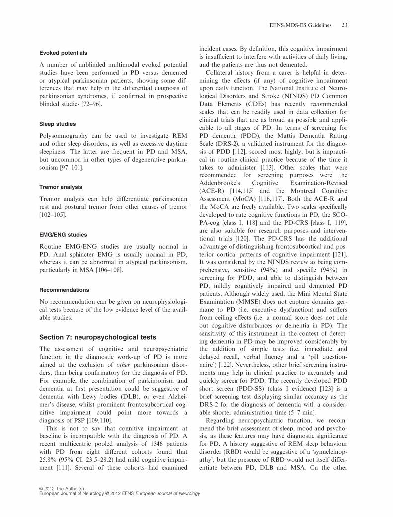
Figure 1 Transcranial sonography of the mesencephalic brain-
stem. The mesencephalic brainstem is scanned parallel to the
orbito-meatal line (a). (b) Depicts the mesencephalic brainstem
(surrounded by dotted line) of a healthy control with normal
echogenicity of the substantia nigra (SN) (encircled ipsilaterally
and marked with arrows contralaterally to the insonating
probe), the area of the red nucleus (asterisk) and the hyperecho-
genic midline raphe (open arrow). In a patient with Parkinson’s
disease (PD), the area of hyperechogenicity at the anatomical
site of the SN is enlarged (c; encircled ipsilaterally and marked
with arrows contralaterally).
© 2012 The Author(s)European Journal of Neurology © 2012 EFNS European Journal of Neurology
24 A. Berardelli et al.
Planimetric measurement of the area of hyperechog-
enicity at the anatomical site of the SN enables the
categorization of SN echogenicity into normal or
abnormal according to cut-off values established with
reference to percentile ranks assessed in healthy
cohorts.
Substantia nigra areas are classified as markedly
hyperechogenic (above the 90th percentile of the
healthy population) or moderately hyperechogenic
(between the 75th and 90th percentile, see also [135–137]). Measurement of the ventricular system and
semiquantitative assessment of the basal ganglia are
primarily important for the differential diagnosis of
PD.
For clinical practice, it is important to realize that
proper evaluation of the SN depends on
• Application of ultrasound-specific cut-offs
• Quality of the temporal bone window – in about
10% of the Caucasian population, the transtemporal
bone window is not sufficient to depict the relevant
structures
• To some extent experience of the investigator (see
also [141]).
The current evidence suggests that TCS is useful in
the diagnosis of parkinsonian syndromes, especially
with regard to
• Differentiation of atypical parkinsonian syndromes
(APS; class I evidence, Level A) [142]
• Differentiation of secondary parkinsonian syn-
dromes (class I and II evidence, Level A – for
sensitivity, specificity and predictive value of
parameters) [132]
• Early diagnosis of PD, in clinically unclear cases
(class II evidence) [143]
• Detection of subjects at risk for PD (class I) includ-
ing asymptomatic mutation carriers for monogenic
forms of PD [133–150].Substantia nigra hyperechogenicity occurs in
about 10% of the healthy population [151,152], a
proportion much larger than the prevalence of PD,
and may occur in a much smaller proportion in
subjects with other neurodegenerative disorders
[133,153–156], frequently in heavy metal storing dis-
eases [157–159] and sometimes in neuroinflammatory
disorders [160]. Because specificity for the develop-
ment of incident PD is about 80%, the application
of TCS should be therefore combined with other
screening procedures.
Stability of SN hyperechogenicity during the disease
course is still an unclear issue. Most reports argue for
a stable area of echogenicity [126,136,161,162], which,
however, may increase with age [163], thus hindering
its use as progression marker in PD.
Recommendations
Transcranial sonography is recommended (Level A)
for
I Differential diagnosis of PD from APS and second-
ary parkinsonian syndromes
II Early diagnosis of PD
III Detection of subjects at risk for PD
The technique is so far not universally used and
requires some expertise. Because specificity of TCS for
the development of PD is limited, TCS should be used
in conjunction with other screening tests.
Magnetic resonance imaging
In clinical practice, conventional magnetic resonance
imaging (cMRI) with visual assessment of T2- (sensi-
tive to changes in tissue properties, including tissue
damage) and T1-weighted (important for anatomical
details providing good grey matter/white matter con-
trast) imaging is a well-established method for the
exclusion of symptomatic [164] (class IV evidence
studies available – for review, see [164]).
Several findings on conventional structural MRI
have been described as diagnostic markers of MSA
(See Table 2). These include atrophy and signal altera-
tions at 1.5 T in the putamen and several infratentori-
al regions, such as
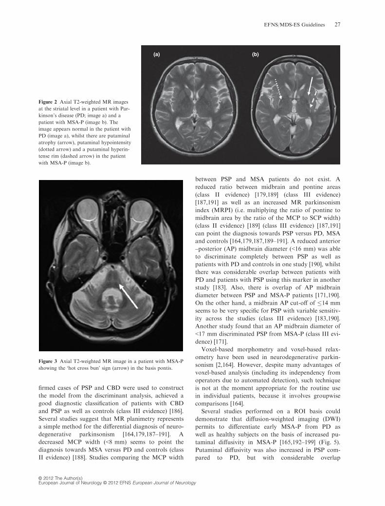
• Hyperintense putaminal rim with or without hypoin-
tensity in the dorsolateral part of the putamen (Fig. 2)
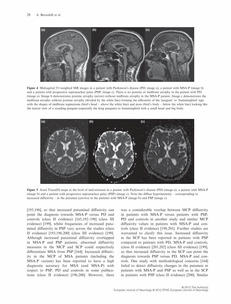
• ‘Hot cross bun’ sign of the pons (Fig. 3)
• Atrophy of the cerebellum
• Hyperintensity in the middle cerebellar peduncle
(MCP)
compared to PD, PSP and controls as well as pontine
and putaminal atrophy compared to PD and controls
(class II evidence) [165–168]; (class III evidence) [167–169,169–175]; (class IV evidence) [176]. Specificity of
the aforementioned abnormalities in differentiating
MSA from PD and healthy controls is considered
quite high, whereas sensitivity – particularly in early
disease stages – seems to be insufficient [164]. How-
ever, sensitivity of signal alterations can be somewhat
improved by modifying technical aspects such as spa-
tial resolution by using thinner slices or modifying
relaxation contrast by using T2*-weighted gradient
echo sequences [168,174].
Other abnormalities in MSA-P, which may provide
a differentiation from PD, PSP and controls, include
• Posterolateral linearization of the putaminal margin
(versus convex in controls) (class III evidence) [177]
• Putaminal hyperintensity on T1-weighted images
(class III evidence) [169]
© 2012 The Author(s)European Journal of Neurology © 2012 EFNS European Journal of Neurology
EFNS/MDS-ES Guidelines 25
Further, visual assessment of
• Atrophy of the superior cerebellar peduncle (SCP)
(class II evidence) [178]
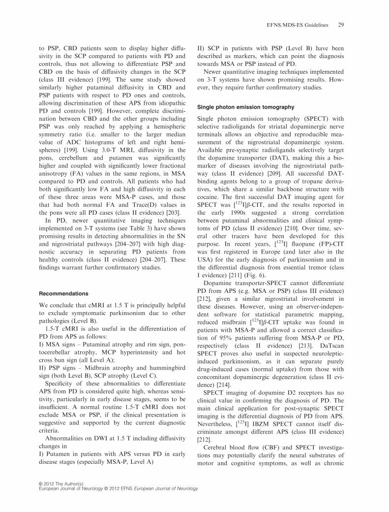
• ‘Penguin silhouette’ or ‘hummingbird’ sign (where
the shapes of the midbrain tegmentum – that is, the
bird’s head – and pons – that is, the bird’s body –resemble a lateral view of a standing king penguin
or hummingbird; Fig. 4) on midsagittal MR images
(class II evidence) [179]; (class III evidence) [180];
(class IV evidence) [181]
• Enlarged 3rd ventricle (class II evidence) [182];
(class III evidence) [171,173]
can point the diagnosis towards PSP versus PD, con-
trols and MSA (Level C), whereas visual assessment
of midbrain atrophy may point out a diagnosis of
PSP versus PD or controls (class III evidence)
[171,173,175].
Putaminal, pontine and midbrain atrophy seems to
occur in both MSA and PSP patients, although pu-
taminal and pontine atrophy is more common in
MSA and midbrain atrophy more common in PSP
[171,173]. Other abnormalities in PSP include an
abnormal superior profile of the midbrain (flat or
concave versus convex aspect in healthy people)
compared to patients with PD and controls [183]
(class III evidence) [183] and increased signal
changes in the SCP on FLAIR images compared to
controls, PD and MSA patients [184] (class III evi-
dence) [183].
Simple quantitative measures of diameters, areas
and volumes including region of interest (ROI)-based
assessment of various structures on MRI have been
applied as an indirect measure of brain structures
known to be atrophic in different parkinsonian disor-
ders for the differential diagnosis of neurodegenerative
parkinsonism. Volume loss of different supratentorial
and infratentorial brain structures, measured by MR
volumetry (MRV) with semi-automatic segmentation
techniques on a ROI approach, has been reported in
patients with APS, whereas most studies were not able
to detect such volume differences between patients with
PD and controls, whilst with advancing disease hippo-
campal atrophy has been reported in patients with PD
compared to healthy controls at a group level [164].
Whereas patients with MSA demonstrated significant
reductions in mean striatal, brainstem and cerebellar
volumes, in patients with PSP significant reductions in
whole brain, striatal, brainstem – especially midbrain –and frontal volumes have been shown [164]. By the
application of stepwise discriminant analysis to MRV
on a ROI basis, there was a good discrimination of
patients with PD and controls from MSA and PSP
patients. On the other hand, separation between
patients with PD and controls as well as between MSA
and PSP patients was insufficient (class II evidence)
[185]. A similar approach, in which post-mortem-con-
Table 2 Routine MRI findings in atypical parkinsonism [2,164]
Multiple system atrophy
• Putaminal atrophy
• Putaminal slit sign
• Putaminal hypointensity
• Pontine and/or bulbar atrophy
• Cerebellar and/or dentate atrophy
• Atrophy of the MCP
• Reduced MCP diameter (<8.0 mm in reference [188])
• Dilatation of the fourth ventricle
• Signal increase in MCP
• Signal increase in cerebellum
• Signal increase in inferior olives
• Signal increase in pontine fibres (hot cross bun sign)
Progressive supranuclear palsya
• Midbrain atrophy
• Indirect signs of midbrain atrophy:
○ Reduced AP midbrain diameter (<14 mm in reference
[190])
○ Abnormal superior midbrain profileb
○ ‘(king) penguin silhouette’ or ‘hummingbird’ signc
○ Reduced ratio between midbrain and pontine areas
○ Increased MRPId
• Dilatation of the third ventricle
• Atrophy of the SCP
• Signal increase in SCP (on FLAIR images)
• Signal increase in globus pallidus
• Signal increase in red nucleus
• Putaminal atrophy
• Frontal and parietal atrophy
Corticobasal degeneratione
• Cortical atrophy (mostly frontoparietal and asymmetric,
sometimes even global and symmetric)
• Putaminal hypointensityf
• Hyperintense signal changes in the motor cortex or subcorti-
cal white matter
Signal changes refer to 1.5-Tesla MRI scanners.
MCP, middle cerebellar peduncle; SCP, superior cerebellar peduncle;
AP anterior–posterior; MRPI, MR parkinsonism index.aAlmost all MRI studies of PSP included patients suffering from the
most reliably identifiable classic picture of PSP (i.e. Richardson’s
syndrome).bFlat or concave versus convex aspect in healthy people.cThe shapes of the midbrain tegmentum (the bird’s head) and pons
(the bird’s body) resemble a lateral view of a standing king penguin
or hummingbird.dMRPI: multiplying the ratio of pontine to midbrain area by the
ratio of the MCP to SCP width.eGiven the pathological heterogeneity of a ‘corticobasal syndrome’,
including CBD and other neurodegenerative causes such as PSP,
Pick’s disease and other frontotemporal lobar degenerations, MRI
studies of clinically defined CBD must be discussed with a grain of
caution.
© 2012 The Author(s)European Journal of Neurology © 2012 EFNS European Journal of Neurology
26 A. Berardelli et al.
firmed cases of PSP and CBD were used to construct
the model from the discriminant analysis, achieved a
good diagnostic classification of patients with CBD
and PSP as well as controls (class III evidence) [186].
Several studies suggest that MR planimetry represents
a simple method for the differential diagnosis of neuro-
degenerative parkinsonism [164,179,187–191]. A
decreased MCP width (<8 mm) seems to point the
diagnosis towards MSA versus PD and controls (class
II evidence) [188]. Studies comparing the MCP width
between PSP and MSA patients do not exist. A
reduced ratio between midbrain and pontine areas
(class II evidence) [179,189] (class III evidence)
[187,191] as well as an increased MR parkinsonism
index (MRPI) (i.e. multiplying the ratio of pontine to
midbrain area by the ratio of the MCP to SCP width)
(class II evidence) [189] (class III evidence) [187,191]
can point the diagnosis towards PSP versus PD, MSA
and controls [164,179,187,189–191]. A reduced anterior
–posterior (AP) midbrain diameter (<16 mm) was able
to discriminate completely between PSP as well as
patients with PD and controls in one study [190], whilst
there was considerable overlap between patients with
PD and patients with PSP using this marker in another
study [183]. Also, there is overlap of AP midbrain
diameter between PSP and MSA-P patients [171,190].
On the other hand, a midbrain AP cut-off of �14 mm
seems to be very specific for PSP with variable sensitiv-
ity across the studies (class III evidence) [183,190].
Another study found that an AP midbrain diameter of
<17 mm discriminated PSP from MSA-P (class III evi-
dence) [171].
Voxel-based morphometry and voxel-based relax-
ometry have been used in neurodegenerative parkin-
sonism [2,164]. However, despite many advantages of
voxel-based analysis (including its independency from
operators due to automated detection), such technique
is not at the moment appropriate for the routine use
in individual patients, because it involves groupwise
comparisons [164].
Several studies performed on a ROI basis could
demonstrate that diffusion-weighted imaging (DWI)
permits to differentiate early MSA-P from PD as
well as healthy subjects on the basis of increased pu-
taminal diffusivity in MSA-P [165,192–199] (Fig. 5).
Putaminal diffusivity was also increased in PSP com-
pared to PD, but with considerable overlap
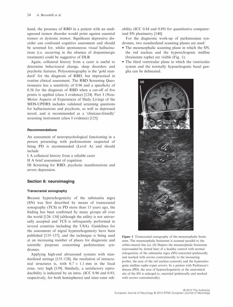
Figure 3 Axial T2-weighted MR image in a patient with MSA-P
showing the ‘hot cross bun’ sign (arrow) in the basis pontis.
(a) (b)
Figure 2 Axial T2-weighted MR images
at the striatal level in a patient with Par-
kinson’s disease (PD; image a) and a
patient with MSA-P (image b). The
image appears normal in the patient with
PD (image a), whilst there are putaminal
atrophy (arrow), putaminal hypointensity
(dotted arrow) and a putaminal hyperin-
tense rim (dashed arrow) in the patient
with MSA-P (image b).
© 2012 The Author(s)European Journal of Neurology © 2012 EFNS European Journal of Neurology
EFNS/MDS-ES Guidelines 27
[193,196], so that increased putaminal diffusivity can
point the diagnosis towards MSA-P versus PD and
controls (class II evidence) [165,192–198] (class III
evidence) [199], whilst frequencies of increased puta-
minal diffusivity in PSP vary across the studies (class
II evidence) [193,196,200] (class III evidence) [199].
Although increased putaminal diffusivity overlapped
in MSA-P and PSP patients, abnormal diffusivity
measures in the MCP and SCP could respectively
differentiate MSA from PSP [164]. Increased diffusiv-
ity in the MCP of MSA patients (including the
MSA-P variant) has been reported to have a high
diagnostic accuracy for MSA (and MSA-P) with
respect to PSP, PD and controls in some publica-
tions (class II evidence) [196,200]. However, there
was a considerable overlap between MCP diffusivity
in patients with MSA-P versus patients with PSP,
PD and controls in another study and similar MCP
diffusivity values in patients with MSA-P and con-
trols (class II evidence) [198,201]. Further studies are
warranted to clarify this issue. Increased diffusivity
in the SCP has been reported in patients with PSP
compared to patients with PD, MSA-P and controls,
(class II evidence) [201,202] (class III evidence) [199],
so that increased diffusivity in the SCP can point the
diagnosis towards PSP versus PD, MSA-P and con-
trols. One study with methodological concerns [164]
failed to detect diffusivity changes in the putamen in
patients with MSA-P and PSP as well as in the SCP
in patients with PSP (class II evidence) [200]. Similar
(a) (b) (c)
Figure 4 Midsagittal T1-weighted MR images in a patient with Parkinson’s disease (PD; image a), a patient with MSA-P (image b)
and a patient with progressive supranuclear palsy (PSP; image c). There is no pontine or midbrain atrophy in the patient with PD
(image a). Image b demonstrates pontine atrophy (arrow) without midbrain atrophy in the MSA-P patient. Image c demonstrates the
midbrain atrophy without pontine atrophy (divided by the white line) forming the silhouette of the ‘penguin’ or ‘hummingbird’ sign
with the shapes of midbrain tegmentum (bird’s head – above the white line) and pons (bird’s body – below the white line) looking like
the lateral view of a standing penguin (especially the king penguin) or hummingbird with a small head and big body.
(a) (b) (c)
Figure 5 Axial Trace(D) maps at the level of mid-striatum in a patient with Parkinson’s disease (PD) (image a), a patient with MSA-P
(image b) and a patient with progressive supranuclear palsy (PSP) (image c). Note the diffuse hyperintensity – corresponding to
increased diffusivity – in the putamen (arrows) in the patients with MSA-P (image b) and PSP (image c)
© 2012 The Author(s)European Journal of Neurology © 2012 EFNS European Journal of Neurology
28 A. Berardelli et al.
to PSP, CBD patients seem to display higher diffu-
sivity in the SCP compared to patients with PD and
controls, thus not allowing to differentiate PSP and
CBD on the basis of diffusivity changes in the SCP
(class III evidence) [199]. The same study showed
similarly higher putaminal diffusivity in CBD and
PSP patients with respect to PD ones and controls,
allowing discrimination of these APS from idiopathic
PD and controls [199]. However, complete discrimi-
nation between CBD and the other groups including
PSP was only reached by applying a hemispheric
symmetry ratio (i.e. smaller to the larger median
value of ADC histograms of left and right hemi-
spheres) [199]. Using 3.0-T MRI, diffusivity in the
pons, cerebellum and putamen was significantly
higher and coupled with significantly lower fractional
anisotropy (FA) values in the same regions, in MSA
compared to PD and controls. All patients who had
both significantly low FA and high diffusivity in each
of these three areas were MSA-P cases, and those
that had both normal FA and Trace(D) values in
the pons were all PD cases (class II evidence) [203].
In PD, newer quantitative imaging techniques
implemented on 3-T systems (see Table 3) have shown
promising results in detecting abnormalities in the SN
and nigrostriatal pathways [204–207] with high diag-
nostic accuracy in separating PD patients from
healthy controls (class II evidence) [204–207]. These
findings warrant further confirmatory studies.
Recommendations
We conclude that cMRI at 1.5 T is principally helpful
to exclude symptomatic parkinsonism due to other
pathologies (Level B).
1.5-T cMRI is also useful in the differentiation of
PD from APS as follows:
I) MSA signs – Putaminal atrophy and rim sign, pon-
tocerebellar atrophy, MCP hyperintensity and hot
cross bun sign (all Level A);
II) PSP signs – Midbrain atrophy and hummingbird
sign (both Level B), SCP atrophy (Level C).
Specificity of these abnormalities to differentiate
APS from PD is considered quite high, whereas sensi-
tivity, particularly in early disease stages, seems to be
insufficient. A normal routine 1.5-T cMRI does not
exclude MSA or PSP, if the clinical presentation is
suggestive and supported by the current diagnostic
criteria.
Abnormalities on DWI at 1.5 T including diffusivity
changes in
I) Putamen in patients with APS versus PD in early
disease stages (especially MSA-P, Level A)
II) SCP in patients with PSP (Level B) have been
described as markers, which can point the diagnosis
towards MSA or PSP instead of PD.
Newer quantitative imaging techniques implemented
on 3-T systems have shown promising results. How-
ever, they require further confirmatory studies.
Single photon emission tomography
Single photon emission tomography (SPECT) with
selective radioligands for striatal dopaminergic nerve
terminals allows an objective and reproducible mea-
surement of the nigrostriatal dopaminergic system.
Available pre-synaptic radioligands selectively target
the dopamine transporter (DAT), making this a bio-
marker of diseases involving the nigrostriatal path-
way (class II evidence) [209]. All successful DAT-
binding agents belong to a group of tropane deriva-
tives, which share a similar backbone structure with
cocaine. The first successful DAT imaging agent for
SPECT was [123I]b-CIT, and the results reported in
the early 1990s suggested a strong correlation
between putaminal abnormalities and clinical symp-
toms of PD (class II evidence) [210]. Over time, sev-
eral other tracers have been developed for this
purpose. In recent years, [123I] fluopane (FP)-CIT
was first registered in Europe (and later also in the
USA) for the early diagnosis of parkinsonism and in
the differential diagnosis from essential tremor (class
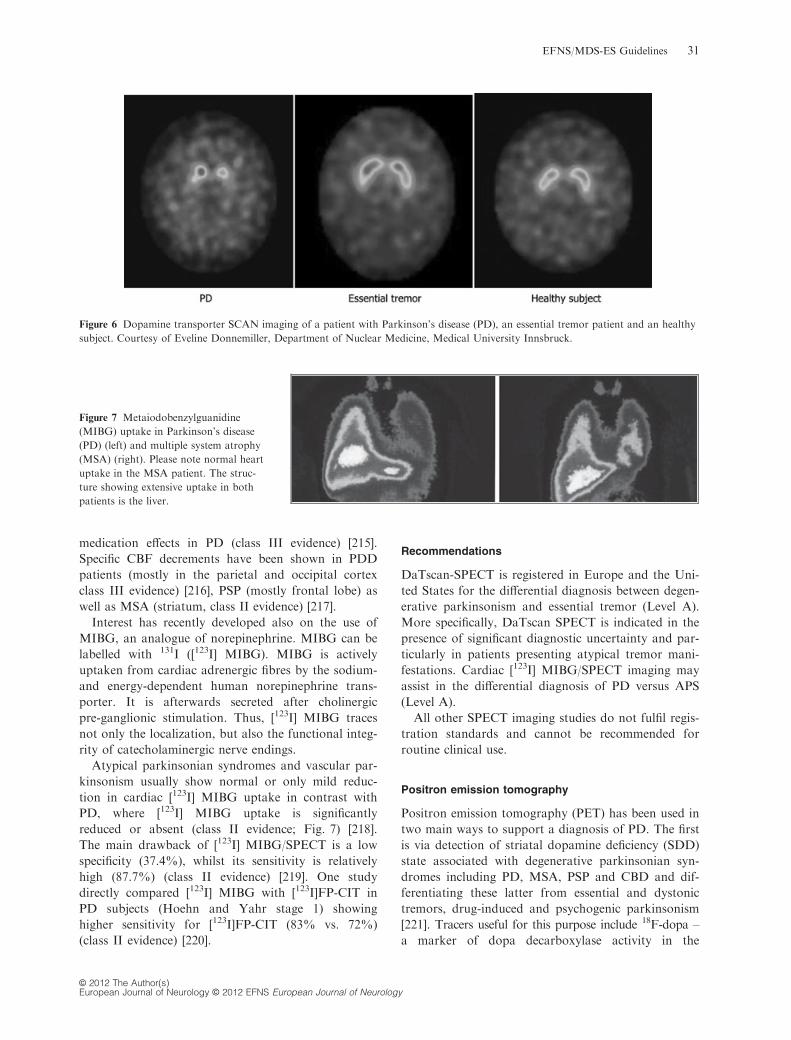
I evidence) [211] (Fig. 6).
Dopamine transporter-SPECT cannot differentiate
PD from APS (e.g. MSA or PSP) (class III evidence)
[212], given a similar nigrostriatal involvement in
these diseases. However, using an observer-indepen-
dent software for statistical parametric mapping,
reduced midbrain [123I]b-CIT uptake was found in
patients with MSA-P and allowed a correct classifica-
tion of 95% patients suffering from MSA-P or PD,
respectively (class II evidence) [213]. DaTscan
SPECT proves also useful in suspected neuroleptic-
induced parkinsonism, as it can separate purely
drug-induced cases (normal uptake) from those with
concomitant dopaminergic degeneration (class II evi-
dence) [214].
SPECT imaging of dopamine D2 receptors has no
clinical value in confirming the diagnosis of PD. The
main clinical application for post-synaptic SPECT
imaging is the differential diagnosis of PD from APS.
Nevertheless, [123I] IBZM SPECT cannot itself dis-
criminate amongst different APS (class III evidence)
[212].
Cerebral blood flow (CBF) and SPECT investiga-
tions may potentially clarify the neural substrates of
motor and cognitive symptoms, as well as chronic
© 2012 The Author(s)European Journal of Neurology © 2012 EFNS European Journal of Neurology
EFNS/MDS-ES Guidelines 29
Table
3Findingswithquantitativeim
agingtechniques
implementedon3-T
MR
system
sin
PD
Author,year
(reference)
Cohort
size
(meanage,
meandisease
duration,rangedisease
duration)/design
MRIMethodology
Marker
Sensitivity(%
)Specificity
(%)
Vaillancourt
2009[204]
•PD
14denovountreatedwith
H&Y
1-2
(57y,16m,433m)/controls14(58y)
•Prospectiveblinded
analysis
DTIoftheSN
(subregions)
DecreasedFA
incaudalSN
100
100
DecreasedFA
inmiddle
SN
100
35
DecreasedFA
inrostralSN
100
7
Menke2009[205]
•PD
10withH&Y
1–3
(64y,
6y,1–14y)/controls10(64y)
•Prospectiveblinded
analysis
Combined
SN
volumetry
with
DTIofSN
(aVCDR)
DecreasedSN
volume
80
70
DecreasedVCDR
b100
70
DecreasedSN
volume+
decreasedVCDR
100
80
Gr€ oger
2011[206]•
PD
9withH&Y
2,5–3,allmedicated
(69y,n.g.,4–25y)/controls8(66y)
•Prospectiveblinded
analysis
3D-M
RSIoftheSN
DecreasedNAA/C
rratio
ofrostralSN
89
50
IncreasedNAA/C
rratio
ofcaudalSN
89
75
Decreasedrostral-to-caudal
ratiosoftheNAA/C
rratio
100
100
Peran2010[207]
•PD
30withH&Y
1–2,allmedicated
(62y,4.5y,n.g.)/controls22(57y)
•Prospectiveblinded
analysis
Multim
odalstudyusingacombination
ofdifferentMR
markersincluding
volumetry,meanR
b,meandiffusivity
andFA
applied
in6deepgreymatter
structures(SN,RN
thalamus,putamen,
caudate
andpallidum)
IncreasedR2bin
the
substantianigra,reduced
FA
inthesubstantianigra
andincreasedmeandiffusivity
intheputamen
orcaudate
nucleus
Discrim
inantpower
c:
•71–83%
inconsideringonly
onemarker
•95–98%
inconsideringthe
three-marker
combinations
Melzer2011[208]
•PD
61with26drug-na€ıve(cognitively
norm
aln=
34:65y,3y;withmildcognitive
impairmentn=
16:70y,9y;withdem
entia,
n=
11:75y,12y)/controls29(69y)
•Prospectiveblinded
analysis
Perfusionim
agingstudywitharterial
spin
labelling
PD-relatedperfusionnetwork
byprincipalcomponentanalysis
AUC
ofROC2:
•ControlsversusPD
0.71
•ControlsversusPD+
MCI
0.94
•ControlsversusPDD
0.99
•Denovopatients
notanaly-
sed
PD,Parkinson’s
disease;PDD,PD
dem
entia;SN,substantianigra;RN,rednucleus;
DTI,
diffusiontensorim
aging;3D-M
RSI,
three-dim
ensionalmagnetic
resonance
spectroscopic
imaging;R2b,
relaxationrates=1/T2b);FA,fractionalanisotropy;ASL,arterialspin
labelling;AUC,areaunder
thecurve;
ROC,receiver
operatingcharacteristicanalysis;n.g.notgiven.
aVCDR
isaDTImarker
representingthenumbersofvoxelsforallconnectivity-defined
subregionswithin
theSN
thresholded
at10%
ofthemaxim
um
connectionprobability.
bReducedleft
andrightSN
toipsilateralthalamusVCDRs.
cSensitivity,specificity
notgiven.
© 2012 The Author(s)European Journal of Neurology © 2012 EFNS European Journal of Neurology
30 A. Berardelli et al.
medication effects in PD (class III evidence) [215].
Specific CBF decrements have been shown in PDD
patients (mostly in the parietal and occipital cortex
class III evidence) [216], PSP (mostly frontal lobe) as
well as MSA (striatum, class II evidence) [217].
Interest has recently developed also on the use of
MIBG, an analogue of norepinephrine. MIBG can be
labelled with 131I ([123I] MIBG). MIBG is actively
uptaken from cardiac adrenergic fibres by the sodium-
and energy-dependent human norepinephrine trans-
porter. It is afterwards secreted after cholinergic
pre-ganglionic stimulation. Thus, [123I] MIBG traces
not only the localization, but also the functional integ-
rity of catecholaminergic nerve endings.
Atypical parkinsonian syndromes and vascular par-
kinsonism usually show normal or only mild reduc-
tion in cardiac [123I] MIBG uptake in contrast with
PD, where [123I] MIBG uptake is significantly
reduced or absent (class II evidence; Fig. 7) [218].
The main drawback of [123I] MIBG/SPECT is a low
specificity (37.4%), whilst its sensitivity is relatively
high (87.7%) (class II evidence) [219]. One study
directly compared [123I] MIBG with [123I]FP-CIT in
PD subjects (Hoehn and Yahr stage 1) showing
higher sensitivity for [123I]FP-CIT (83% vs. 72%)
(class II evidence) [220].
Recommendations
DaTscan-SPECT is registered in Europe and the Uni-
ted States for the differential diagnosis between degen-
erative parkinsonism and essential tremor (Level A).
More specifically, DaTscan SPECT is indicated in the
presence of significant diagnostic uncertainty and par-
ticularly in patients presenting atypical tremor mani-
festations. Cardiac [123I] MIBG/SPECT imaging may
assist in the differential diagnosis of PD versus APS
(Level A).
All other SPECT imaging studies do not fulfil regis-
tration standards and cannot be recommended for
routine clinical use.
Positron emission tomography
Positron emission tomography (PET) has been used in
two main ways to support a diagnosis of PD. The first
is via detection of striatal dopamine deficiency (SDD)
state associated with degenerative parkinsonian syn-
dromes including PD, MSA, PSP and CBD and dif-
ferentiating these latter from essential and dystonic
tremors, drug-induced and psychogenic parkinsonism
[221]. Tracers useful for this purpose include 18F-dopa –a marker of dopa decarboxylase activity in the
Figure 6 Dopamine transporter SCAN imaging of a patient with Parkinson’s disease (PD), an essential tremor patient and an healthy
subject. Courtesy of Eveline Donnemiller, Department of Nuclear Medicine, Medical University Innsbruck.
Figure 7 Metaiodobenzylguanidine
(MIBG) uptake in Parkinson’s disease
(PD) (left) and multiple system atrophy
(MSA) (right). Please note normal heart
uptake in the MSA patient. The struc-
ture showing extensive uptake in both
patients is the liver.
© 2012 The Author(s)European Journal of Neurology © 2012 EFNS European Journal of Neurology
EFNS/MDS-ES Guidelines 31
dopaminergic terminal [222], 11C- and 18F-dihydrotet-
rabenazine (DTBZ) – both markers of monoamine
vesicular transporter binding [223,224], and 11C-meth-
ylphenidate [225], 18F-CFT [226], 11C-RTI-32 [227] 7
and 18F-FP-CIT [228] – all markers of DAT availabil-
ity (Fig. 8). It has been demonstrated that levels of
SDD correlate well with locomotor disability [229]
and progression of disease can be detected in longitu-
dinal studies [226,230,231]. Additionally, pre-motor
SDD can be detected in some adult subjects at risk
for PD, such as LRRK2 gene carriers [232], asymp-
tomatic relatives of familial PD cases [233] and homo-
zygous twins [234].
Whilst these PET tracers have high sensitivity for
detecting SDD, they do not reliably discriminate
between parkinsonian syndromes, although PD is
associated with a rostro-caudal putamen gradient of
dopamine dysfunction compared with the more uni-
form dysfunction evident in PSP and CBD [235]. In
PD, putamen dopamine D2 receptor availability is
preserved or even mildly increased in de novo cases.
This can be demonstrated with PET benzamide tracers
such as 11C-raclopride [236]. In contrast, striatal D2
binding is reduced in MSA, PSP and CBD, although
significant decreases are only present in around 50%
of individuals [237,238].
The second approach involves 18F-FDG PET which
allows the patterns of resting brain glucose metabo-
lism (rCMRGlc) to be determined. In PD, covariance
analysis reveals a characteristic profile where lentiform
rCMRGlc is relatively increased and frontal
rCMRGlc lowered. This has been indicated as the
PD-related profile (PDRP; Fig. 9) [239]. The PDRP
correlates with locomotor disability when patients are
temporarily withdrawn from medication and can be
normalized by the treatment with dopaminergic agents
[240]. In contrast, APS characteristically show reduced
striatal rCMRGlc and can be sensitively discriminated
from typical PD [241]. It may also be possible to dis-
criminate between the atypical syndromes by the pat-
terns of brainstem and cortical involvement [242].
In summary, PET imaging cannot directly diagnose
PD or APS. The patterns of dysfunction it reveals can
help supporting or refuting clinical impressions. If
neuroprotective or restorative therapies become avail-
able, PET can also reveal subclinical dysfunction in
at-risk subjects for PD.
Recommendations
None of the reviewed PET studies has been per-
formed according to regulatory standards with the
exception of the study by Whone et al. [230]. There-
fore, we cannot make any formal recommendation
for the routine use of PET studies in the diagnostic
work-up of PD.
Figure 8 Images of striatal 11C-RTI-32 positron emission tomography (PET) dopamine transporter (DAT), 11C-DTBZ PET (VMAT2)
and 18F-dopa PET (dopa decarboxylase) uptake in healthy volunteers and early Parkinson’s disease (PD). It can be seen that the three
PET imaging modalities all show asymmetrically reduced posterior putamen dopaminergic function in PD.
Figure 9 Covariance analysis of FDG positron emission tomog-
raphy (PET) showing the Parkinson’s disease (PD)-related pro-
file of raised lentiform and reduced frontal metabolism
(Eidelberg 1990).
© 2012 The Author(s)European Journal of Neurology © 2012 EFNS European Journal of Neurology
32 A. Berardelli et al.
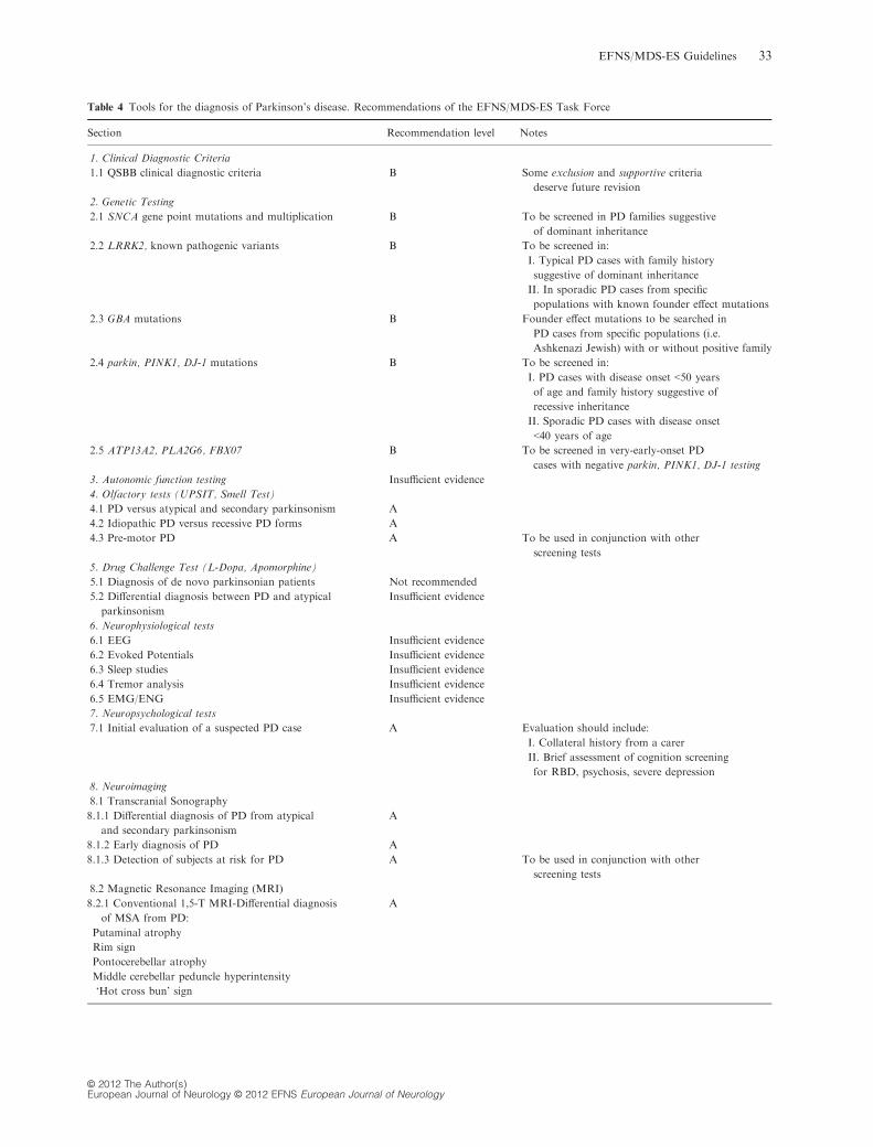
Table 4 Tools for the diagnosis of Parkinson’s disease. Recommendations of the EFNS/MDS-ES Task Force
Section Recommendation level Notes
1. Clinical Diagnostic Criteria
1.1 QSBB clinical diagnostic criteria B Some exclusion and supportive criteria
deserve future revision
2. Genetic Testing
2.1 SNCA gene point mutations and multiplication B To be screened in PD families suggestive
of dominant inheritance
2.2 LRRK2, known pathogenic variants B To be screened in:
I. Typical PD cases with family history
suggestive of dominant inheritance
II. In sporadic PD cases from specific
populations with known founder effect mutations
2.3 GBA mutations B Founder effect mutations to be searched in
PD cases from specific populations (i.e.
Ashkenazi Jewish) with or without positive family
2.4 parkin, PINK1, DJ-1 mutations B To be screened in:
I. PD cases with disease onset <50 years
of age and family history suggestive of
recessive inheritance
II. Sporadic PD cases with disease onset
<40 years of age
2.5 ATP13A2, PLA2G6, FBX07 B To be screened in very-early-onset PD
cases with negative parkin, PINK1, DJ-1 testing
3. Autonomic function testing Insufficient evidence
4. Olfactory tests (UPSIT, Smell Test)
4.1 PD versus atypical and secondary parkinsonism A
4.2 Idiopathic PD versus recessive PD forms A
4.3 Pre-motor PD A To be used in conjunction with other
screening tests
5. Drug Challenge Test (L-Dopa, Apomorphine)
5.1 Diagnosis of de novo parkinsonian patients Not recommended
5.2 Differential diagnosis between PD and atypical
parkinsonism
Insufficient evidence
6. Neurophysiological tests
6.1 EEG Insufficient evidence
6.2 Evoked Potentials Insufficient evidence
6.3 Sleep studies Insufficient evidence
6.4 Tremor analysis Insufficient evidence
6.5 EMG/ENG Insufficient evidence
7. Neuropsychological tests
7.1 Initial evaluation of a suspected PD case A Evaluation should include:
I. Collateral history from a carer
II. Brief assessment of cognition screening
for RBD, psychosis, severe depression
8. Neuroimaging
8.1 Transcranial Sonography
8.1.1 Differential diagnosis of PD from atypical
and secondary parkinsonism
A
8.1.2 Early diagnosis of PD A
8.1.3 Detection of subjects at risk for PD A To be used in conjunction with other
screening tests
8.2 Magnetic Resonance Imaging (MRI)
8.2.1 Conventional 1,5-T MRI-Differential diagnosis
of MSA from PD:
A
Putaminal atrophy
Rim sign
Pontocerebellar atrophy
Middle cerebellar peduncle hyperintensity
‘Hot cross bun’ sign
© 2012 The Author(s)European Journal of Neurology © 2012 EFNS European Journal of Neurology
EFNS/MDS-ES Guidelines 33
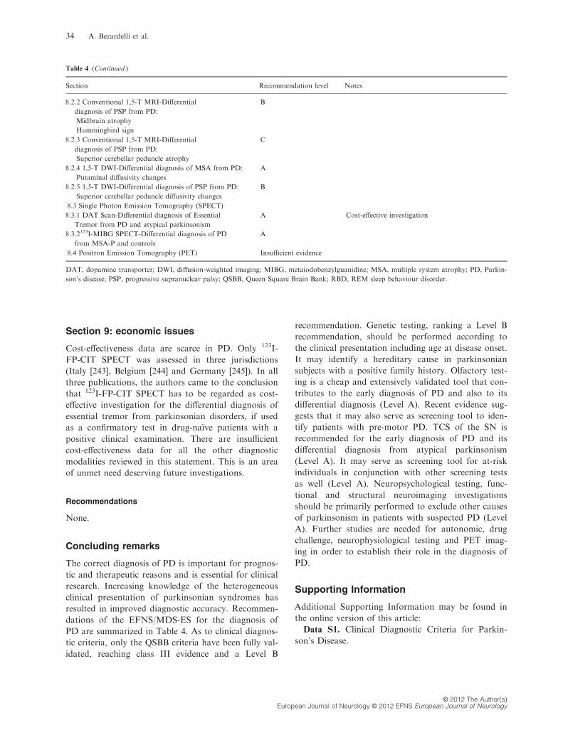
Section 9: economic issues
Cost-effectiveness data are scarce in PD. Only 123I-
FP-CIT SPECT was assessed in three jurisdictions
(Italy [243], Belgium [244] and Germany [245]). In all
three publications, the authors came to the conclusion
that 123I-FP-CIT SPECT has to be regarded as cost-
effective investigation for the differential diagnosis of
essential tremor from parkinsonian disorders, if used
as a confirmatory test in drug-na€ıve patients with a
positive clinical examination. There are insufficient
cost-effectiveness data for all the other diagnostic
modalities reviewed in this statement. This is an area
of unmet need deserving future investigations.
Recommendations
None.
Concluding remarks
The correct diagnosis of PD is important for prognos-
tic and therapeutic reasons and is essential for clinical
research. Increasing knowledge of the heterogeneous
clinical presentation of parkinsonian syndromes has
resulted in improved diagnostic accuracy. Recommen-
dations of the EFNS/MDS-ES for the diagnosis of
PD are summarized in Table 4. As to clinical diagnos-
tic criteria, only the QSBB criteria have been fully val-
idated, reaching class III evidence and a Level B
recommendation. Genetic testing, ranking a Level B
recommendation, should be performed according to
the clinical presentation including age at disease onset.
It may identify a hereditary cause in parkinsonian
subjects with a positive family history. Olfactory test-
ing is a cheap and extensively validated tool that con-
tributes to the early diagnosis of PD and also to its
differential diagnosis (Level A). Recent evidence sug-
gests that it may also serve as screening tool to iden-
tify patients with pre-motor PD. TCS of the SN is
recommended for the early diagnosis of PD and its
differential diagnosis from atypical parkinsonism
(Level A). It may serve as screening tool for at-risk
individuals in conjunction with other screening tests
as well (Level A). Neuropsychological testing, func-
tional and structural neuroimaging investigations
should be primarily performed to exclude other causes
of parkinsonism in patients with suspected PD (Level
A). Further studies are needed for autonomic, drug
challenge, neurophysiological testing and PET imag-
ing in order to establish their role in the diagnosis of
PD.
Supporting Information
Additional Supporting Information may be found in
the online version of this article:
Data S1. Clinical Diagnostic Criteria for Parkin-
son’s Disease.
Table 4 (Continued )
Section Recommendation level Notes
8.2.2 Conventional 1,5-T MRI-Differential
diagnosis of PSP from PD:
Midbrain atrophy
Hummingbird sign
B
8.2.3 Conventional 1,5-T MRI-Differential
diagnosis of PSP from PD:
Superior cerebellar peduncle atrophy
C
8.2.4 1,5-T DWI-Differential diagnosis of MSA from PD:
Putaminal diffusivity changes
A
8.2.5 1,5-T DWI-Differential diagnosis of PSP from PD:
Superior cerebellar peduncle diffusivity changes
B
8.3 Single Photon Emission Tomography (SPECT)
8.3.1 DAT Scan-Differential diagnosis of Essential
Tremor from PD and atypical parkinsonism
A Cost-effective investigation
8.3.2123I-MIBG SPECT-Differential diagnosis of PD
from MSA-P and controls
A
8.4 Positron Emission Tomography (PET) Insufficient evidence
DAT, dopamine transporter; DWI, diffusion-weighted imaging; MIBG, metaiodobenzylguanidine; MSA, multiple system atrophy; PD, Parkin-
son’s disease; PSP, progressive supranuclear palsy; QSBB, Queen Square Brain Bank; RBD, REM sleep behaviour disorder.
© 2012 The Author(s)European Journal of Neurology © 2012 EFNS European Journal of Neurology
34 A. Berardelli et al.
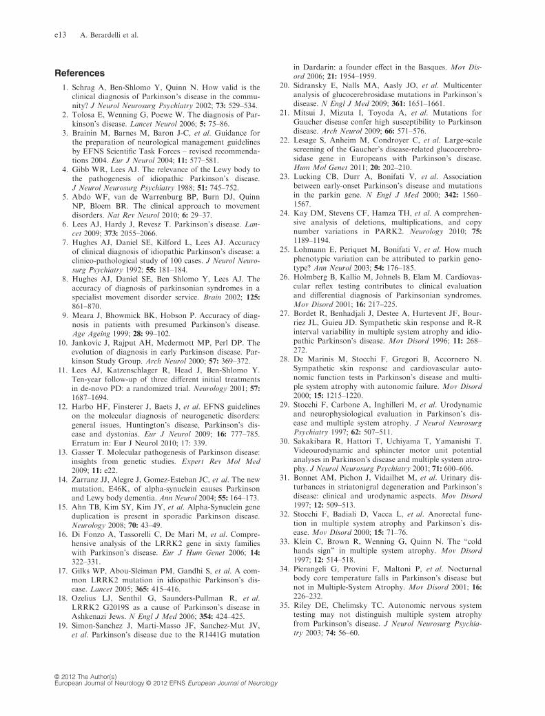
References
1. Schrag A, Ben-Shlomo Y, Quinn N. How valid is theclinical diagnosis of Parkinson’s disease in the commu-nity? J Neurol Neurosurg Psychiatry 2002; 73: 529–534.
2. Tolosa E, Wenning G, Poewe W. The diagnosis of Par-kinson’s disease. Lancet Neurol 2006; 5: 75–86.
3. Brainin M, Barnes M, Baron J-C, et al. Guidance forthe preparation of neurological management guidelinesby EFNS Scientific Task Forces – revised recommenda-tions 2004. Eur J Neurol 2004; 11: 577–581.
4. Gibb WR, Lees AJ. The relevance of the Lewy body tothe pathogenesis of idiopathic Parkinson’s disease.J Neurol Neurosurg Psychiatry 1988; 51: 745–752.
5. Abdo WF, van de Warrenburg BP, Burn DJ, QuinnNP, Bloem BR. The clinical approach to movementdisorders. Nat Rev Neurol 2010; 6: 29–37.
6. Lees AJ, Hardy J, Revesz T. Parkinson’s disease. Lan-cet 2009; 373: 2055–2066.
7. Hughes AJ, Daniel SE, Kilford L, Lees AJ. Accuracyof clinical diagnosis of idiopathic Parkinson’s disease: aclinico-pathological study of 100 cases. J Neurol Neuro-surg Psychiatry 1992; 55: 181–184.
8. Hughes AJ, Daniel SE, Ben Shlomo Y, Lees AJ. Theaccuracy of diagnosis of parkinsonian syndromes in aspecialist movement disorder service. Brain 2002; 125:
861–870.9. Meara J, Bhowmick BK, Hobson P. Accuracy of diag-
nosis in patients with presumed Parkinson’s disease.Age Ageing 1999; 28: 99–102.
10. Jankovic J, Rajput AH, Mcdermott MP, Perl DP. Theevolution of diagnosis in early Parkinson disease. Par-kinson Study Group. Arch Neurol 2000; 57: 369–372.
11. Lees AJ, Katzenschlager R, Head J, Ben-Shlomo Y.Ten-year follow-up of three different initial treatmentsin de-novo PD: a randomized trial. Neurology 2001; 57:1687–1694.
12. Harbo HF, Finsterer J, Baets J, et al. EFNS guidelineson the molecular diagnosis of neurogenetic disorders:general issues, Huntington’s disease, Parkinson’s dis-ease and dystonias. Eur J Neurol 2009; 16: 777–785.Erratum in: Eur J Neurol 2010; 17: 339.
13. Gasser T. Molecular pathogenesis of Parkinson disease:insights from genetic studies. Expert Rev Mol Med2009; 11: e22.
14. Zarranz JJ, Alegre J, Gomez-Esteban JC, et al. The newmutation, E46K, of alpha-synuclein causes Parkinsonand Lewy body dementia. Ann Neurol 2004; 55: 164–173.
15. Ahn TB, Kim SY, Kim JY, et al. Alpha-Synuclein geneduplication is present in sporadic Parkinson disease.Neurology 2008; 70: 43–49.
16. Di Fonzo A, Tassorelli C, De Mari M, et al. Compre-hensive analysis of the LRRK2 gene in sixty familieswith Parkinson’s disease. Eur J Hum Genet 2006; 14:
322–331.17. Gilks WP, Abou-Sleiman PM, Gandhi S, et al. A com-
mon LRRK2 mutation in idiopathic Parkinson’s dis-ease. Lancet 2005; 365: 415–416.
18. Ozelius LJ, Senthil G, Saunders-Pullman R, et al.LRRK2 G2019S as a cause of Parkinson’s disease inAshkenazi Jews. N Engl J Med 2006; 354: 424–425.
19. Simon-Sanchez J, Marti-Masso JF, Sanchez-Mut JV,et al. Parkinson’s disease due to the R1441G mutation
in Dardarin: a founder effect in the Basques. Mov Dis-ord 2006; 21: 1954–1959.
20. Sidransky E, Nalls MA, Aasly JO, et al. Multicenteranalysis of glucocerebrosidase mutations in Parkinson’sdisease. N Engl J Med 2009; 361: 1651–1661.
21. Mitsui J, Mizuta I, Toyoda A, et al. Mutations forGaucher disease confer high susceptibility to Parkinsondisease. Arch Neurol 2009; 66: 571–576.
22. Lesage S, Anheim M, Condroyer C, et al. Large-scalescreening of the Gaucher’s disease-related glucocerebro-sidase gene in Europeans with Parkinson’s disease.Hum Mol Genet 2011; 20: 202–210.
23. Lucking CB, Durr A, Bonifati V, et al. Associationbetween early-onset Parkinson’s disease and mutationsin the parkin gene. N Engl J Med 2000; 342: 1560–1567.
24. Kay DM, Stevens CF, Hamza TH, et al. A comprehen-sive analysis of deletions, multiplications, and copynumber variations in PARK2. Neurology 2010; 75:
1189–1194.25. Lohmann E, Periquet M, Bonifati V, et al. How much
phenotypic variation can be attributed to parkin geno-type? Ann Neurol 2003; 54: 176–185.
26. Holmberg B, Kallio M, Johnels B, Elam M. Cardiovas-cular reflex testing contributes to clinical evaluationand differential diagnosis of Parkinsonian syndromes.Mov Disord 2001; 16: 217–225.
27. Bordet R, Benhadjali J, Destee A, Hurtevent JF, Bour-riez JL, Guieu JD. Sympathetic skin response and R-Rinterval variability in multiple system atrophy and idio-pathic Parkinson’s disease. Mov Disord 1996; 11: 268–272.
28. De Marinis M, Stocchi F, Gregori B, Accornero N.Sympathetic skin response and cardiovascular auto-nomic function tests in Parkinson’s disease and multi-ple system atrophy with autonomic failure. Mov Disord2000; 15: 1215–1220.
29. Stocchi F, Carbone A, Inghilleri M, et al. Urodynamicand neurophysiological evaluation in Parkinson’s dis-ease and multiple system atrophy. J Neurol NeurosurgPsychiatry 1997; 62: 507–511.
30. Sakakibara R, Hattori T, Uchiyama T, Yamanishi T.Videourodynamic and sphincter motor unit potentialanalyses in Parkinson’s disease and multiple system atro-phy. J Neurol Neurosurg Psychiatry 2001; 71: 600–606.
31. Bonnet AM, Pichon J, Vidailhet M, et al. Urinary dis-turbances in striatonigral degeneration and Parkinson’sdisease: clinical and urodynamic aspects. Mov Disord1997; 12: 509–513.
32. Stocchi F, Badiali D, Vacca L, et al. Anorectal func-tion in multiple system atrophy and Parkinson’s dis-ease. Mov Disord 2000; 15: 71–76.
33. Klein C, Brown R, Wenning G, Quinn N. The “coldhands sign” in multiple system atrophy. Mov Disord1997; 12: 514–518.
34. Pierangeli G, Provini F, Maltoni P, et al. Nocturnalbody core temperature falls in Parkinson’s disease butnot in Multiple-System Atrophy. Mov Disord 2001; 16:226–232.
35. Riley DE, Chelimsky TC. Autonomic nervous systemtesting may not distinguish multiple system atrophyfrom Parkinson’s disease. J Neurol Neurosurg Psychia-try 2003; 74: 56–60.
© 2012 The Author(s)European Journal of Neurology © 2012 EFNS European Journal of Neurology
e13 A. Berardelli et al.
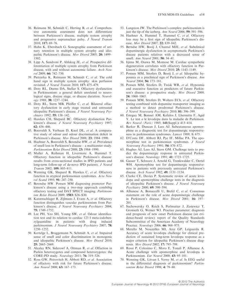
36. Reimann M, Schmidt C, Herting B, et al. Comprehen-sive autonomic assessment does not differentiatebetween Parkinson’s disease, multiple system atrophyand progressive supranuclear palsy. J Neural Transm2010; 117: 69–76.
37. Hahn K, Ebersbach G. Sonographic assessment of uri-nary retention in multiple system atrophy and idio-pathic Parkinson’s disease. Mov Disord 2005; 20: 1499–1502.
38. Lipp A, Sandroni P, Ahlskog JE, et al. Prospective dif-ferentiation of multiple system atrophy from Parkinsondisease, with and without autonomic failure. Arch Neu-rol 2009; 66: 742–750.
39. Pietzarka K, Reimann M, Schmidt C, et al. The coldhand sign in multiple system atrophy: skin perfusionrevisited. J Neural Transm 2010; 117: 475–479.
40. Doty RL, Deems DA, Stellar S. Olfactory dysfunctionin Parkinsonism: a general deficit unrelated to neuro-logical signs, disease stage, or disease duration. Neurol-ogy 1988; 38: 1237–1244.
41. Doty RL, Stern MB, Pfeiffer C, et al. Bilateral olfac-tory dysfunction in early stage treated and untreatedidiopathic Parkinson’s disease. J Neurol Neurosurg Psy-chiatry 1992; 55: 138–142.
42. Hawkes CH, Shepard BC. Olfactory dysfunction Par-kinson’s disease. J Neurol Neurosurg Psychiatry 1997;62: 436–446.
43. Boesveldt S, Verbaan D, Knol DL, et al. A compara-tive study of odour and odour discrimination deficit inParkinson’s disease. Mov Disord 2008; 23: 1984–1990.
44. Haehner A, Boesveldt S, Berendse HW, et al. Prevalenceof smell loss in Parkinson’s disease – a multicenter study.Parkinsonism Relat Disord 2008; 23: 1984–1990.
45. Muller A, Reihman H, Livermore A, Hummel T.Olfactory function in idiopathic Parkinson’s disease:results from cross-sectional studies in IPD patients andlong-term follow-up of de-novo IPD patients. J NeuralTransm 2002; 109: 805–811.
46. Wenning GK, Shepard B, Hawkes C, et al. Olfactoryfunction in atypical parkinsonian syndromes. Acta Neu-rol Scand 1995; 91: 247–250.
47. Berendse HW. Ponsen MM diagnosing premotor Par-kinson’s disease using a two-step approach combibigolfactory testing and DAT SPECT imaging. Parkinson-ism Relat Disord 2009; 1583: S26–S30.
48. Katzenschlager R, Zijlmans J, Evans A, et al. Olfactoryfunction distinguishes vascular parkinsonism from Par-kinson’s disease. J Neurol Neurosurg Psychiatry 2004;75: 1749–1752.
49. Lee PH, Yeo SH, Young SW, et al. Odour identifica-tion test and its relation to cardiac 123 I meta-iodoben-zylguanidine in patients with drug inducedparkinsonism. J Neurol Neurosurg Psychiatry 2007; 78:1250–1252.
50. Kertelge L, Bruggemann N, Schmidt A, et al. Impairedsense of smell and color discrimination in monogenicand idiopathic Parkinson’s disease. Mov Disord 2010;25: 2665–2669.
51. Alcalay RN, Siderowf A, Ottman R, et al. Olfaction inParkin heterozygotes and compound heterozygotes: theCORE-PD study. Neurology 2011; 76: 319–326.
52. Ross GW, Petrovitch H, Abbott RD, et al. Associationof olfactory with risk for future Parkinson’s disease.Ann Neurol 2008; 63: 167–173.
53. Langston JW. The Parkinson’s complex: parkinsonism isjust the tip of the iceberg. Ann Neurol 2006; 59: 591–596.
54. Haehner A, Hummel T, Hummel C, et al. Olfactoryloss may be a first sign of idiopathic Parkinson’s dis-ease. Mov Disord 2007; 22: 839–842.
55. Berndse HW, Booij J, Chantal MJE, et al. Subclinicaldopaminergic dysfunction in asymptomatic Parkinson’sdisease patients relatives with a decreased sense ofsmell. Ann Neurol 2001; 50: 34–41.
56. Iijima M, Osawa M, Momose M. Cardiac sympatheticdegeneration correlates with olfactory function in Par-kinson’s disease. Mov Disord 2010; 25: 1143–1149.
57. Ponsen MM, Sttofers D, Booij J, et al. Idiopathic hy-posmia as a preclinical sign of Parkinson’s disease. AnnNeurol 2004; 56: 173–181.
58. Ponsen MM, Sttofers D, Twisk WR, et al. Hyposmiaand executive function as predictors of future Parkin-son’s disease: a prospective study. Mov Disord 2009;24: 1060–1065.
59. Ponsen MM, Sttofers D, Wolters ECh, et al. Olfactorytesting combined with dopamine transporter imaging asa method to detect prodromal Parkinson’s disease.J Neurol Neurosurg Psychiatry 2010; 81: 396–399.
60. Esteguy M, Bonnet AM, Kefalos J, Lhermitte F, AgidY. Le test a le levodopa dans la maladie de Parkinson.Rev Neurol (Paris) 1985; 141(Suppl.): 413–414.
61. Barker R, Duncan J, Lees AJ. Subcutaneous apomor-phine as a diagnostic test for dopaminergic responsive-ness in parkinsonian syndromes. Lancet 1989; 1: 675.
62. D’Costa DF, Abbott RJ, Pye IF, Millac PA. The apo-morphine test in parkinsonian syndromes. J NeurolNeurosurg Psychiatry 1991; 54: 870–872.
63. Hughes AJ, Lees AJ, Stern GM. Challenge tests to pre-dict the dopaminergic response in untreated Parkin-son’s disease. Neurology 1991; 41: 1723–1725.
64. Gasser T, Schwarz J, Arnold G, Trenkwalder C, OertelWH. Apomorphine test for dopaminergic responsive-ness in patients with previously untreated Parkinson’sdisease. Arch Neurol 1992; 49: 1131–1134.
65. Clarke CE, Davies P. Systematic review of acute levo-dopa and apomorphine challenge tests in the diagnosisof idiopathic Parkinson’s disease. J Neurol NeurosurgPsychiatry 2000; 69: 590–594.
66. Albanese A, Bonuccelli U, Brefel C, et al. Consensusstatement on the role of acute dopaminergic challengein Parkinson’s disease. Mov Disord 2001; 16: 197–201.
67. Suchowersky O, Reich S, Perlmutter J, Zesiewicz T,Gronseth G, Weiner WJ. Practice parameter: diagnosisand prognosis of new onset Parkinson disease (an evi-dence-based review): report of the Quality StandardsSubcommittee of the American Academy of NeurologyPractice. Neurology 2006; 66: 968–975.
68. Merello M, Nouzeilles MI, Arce GP, Leiguarda R.Accuracy of acute levodopa challenge for clinical pre-diction of sustained long-term levodopa response as amajor criterion for idiopathic Parkinson’s disease diag-nosis. Mov Disord 2002; 17: 795–798.
69. Rossi P, Colosimo C, Moro E, Tonali P, Albanese A.Acute challenge with apomorphine and levodopa inParkinsonism. Eur Neurol 2000; 43: 95–101.
70. Wenning GK, Litvan I, Verny M, et al. Is EEG usefulin the differential diagnosis of parkinsonism? Parkin-sonism Relat Disord 1998; 4: 79–80.
© 2012 The Author(s)European Journal of Neurology © 2012 EFNS European Journal of Neurology
EFNS/MDS-ES Guidelines e14
71. Bonanni L, Thomas A, Tiraboschi P, Perfetti B, Vara-nese S, Onofrj M. EEG comparisons in early Alzhei-mer′s disease, dementia with Lewy bodies andParkinson′s disease with dementia patients with a2-year follow-up. Brain 2008; 131: 690–705.
72. Wang L, Kuroiwa Y, Kamitani T, et al. Visual event-related potentials in progressive supranuclear palsy,corticobasal degeneration, striatonigral degenerationand Parkinson′s disease. J Neurol 2000; 247: 356–363.
73. Nightingale S, Mitchell KW, Howe JW. Visual evokedcortical potentials and pattern electroretinograms inParkinson′s disease and control subjects. J Neurol Neu-rosurg Psychiatry 1986; 49: 1280–1287.
74. Takeda M, Tachibana H, Okuda B, et al. Event-relatedpotential and visual evoked potential in patients withParkinson′s disease. Nihon Ronen Igakkai Zasshi 1993;30: 363–368.
75. Okuda B, Tachibana H, Kawabata K, et al. Visualevoked potentials in Parkinson′s disease: correlation ofpattern VEPs abnormality with dementia. AlzheimerDis Assoc Disord 1995; 9: 68–72.
76. Rossini PM, Babiloni F, Bernardi G, et al. Abnormali-ties of short-latency somatosensory evoked potentialsin parkinsonian patients. Electroencephalogr Clin Neu-rophysiol 1989; 74: 277–289.
77. Garcia PA, Aminoff MJ, Goodin DS. The frontla N30component of the median-derived SEP in patients withpredominantly unilateral Parkinson′s disease. Neurology1995; 45: 989–992.
78. Onofrj M, Fulgente T, Malatesta G, et al. The abnor-mality of N30 somatosensory evoked potential in idio-pathic Parkinson′s disease is unrelated to disease stageor clinical scores and insensitive to dopamine manipu-lations. Mov Disord 1995; 10: 71–80.
79. Bostantjopoulou S, Katsarou Z, Zafiriou D, et al.Abnormality of N30 somatosensory evoked potentialsin Parkinson′s disease: a multidisciplinary approach.Neurophysiol Clin 2000; 30: 368–376.
80. Rossini PM, Basseti MA, Paqualetti P. Median nervesomatosensory evoked potentials. Apomorphine-induced transient potentiation of frontal components inParkinson′s disease and parkinsonism. Electroencepha-logr Clin Neurophysiol 1995; 96: 236–247.
81. Tinazzi M, Fiaschi A, Idone D, et al. Parkinson′s dis-ease and lower limb somatosensory evoked potentials:apomorphine-induced relief of the akinetic-rigid syn-drome and vertex P37–N50 potentials. J Neurol Sci1999; 164: 163–171.
82. Defebvre L. Extrapyramidal disorders: interest ofmyoclonus analysis. Rev Neurol 2007; 163: 1115–1121.
83. Van de Wassenberg WJ, van der Hoeven JH, LeendersKL. 128-Channel somatosensory evoked potentials inthe differential diagnosis of parkinsonian disorders.Parkinsonism Relat Disord 2009; 15: 564–571.
84. Tachibana H, Takeda M, Sugita M. Short-latencysomatosensory and brainstem auditory evoked poten-tials in patients with Parkinson′s disease. Int J Neurosci1989; 44: 321–326 (class III study).
85. Kodama Y, Ieda T, Hirayama M, et al. Auditorybrainstem responses in patients with autonomic failurewith Parkinson′s disease and multiple system atrophy.J Auton Nerv Syst 1999; 77: 184–189 (class III study).
86. Laffront F, Agar N, Zuber M, et al. Auditory evokedresponses (AER) and augmenting – reducing phenome-
non in patients with progressive supranuclear palsy(PSP). Neurophysiol Clin 1991; 21: 149–160 (class IIIstudy).
87. Tyvaert L, Cassim F, Derambure P, Lefebvre L. Neu-rophysiology of corticobasal degeneration. Rev Neurol2007; 163: 779–791 (review).
88. Cantello R, Gianelli M, Bettucci D, et al. Parkinson′sdisease rigidity: magnetic motor evoked potentials insmall hand muscle. Neurology 1991; 41: 1449–1456.
89. Priori A, Berardelli A, Inghilleri M, et al. Motor corti-cal inhibition and the dopaminergic syst�em. Pharmaco-logical changes in the silent period after transcranialbrain stimulation in normal subjects, patients with Par-kinson′s disease and drug-induced parkinsonism. Brain1994; 117: 317–323.
90. Cantello R, Tarletti R, Civardi C. Transcranial mag-netic stimulation and Parkinson′s disease. Brain ResBrain Res Rev 2002; 38: 309–327.
91. De Rosa A, Volpe G, Marcantonio L, et al. Neuro-physiological evidence of corticospinal tract abnormal-ity in patients with Parkin mutations. J Neurol 2006;253: 275–279.
92. Abbruzzese G, Marchese R, Trompetto C. Sensory andmotor evoked potentials in multiple system atrophy: acomparative study with Parkinson′s disease. Mov Dis-ord 1997; 12: 315–321.
93. Wenning GK, Smith SJ. Magnetic brain stimulation inmultiple system atrophy. Mov Disord 1997; 12: 452–453.
94. Morita Y, Osaki Y, Doi Y. Transcranial magneticstimulation for differential diagnostics in patients withparkinsonism. Acta Neurol Scand 2008; 118: 159–163.
95. Vidailhet M, Rothwell JC, Thompson PD, Lees AJ,Marsden CD. The auditory startle response in theSteele-Richardson-Olszewski syndrome and Parkinson′sdisease. Brain 1992; 115: 1181–1192.
96. Kofler M, M€uller J, Wenning GK, et al. The auditorystartle reaction in parkinsonian disorders. Mov Disord2001; 16: 62–71.
97. Plazzi G, Corsini R, Provini F, et al. REM sleepbehavior disorders in multiple system atrophy. Neurol-ogy 1997; 48: 1094–1097.
98. Boeve BF, Silber MH, Ferman TJ, Lucas JA, ParisiJE. Association of REM sleep behavior disorder andneurodegenerative disease may reflect an underlyingsynucleinopathy. Mov Disord 2001; 16: 622–630.
99. Menza M, Dobkin RD, Marin H, Bienfait K. Sleepdisturbances in Parkinson′s disease. Mov Disord 2010;25(Suppl. 1): S117–S122.
100. Moreno-Lopez C, Santamaria J, Salamero M, DelSorbo F, Albanese A, et al. Excessive daytime sleepi-ness in jultiple system atrophy (SLEEMSA study).Arch Neurol 2011; 68: 223–230.
101. Schulte EC, Winkelmann J. When Parkinson′s diseasepatients go to sleep: specific sleep disturbances relatedto Parkinson′s disease. J Neurol 2011; 258(Suppl. 2):S328–S335.
102. Deuschl G, Krack P, Lauk M, et al. Clinical neurophysi-ology of tremor. J Clin Neurophysiol 1996; 13: 110–121.
103. Deuschl G. Differential diagnosis of tremor. J NeuralTransm Suppl 1999; 56: 211–220.
104. Milanov I. Electromyographic differentiation of trem-ors. Clin Neurophysiol 2001; 112: 1626–1632.
© 2012 The Author(s)European Journal of Neurology © 2012 EFNS European Journal of Neurology
e15 A. Berardelli et al.
105. Breit S, Spieker S, Schulz JB, et al. Long-term EMGrecordings differentiate between parkinsonian andEssentials tremor. J Neurol 2008; 255: 103–111.
106. Eardley I, Quinn NP, Fowler CJ, et al. The value of ure-thral sphincter electromyography in the differential diag-nosis of parkinsonism. Br J Urol 1989; 64: 360–362.
107. Pramstaller PP, Wenning GK, Smith SJ, Beck RO,Quinn NP, Fowler J. Nerve conduction studies, skeletalmuscle EMG, and sphincter EMG in multiple systematrophy. J Neurol Neurosurg Psychiatry 1995; 58: 618–621.
108. Winge K, Jennum P, Lokkegaard A, et al. Analsphincter EMG in the diagnosis of parkinsonian syn-dromes. Acta Neurol Scand 2010; 121: 198–203.
109. McKeith I, Dickson D, Lowe J, et al. Diagnosis andmanagement of dementia with Lewy bodies: thirdreport of the DLB Consortium. Neurology 2005; 65:
1863–1872.110. Litvan I, Bhatia KP, Burn DJ, et al. SIC task force
appraisal of clinical diagnostic criteria for parkinsoniandisorders. Mov Disord 2003; 18: 467–486.
111. Aarsland D, Bronnick K, Williams-Gray C, et al. Mildcognitive impairment in Parkinson disease: a multicen-ter pooled analysis. Neurology 2010; 75: 1062–1069.
112. Llebaria G, Pagonabarraga J, Kulisevsky J, et al. Cut-off score of the Mattis Dementia Rating Scale forscreening dementia in Parkinson’s disease. Mov Disord2008; 23: 1546–1550.
113. Aarsland D, Litvan I, Salmon D, Galakso D, Wentzel-Larsen T, Larsen JP. Performance on the dementia rat-ing scale in Parkinson’s disease with dementia anddementia with Lewy bodies: comparison with progres-sive supranuclear palsy and Alzheimer’s disease. J Neu-rol Neurosurg Psychiatry 2003; 74: 1215–1220.
114. Mioshi E, Dawson K, Mitchell J, Arnold R, HodgesJR. The Addenbrooke’s Cognitive ExaminationRevised (ACE-R): a brief cognitive test battery fordementia screening. Int J Geriatr Psychiatry 2006; 21:1078–1085.
115. Bak TH, Mioshi E. A cognitive bedside assessmentbeyond the MMSE: the Addenbrooke’s CognitiveExamination. Pract Neurol 2007; 7: 245–249.
116. Hoops S, Nazem S, Siderowf AD, et al. Validity of theMoCA and MMSE in the detection of MCI anddementia in Parkinson disease. Neurology 2009; 73:
1738–1745.117. Dalrymple-Alford JC, MacAskill MR, Nakas CT, et al.
The MoCA: well-suited screen for cognitive impairmentin Parkinson disease. Neurology 2010; 75: 1717–1725.
118. Marinus J, Visser M, Verwey NA, et al. Assessment ofcognition in Parkinson’s disease. Neurology 2003; 61:
1222–1228.119. Pagonabarraga J, Kulisevsky J, Llebaria G, Garcia-
Sanchez C, Pascual-Sedano B, Gironell A. Parkinson’sdisease-cognitive rating scale: a new cognitive scale spe-cific for Parkinson’s disease. Mov Disord 2008; 23: 998–1005.
120. Kulisevsky J, Pagonabarraga J. Cognitive impairmentin Parkinson’s disease: tools for diagnosis and assess-ment. Mov Disord 2009; 24: 1103–1110.
121. Williams-Gray CH, Evans JR, Goris A, et al. The dis-tinct cognitive syndromes of Parkinson’s disease: 5 yearfollow-up of the CamPaIGN cohort. Brain 2009; 132:2958–2969.
122. Dujardin K, Dubois B, Tison F, et al. Parkinson’s dis-ease dementia can be easily detected in routine clinicalpractice. Mov Disord 2010; 25: 2769–2776.
123. Pagonabarraga J, Kulisevsky J, Llebaria G, et al.PDD-Short Screen: a brief cognitive test for screeningdementia in Parkinson’s disease. Mov Disord 2010; 25:440–446.
124. Stiasny-Kolster K, Mayer G, Schafer S, Carsten MollerJ, Heinzel-Gutenbrunner M, Oertel WH. The REMsleep behavior disorder screening questionnaire: a newdiagnostic instrument. Mov Disord 2007; 22: 2386–2393.
125. Goetz CG, Tilley BC, Shaftman SR, et al. MovementDisorder Society-sponsored revision of the Unified Par-kinson’s Disease Rating Scale (MDS-UPDRS): scalepresentation and clinimetric testing results. Mov Disord2008; 23: 2129–2170.
126. Berg D, Siefker C, Becker G. Echogenicity of the sub-stantia nigra in Parkinson’s disease and its relation toclinical findings. J Neurol 2001; 248: 684–689.
127. Walter U, Wittstock M, Benecke R, Dressler D. Sub-stantia nigra echogenicity is normal in non-extrapyra-midal cerebral disorders but increased in Parkinson’sdisease. J Neural Transm 2002; 109: 191–196.
128. Huang YW, Jeng JS, Tsai CF, Chen LL, Wu RM.Transcranial imaging of substantia nigra hyperechoge-nicity in a Taiwanese cohort of Parkinson’s disease.Mov Disord 2007; 22: 550–555.
129. Kim JY, Kim ST, Jeon SH, Lee WY. Midbrain trans-cranial sonography in Korean patients with Parkin-son’s disease. Mov Disord 2007; 22: 1922–1926.
130. Okawa M, Miwa H, Kajimoto Y, et al. Transcranialsonography of the substantia nigra in Japanese patientswith Parkinson’s disease or atypical parkinsonism: clin-ical potential and limitations. Intern Med 2007; 46:
1527–1531.131. Ressner P, Skoloudik D, Hlustik P, Kanovsky P. Hy-
perechogenicity of the substantia nigra in Parkinson’sdisease. J Neuroimaging 2007; 17: 164–167.
132. Tsai CF, Wu RM, Huang YW, Chen LL, Yip PK,Jeng JS. Transcranial color-coded sonography helpsdifferentiation between idiopathic Parkinson’s diseaseand vascular parkinsonism. J Neurol 2007; 254: 501–507.
133. Mijajlovic M, Dragasevic N, Stefanova E, Petrovic I,Svetel M, Kostic VS. Transcranial sonography in spinoc-erebellar ataxia type 2. J Neurol 2008; 255: 1164–1167.
134. Budisic M, Trkanjec Z, Bosnjak J, Lovrencic-HuzjanA, Vukovic V, Demarin V. Distinguishing Parkinson’sdisease and essential tremor with transcranial sonogra-phy. Acta Neurol Scand 2009; 119: 17–21.
135. Berg D, Behnke S, Walter U. Application of transcra-nial sonography in extrapyramidal disorders: updatedrecommendations. Ultraschall Med 2006; 27: 12–19.
136. Walter U, Behnke S, Eyding J, et al. Transcranialbrain parenchyma sonography in movement disorders:state of the art. Ultrasound Med Biol 2007; 33: 15–25.
137. Berg D, Godau J, Walter U. Transcranial sonographyin movement disorders. Lancet Neurol 2008; 7: 1044–1055.
138. Skoloudik D, Walter U. Method and validity of trans-cranial sonography in movement disorders. Int RevNeurobiol 2010; 90: 7–34.
139. Walter U, Kanowski M, Kaufmann J, Grossmann A,Benecke R, Niehaus L. Contemporary ultrasound sys-
© 2012 The Author(s)European Journal of Neurology © 2012 EFNS European Journal of Neurology
EFNS/MDS-ES Guidelines e16
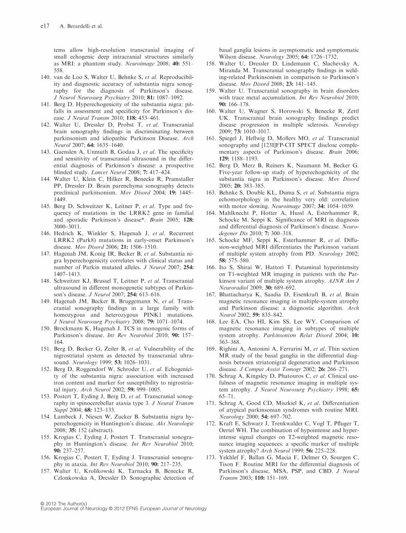
tems allow high-resolution transcranial imaging ofsmall echogenic deep intracranial structures similarlyas MRI: a phantom study. Neuroimage 2008; 40: 551–558.
140. van de Loo S, Walter U, Behnke S, et al. Reproducibil-ity and diagnostic accuracy of substantia nigra sonog-raphy for the diagnosis of Parkinson’s disease.J Neurol Neurosurg Psychiatry 2010; 81: 1087–1092.
141. Berg D. Hyperechogenicity of the substantia nigra: pit-falls in assessment and specificity for Parkinson’s dis-ease. J Neural Transm 2010; 118: 453–461.
142. Walter U, Dressler D, Probst T, et al. Transcranialbrain sonography findings in discriminating betweenparkinsonism and idiopathic Parkinson Disease. ArchNeurol 2007; 64: 1635–1640.
143. Gaenslen A, Unmuth B, Godau J, et al. The specificityand sensitivity of transcranial ultrasound in the differ-ential diagnosis of Parkinson’s disease: a prospectiveblinded study. Lancet Neurol 2008; 7: 417–424.
144. Walter U, Klein C, Hilker R, Benecke R, PramstallerPP, Dressler D. Brain parenchyma sonography detectspreclinical parkinsonism. Mov Disord 2004; 19: 1445–1449.
145. Berg D, Schweitzer K, Leitner P, et al. Type and fre-quency of mutations in the LRRK2 gene in familialand sporadic Parkinson’s disease*. Brain 2005; 128:
3000–3011.146. Hedrich K, Winkler S, Hagenah J, et al. Recurrent
LRRK2 (Park8) mutations in early-onset Parkinson’sdisease. Mov Disord 2006; 21: 1506–1510.
147. Hagenah JM, Konig IR, Becker B, et al. Substantia ni-gra hyperechogenicity correlates with clinical status andnumber of Parkin mutated alleles. J Neurol 2007; 254:1407–1413.
148. Schweitzer KJ, Brussel T, Leitner P, et al. Transcranialultrasound in different monogenetic subtypes of Parkin-son’s disease. J Neurol 2007; 254: 613–616.
149. Hagenah JM, Becker B, Bruggemann N, et al. Trans-cranial sonography findings in a large family withhomozygous and heterozygous PINK1 mutations.J Neurol Neurosurg Psychiatry 2008; 79: 1071–1074.
150. Brockmann K, Hagenah J. TCS in monogenic forms ofParkinson’s disease. Int Rev Neurobiol 2010; 90: 157–164.
151. Berg D, Becker G, Zeiler B, et al. Vulnerability of thenigrostriatal system as detected by transcranial ultra-sound. Neurology 1999; 53: 1026–1031.
152. Berg D, Roggendorf W, Schroder U, et al. Echogenici-ty of the substantia nigra: association with increasediron content and marker for susceptibility to nigrostria-tal injury. Arch Neurol 2002; 59: 999–1005.
153. Postert T, Eyding J, Berg D, et al. Transcranial sonog-raphy in spinocerebellar ataxia type 3. J Neural TransmSuppl 2004; 68: 123–133.
154. Lambeck J, Niesen W, Zucker B. Substantia nigra hy-perechogenicity in Huntington’s disease. Akt Neurologie2008; 35: 152 (abstract).
155. Krogias C, Eyding J, Postert T. Transcranial sonogra-phy in Huntington’s disease. Int Rev Neurobiol 2010;90: 237–257.
156. Krogias C, Postert T, Eyding J. Transcranial sonogra-phy in ataxia. Int Rev Neurobiol 2010; 90: 217–235.
157. Walter U, Krolikowski K, Tarnacka B, Benecke R,Czlonkowska A, Dressler D. Sonographic detection of
basal ganglia lesions in asymptomatic and symptomaticWilson disease. Neurology 2005; 64: 1726–1732.
158. Walter U, Dressler D, Lindemann C, Slachevsky A,Miranda M. Transcranial sonography findings in weld-ing-related Parkinsonism in comparison to Parkinson’sdisease. Mov Disord 2008; 23: 141–145.
159. Walter U. Transcranial sonography in brain disorderswith trace metal accumulation. Int Rev Neurobiol 2010;90: 166–178.
160. Walter U, Wagner S, Horowski S, Benecke R, ZettlUK. Transcranial brain sonography findings predictdisease progression in multiple sclerosis. Neurology2009; 73: 1010–1017.
161. Spiegel J, Hellwig D, Mollers MO, et al. Transcranialsonography and [123I]FP-CIT SPECT disclose comple-mentary aspects of Parkinson’s disease. Brain 2006;129: 1188–1193.
162. Berg D, Merz B, Reiners K, Naumann M, Becker G.Five-year follow-up study of hyperechogenicity of thesubstantia nigra in Parkinson’s disease. Mov Disord2005; 20: 383–385.
163. Behnke S, Double KL, Duma S, et al. Substantia nigraechomorphology in the healthy very old: correlationwith motor slowing. Neuroimage 2007; 34: 1054–1059.
164. Mahlknecht P, Hotter A, Hussl A, Esterhammer R,Schocke M, Seppi K. Significance of MRI in diagnosisand differential diagnosis of Parkinson’s disease. Neuro-degener Dis 2010; 7: 300–318.
165. Schocke MF, Seppi K, Esterhammer R, et al. Diffu-sion-weighted MRI differentiates the Parkinson variantof multiple system atrophy from PD. Neurology 2002;58: 575–580.
166. Ito S, Shirai W, Hattori T. Putaminal hyperintensityon T1-weighted MR imaging in patients with the Par-kinson variant of multiple system atrophy. AJNR Am JNeuroradiol 2009; 30: 689–692.
167. Bhattacharya K, Saadia D, Eisenkraft B, et al. Brainmagnetic resonance imaging in multiple-system atrophyand Parkinson disease: a diagnostic algorithm. ArchNeurol 2002; 59: 835–842.
168. Lee EA, Cho HI, Kim SS, Lee WY. Comparison ofmagnetic resonance imaging in subtypes of multiplesystem atrophy. Parkinsonism Relat Disord 2004; 10:
363–368.169. Righini A, Antonini A, Ferrarini M, et al. Thin section
MR study of the basal ganglia in the differential diag-nosis between striatonigral degeneration and Parkinsondisease. J Comput Assist Tomogr 2002; 26: 266–271.
170. Schrag A, Kingsley D, Phatouros C, et al. Clinical use-fulness of magnetic resonance imaging in multiple sys-tem atrophy. J Neurol Neurosurg Psychiatry 1998; 65:65–71.
171. Schrag A, Good CD, Miszkiel K, et al. Differentiationof atypical parkinsonian syndromes with routine MRI.Neurology 2000; 54: 697–702.
172. Kraft E, Schwarz J, Trenkwalder C, Vogl T, Pfluger T,Oertel WH. The combination of hypointense and hyper-intense signal changes on T2-weighted magnetic reso-nance imaging sequences: a specific marker of multiplesystem atrophy? Arch Neurol 1999; 56: 225–228.
173. Yekhlef F, Ballan G, Macia F, Delmer O, Sourgen C,Tison F. Routine MRI for the differential diagnosis ofParkinson’s disease, MSA, PSP, and CBD. J NeuralTransm 2003; 110: 151–169.
© 2012 The Author(s)European Journal of Neurology © 2012 EFNS European Journal of Neurology
e17 A. Berardelli et al.
174. Kraft E, Trenkwalder C, Auer DP. T2*-weighted MRIdifferentiates multiple system atrophy from Parkinson’sdisease. Neurology 2002; 59: 1265–1267.
175. Savoiardo M, Strada L, Girotti F, et al. MR imagingin progressive supranuclear palsy and Shy-Drager syn-drome. J Comput Assist Tomogr 1989; 13: 555–560.
176. Watanabe H, Saito Y, Terao S, et al. Progression andprognosis in multiple system atrophy: an analysis of230 Japanese patients. Brain 2002; 125: 1070–1083.
177. Ito S, Shirai W, Hattori T. Evaluating posterolateral lin-earization of the putaminal margin with magnetic reso-nance imaging to diagnose the Parkinson variant ofmultiple system atrophy. Mov Disord 2007; 22: 578–581.
178. Paviour DC, Price SL, Stevens JM, Lees AJ, Fox NC.Quantitative MRI measurement of superior cerebellarpeduncle in progressive supranuclear palsy. Neurology2005; 64: 675–679.
179. Oba H, Yagishita A, Terada H, et al. New and reliableMRI diagnosis for progressive supranuclear palsy. Neu-rology 2005; 64: 2050–2055.
180. Kato N, Arai K, Hattori T. Study of the rostral mid-brain atrophy in progressive supranuclear palsy. J Neu-rol Sci 2003; 210: 57–60.
181. Graber JJ, Staudinger R. Teaching neuroimages: “Pen-guin” or “hummingbird” sign and midbrain atrophy inprogressive supranuclear palsy. Neurology 2009; 72: e81.
182. Barsottini OG, Ferraz HB, Maia AC Jr, Silva CJ, Ro-cha AJ. Differentiation of Parkinson’s disease and pro-gressive supranuclear palsy with magnetic resonanceimaging: the first Brazilian experience. ParkinsonismRelat Disord 2007; 13: 389–393.
183. Righini A, Antonini A, De NR, et al. MR imaging ofthe superior profile of the midbrain: differential diagno-sis between progressive supranuclear palsy and Parkin-son disease. AJNR Am J Neuroradiol 2004; 25: 927–932.
184. Kataoka H, Tonomura Y, Taoka T, Ueno S. Signalchanges of superior cerebellar peduncle on fluid-attenu-ated inversion recovery in progressive supranuclearpalsy. Parkinsonism Relat Disord 2008; 14: 63–65.
185. Schulz JB, Skalej M, Wedekind D, et al. Magnetic res-onance imaging-based volumetry differentiates idio-pathic Parkinson’s syndrome from multiple systematrophy and progressive supranuclear palsy. Ann Neu-rol 1999; 45: 65–74.
186. Groschel K, Hauser TK, Luft A, et al. Magnetic reso-nance imaging-based volumetry differentiates progres-sive supranuclear palsy from corticobasal degeneration.Neuroimage 2004; 21: 714–724.
187. Hussl A, Mahlknecht P, Scherfler C, et al. Diagnosticaccuracy of the magnetic resonance Parkinsonism indexand the midbrain-to-pontine area ratio to differentiateprogressive supranuclear palsy from Parkinson’s diseaseand the Parkinson variant of multiple system atrophy.Mov Disord 2010; 25: 2444–2449.
188. Nicoletti G, Fera F, Condino F, et al. MR imaging ofmiddle cerebellar peduncle width: differentiation ofmultiple system atrophy from Parkinson disease. Radi-ology 2006; 239: 825–830.
189. Quattrone A, Nicoletti G, Messina D, et al. MR imag-ing index for differentiation of progressive supranuclearpalsy from Parkinson disease and the Parkinson vari-ant of multiple system atrophy. Radiology 2008; 246:
214–221.
190. Warmuth-Metz M, Naumann M, Csoti I, Solymosi L.Measurement of the midbrain diameter on routinemagnetic resonance imaging: a simple and accuratemethod of differentiating between Parkinson diseaseand progressive supranuclear palsy. Arch Neurol 2001;58: 1076–1079.
191. Longoni G, Agosta F, Kostic VS, et al. MRI measure-ments of brainstem structures in patients with Richard-son’s syndrome, progressive supranuclear palsy-parkinsonism, and Parkinson’s disease. Mov Disord2011; 26: 247–255.
192. Schocke MF, Seppi K, Esterhammer R, et al. Trace ofdiffusion tensor differentiates the Parkinson variant ofmultiple system atrophy and Parkinson’s disease. Neu-roimage 2004; 21: 1443–1451.
193. Seppi K, Schocke MF, Esterhammer R, et al. Diffu-sion-weighted imaging discriminates progressive supra-nuclear palsy from PD, but not from the parkinsonvariant of multiple system atrophy. Neurology 2003; 60:922–927.
194. Seppi K, Schocke MF, Donnemiller E, et al. Compari-son of diffusion-weighted imaging and [123I]IBZM-SPECT for the differentiation of patients with the Par-kinson variant of multiple system atrophy from thosewith Parkinson’s disease. Mov Disord 2004; 19: 1438–1445.
195. Seppi K, Schocke MF, Prennschuetz-Schuetzenau K,et al. Topography of putaminal degeneration in multi-ple system atrophy: a diffusion magnetic resonancestudy. Mov Disord 2006; 21: 847–852.
196. Nicoletti G, Lodi R, Condino F, et al. Apparent diffu-sion coefficient measurements of the middle cerebellarpeduncle differentiate the Parkinson variant of MSAfrom Parkinson’s disease and progressive supranuclearpalsy. Brain 2006; 129: 2679–2687.
197. Kollensperger M, Seppi K, Liener C, et al. Diffusionweighted imaging best discriminates PD from MSA-P:a comparison with tilt table testing and heart MIBGscintigraphy. Mov Disord 2007; 22: 1771–1776.
198. Pellecchia MT, Barone P, Mollica C, et al. Diffusion-weighted imaging in multiple system atrophy: a com-parison between clinical subtypes. Mov Disord 2009;24: 689–696.
199. Rizzo G, Martinelli P, Manners D, et al. Diffusion-weighted brain imaging study of patients with clinicaldiagnosis of corticobasal degeneration, progressivesupranuclear palsy and Parkinson’s disease. Brain 2008;131: 2690–2700.
200. Paviour DC, Thornton JS, Lees AJ, Jager HR. Diffu-sion-weighted magnetic resonance imaging differentiatesParkinsonian variant of multiple-system atrophy fromprogressive supranuclear palsy. Mov Disord 2007; 22:
68–74.201. Blain CR, Barker GJ, Jarosz JM, et al. Measuring
brain stem and cerebellar damage in parkinsonian syn-dromes using diffusion tensor MRI. Neurology 2006;67: 2199–2205.
202. Nicoletti G, Tonon C, Lodi R, et al. Apparent diffu-sion coefficient of the superior cerebellar peduncle dif-ferentiates progressive supranuclear palsy fromParkinson’s disease. Mov Disord 2008; 23: 2370–2376.
203. Ito M, Watanabe H, Kawai Y, et al. Usefulness ofcombined fractional anisotropy and apparent diffusioncoefficient values for detection of involvement in multi-
© 2012 The Author(s)European Journal of Neurology © 2012 EFNS European Journal of Neurology
EFNS/MDS-ES Guidelines e18
ple system atrophy. J Neurol Neurosurg Psychiatry2007; 78: 722–728.
204. Vaillancourt DE, Spraker MB, Prodoehl J, et al. High-resolution diffusion tensor imaging in the substantia ni-gra of de novo Parkinson disease. Neurology 2009; 72:1378–1384.
205. Menke RA, Scholz J, Miller KL, et al. MRI character-istics of the substantia nigra in Parkinson’s disease: acombined quantitative T1 and DTI study. Neuroimage2009; 47: 435–441.
206. Groger A, Chadzynski G, Godau J, Berg D, Klose U.Three-dimensional magnetic resonance spectroscopicimaging in the substantia nigra of healthy controls andpatients with Parkinson’s disease. Eur Radiol 2011; 21:1962–1969.
207. Peran P, Cherubini A, Assogna F, et al. Magnetic reso-nance imaging markers of Parkinson’s disease nigrostri-atal signature. Brain 2010; 133: 3423–3433.
208. Melzer TR, Watts R, MacAskill MR, et al. Arterial spinlabelling reveals an abnormal cerebral perfusion patternin Parkinson’s disease. Brain 2011; 134: 845–855.
209. Antonini A. Imaging for early differential diagnosis ofparkinsonism. Lancet Neurol 2010; 9: 130–131.
210. Scherfler C, Schwarz J, Antonini A, et al. Role ofDAT-SPECT in the diagnostic work up of parkinson-ism. Mov Disord 2007; 22: 1229–1238.
211. Benamer HTS, Patterson J, Grosset DG. Accurate dif-ferentiation of parkinsonism and essential tremor usingvisual assessment of [123I]FP-CIT SPECT imaging: the[123I]FP-CIT SPECT study group. Mov Disord 2000;15: 503–510.
212. Vlaar AM, de Nijs T, Kessel AG, et al. Diagnosticvalue of 123I-ioflupane and 123I-iodobenzamideSPECT scans in 248 patients with parkinsonian syn-dromes. Eur Neurol 2008; 59: 258–266.
213. Scherfler C, Seppi K, Donnemiller E, et al. Voxel-wiseanalysis of [123I]-CIT SPECT differentiates the Parkin-son variant of multiple system atrophy from idiopathicParkinson’s disease. Brain 2005; 128: 1605–1612.
214. Tinazzi M, Antonini A, Bovi T, et al. Clinical and 123IFP-CIT SPET imaging follow-up in patients with drug-induced parkinsonism. J Neurol 2009; 256: 910–915.
215. Eckert T, Tang C, Eidelberg D. Assessment of the pro-gression of Parkinson’s disease: a metabolic networkapproach. Lancet Neurol 2007; 6: 926–932.
216. Chang CC, Liu JS, Chang YY, Chang WN, Chen SS,Lee CH. (99m)Tc-ethyl cysteinate dimer brain SPECTfindings in early stage of dementia with Lewy bodiesand Parkinson’s disease patients: a correlation withneuropsychological tests. Eur J Neurol 2008; 15: 61–65.
217. Feigin A, Antonini A, Fukuda M, et al. Tc-99m ethyl-ene cysteinate dimer SPECT in the differential diagno-sis of parkinsonism. Mov Disord 2002; 17: 1265–1270.
218. Courbon F, Brefel-Courbon C, Thalamas C, et al. Car-diac MIBG scintigraphy is a sensitive tool for detectingcardiac sympathetic denervation in Parkinson’s disease.Mov Disord 2003; 18: 890–897.
219. Nagayama H, Hamamoto M, Ueda M, Nagashima J,Katayama Y. Reliability of MIBG myocardial scintig-raphy in the diagnosis of Parkinson’s disease. J NeurolNeurosurg Psychiatry 2005; 76: 249–251.
220. Spiegel J, M€ollers MO, Jost WH, et al. FP-CIT andMIBG scintigraphy in early Parkinson’s disease. MovDisord 2005; 20: 552–561.
221. Brooks DJ. Technology insight: imaging neurodegener-ation in Parkinson’s disease. Nat Clin Pract Neurol2008; 4: 267–277.
222. Dhawan V, Ma Y, Pillai V, et al. Comparative analysisof striatal FDOPA uptake in Parkinson’s disease: ratiomethod versus graphical approach. J Nucl Med 2002;43: 1324–1330.
223. Bohnen NI, Albin RL, Koeppe RA, et al. Positronemission tomography of monoaminergic vesicular bind-ing in aging and Parkinson disease. J Cereb Blood FlowMetab 2006; 26: 1198–1212.
224. Okamura N, Villemagne V, Drago J, et al. In vivomeasurement of vesicular monoamine transporter type2 density in Parkinson disease with (18)F-AV-133. JNucl Med 2010; 51: 223–228.
225. Lee CS, Samii A, Sossi V, et al. In vivo positron emis-sion tomographic evidence for compensatory changesin presynaptic dopaminergic nerve terminals in Parkin-son’s disease. Ann Neurol 2000; 47: 493–503.
226. Nurmi E, Bergman J, Eskola O, et al. Progression ofdopaminergic hypofunction in striatal subregions inParkinson’s disease using [18F]CFT PET. Synapse2003; 48: 109–115.
227. Guttman M, Burkholder J, Kish SJ, et al. [11C]RTI-32PET studies of the dopamine transporter in early dopa-naive Parkinson’s disease: implications for the symp-tomatic threshold. Neurology 1997; 48: 1578–1583.
228. Ishikawa T, Dhawan V, Kazumata K, et al. Compara-tive nigrostriatal dopaminergic imaging with iodine-123beta-CIT-FP/SPECT and Fluorine-18 FDOPA/PET.J Nucl Med 1996; 37: 1760–1765.
229. Vingerhoets FJG, Schulzer M, Calne DB, Snow BJ.Which clinical sign of Parkinson’s disease best reflectsthe nigrostriatal lesion? Ann Neurol 1997; 41: 58–64.
230. Whone AL, Watts RL, Stoessl J, et al. Slower progres-sion of PD with ropinirol versus L-dopa: the REAL-PET study. Ann Neurol 2003; 54: 93–101.
231. Bruck A, Aalto S, Rauhala E, Bergman J, Marttila R,Rinne JO. A follow-up study on 6-[18F]fluoro-L-dopauptake in early Parkinson’s disease shows nonlinearprogression in the putamen. Mov Disord 2009; 24: 1009–1015.
232. Adams JR, van Netten H, Schulzer M, et al. PET inLRRK2 mutations: comparison to sporadic Parkin-son’s disease and evidence for presymptomatic compen-sation. Brain 2005; 128: 2777–2785.
233. Piccini P, Morrish PK, Turjanski N, et al. Dopaminergicfunction in familial Parkinson’s disease: a clinical and18F-dopa PET study. Ann Neurol 1997; 41: 222–229.
234. Piccini P, Burn DJ, Ceravalo R, Maraganore DM,Brooks DJ. The role of inheritance in sporadic Parkin-son’s disease: evidence from a longitudinal study ofdopaminergic function in twins. Ann Neurol 1999; 45:577–582.
235. Brooks DJ, Iba~nez V, Sawle GV, et al. Differing pat-terns of striatal 18F-dopa uptake in Parkinson’s dis-ease, multiple system atrophy and progressivesupranuclear palsy. Ann Neurol 1990; 28: 547–555.
236. Antonini A, Schwarz J, Oertel WH, Beer HF, MadejaUD, Leenders KL. [11C]raclopride and positron emis-sion tomography in previously untreated patients withParkinson’s disease: influence of L-dopa and lisuridetherapy on striatal dopamine D2-receptors. Neurology1994; 44: 1325–1329.
© 2012 The Author(s)European Journal of Neurology © 2012 EFNS European Journal of Neurology
e19 A. Berardelli et al.
237. Antonini A, Leenders KL, Vontobel P, et al. Comple-mentary PET studies of striatal neuronal function inthe differential diagnosis between multiple system atro-phy and Parkinson’s disease. Brain 1997; 120: 2187–2195.
238. Brooks DJ, Ibanez V, Sawle GV, et al. Striatal D2receptor status in Parkinson’s disease, striatonigraldegeneration, and progressive supranuclear palsy, mea-sured with 11C-raclopride and PET. Ann Neurol 1992;31: 184–192.
239. Eidelberg D, Moeller JR, Dhawan V, et al. The metabolicanatomy of Parkinson’s disease: complementary [18F]flu-orodeoxyglucose and [18F]fluorodopa positron emissiontomographic studies.Mov Disord 1990; 5: 203–213.
240. Feigin A, Fukuda M, Dhawan V, et al. Metabolic cor-relates of levodopa response in Parkinson’s disease.Neurology 2001; 57: 2083–2088.
241. Eidelberg D, Takikawa S, Moeller JR, et al. Striatalhypometabolism distinguishes striatonigral degenera-
tion from Parkinson’s disease. Ann Neurol 1993; 33:
518–527.242. Eckert T, Tang C, Ma Y, et al. Abnormal metabolic
networks in atypical parkinsonism. Mov Disord 2008;23: 727–733.
243. Antonini A, Berto P, Lopatriello S, Tamma F, Anne-mans L, Chambers M. Cost-effectiveness of 123I-FP-CIT SPECT in the differential diagnosis of essentialtremor and Parkinson’s disease in Italy. Mov Disord2008; 23: 2202–2209.
244. Van Laere K, Everaert L, Annemans L, Gonce M,Vandenberghe W, Vander Borght T. The cost effective-ness of 123I-FP-CIT SPECT imaging in patients withan uncertain clinical diagnosis of parkinsonism. Eur JNucl Med Mol Imaging 2008; 35: 1367–1376.
245. Dodel RC, H€offken H, M€oller JC, et al. Dopaminetransporter imaging and SPECT in diagnostic work-upof Parkinson’s disease: a decision-analytic approach.Mov Disord 2003; 18(Suppl. 7): S52–S62.
© 2012 The Author(s)European Journal of Neurology © 2012 EFNS European Journal of Neurology
EFNS/MDS-ES Guidelines e20