Embed Size (px)
Citation preview
1
Electronic Supplementary Information
Efficient Synthesis of Narrowly Dispersed Molecularly Imprinted Polymer
Microspheres with Multiple Stimuli-Responsive Template Binding Properties in
Aqueous Media
Yue Ma, Ying Zhang, Man Zhao, Xianzhi Guo and Huiqi Zhang*
Key Laboratory of Functional Polymer Materials (Nankai University), Ministry of Education,
Department of Chemistry, Nankai University, Tianjin 300071 (P. R. China)
Materials
4-Vinylpyridine (4-VP, Alfa Aesar, 96%), ethylene glycol dimethacrylate (EGDMA, Alfa Aesar, 98%),
2-(dimethylamino)ethyl methacrylate (DMAEMA, Tianjin heowns Biochemical Technology Co., Ltd.,
China, 97%), methacrylic acid (MAA, Tianjin Jiangtian Chemicals, China, 99%), and
N,N-dimethylformamide (DMF, Tianjin Jiangtian Chemicals, Analytical grade (AR)) were purified by
distillation under vacuum. Methanol (Tianjin Jiangtian Chemicals, AR) was distilled prior to use.
Tetrahydrofuran (THF, Tianjin Jiangtian Chemicals, 99%) was refluxed over sodium and then distilled.
Acetonitrile (Tianjin Concord Chemicals, China, AR) was refluxed over calcium hydride (CaH2) and
then distilled. N-Isopropylacrylamide (NIPAAm, Acros, 99%) was recrystallized from hexane.
Azobisisobutyronitrile (AIBN, Chemical Plant of Nankai University, AR) was recrystallized from
ethanol. Cumyl dithiobenzoate (CDB) was prepared according to a literature procedure (T. P. Le, G.
Moad, E. Rizzardo and S. H. Thang, PCT Int. Appl. WO 98/01478; Chem. Abstr. 1998, 128, 115390).
Methacrylic chloride was prepared by the reaction between methacrylic acid and the freshly distilled
thionyl chloride (Tianjin Jiangtian Chemicals, 99%) following a literature procedure (Y. Feng, J.
Huang and C. Li, Chem. World 1997, 38, 258-260). Methacrylic anhydride was prepared by reacting
methacrylic acid with methacrylic chloride in the presence of an aqueous solution of sodium hydroxide
following a literature procedure (R. A. Barabashina and O. M. Sleptsova, Z. Prikl. Fiz. 1967, 40, 696).
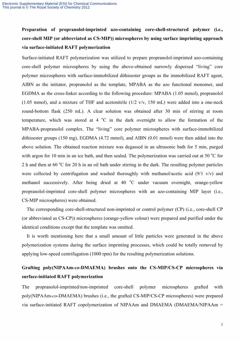
4-((4-Methacryloyloxy)phenylazo)benzoic acid (MPABA) was prepared following our previously
reported procedure (Scheme S1. X. Li, R. Wen, Y. Zhang, L. Zhu, B. Zhang and H. Zhang, J. Mater.
Chem. 2009, 19, 236-245). (±)-Propranolol hydrochloride (Alfa Aesar, 99%) was converted into its
free base form (Scheme S2) before use following the previously reported procedure (L. Schweitz, P.
Spégel and S. Nilsson, Analyst 2000, 125, 1899-1901). Atenolol (National Institute for the Control of
Pharmaceutical and Biological Products, China, Chemical reference substance, Scheme S2) was dried
Electronic Supplementary Material (ESI) for Chemical CommunicationsThis journal is © The Royal Society of Chemistry 2012
2
at 105 oC for 3 h before use. 4-(Dimethylamino)pyridine (DMAP, Merck, AR), triethylamine (TEA,
Tianjin Jiangtian Chemicals, 99%), and all the other reagents were commercially available and used as
received.
Scheme S1. Synthetic route for the azobenzene functional monomer
4-((4-methacryloyloxy)phenylazo)benzoic acid (MPABA).
Scheme S2. The chemical structures of propranolol and atenolol.
Preparation of narrowly dispersed “living” core polymer microspheres with
surface-immobilized dithioester groups via RAFT precipitation polymerization
4-VP (0.611 mmol), EGDMA (2.445 mmol), CDB (0.165 mmol), AIBN (0.055 mmol), and a mixture
of methanol and water (4/1 v/v, 60 mL) were added into a one-neck round-bottom flask (100 mL)
successively. A clear purple reaction mixture was obtained after 10 min of stirring at room temperature,
which was then purged with argon for 30 min, sealed, and submerged in an oil bath. The temperature
of the oil bath was heated from room temperature to 70 oC over 2 h and the polymerization was
conducted at 70 oC for 24 h under stirring. The resulting polymer particles were collected by filtration
and subsequently washed with methanol three times. After being dried at 40 oC under vacuum for 48 h,
a light pink powder was obtained in a yield of 59%.
Electronic Supplementary Material (ESI) for Chemical CommunicationsThis journal is © The Royal Society of Chemistry 2012
3
Preparation of propranolol-imprinted azo-containing core-shell-structured polymer (i.e.,
core-shell MIP (or abbreviated as CS-MIP)) microspheres by using surface imprinting approach
via surface-initiated RAFT polymerization
Surface-initiated RAFT polymerization was utilized to prepare propranolol-imprinted azo-containing
core-shell polymer microspheres by using the above-obtained narrowly dispersed “living” core
polymer microspheres with surface-immobilized dithioester groups as the immobilized RAFT agent,
AIBN as the initiator, propranolol as the template, MPABA as the azo functional monomer, and
EGDMA as the cross-linker according to the following procedure: MPABA (1.05 mmol), propranolol
(1.05 mmol), and a mixture of THF and acetonitrile (1/2 v/v, 150 mL) were added into a one-neck
round-bottom flask (250 mL). A clear solution was obtained after 30 min of stirring at room
temperature, which was stored at 4 oC in the dark overnight to allow the formation of the
MPABA-propranolol complex. The “living” core polymer microspheres with surface-immobilized
dithioester groups (150 mg), EGDMA (4.72 mmol), and AIBN (0.01 mmol) were then added into the
above solution. The obtained reaction mixture was degassed in an ultrasonic bath for 5 min, purged
with argon for 10 min in an ice bath, and then sealed. The polymerization was carried out at 50 oC for
2 h and then at 60 oC for 20 h in an oil bath under stirring in the dark. The resulting polymer particles
were collected by centrifugation and washed thoroughly with methanol/acetic acid (9/1 v/v) and
methanol successively. After being dried at 40 oC under vacuum overnight, orange-yellow
propranolol-imprinted core-shell polymer microspheres with an azo-containing MIP layer (i.e.,
CS-MIP microspheres) were obtained.
The corresponding core-shell-structured non-imprinted or control polymer (CP) (i.e., core-shell CP
(or abbreviated as CS-CP)) microspheres (orange-yellow colour) were prepared and purified under the
identical conditions except that the template was omitted.
It is worth mentioning here that a small amount of little particles were generated in the above
polymerization systems during the surface imprinting processes, which could be totally removed by
applying low-speed centrifugation (1000 rpm) for the resulting polymerization solutions.
Grafting poly(NIPAAm-co-DMAEMA) brushes onto the CS-MIP/CS-CP microspheres via
surface-initiated RAFT polymerization
The propranolol-imprinted/non-imprinted core-shell polymer microspheres grafted with
poly(NIPAAm-co-DMAEMA) brushes (i.e., the grafted CS-MIP/CS-CP microspheres) were prepared
via surface-initiated RAFT copolymerization of NIPAAm and DMAEMA (DMAEMA/NIPAAm =
Electronic Supplementary Material (ESI) for Chemical CommunicationsThis journal is © The Royal Society of Chemistry 2012
4
5/100, molar ratio) by using the above-obtained (ungrafted) CS-MIP/CS-CP microspheres as the
immobilized RAFT agent according to the following procedure: The CS-MIP/CS-CP microspheres (50
mg), NIPAAm (13.27 mmol), DMAEMA (0.66 mmol), CDB (0.01 mmol), AIBN (0.003 mmol), and
DMF (2.5 mL) were added into a two-neck round-bottom flask (25 mL) successively. After being
degassed with five freeze-pump-thaw cycles, the flask was sealed and then immersed in a
thermostatted oil bath at 70 oC and stirred for 24 h. The resulting polymer products were collected by
centrifugation and thoroughly washed with methanol until no white sediment was detectable when
ether was added into the washing solutions, which were then dried at 30 oC under vacuum to the
constant weights, leading to orange-yellow grafted CS-MIP and CS-CP microspheres with a weight
increase of 11.3 and 11.5% in comparison with the corresponding ungrafted ones, respectively.
It is important to stress here that the increased weights of the grafted CS-MIP/CS-CP particles
should be mainly stemmed from the surface-grafted polymer brushes because the rather high
cross-linking densities (around 80%) of the CS-MIP/CS-CP particles would prevent them from
swelling in the reaction media and only allow the occurrence of surface polymerization, just as
reported by Tirelli and coworkers (D. Bontempo, N. Tirelli, K. Feldman, G. Masci, V. Crescenzi and J.
A. Hubbell, Adv. Mater. 2002, 14, 1239-1241) and by our group (J. Jiang, Y. Zhang, X. Guo and H.
Zhang, Macromolecules 2011, 44, 5893-5904; G. Pan, Y. Ma, Y. Zhang, X. Guo, C. Li and H. Zhang,
Soft Matter 2011, 7, 8428-8439).
The addition of CDB into the above polymerization systems also led to the generation of free
poly(NIPAAm-co-DMAEMA) in the reaction solutions, which were obtained by precipitating the
supernatant solutions (after the centrifugation of the reaction mixtures) into pentane, filtered, and then
dried at 40 oC under vacuum for 48 h, finally resulting in light pink polymer products.
Characterization of the morphologies, particle sizes, and size distributions of the polymer
microspheres
The morphologies, particle sizes, and size distributions of the above-obtained “living” core polymer
microspheres, the ungrafted CS-MIP/CS-CP microspheres, and the grafted CS-MIP/CS-CP
microspheres were characterized with a scanning electron microscope (SEM, Shimadzu SS-550)
(Table S1). All of the SEM size data reflect the averages of about 200 particles, which are calculated
by the following formulas:
k k k k Dn = niDi/ ni; Dw� = niDi
4/ niDi3; U = Dw/Dn
i=1 i=1 i=1 i=1
Electronic Supplementary Material (ESI) for Chemical CommunicationsThis journal is © The Royal Society of Chemistry 2012
5
where Dn is the number-average diameter, Dw the weight-average diameter, k the total number of the
measured particles, Di the particle diameter of the ith polymer microsphere, and ni the number of the
microspheres with a diameter Di, U the size distribution index.
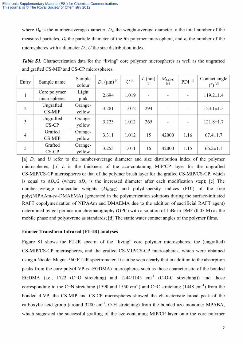
Table S1. Characterization data for the “living” core polymer microspheres as well as the ungrafted
and grafted CS-MIP and CS-CP microspheres.
Entry Sample name Sample colour
Dn (μm) [a] U [a] L (nm)
[b] Mn,GPC
[c] PDI [c]
Contact angle (°) [d]
1 Core polymer microspheres
Light pink
2.694 1.019 - - - 119.2±1.4
2 Ungrafted CS-MIP
Orange- yellow
3.281 1.012 294 - - 123.1±1.5
3 Ungrafted
CS-CP Orange- yellow
3.223 1.012 265 - - 121.8±1.7
4 Grafted CS-MIP
Orange- yellow
3.311 1.012 15 42000 1.16 67.4±1.7
5 Grafted CS-CP
Orange- yellow
3.255 1.011 16 42000 1.15 66.5±1.1
[a] Dn and U refer to the number-average diameter and size distribution index of the polymer
microspheres; [b] L is the thickness of the azo-containing MIP/CP layer for the ungrafted
CS-MIP/CS-CP microspheres or that of the polymer brush layer for the grafted CS-MIP/CS-CP, which
is equal to ∆Dn/2 (where ∆Dn is the increased diameter after each modification step); [c] The
number-average molecular weights (Mn,GPC) and polydispersity indices (PDI) of the free
poly(NIPAAm-co-DMAEMA) (generated in the polymerization solutions during the surface-initiated
RAFT copolymerization of NIPAAm and DMAEMA due to the addition of sacrificial RAFT agent)
determined by gel permeation chromatography (GPC) with a solution of LiBr in DMF (0.05 M) as the
mobile phase and polystyrene as standards; [d] The static water contact angles of the polymer films.
Fourier Transform Infrared (FT-IR) analyses
Figure S1 shows the FT-IR spectra of the “living” core polymer microspheres, the (ungrafted)
CS-MIP/CS-CP microspheres, and the grafted CS-MIP/CS-CP microspheres, which were obtained
using a Nicolet Magna-560 FT-IR spectrometer. It can be seen clearly that in addition to the absorption
peaks from the core poly(4-VP-co-EGDMA) microspheres such as those characteristic of the bonded
EGDMA (i.e., 1722 (C=O stretching) and 1244/1145 cm-1 (C-O-C stretching)) and those
corresponding to the C=N stretching (1590 and 1550 cm-1) and C=C stretching (1448 cm-1) from the
bonded 4-VP, the CS-MIP and CS-CP microspheres showed the characteristic broad peak of the
carboxylic acid group (around 3280 cm-1, O-H stretching) from the bonded azo monomer MPABA,
which suggested the successful grafting of the azo-containing MIP/CP layer onto the core polymer
Electronic Supplementary Material (ESI) for Chemical CommunicationsThis journal is © The Royal Society of Chemistry 2012
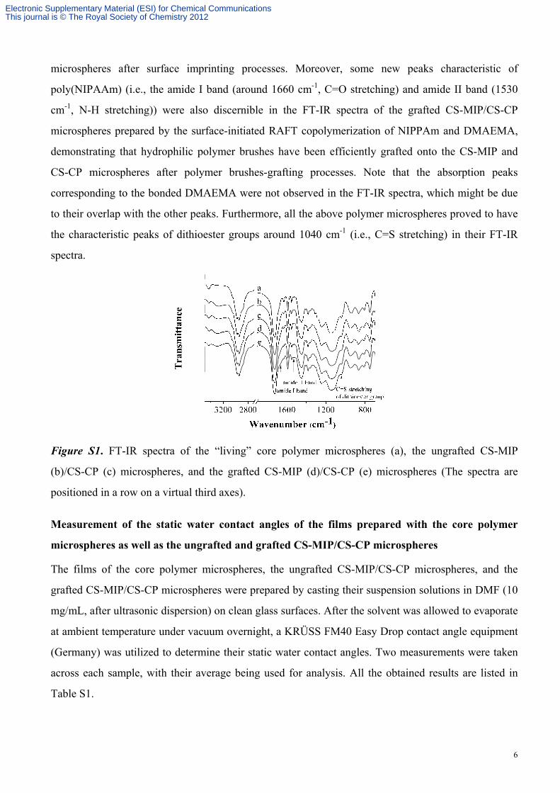
6
microspheres after surface imprinting processes. Moreover, some new peaks characteristic of
poly(NIPAAm) (i.e., the amide I band (around 1660 cm-1, C=O stretching) and amide II band (1530
cm-1, N-H stretching)) were also discernible in the FT-IR spectra of the grafted CS-MIP/CS-CP
microspheres prepared by the surface-initiated RAFT copolymerization of NIPPAm and DMAEMA,
demonstrating that hydrophilic polymer brushes have been efficiently grafted onto the CS-MIP and
CS-CP microspheres after polymer brushes-grafting processes. Note that the absorption peaks
corresponding to the bonded DMAEMA were not observed in the FT-IR spectra, which might be due
to their overlap with the other peaks. Furthermore, all the above polymer microspheres proved to have
the characteristic peaks of dithioester groups around 1040 cm-1 (i.e., C=S stretching) in their FT-IR
spectra.
Figure S1. FT-IR spectra of the “living” core polymer microspheres (a), the ungrafted CS-MIP
(b)/CS-CP (c) microspheres, and the grafted CS-MIP (d)/CS-CP (e) microspheres (The spectra are
positioned in a row on a virtual third axes).
Measurement of the static water contact angles of the films prepared with the core polymer
microspheres as well as the ungrafted and grafted CS-MIP/CS-CP microspheres
The films of the core polymer microspheres, the ungrafted CS-MIP/CS-CP microspheres, and the
grafted CS-MIP/CS-CP microspheres were prepared by casting their suspension solutions in DMF (10
mg/mL, after ultrasonic dispersion) on clean glass surfaces. After the solvent was allowed to evaporate
at ambient temperature under vacuum overnight, a KRÜSS FM40 Easy Drop contact angle equipment
(Germany) was utilized to determine their static water contact angles. Two measurements were taken
across each sample, with their average being used for analysis. All the obtained results are listed in
Table S1.
Electronic Supplementary Material (ESI) for Chemical CommunicationsThis journal is © The Royal Society of Chemistry 2012
7
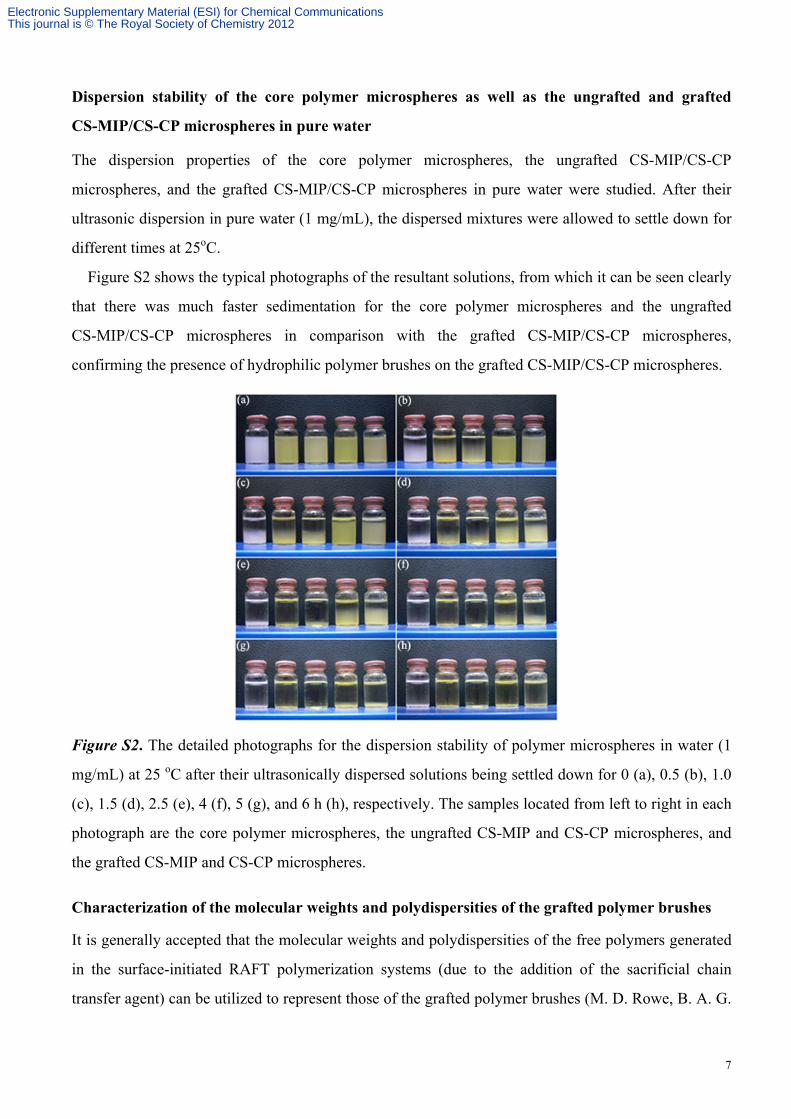
Dispersion stability of the core polymer microspheres as well as the ungrafted and grafted
CS-MIP/CS-CP microspheres in pure water
The dispersion properties of the core polymer microspheres, the ungrafted CS-MIP/CS-CP
microspheres, and the grafted CS-MIP/CS-CP microspheres in pure water were studied. After their
ultrasonic dispersion in pure water (1 mg/mL), the dispersed mixtures were allowed to settle down for
different times at 25oC.
Figure S2 shows the typical photographs of the resultant solutions, from which it can be seen clearly
that there was much faster sedimentation for the core polymer microspheres and the ungrafted
CS-MIP/CS-CP microspheres in comparison with the grafted CS-MIP/CS-CP microspheres,
confirming the presence of hydrophilic polymer brushes on the grafted CS-MIP/CS-CP microspheres.
Figure S2. The detailed photographs for the dispersion stability of polymer microspheres in water (1
mg/mL) at 25 oC after their ultrasonically dispersed solutions being settled down for 0 (a), 0.5 (b), 1.0
(c), 1.5 (d), 2.5 (e), 4 (f), 5 (g), and 6 h (h), respectively. The samples located from left to right in each
photograph are the core polymer microspheres, the ungrafted CS-MIP and CS-CP microspheres, and
the grafted CS-MIP and CS-CP microspheres.
Characterization of the molecular weights and polydispersities of the grafted polymer brushes
It is generally accepted that the molecular weights and polydispersities of the free polymers generated
in the surface-initiated RAFT polymerization systems (due to the addition of the sacrificial chain
transfer agent) can be utilized to represent those of the grafted polymer brushes (M. D. Rowe, B. A. G.
Electronic Supplementary Material (ESI) for Chemical CommunicationsThis journal is © The Royal Society of Chemistry 2012
8
Hammer and S. G. Boyes, Macromolecules 2008, 41, 4147-4157). Therefore, the free polymers
obtained in our study were characterized with gel permeation chromatography (GPC), from which the
number-average molecular weights (Mn,GPC) of the grafted polymer brushes on both the CS-MIP and
CS-CP microspheres were evaluated to be 42000 and their PDIs were found to be 1.16 and 1.15,
respectively (Table S1).
A Waters GPC was utilized in the above measurements, which was equipped with a Waters 2414
series manual injector, a Waters 1515 high-performance liquid chromatography (HPLC) pump, a
Waters 2414 refractive index detector, and three Waters UltraStyragel columns with 5 K-600 K,
500-30 K, and 100-10 K molecular ranges (the temperature of the column oven was set at 40 oC). A
solution of LiBr in DMF (0.05 M) was used as the mobile phase with a flow rate of 1.0 mL/min, and
the calibration curve was obtained by using polystyrene standards.
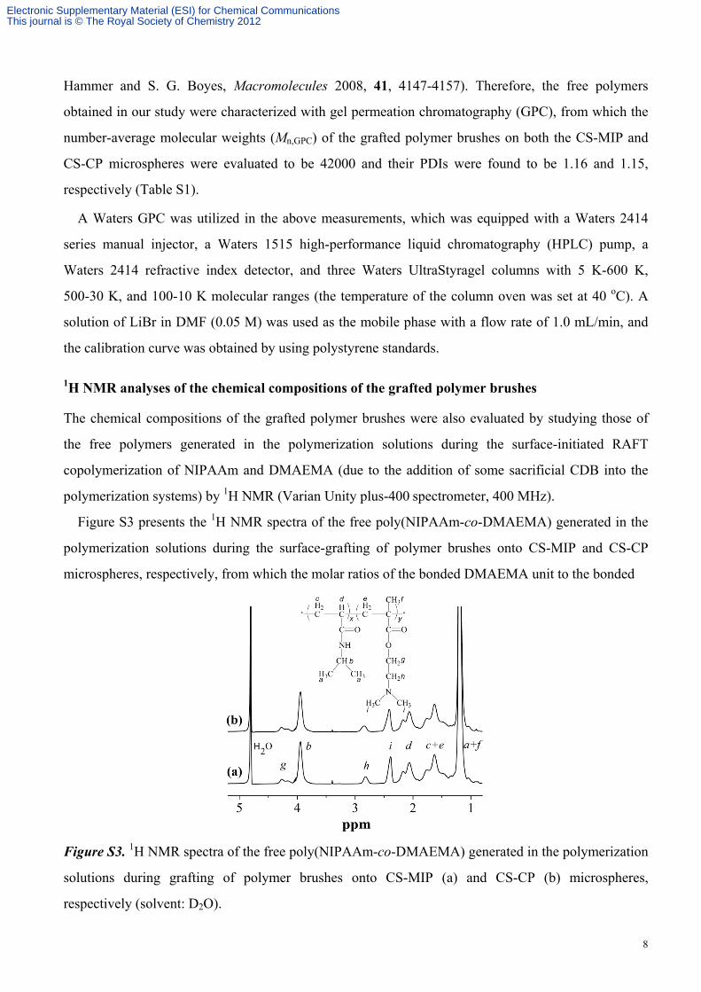
1H NMR analyses of the chemical compositions of the grafted polymer brushes
The chemical compositions of the grafted polymer brushes were also evaluated by studying those of
the free polymers generated in the polymerization solutions during the surface-initiated RAFT
copolymerization of NIPAAm and DMAEMA (due to the addition of some sacrificial CDB into the
polymerization systems) by 1H NMR (Varian Unity plus-400 spectrometer, 400 MHz).
Figure S3 presents the 1H NMR spectra of the free poly(NIPAAm-co-DMAEMA) generated in the
polymerization solutions during the surface-grafting of polymer brushes onto CS-MIP and CS-CP
microspheres, respectively, from which the molar ratios of the bonded DMAEMA unit to the bonded
Figure S3. 1H NMR spectra of the free poly(NIPAAm-co-DMAEMA) generated in the polymerization
solutions during grafting of polymer brushes onto CS-MIP (a) and CS-CP (b) microspheres,
respectively (solvent: D2O).
Electronic Supplementary Material (ESI) for Chemical CommunicationsThis journal is © The Royal Society of Chemistry 2012
9
NIPAAm unit in both poly(NIPAAm-co-DMAEMA) brushes grafted on CS-MIP and CS-CP
microspheres were determined to be 8% by comparing the integral of the peak h around 2.76 ppm with
that of the peak b around 3.93 ppm, which are close to the initial comonomer ratios (i.e., 5 mol%) in
the surface-initiated RAFT copolymerization systems.
Determination of the lower critical solution temperatures (LCSTs) of the grafted polymer
brushes in pure water and in Britton-Robison buffer (with different pH values)
The LCSTs of the grafted polymer brushes were evaluated by studying those of the free
poly(NIPAAm-co-DMAEMA) generated in the polymerization solutions during surface-initiated
RAFT polymerization processes. An UV-Vis scanning spectrophotometer (TU1900, Beijing Purkinje
General Instrument Co., Ltd) equipped with a thermostatic cell holder (CH19-1) was utilized for this
purpose. The polymer solutions (1.5 mg/mL) in the quartz sample cells were heated from 25 to 60 oC
at a rate of 0.2 oC/min and the LCSTs of the polymers were determined at 50% of the transmittance of
a 600 nm light beam through the polymer solutions (Y. Xia, N. A. D. Burke and H. D. H. Stöver,
Macromolecules 2006, 39, 2275-2283; C. Luo, Y. Liu and Z. Li, Macromolecules 2010, 43,
8101-8108).
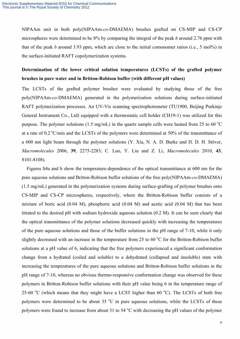
Figures S4a and b show the temperature-dependence of the optical transmittance at 600 nm for the
pure aqueous solutions and Britton-Robison buffer solutions of the free poly(NIPAAm-co-DMAEMA)
(1.5 mg/mL) generated in the polymerization systems during surface-grafting of polymer brushes onto
CS-MIP and CS-CP microspheres, respectively, where the Britton-Robison buffer consists of a
mixture of boric acid (0.04 M), phosphoric acid (0.04 M) and acetic acid (0.04 M) that has been
titrated to the desired pH with sodium hydroxide aqueous solution (0.2 M). It can be seen clearly that
the optical transmittance of the polymer solutions decreased quickly with increasing the temperatures
of the pure aqueous solutions and those of the buffer solutions in the pH range of 7-10, while it only
slightly decreased with an increase in the temperature from 25 to 60 oC for the Britton-Robison buffer
solutions at a pH value of 6, indicating that the free polymers experienced a significant conformation
change from a hydrated (coiled and soluble) to a dehydrated (collapsed and insoluble) state with
increasing the temperatures of the pure aqueous solutions and Britton-Robison buffer solutions in the
pH range of 7-10, whereas no obvious thermo-responsive conformation change was observed for these
polymers in Britton-Robison buffer solutions with their pH value being 6 in the temperature range of
25-60 oC (which means that they might have a LCST higher than 60 oC). The LCSTs of both free
polymers were determined to be about 35 oC in pure aqueous solutions, while the LCSTs of these
polymers were found to increase from about 31 to 54 oC with decreasing the pH values of the polymer
Electronic Supplementary Material (ESI) for Chemical CommunicationsThis journal is © The Royal Society of Chemistry 2012
10
buffer solutions from 10 to 7 (Figure S4c), which is easily understandable because the protonation of
DMAEMA units at lower pH conditions improved the hydrophilicity of the polymer chains (M. S.
Jones, Eur. Polym. J. 1999, 35, 795-801; V. Bulmus, Z. Ding, C. J. Long, P. S. Stayton and A. S.
Hoffman, Bioconjugate Chem. 2000, 11, 78-83). Furthermore, it is worth noting that the LCSTs of
both free polymers generated in the polymerization systems during the surface-grafting of polymer
brushes onto CS-MIP and CS-CP microspheres were essentially the same for their pure aqueous
solutions and aqueous buffer solutions in the studied pH range, which is in good consistency with their
almost same chemical compositions, molecular weights, and polydispersities.
Figure S4. (a,b) Temperature-dependence of the optical transmittance at 600 nm obtained for the pure
aqueous solutions (★) and Britton-Robison buffer solutions (pH 6 (■), 7 (●), 8 (◆), 9 (◄), and 10
(▲)) of the free poly(NIPAAm-co-DMAEMA) generated in the polymerization systems during
surface-grafting of polymer brushes onto CS-MIP (a) and CS-CP (b) microspheres, respectively
(polymer concentration: 1.5 mg/mL); (c) pH-dependence of LCSTs obtained for the Britton-Robison
buffer solutions of free poly(NIPAAm-co-DMAEMA) generated in the polymerization systems during
surface-grafting of polymer brushes onto CS-MIP (▲) and CS-CP (●) microspheres, respectively
(polymer concentration: 1.5 mg/mL).
Equilibrium template binding experiments with the ungrafted and grafted CS-MIP/CS-CP
microspheres
Equilibrium template binding experiments were performed by incubating a propranolol solution in
acetonitrile (0.5 mL, 0.05 mM) or in pure water (0.5 mL, 0.05 mM) with different amounts of the
ungrafted or grafted CS-MIP/CS-CP microspheres at 25 oC for 16 h. After centrifugation, the amounts
of the template remaining in the supernatants were then quantified by using a HPLC (Scientific System
Electronic Supplementary Material (ESI) for Chemical CommunicationsThis journal is © The Royal Society of Chemistry 2012
11
Inc., USA) equipped with a UV-Vis detector, from which the amounts of the template bound to the
ungrafted and grafted CS-MIP/CS-CP microspheres could be obtained (Figures 2a,b). The wavelength
used for the determination of propranolol was 293 nm. A mixture of acetonitrile and 0.4% aqueous
solution of triethylamine (7/3 v/v) was used as the mobile phase at a flow rate of 1 mL/min. All the
above binding analyses were performed in duplicate and the mean values were used.
In this context, it is worth mentioning that the specific template bindings of the ungrafted and
grafted CS-MIP microspheres (i.e., the template bindings by the imprinted binding sites of the studied
MIPs) could be derived by using the following equation:
Specific template binding = BMIP – BCP
where BMIP and BCP are the equilibrium template bindings of the studied MIP and its corresponding CP,
respectively.
Competitive binding experiments
The binding selectivity of the ungrafted and grafted CS-MIP/CS-CP microspheres was evaluated by
measuring their competitive binding capacities towards propranolol and its structurally related
compound atenolol as follows: 0.5 mg of ungrafted or grafted CS-MIP/CS-CP microspheres were
incubated with 0.5 mL of a mixed solution of propranolol and atenolol in acetonitrile (Cpropranolol or
atenolol = 0.05 mM) or in pure water (Cpropranolol or atenolol = 0.05 mM) at 25 oC for 16 h and the amounts of
propranolol and atenolol bound to the ungrafted and grafted CS-MIPs/CS-CPs were quantified by
HPLC. The wavelength used for the determination of the mixed solution of propranolol and atenolol
was 275 nm. A mixture of acetonitrile and 0.4% aqueous solution of triethylamine (7/3 v/v) was used
as the mobile phase at a flow rate of 1 mL/min. All the above binding analyses were performed in
duplicate and the mean values were used.
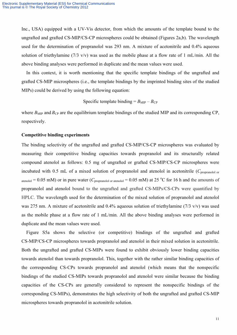
Figure S5a shows the selective (or competitive) bindings of the ungrafted and grafted
CS-MIP/CS-CP microspheres towards propranolol and atenolol in their mixed solution in acetonitrile.
Both the ungrafted and grafted CS-MIPs were found to exhibit obviously lower binding capacities
towards atenolol than towards propranolol. This, together with the rather similar binding capacities of
the corresponding CS-CPs towards propranolol and atenolol (which means that the nonspecific
bindings of the studied CS-MIPs towards propranolol and atenolol were similar because the binding
capacities of the CS-CPs are generally considered to represent the nonspecific bindings of the
corresponding CS-MIPs), demonstrates the high selectivity of both the ungrafted and grafted CS-MIP
microspheres towards propranolol in acetonitrile solution.
Electronic Supplementary Material (ESI) for Chemical CommunicationsThis journal is © The Royal Society of Chemistry 2012
12
Figure S5. Selective bindings of the ungrafted and grafted CS-MIP/CS-CP microspheres towards
propranolol and atenolol in their mixed solution in acetonitrile (a) and in pure water (b) (CPropranolol or
atenolol = 0.05 mM), respectively (polymer concentration: 1 mg/mL).
Figure S5b presents the selective bindings of the ungrafted and grafted CS-MIP/CS-CP
microspheres towards propranolol and atenolol in their mixed pure aqueous solutions. It can be seen
that although both the ungrafted and grafted CS-MIPs exhibited higher binding capacities towards
propranolol than towards atenolol in pure aqueous solutions, the binding capacities of both the
ungrafted and grafted CS-CPs towards propranolol were also higher than towards atenolol. The above
results suggested that the nonspecific bindings of the studied CS-MIPs towards propranolol and
atenolol were different, which makes it inappropriate to evaluate the selectivity (or specificity) of the
ungrafted and grafted CS-MIPs by directly comparing their binding capacities towards propranolol and
atenolol.
In the above case, the “imprinting-induced promotion of binding” (IPB) has proven to be an useful
parameter for evaluating the MIPs’ selectivity because the difference in the intrinsic nonspecific
bindings of the MIPs towards different analytes is normalized (T. Hishiya, M. Shibata, M. Kakazu, H.
Asanuma and M. Komiyama, Macromolecules 1999, 32, 2265-2269; J. H. Zhang, M. Jiang, L. Zou, D.
Shi, S. R. Mei, Y. X. Zhu, Y. Shi, K. Dai and B. Lu, Anal. Bioanal. Chem. 2006, 385, 780-786; G. Pan,
Y. Zhang, Y. Ma, C. Li and H. Zhang, Angew. Chem. Int. Ed. 2011, 50, 11731-11734). IPB can be
defined by the following equation:
IPB = (BMIP − BCP)/BCP
where BMIP and BCP are the equilibrium bindings of the studied MIP and its corresponding CP towards
an analyte, respectively. The larger the IPB value of the MIP towards the analyte, the better the
selectivity of the MIP.
Electronic Supplementary Material (ESI) for Chemical CommunicationsThis journal is © The Royal Society of Chemistry 2012
13
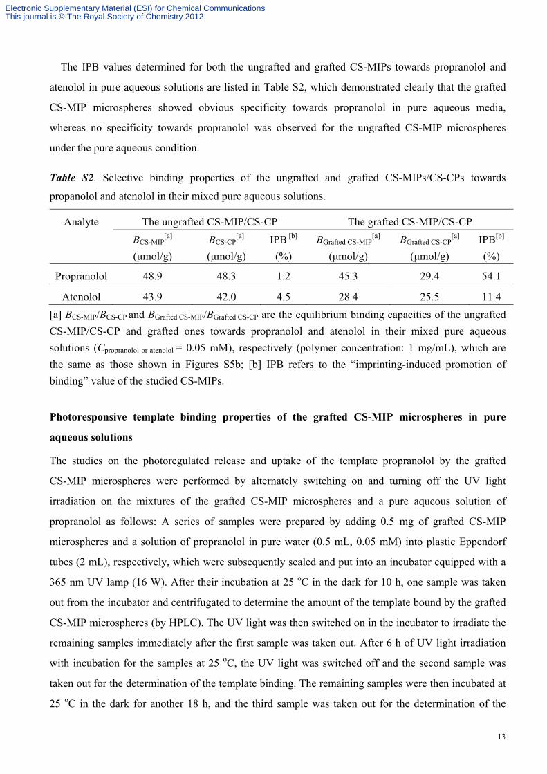
The IPB values determined for both the ungrafted and grafted CS-MIPs towards propranolol and
atenolol in pure aqueous solutions are listed in Table S2, which demonstrated clearly that the grafted
CS-MIP microspheres showed obvious specificity towards propranolol in pure aqueous media,
whereas no specificity towards propranolol was observed for the ungrafted CS-MIP microspheres
under the pure aqueous condition.
Table S2. Selective binding properties of the ungrafted and grafted CS-MIPs/CS-CPs towards
propanolol and atenolol in their mixed pure aqueous solutions.
Analyte The ungrafted CS-MIP/CS-CP The grafted CS-MIP/CS-CP
BCS-MIP[a]
(μmol/g)
BCS-CP[a]
(μmol/g)
IPB [b]
(%)
BGrafted CS-MIP[a]
(μmol/g)
BGrafted CS-CP[a]
(μmol/g)
IPB[b]
(%)
Propranolol 48.9 48.3 1.2 45.3 29.4 54.1
Atenolol 43.9 42.0 4.5 28.4 25.5 11.4
[a] BCS-MIP/BCS-CP and BGrafted CS-MIP/BGrafted CS-CP are the equilibrium binding capacities of the ungrafted
CS-MIP/CS-CP and grafted ones towards propranolol and atenolol in their mixed pure aqueous
solutions (Cpropranolol or atenolol = 0.05 mM), respectively (polymer concentration: 1 mg/mL), which are
the same as those shown in Figures S5b; [b] IPB refers to the “imprinting-induced promotion of
binding” value of the studied CS-MIPs.
Photoresponsive template binding properties of the grafted CS-MIP microspheres in pure
aqueous solutions
The studies on the photoregulated release and uptake of the template propranolol by the grafted
CS-MIP microspheres were performed by alternately switching on and turning off the UV light
irradiation on the mixtures of the grafted CS-MIP microspheres and a pure aqueous solution of
propranolol as follows: A series of samples were prepared by adding 0.5 mg of grafted CS-MIP
microspheres and a solution of propranolol in pure water (0.5 mL, 0.05 mM) into plastic Eppendorf
tubes (2 mL), respectively, which were subsequently sealed and put into an incubator equipped with a
365 nm UV lamp (16 W). After their incubation at 25 oC in the dark for 10 h, one sample was taken
out from the incubator and centrifugated to determine the amount of the template bound by the grafted
CS-MIP microspheres (by HPLC). The UV light was then switched on in the incubator to irradiate the
remaining samples immediately after the first sample was taken out. After 6 h of UV light irradiation
with incubation for the samples at 25 oC, the UV light was switched off and the second sample was
taken out for the determination of the template binding. The remaining samples were then incubated at
25 oC in the dark for another 18 h, and the third sample was taken out for the determination of the
Electronic Supplementary Material (ESI) for Chemical CommunicationsThis journal is © The Royal Society of Chemistry 2012
14
template binding. The UV light was then switched on again to irradiate the remaining samples at 25 oC
immediately after the third sample was taken out. The above photoswitching cycles (i.e., UV light on
for 6 h and off for 18 h alternately) were repeated until all the other samples were measured.
The photoregulated release and uptake of propranolol by the grafted CS-CP microspheres were also
studied similarly to provide control results for the grafted CS-MIP microspheres.
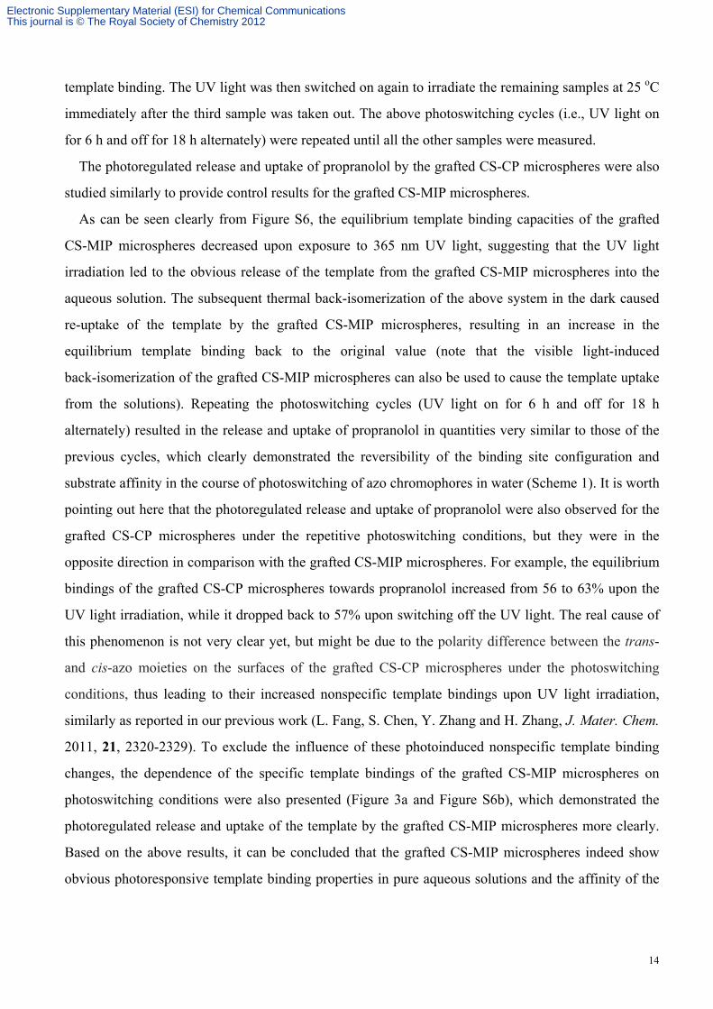
As can be seen clearly from Figure S6, the equilibrium template binding capacities of the grafted
CS-MIP microspheres decreased upon exposure to 365 nm UV light, suggesting that the UV light
irradiation led to the obvious release of the template from the grafted CS-MIP microspheres into the
aqueous solution. The subsequent thermal back-isomerization of the above system in the dark caused
re-uptake of the template by the grafted CS-MIP microspheres, resulting in an increase in the
equilibrium template binding back to the original value (note that the visible light-induced
back-isomerization of the grafted CS-MIP microspheres can also be used to cause the template uptake
from the solutions). Repeating the photoswitching cycles (UV light on for 6 h and off for 18 h
alternately) resulted in the release and uptake of propranolol in quantities very similar to those of the
previous cycles, which clearly demonstrated the reversibility of the binding site configuration and
substrate affinity in the course of photoswitching of azo chromophores in water (Scheme 1). It is worth
pointing out here that the photoregulated release and uptake of propranolol were also observed for the
grafted CS-CP microspheres under the repetitive photoswitching conditions, but they were in the
opposite direction in comparison with the grafted CS-MIP microspheres. For example, the equilibrium
bindings of the grafted CS-CP microspheres towards propranolol increased from 56 to 63% upon the
UV light irradiation, while it dropped back to 57% upon switching off the UV light. The real cause of
this phenomenon is not very clear yet, but might be due to the polarity difference between the trans-
and cis-azo moieties on the surfaces of the grafted CS-CP microspheres under the photoswitching
conditions, thus leading to their increased nonspecific template bindings upon UV light irradiation,
similarly as reported in our previous work (L. Fang, S. Chen, Y. Zhang and H. Zhang, J. Mater. Chem.
2011, 21, 2320-2329). To exclude the influence of these photoinduced nonspecific template binding
changes, the dependence of the specific template bindings of the grafted CS-MIP microspheres on
photoswitching conditions were also presented (Figure 3a and Figure S6b), which demonstrated the
photoregulated release and uptake of the template by the grafted CS-MIP microspheres more clearly.
Based on the above results, it can be concluded that the grafted CS-MIP microspheres indeed show
obvious photoresponsive template binding properties in pure aqueous solutions and the affinity of the
Electronic Supplementary Material (ESI) for Chemical CommunicationsThis journal is © The Royal Society of Chemistry 2012
15
binding sites in the grafted CS-MIP microspheres towards the template can be easily tuned by the
simple photoswitching.
Figure S6. (a) Photoregulated release and uptake of propranolol by the grafted CS-MIP (filled
circle)/CS-CP (open circle) microspheres in pure aqueous solutions under photoswitching conditions
(UV light on for 6 h and off for 18 h alternately at 25 oC); (b) The photoresponsive specific template
bindings of the grafted CS-MIP microspheres in pure aqueous solutions under photoswitching
conditions. The concentration of the grafted CS-MIP/CS-CP microspheres was 1.0 mg/mL and the
initial concentration of propranolol in the aqueous solution was 0.05 mM.
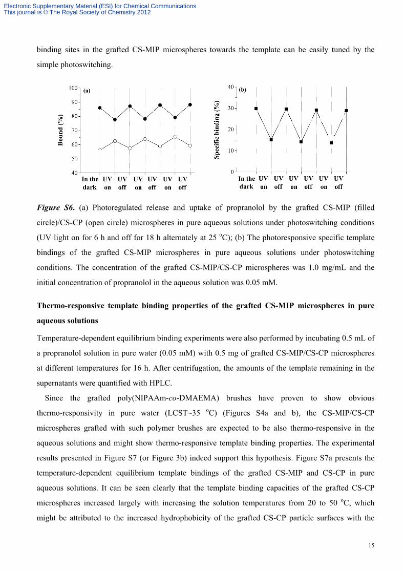
Thermo-responsive template binding properties of the grafted CS-MIP microspheres in pure
aqueous solutions
Temperature-dependent equilibrium binding experiments were also performed by incubating 0.5 mL of
a propranolol solution in pure water (0.05 mM) with 0.5 mg of grafted CS-MIP/CS-CP microspheres
at different temperatures for 16 h. After centrifugation, the amounts of the template remaining in the
supernatants were quantified with HPLC.
Since the grafted poly(NIPAAm-co-DMAEMA) brushes have proven to show obvious
thermo-responsivity in pure water (LCST~35 oC) (Figures S4a and b), the CS-MIP/CS-CP
microspheres grafted with such polymer brushes are expected to be also thermo-responsive in the
aqueous solutions and might show thermo-responsive template binding properties. The experimental
results presented in Figure S7 (or Figure 3b) indeed support this hypothesis. Figure S7a presents the
temperature-dependent equilibrium template bindings of the grafted CS-MIP and CS-CP in pure
aqueous solutions. It can be seen clearly that the template binding capacities of the grafted CS-CP
microspheres increased largely with increasing the solution temperatures from 20 to 50 oC, which
might be attributed to the increased hydrophobicity of the grafted CS-CP particle surfaces with the
Electronic Supplementary Material (ESI) for Chemical CommunicationsThis journal is © The Royal Society of Chemistry 2012
16
polymer brushes changing from the hydrophilic soluble state to hydrophobic insoluble state following
the temperature changes, thus leading to their higher nonspecific template bindings with the increase in
the solution temperatures. In comparison, the template binding capacities of the grafted CS-MIP
microspheres showed much less increase with an increase in the solution temperatures. The above
results demonstrated that the specific template bindings of the grafted CS-MIP microspheres (which
could be derived by subtracting the binding capacities of the grafted CS-CP from those of the grafted
CS-MIP) decreased obviously with increasing the solution temperatures (Figure S7b, Figure 3b),
which might be ascribed to the collapse of the polymer brushes at higher temperatures, thus resulting
in the blocking of the imprinted binding sites in the grafted CS-MIP (Scheme 1), just as reported by
the groups of Tatsuma and Hoffman in a peptide electrode and a protein system, respectively (K.
Komori, K. Takada and T. Tatsuma, Anal. Sci. 2005, 21, 351-353; P. S. Stayton, T. Shimoboji, C.
Long, A. Chilkoti, G. Chen, J. M. Harris and A. S. Hoffman, Nature 1995, 378, 472-474).
Figure S7. (a) Temperature-dependent equilibrium bindings of the grafted CS-MIP (filled
circle)/CS-CP (open circle) microspheres towards propranolol in pure water; (b)
Temperature-dependent specific bindings of the grafted CS-MIP microspheres towards propranolol in
pure water. The concentration of the grafted CS-MIP/CS-CP microspheres was 1.0 mg/mL and the
initial concentration of the propranolol solution in pure water was 0.05 mM.
pH-responsive template binding properties of the grafted CS-MIP microspheres in
Britton-Robison buffer solutions
Equilibrium binding experiments were further carried out by incubating 0.5 mg of grafted
CS-MIP/CS-CP microspheres with a propranolol solution in Britton-Robison buffer with different pH
values (0.5 mL, 0.05 mM) at 40 oC for 16 h. After centrifugation, the amounts of the template
remaining in the supernatants were then quantified with HPLC. A temperature of 40 oC was chosen
Electronic Supplementary Material (ESI) for Chemical CommunicationsThis journal is © The Royal Society of Chemistry 2012
17
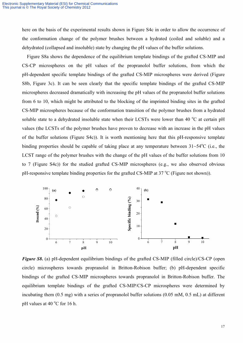
here on the basis of the experimental results shown in Figure S4c in order to allow the occurrence of
the conformation change of the polymer brushes between a hydrated (coiled and soluble) and a
dehydrated (collapsed and insoluble) state by changing the pH values of the buffer solutions.
Figure S8a shows the dependence of the equilibrium template bindings of the grafted CS-MIP and
CS-CP microspheres on the pH values of the propranolol buffer solutions, from which the
pH-dependent specific template bindings of the grafted CS-MIP microspheres were derived (Figure
S8b, Figure 3c). It can be seen clearly that the specific template bindings of the grafted CS-MIP
microspheres decreased dramatically with increasing the pH values of the propranolol buffer solutions
from 6 to 10, which might be attributed to the blocking of the imprinted binding sites in the grafted
CS-MIP microspheres because of the conformation transition of the polymer brushes from a hydrated
soluble state to a dehydrated insoluble state when their LCSTs were lower than 40 oC at certain pH
values (the LCSTs of the polymer brushes have proven to decrease with an increase in the pH values
of the buffer solutions (Figure S4c)). It is worth mentioning here that this pH-responsive template
binding properties should be capable of taking place at any temperature between 31~54oC (i.e., the
LCST range of the polymer brushes with the change of the pH values of the buffer solutions from 10
to 7 (Figure S4c)) for the studied grafted CS-MIP microspheres (e.g., we also observed obvious
pH-responsive template binding properties for the grafted CS-MIP at 37 oC (Figure not shown)).
Figure S8. (a) pH-dependent equilibrium bindings of the grafted CS-MIP (filled circle)/CS-CP (open
circle) microspheres towards propranolol in Britton-Robison buffer; (b) pH-dependent specific
bindings of the grafted CS-MIP microspheres towards propranolol in Britton-Robison buffer. The
equilibrium template bindings of the grafted CS-MIP/CS-CP microspheres were determined by
incubating them (0.5 mg) with a series of propranolol buffer solutions (0.05 mM, 0.5 mL) at different
pH values at 40 oC for 16 h.
Electronic Supplementary Material (ESI) for Chemical CommunicationsThis journal is © The Royal Society of Chemistry 2012