Embed Size (px)
Citation preview

Efficient Protein Digestion at Elevated Temperature in the Presence of SDS and Calcium Ions for Membrane Proteomics
Jessica Loraine†$, Ohoud Alhumaidan†§‡$, Andrew R. Bottrill⊥∥, Sharad C. Mistry⊥,
Peter Andrew†,‡, Galina V. Mukamolova†,‡ and Obolbek Turapov†,‡*
†Department of Infection, Immunity and Inflammation, University of Leicester,
Leicester, LE1 9HN, UK
‡Department of Respiratory Sciences, University of Leicester, Leicester, LE1 9HN,
UK
§Department of Clinical Laboratory Sciences, King Saud University. Riyadh, Saudi
Arabia
⊥Protein Nucleic Acid Laboratory, University of Leicester, Leicester, LE1 7RH, UK,
∥Current address: Proteomics Research Technology Platform, School of Life Sciences, University of Warwick, CV4 7AL, UK
AUTHOR INFORMATION
$These authors contributed equally to this work
Corresponding Author
*E-mail: [email protected]
ABSTRACT
Growing significance of membrane proteins inspires continuous development and
improvement of methods for robust membrane proteomics. Here, we developed a very
simple and efficient method for membrane protein digestion using an ionic detergent
sodium dodecyl sulphate (SDS) at high temperature, conditions where trypsin is
normally inactivated. Our results suggest that trypsin can be stabilized by a
combination of calcium ions and sodium chloride which enables protein digestion at
elevated temperature in the presence of strong ionic detergents such as SDS. Finding
conditions for stabilisation of trypsin offers novel opportunities for application of
detergents for investigation of membrane proteins.

INTRODUCTION
Membrane proteins play an essential biological role in all genera of life. These proteins
are critical for cell-cell communication, transport of chemicals, sensing environmental
changes and pathogenesis. The membrane proteins of microorganisms are in the
frontline of the host-parasite/symbiont interactions.1,2 Understanding the role and
function of these membrane proteins is crucial for environmental and medical biology;
however, they are challenging to handle and digest for proteomics analysis.3 For
analytical purposes membrane proteins are extracted from the membrane and
identified using a variety of analytical and biochemical techniques. For the proteomic
analysis proteins are digested with a suitable enzyme and generated fragments are
analysed by mass spectrometry.3,4 To achieve efficient digestion, proteins are
chemically denatured prior to proteinase digestion.4-7 The traditional methods require
removal of denaturing agents and may result in loss of valuable samples.7,8 Many
alternative methods for improvement of protein digestion have been developed
including application of high temperature,7 microwave irradiation technology,9
pressure-assisted treatment,10 focused ultrasound technique11. However, these
techniques have been mainly used for digestion of soluble cytoplasmic proteins, while
processing of membrane and cell wall proteins for down-stream analysis remains
problematic.
Trypsin is the most popular enzyme used in proteomics because of its exceptional
specificity and ability to cleave proteins into the fragments in the ideal range for mass
spectrometry.12 At the same time, this is a powerful enzyme with a comparatively good
stability under various conditions.13 Improving stability of the proteolytic enzyme
enables exploration of more potent conditions for membrane protein digestion.
Here, we developed a method of stabilisation of trypsin in the presence of strong
ionic detergents. We evaluated this method initially for digestion of ovalbumin, which
is highly resistant to proteolytic cleavage. Then digestion/identification of membrane
proteins from Listeria monocytogenes, a Gram positive pathogen, was performed
using this method. Our results suggest that trypsin can be stabilized by Ca2+ ions and
used for protein digestion at elevated temperature in the presence of strong ionic
detergent, SDS. Formation of calcium dodecyl sulphate precipitates can be prevented
by NaCl. Application of this method resulted in higher efficiency of digestion and
improved identification of membrane proteins. Our simple and efficient method opens
novel directions for optimisation of methods for digestion and analysis of membrane
proteins.
EXPERIMENTAL SECTION Materials and Reagents Ovalbumin (A5503-1G), analytical grade Tris-HCl, NaCl, HCl, CaCl2, SDS, DTT, water
and glass beads were purchased from Sigma-Aldrich, UK. Proteomics grade trypsin
(#P1228-1000, 1 mg) was purchased from BioVision Incorporated. PAGE instruments
were from Geneflow (England). Low Protein Binding Collection Tubes (#90410,
Thermo Fisher Scientific) were used in all experiments.
Experimental Procedures. Preparation of trypsin. Trypsin was reconstituted in diluted 0.037% (w/v) HCl
solution, pH 2.0 to a final concentration of 1 mg per mL.
Preparation of bacterial culture. L. monocytogenes EGD-e was inoculated into 5 ml
of Brain Heart Infusion broth from a frozen stock and incubated overnight at 37°C in a
shaking incubator. The starter culture (1 ml) was inoculated into 500 mL BHI medium
and incubated at 37°C to OD600 nm ~ 0.9. Bacteria were pelleted by centrifugation at
5000 x g for 20 minutes and used for isolation of membrane fractions.
Isolation of membrane proteins. The membrane fraction was isolated as described
before.14,15 Briefly, L. monocytogenes cell pellets were washed twice in lysis buffer
(Tris HCl, 20 mM, pH 8.5, NaCl 150 mM, MgCl2 10 mM) and resuspended in the same
buffer containing cOmplete™ Mini EDTA-free Protease Inhibitor Cocktail (Roche). The
cells were lysed in a Minilys Personal Homogenizer (Bertin Instruments) using acid-
washed glass beads (150-212 μm, Sigma) at maximum amplitude for 45 sec. The
procedure was repeated 3 times and the cell lysates were centrifuged at 2500 x g for
15 min. The supernatant was then centrifuged at 27 000 x g for 30 min (pellet
discarded) and at 100 000 x g for 60 min. This pellet was then washed (100,000 x g
for 60 min) once in lysis buffer without magnesium. This was then washed twice in
carbonate buffer (pH 10) and once in water.
Ovalbumin digestion at elevated temperature (DIET). Ovalbumin was digested
without denaturation or alkylation. In each experiment 2.5 μg of ovalbumin was used
in 15 μL of 10 mM Tris HCl buffer (pH as indicated) containing 5 mM dithiothreitol,
0.1% (w/v) SDS and 200 mM NaCl. Calcium chloride was added to a final

concentration of 10 mM prior to digestion and samples were carefully mixed at room
temperature. The necessary amount of trypsin was added (~1 molecule of trypsin to
50 molecule of substrate) to the samples immediately before placing them in a heating
block. The samples were digested at 20, 35, 45, 50, 52, 54, 56, 58, 60, 68 and 70°C
for 30 min.
Membrane protein digestion using DIET method. Membrane preparations were
digested without denaturation or alkylation. In each experiment, approximately 250 μg
of membrane proteins were used in a final volume of 50 μL. Each sample contained
10 mM Tris HCl buffer, pH 8.5, 5 mM dithiothreitol, 0.1% (w/v) SDS and 10 mM CaCl2
with NaCl (200 mM) or without. Trypsin to substrate ratio was 1:50. The samples were
incubated at 52°C for 30 min.
Detection of SDS using Hayashi test. Hayashi test was done as previously
described16. Briefly, an SDS-containing sample (335 µL) was mixed with a solution
containing 7 µL of 0.5% methylene blue and 170 µL of sodium phosphate (0.7 M, pH
7.2). Chloroform (1 mL) was added to this solution and vortexed immediately. The
solution was left for 5 min to form phase partitioning. A small volume (100 µL) of the
lower part was used to measure absorbance at 665 nm. Absorbance of 0.1% (w/v)
SDS solution (without NaCl and CaCl2) was defined as 100%.
Detection of SDS in solutions containing NaCl. NaCl was added to the 0.1 % of
SDS solution containing 10 mM CaCl2. The final concentrations of NaCl was 0, 25, 50,
75, 100, 150 and 200 mM. Triplicates of the solutions were incubated for 30 min at
22°C and 52°C and centrifuged immediately at 15 000 x g for 3 min. The supernatants
were used for Hayashi test without delay as described above.
Removal of SDS after protein digestion. For removal of SDS digested mixtures
were frozen at -20°C, defrosted and centrifuged at 15 000 x g for 10 min. The pellets
were discarded and the absence of SDS in supernatants was confirmed by Hayashi
test. SDS-free supernatants were used for mass spectrometry and SDS PAGE
analyses.
Digestion of membrane preparations using acid labile detergent Rapigest. Membrane proteins were digested following manufacturer’s protocol. Briefly, protein
sample (250 μg in 50 μL) containing 0.1 % (w/v) Rapigest and 5 mM DTT was
incubated for 30 min at 60°C. The sample was cooled down and 15 mM iodoacetamide
was added. This solution was incubated in the dark for 30 min. Trypsin was added in
the ratio of 1 molecule of enzyme to 50 molecules of the substrate. The mixture was

incubated at 37°C overnight. After overnight incubation Rapigest was cleaved using
TFA (pH 2) and the pellet was removed by centrifugation. Supernatant was used for
mass spectrometry and SDS PAGE analyses.
Preparation of membrane peptides using conventional protein digestion method. Membrane preparations were re-suspended in 8 M Urea/50 mM
triethylammonium bicarbonate, reduced with dithiothreitol and alkylated with
iodoacetamide. Samples were then diluted 8x with 50 mM triethylammonium
bicarbonate, trypsin was added to make a protein/trypsin ratio of 50/1 and incubated
at 37°C overnight. Peptides were desalted using a SepPak Light cartridge (Waters
Corporation) and dried to the volume of 20 µL.
Mass-spectrometry analysis and data processing. Proteomics was carried out by
the University of Leicester Proteomics Facility (PNACL, University of Leicester). LC-
MS/MS was carried out using a RSLCnano HPLC system (Dionex, UK) and LTQ-
Orbitrap-Velos mass spectrometer (Thermo Scientific). Samples were loaded at high
flow rate onto a reverse-phase trap column (0.3 mm i.d. x 1 mm), containing 5 μm C18
300 Å Acclaim PepMap media (Dionex) maintained at a temperature of 37°C. The
loading buffer contained 0.1% formic acid / 0.05% trifluoroacetic acid/2% acetonitrile. Peptides were eluted from the trap column at a flow rate of 0.3 µl/min and through a
reverse-phase capillary column (75 μm i.d. x 250 mm) containing Symmetry C18 100
Å media (Waters, UK) that was manufactured in-house using a high pressure packing
device (Proxeon Biosystems, Denmark). The output from the column was sprayed
directly into the nanospray ion source of the LTQ-Orbitrap-Velos mass spectrometer.
The LTQ-Orbitrap-Velos mass spectrometer was set to acquire a 1 microscan FTMS
scan event at 60 000 resolution over the m/z range 300-2 000 Da in positive ion mode.
The maximum injection time for MS was 500 ms and the AGC target setting was 1e6.
Accurate calibration of the FTMS scan was achieved using a background ion lock
mass for C6H10O14S3 (401.922718 Da). Subsequently up to 10 data dependent HCD
MS/MS were triggered from the FTMS scan. The isolation width was 2.0 Da,
normalized collision energy 42.5. Dynamic exclusion was enabled. The maximum
injection time for MS/MS was 250 ms and the AGC target setting was 5e4.
The raw data file obtained from each LC-MS/MS acquisition was processed using
Proteome Discoverer (version 1.4, Thermo Scientific), searching each file in turn using
Mascot17 (version 2.2.04, Matrix Science Ltd.) against the L. monocytogenes
reference proteome downloaded from UniProtKB18 (Proteome ID: UP000000817). The

peptide tolerance was set to 10 ppm and the MS/MS tolerance was set to 0.05 Da.
Fixed modifications were set as carbamidomethyl (C) with variable modification of
oxidation (M). Trypsin was selected as the enzyme and up to 3 missed cleavages were
allowed. A decoy database search was performed. The output from Proteome Discoverer was further processed using Scaffold Q+S19
(version 4.0.5, Proteome Software). Upon import, the data was searched using
X!Tandem20 (The Global Proteome Machine Organization). PeptideProphet21 and
ProteinProphet22 (Institute for Systems Biology) probability thresholds of 95% were
calculated from the decoy searches and Scaffold was used to calculate an improved
95% peptide and protein probability threshold based on the data from the two different
search algorithms
SDS-PAGE Analysis of Digested Proteins. A solution (15 μL) containing ovalbumin
and trypsin was placed on ice immediately after completion of digestion. Laemmli
sample buffer (5 µL) was added to the samples and incubated at 95°C for 5 min. These
protein digests were analysed on 12% SERVA gels using GeneFlow PAGE
instrument.
Results SDS solubility in the presence of calcium ions depends on temperature. SDS is
a mass spectrometry incompatible detergent and its removal is critical for further
analysis. Figure 1 shows results of Hayashi test performed on SDS-containing
samples. At room temperature SDS precipitated in the presence of CaCl2, however,
elevation of temperature to 52°C completely abolished precipitation of SDS even in
the presence of 50 mM NaCl. (Figure 1). Therefore, cooling down the mixture to room
temperature can be used for removal of SDS from trypsin-digested samples.
Establishment of protocol for protein digestion by trypsin at elevated temperature. Ovalbumin is resistant to proteinase cleavage and represents an ideal
model substrate for development of digestion methods. We hypothesised that elevated
temperature may improve ovalbumin digestion by trypsin and used a range of
temperatures for validation experiments. We also evaluated buffers with different pH.
Digestion buffers contained 200 mM NaCl, 5 mM DTT and 20 mM Tris-HCl, at pH 7.0,
7.5, 8.0 and 8.5. Efficiency of ovalbumin digestion was assessed using polyacrylamide
gel electrophoresis and quantitation of the protein bands was done using ImageJ
software (https://imagej.nih.gov/ij/). This protein was not digested at the different range
of temperatures tested (20 to 75°C). Previously it has been reported that addition of
solvents promoted cleavage of proteins,23 however application of various
concentrations of acetonitrile, dimethyl sulfoxide and methanol did not improve
digestion of ovalbumin in our experiments. Similarly, supplementation of digestion
buffer with detergents such as Triton x 100, Tween 20 and 80 and Brij series had no
effect on ovalbumin digestion. Interestingly, addition of sodium dodecyl sulphate
(SDS) to the buffer dramatically improved digestion of ovalbumin at elevated
temperature, giving the best results at 50-52°C (Figure 2A, B). Densitometry analysis
showed that 50% of protein remained undigested, indicating potential inactivation of
trypsin at high temperature. Addition of Ca2+ ions to the buffer stabilised trypsin. As
shown in Figures 2C and D ovalbumin could be completely digested at 50-56°C in the
presence of calcium chloride. Buffer was also critical for efficient digestion; trypsin had
poor activity at lower pH but increasing the pH of the buffer had a positive effect on
digestion. Figures 2C and D demonstrate that the maximum efficiency of digestion
was achieved at 52-56°C in buffer with pH 8.5. Further increase of temperature
gradually decreased the efficiency of digestion, suggesting that at temperatures
exceeding 58°C trypsin was inactivated and failed to digest ovalbumin.
Validation of DIET for detection of membane proteins from L. monocytogenes. Membrane fractions of L. monocytogenes were prepared in three biological replicates,
and each replicate was split in two parts where efficiency of two different methods of
digestion was compared against each other. As shown in Figure 3 the total number of
proteins identified using the conventional method of digestion was 304 with 51
uniquely identified proteins. The amount of proteins identified by DIET was 401 in total,
with 148 uniquely identified proteins (Figure 3).
Statistical analysis of the membrane proteins identified by two methods. The
volcano plot in Figure 4 shows the results of differentially identified proteins based on
fold change versus t-test probability. The plot was obtained by comparing proteins
derived from the traditional method with proteins derived from DIET. Figure 4 shows
data inferred from the experiment with samples (three biological replicates) treated
with the traditional method and samples (three biological replicates) digested with
DIET, with 401 proteins identified in total and quantified by 2 or more peptides.
Proteins showing high p-values are marked as green squares enclosed in a red
rounded square (Significantly Different), where the p<0.05 is set as a threshold. Here,

above this threshold only proteins identified with the negative values of fold change
(ratio of traditional method/DIET) can be observed. This clearly shows a high efficiency
of the DIET method. Proteins showing high p-values and not identified by the
traditional method of digestion are marked as green triangles (Significant Outlier).
Once again, the Significant Outlier is the set of proteins that can only be identified by
DIET.
DIET method can be used for efficient digestion and identification of membrane/cell wall and transmembrane proteins. Proteins shown with the high p-
value (Figure 4) assigned as Significantly Different and Significant Outlier were
analysed using Uniprot database. The results are given in the Tables S1 and S2. In
the Table S1 set of proteins (21) are shown. Those were identified by DIET only and
no peptide was identified at all by the traditional method of digestion (Significant
Outlier). Here we see 17 transmembrane or membrane/cell wall associated proteins,
three uncharacterised and only one cytoplasmic protein (30S ribosomal protein S21).
In Table S2 a total 38 proteins are shown, 26 of them are transmembrane or
membrane/cell wall associated proteins and 5 uncharacterised proteins. Only 7
proteins in this table are cytosolic proteins (4 ribosomal, 1 DNA polymerase and 2
likely cytoplasmic proteins). These results show that the membrane proteins
enrichment was successful and identification of such proteins mostly depend on
digestion procedure.
Digestion of membrane proteins using acid labile detergent. Rapigest is one of
the most popular mass spectrometry and trypsin compatible surfactants which has
been recently developed for improvement of membrane protein digestion. We
therefore used Rapigest for digestion of L. monocytogenes membrane preparations.
As Figure 5 shows 948 proteins could be identified using the Rapigest method
compared with 528 proteins identified by the DIET method.
Discussion All the biological substances are enclosed in the membrane, and it allows life forms
on Earth to exist as we know them today. Proteins associated with or embedded in the
membrane form the machinery that enable cells to sense and interact with the world,
transport compounds and ions in and out, build cell walls and do many other essential
tasks. Symbiotic and pathogenic bacteria have evolved numerous strategies to
interact with their hosts, and hosts in response evolved their own systems to interact
with bacteria.1,2 The host–microbe interactions involve protein–protein recognition, yet
our current understanding of these interactions is limited. The role of the membrane
proteins in host-pathogen interaction has been extensively investigated for decades.
Significance of the subject forced proteomics researchers to develop new and efficient
methods for membrane protein study. Overall, mass spectrometry has become the
key technology in protein research and isolation and effective digestion of membrane
proteins are the essential part for such research.
Previously we have shown that cytoplasmic proteins can be digested by simple
incubation at high temperature without chemical denaturation, reduction and alkylation
of the proteins.7 In the current project we used a proteolysis resistant protein
ovalbumin as a model of difficult to digest protein. We found that many chemicals
(acetonitrile, dimethyl sulfoxide, methanol, ethanol and detergents of Triton, Tween
and Brij series) have a limited effect on denaturation and digestion of ovalbumin at
high temperature. However, strong ionic detergent SDS dramatically improved
digestion of this protein. In Figure 1 we showed that protein digestion is pH dependent,
pH 8.5 being ideal. Gradually increasing the temperature resulted in partially digested
protein (at 50 and 52°C). However, further increase of temperature prevented
digestion of protein. Ascribing this effect to the denaturation of trypsin itself at high
temperature we attempted to stabilise it. Sipos and Merkel shown in 1970s that
calcium stabilise trypsin at higher temperature.24 However, there are complications in
using calcium in combination with detergents and certain buffers. The most common
buffer used in proteomics is ammonium bicarbonate and it is known that bicarbonate
(HCO-3) ions react with Ca2+ ions, and form insoluble carbonic acid calcium salt
(CaCO3). Therefore, we recommend avoiding the use of carbonate buffers for this type
of experiments. Another challenge is a calcium dodecyl sulphate precipitation.25
Calcium ions are precipitated in SDS and removed from the solution. This problem
can be solved by addition of sodium chloride, as NaCl increases tolerance toward
calcium26 preventing formation of calcium dodecyl sulphate and precipitation. Thus,
we digested ovalbumin with trypsin in a solution that contained SDS, CaCl2 and NaCl.
As shown in Figure 1 B the maximum efficiency of digestion occurred at 52-56°C.
Densitometry evaluation27 shows that ovalbumin is fully digested at 52°C, with 56°C
showing slightly lower productivity. Results of this experiment suggest that the most
effective temperature range for treatment is 52-54°C. Previously we have shown that
incubation at elevated temperature for a long period of time causes decrease in
peptide recovery,7 therefore it is desirable to incubate for 30 minutes only.
The pilot experiments with the membrane proteins were carried out with the
partially purified membrane fractions of L. monocytogenes. The results show
superiority of the DIET over the traditional method of digestion. Thus, the total amount
of proteins identified using DIET was 401, while that of the traditional method was only
304 (Figure 3). The sequence coverage of individual proteins was much higher in case
of DIET.
Next, bacterial membrane fractions were isolated from a large volume of culture
in triplicate so that statistical analysis could be carried out. The membrane fractions
were split into two sets, one of which was treated using DIET and the second set was
treated by the traditional method of digestion. Statistical analysis of the membrane
proteins showed that proteins identified by DIET are predominantly membrane and
transmembrane proteins. Figure 4 shows the result of quantification by 2 or more
peptides as it gives the highest probability of identification. In Figure 4 we show the
volcano plot where the x-axis is a fold change of peptides identified by traditional
method of digestion compared to that of DIET and the statistical significance is on the
y-axis, that is -log10 of the p-value. Here proteins identified with the larger magnitude
fold changes in DIET are farther to the left, while highly significant changes appear
higher on the y-axis. We see here that peptides assigned with the high p-value are on
the left part of the plot only. Those are the sets of peptides identified by DIET,
designated as Significantly Different. A set of peptides showing high p-values and not
identified by the traditional method of digestion are marked as Significant Outlier.
Proteins assigned as Significantly Different and Significant Outlier on Figure 4
were analysed on Uniprot database. In Table S1 there are 21 proteins in total, 17 of
them are transmembrane or membrane/cell wall associated proteins and three
uncharacterised proteins. Here only one cytoplasmic protein is present, 30S ribosomal
protein S21. Those proteins were identified by DIET only and no peptide was identified
at all by the traditional method of digestion (Significant Outlier in Figure 4). Next, in
Table S2 we show 38 proteins with 26 transmembrane or membrane/cell wall
associated proteins and 5 uncharacterised proteins. Only 7 proteins here are cytosolic
proteins (4 ribosomal, 1 DNA polymerase and 2 likely cytoplasmic proteins). Proteins
in this table are identified with high mascot scores and with larger protein coverage by
DIET, while a traditional method of digestion failed to identify them with high
confidence. As we can see here the sets of proteins identified by DIET, but not
identified by the traditional method are dominated by membrane/cell wall and
transmembrane proteins.
However, comparison of the DIET method with the Rapigest method showed that
further optimisation is required to improve protein digestion in the presence of SDS.
Rapigest is one of the most potent surfactants used for the mass spectrometry
analysis of membrane proteins28, nevertheless the Rapigest method involves several
steps which have to be carefully followed for successful digestion. Moreover, the
method requires expensive reagents and might not be suitable for express validation
of samples. The DIET method is simple, cheap and easy to use and as such it can be
adapted for a high throughput analysis of membrane proteins. Protein digestion using
this method takes only 30 min and does not require any pre or post treatment of the
substrates. Finally, the concept of trypsin stabilisation can widen a list of surfactants
that can be used to improve digestion of “difficult” proteins.
Notes
The authors declare no competing financial interest
ACKNOWLEDGMENTS The project was supported by the UK Biotechnology and Biological Sciences
Research Council grants BB/H008586/1 and BB/K000330/1 (to G.V.M.), MRC-DTG (to
J.L.). Ministry of Education in Saudi Arabia. Riyadh, Saudi Arabia.
Reference List (1) Schweppe D. K.; Harding C,; Chavez J. D.; Wu X.; Ramage E.; Singh P. K.; Manoil
C.; Bruce J. E. Host-microbe protein interactions during bacterial infection. Chem Biol.
2015, 22, 1521-1530.
(2) Jiggins F. M.; Hurst G. D. D.; Yang Z. Host-symbiont conflicts: positive selection
on an outer membrane protein of parasitic but not mutualistic Rickettsiaceae Mol. Biol.
Evol. 2002, 19, 1341–1349.
(3) Moore S. M.; Hess S. M.; Jorgenson J. W. Extraction, enrichment, solubilization,
and digestion techniques for membrane proteomics. J. Proteome Res. 2016, 15,
1243–1252
(4) Aebersold R.; Mann M. Mass spectrometry-based proteomics. Nature 2003, 422,
198–207.
(5) Domon B.; Aebersold R. Mass spectrometry and protein analysis. Science 2006,
312, 212–217.
(6) Medzihradszky K. F. Peptide sequence analysis. Methods Enzymol. 2005, 405,
50–65.
(7) Turapov O.; Mukamolova G.V.; Bottrill A.R.; Pangburn M.K. Digestion of native
proteins for proteomics using a thermocycler. Anal. Chem. 2008, 80, 6093–6099.
(8) Park Z.Y.; Russell D.H. Thermal denaturation: a useful technique in peptide mass
mapping. Anal. Chem. 2000, 72, 2667–2670.
(9) Pramanik B. N.; Mirza U.A.; Ing Y.H.; Liu Y.H.; Bartner P.L.; Weber P.C.; Bose A.
K. Microwave-enhanced enzyme reaction for protein mapping by mass spectrometry:
A new approach to protein digestion in minutes. Protein Sci. 2002, 11, 2676–2687.
(10) Olszowy P. P.; Burns A.; Ciborowskia P. S. Pressure-assisted sample preparation
for proteomic analysis. Anal. Biochem. 2013, 438, 67–72.
(11) López-Ferrer D.; Capelo J. L.; Vázquez J. Ultra fast trypsin digestion of proteins
by high intensity focused ultrasound. J. Proteome Res. 2005, 4, 1569-1574.
(12) Tsiatsiani L.; Heck A. J. Proteomics beyond trypsin. FEBS J. 2015, 282, 2612-
2626
(13) Walsh, K. A. Trypsinogens and trypsins of various species. Meth. Enzymol. 1970,
19, 41-63.
(14) Turapov O.; Loraine J.; Jenkins C. H.; Barthe P.; McFeely D.; Forti F.; Ghisotti D.;
Hesek D.; Lee M.; Bottrill A. R.; Vollmer W.; Mobashery S.; Cohen-Gonsaud M.;
Mukamolova G. V. The external PASTA domain of the essential serine/threonine
protein kinase PknB regulates mycobacterial growth. Open Biol. 2015, 5, 150025.
(15) Turapov O.; Forti F.; Kadhim B.; Ghisotti D.; Sassine J.; Straatman-Iwanowska
A.; Bottrill A. R.; Moynihan P. J.; Wallis R.; Barthe P.; Cohen-Gonsaud M.; Ajuh P.;
Vollmer W.; Mukamolova G. V. Two faces of CwlM, an essential PknB substrate, in
Mycobacterium tuberculosis. Cell Rep. 2018, 25, 57-67
(16) Hayashi K. A rapid determination of sodium dodecyl sulfate with methylene blue.
Analytical Biochemistry. 1975, 67. 503-506
(17) Perkins D. N.; Pappin D. J. C.; Creasy D. M.; Cottrell J. S. Probability-based
protein identification by searching sequence databases using mass spectrometry
data. Electrophoresis 1999, 20, 3551–3567.
(18) UniProt Consortium. The Universal Protein Resource (UniProt) in 2010. Nucleic
Acids Res. 2010, 38, D142–158.

(19) Searle B. C. Scaffold: a bioinformatic tool for validating MS/MS-based proteomic
studies. Proteomics 2010, 1265–1269.
(20) Craig R.; Beavis R. C. TANDEM: matching proteins with tandem mass spectra.
Bioinformatics 2004, 20, 1466–1467.
(21) Keller A.; Nesvizhskii A. I.; Kolker E.; Aebersold R. Empirical statistical model to
estimate the accuracy of peptide identifications made by MS/MS and database
search. Anal. Chem. 2002, 74, 5383–5392.
(22) Nesvizhskii, A. I., Keller, A., Kolker, E. & Aebersold, R. A statistical model for
identifying proteins by tandem mass spectrometry. Anal. Chem. 2003, 75, 4646–
4658.
(23) Russell W. K.; Park Z-Y.; Russell D. H. Proteolysis in mixed organic-aqueous
solvent systems: applications for peptide mass mapping using mass spectrometry.
Anal. Chem. 2001, 73, 2682-2685.
(24) Sipos T.; Merkel J. R. Effect of calcium ions on the activity, heat stability, and
structure of trypsin. Biochemistry 1970, 9, 2766-2775.
(25) Maneedaeng A.; Flood A. E.; Haller K. J.; Scamehorn J. F. Temperature
dependence of the solubility product of calcium dodecyl sulfate and modeling of the
phase boundary. BIWIC 2007, 135 – 142.
(26) Baviere M.; Bazin B.; Aude R. Interactions of surfactants with polyvalent cations
in solution and at the mineral-water interface. Journal of Colloid and Interface Science
1983, 92, 580-583.
(27) Gassmann M.; Grenacher B.; Rohde B.; Vogel J. Quantifying Western blots:
pitfalls of densitometry. Electrophoresis 2009, 30, 1845-1855.
(28) Vit O, Petrak J.. Integral membrane proteins in proteomics. How to break open
the black box? J Proteomics. 2017. 153, 8-20.)

Figure 1. Measurement of SDS using Hayashi test. Solutions containing 0.1% SDS,
10 mM CaCl2 and various concentration of NaCl was incubated for 30 min at 22°C
(dark blue bars) and 52°C (red bars). SDS precipitates were removed by
centrifugation, and soluble part was used for Hayashi test. A small volume (100 µL) of
the organic phase was used to measure absorbance at 665 nm. Absorbance of 0.1%
(w/v) SDS solution (without NaCl and CaCl2) was defined as 100%.

Figure 2. Digestion of ovalbumin by trypsin. Ovalbumin was digested in the presence
of SDS without calcium ions. Protein was digested under different pH at indicated
temperatures. SDS PAGE gel bands were analysed and quantified using ImageJ
software as described by Gassmann M. et al.27 (B) Ovalbumin was digested at pH 7.0,
7.5, 8.0 and 8.5 in the presence of SDS without calcium ions. Protein was digested for
30 min at indicated temperatures and the reaction was stopped by adding Laemmli
sample buffer. The protein digests were analysed on 12% SERVA gels using
GeneFlow PAGE instrument. (C) Ovalbumin was digested in the presence of both
SDS and calcium ions. Protein was digested under different pH at indicated
temperatures. SDS PAGE gel bands were analysed and quantified using ImageJ
software. (D) Ovalbumin was digested at pH 7.0, 7.5, 8.0 and 8.5 in the presence of
both SDS and calcium ions. Protein was digested for 30 min at indicated temperatures
and the digested protein samples were analysed using SDS PAGE. Experiments were
performed at least three times, error bars show a standard deviation

Figure 3. Investigation of membrane proteins from L. monocytogenes. Membrane
fractions of L. monocytogenes were prepared and split in two parts. One part of the
sample was digested using a traditional method of digestion and another one was
digested using DIET method. Digested proteins were analysed by mass spectrometry.
A total number of proteins identified using the conventional method of digestion was
304 while 401 proteins were identified by DIET. The amount of uniquely identified
proteins by DIET was 148, while that of traditional method was 51.

Figure 4. Statistical analysis of the membrane proteins identified by traditional method
of digestion and DIET. The volcano plot shows the results of differentially identified
proteins based on fold change versus t-test probability. Proteins with high p-values are
shown in green squares enclosed in a red rounded square (Significantly Different).
Here only proteins identified with the negative values of fold change (ratio of traditional
method/DIET) are shown. Proteins showing high p-values and not identified by the
traditional method of digestion are marked as green triangles (Significant Outlier)

Figure 5. Digestion of membrane proteins using Rapigest and DIET method. Acid
labile detergent Rapigest was used for digestion of L. monocytogenes membrane
proteins. Using this method 948 proteins were identified. Following it DIET method
was used to digest and analyse membrane proteins. This method identified 528
proteins.
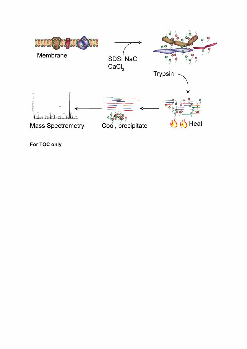
For TOC only