Embed Size (px)
Citation preview
Effects of the gut microbiota on host adiposityare modulated by the short-chain fatty-acidbinding G protein-coupled receptor, Gpr41Buck S. Samuel*†, Abdullah Shaito†‡, Toshiyuki Motoike§, Federico E. Rey*, Fredrik Backhed*¶, Jill K. Manchester*,Robert E. Hammer�, S. Clay Williams**, Jan Crowley††, Masashi Yanagisawa‡§**‡‡, and Jeffrey I. Gordon*‡‡
*Center for Genome Sciences and ††Department of Medicine, Washington University School of Medicine, St. Louis, MO 63108; Departments of ‡Biophysicsand Molecular Genetics, and �Biochemistry, University of Texas Southwestern Medical Center, Dallas, TX 75390; §Yanagisawa Orphan Receptor Project,Exploratory Research for Advanced Technology, Japan Science and Technology Agency, Tokyo 135-0064, Japan; and **Howard Hughes Medical Institute,4000 Jones Bridge Road, Chevy Chase, MD 20815-6789
Contributed by Jeffrey I. Gordon, August 29, 2008 (sent for review July 2, 2008)
The distal human intestine harbors trillions of microbes that allow usto extract calories from otherwise indigestible dietary polysaccha-rides. The products of polysaccharide fermentation include short-chain fatty acids that are ligands for Gpr41, a G protein-coupledreceptor expressed by a subset of enteroendocrine cells in the gutepithelium. To examine the contribution of Gpr41 to energy balance,we compared Gpr41�/� and Gpr41�/� mice that were either con-ventionally-raised with a complete gut microbiota or were rearedgerm-free and then cocolonized as young adults with two prominentmembers of the human distal gut microbial community: the saccha-rolytic bacterium, Bacteroides thetaiotaomicron and the methano-genic archaeon, Methanobrevibacter smithii. Both conventionally-raised and gnotobiotic Gpr41�/� mice colonized with the modelfermentative community are significantly leaner and weigh less thantheir WT (�/�) littermates, despite similar levels of chow consump-tion. These differences are not evident when germ-free WT andgerm-free Gpr41 knockout animals are compared. Functionalgenomic, biochemical, and physiologic studies of germ-free andcocolonized Gpr41�/� and �/� littermates disclosed that Gpr41-deficiency is associated with reduced expression of PYY, an enteroen-docrine cell-derived hormone that normally inhibits gut motility,increased intestinal transit rate, and reduced harvest of energy(short-chain fatty acids) from the diet. These results reveal that Gpr41is a regulator of host energy balance through effects that are depen-dent upon the gut microbiota.
host-microbial interactions � energy balance �enteroendocrine cells � nutrient sensing � polysaccharide fermentation
Our ability to effectively digest food reflects the combinedactivities of enzymes encoded in our primate genome and in
the genomes of the trillions of microbes that reside in our distal guts.This microbial community, or microbiota, affects both sides of theenergy-balance equation, influencing both the harvest of caloriesand the activity of host genes involved in the metabolism andstorage of absorbed energy (e.g., ref. 1).
Our proteome has a very limited repertoire of glycosidehydrolases needed to digest complex dietary plant polysaccha-rides: the microbiota synthesizes a large arsenal of these enzymes(2), and allows us to process complex dietary carbohydrates toshort-chain fatty acids (SCFAs), principally acetate, propionate,and butyrate. Host recovery of SCFAs is generally efficient andoccurs by both passive diffusion and via mono-carboxylic acidtransporters (e.g., MCT1 in the case of butyrate and lactate) (3).Butyrate is the preferred source of energy for colonic epithelialcells. Absorbed acetate and propionate are delivered to hepa-tocytes, which consume most of the propionate for gluconeo-genesis. Although acetate can be used for lipogenesis in colono-cytes, hepatocytes and adipocytes are the principal sites for denovo lipogenesis, at least in rodents.
Studies in gnotobiotic mice have emphasized the contributionsof the gut microbiota and microbial fermentation of dietarypolysaccharides to host energy balance. Adult germ-free (GF)mice are leaner than their age- and gender-matched conven-tionally raised (CONV-R) counterparts who have acquired amicrobiota beginning at birth (1). Transplantation of an unfrac-tionated gut microbiota from a CONV-R donor to an adult GFrecipient results in an increase in adiposity. This increase isgreater if the donors are obese because they are homozygous fora null allele in the leptin gene (ob/ob), or if they have diet-induced obesity (4, 5). Comparative metagenomic studies ofdistal gut (cecal) microbial community DNA prepared frommice with either form of obesity and from lean controls, haveshown that the obesity-associated microbiomes have a greatercapacity to ferment carbohydrates to SCFAs (4, 5). In addition,colonization of adult GF mice, fed a standard polysaccharide-rich chow diet, with two organisms—Bacteroides thetaiotaomi-cron, a prominent saccharolytic bacterium in the normal distalhuman gut microbiota and an adept adaptive forager of poly-saccharides (6), plus Methanobrevibacter smithii, a dominantmethanogenic archaeon in this community (7) that promotespolysaccharide fermentation by removing the H2 end product—results in higher levels of SCFAs in the colon, and significantlygreater host adiposity than colonization of GF animals witheither organism alone (8).
SCFAs also act as signaling molecules. Propionate, acetate, andto a lesser extent butyrate and pentanoate, are ligands for at leasttwo G protein-coupled receptors (GPCRs), Gpr41 and Gpr43(9–11). Both GPCRs are broadly expressed, including in the distalsmall intestine, colon, and adipocytes (9–11). SCFAs (C1-C6),which are ligands for Gpr41, stimulate expression of leptin, apolypeptide hormone with pleiotropic effects on appetite andenergy metabolism, in mouse-cultured adipocytes (11). However,little is known about the regulation of these GPCRs, their mech-anism of action, and whether they represent a class of molecules,strategically placed in certain gut epithelial cell lineages that sensethe biochemical milieu of the gut and ‘‘transduce’’ information
Author contributions: B.S.S., A.S., M.Y., and J.I.G. designed research; B.S.S., A.S., T.M., F.E.R.,F.B., J.K.M., R.E.H., S.C.W., and J.C. performed research; B.S.S., A.S., T.M., F.E.R., M.Y., andJ.I.G. analyzed data; and B.S.S., A.S., F.E.R., M.Y., and J.I.G. wrote the paper.
The authors declare no conflict of interest.
Freely available online through the PNAS open access option.
†B.S.S. and A.S. contributed equally.
¶Present address: Center for Cardiovascular and Metabolic Research, Wallenberg Labora-tory, Sahlgrenska University Hospital, SE 413 45 Goteborg, Sweden.
‡‡To whom correspondence may be addressed: E-mail: [email protected] [email protected].
This article contains supporting information online at www.pnas.org/cgi/content/full/0808567105/DCSupplemental.
© 2008 by The National Academy of Sciences of the USA
www.pnas.org�cgi�doi�10.1073�pnas.0808567105 PNAS � October 28, 2008 � vol. 105 � no. 43 � 16767–16772
MIC
ROBI
OLO
GY
about key metabolic activities of the microbiota, such as polysac-charide fermentation, in ways that impact host physiology, includingenergy balance.
In this report, we compare GF wild-type and Gpr41-deficientmice with and without a model fermentative microbial commu-nity composed of B. thetaiotaiomicron (Bt) and M. smithii (Ms).The results reveal a pivotal role for Gpr41 in a microbiota-dependent metabolic circuit that regulates the flow of caloriesbetween the diet and the host. Our results suggest that inhibitionof SCFA activation of Gpr41 is a potential therapeutic target formodulating the efficiency of caloric extraction from a polysac-charide-rich diet.
Results and DiscussionGpr41 Is Expressed in Enteroendocrine Cells. Analysis of the tissuedistribution of Gpr41 mRNA in CONV-R adult mice indicatedthat highest levels are present in the distal small intestine (ileum)and colon [supporting information (SI) Fig. S1]. Enteroendo-crine cells are strategically positioned to transduce informationabout the nutrient milieu of the gut and the metabolic activity ofthe microbiota to the host: they produce different sets of peptidehormones, depending upon their location along the length of thegastrointestinal tract (12). These neuroactive and endocrinefactors are secreted basolaterally into the portal and systemiccirculation where they influence a wide variety of extraintestinalphysiological activities.
In situ hybridization studies indicated that Gpr41 is expressedin cells with the morphologic appearance of enteroendocrinecells (Fig. S2 A). Cholecystokinin (CCK) is a known biomarkerof this gut epithelial-cell lineage. Therefore, we used flow-assisted cell sorting (FACS) to purify CCK-positive cells fromthe small intestines of CONV-R transgenic mice engineered toexpress GFP in this enteroendocine subpopulation (Fig. S2 Band C). qRT-PCR assays of the expression of Gpr41 and sevenother known enteroendocrine biomarkers in the crude startingmaterial and in the FACS-purified population confirmed thatGpr41 is expressed in this enteroendocrine subset (Fig. S2 D andE). A similar approach was used in different pedigrees oftransgenic mice engineered to express GFP in NeuroD- andNeurogenin3-producing enteroendocrine subpopulations toshow that Gpr41 is also localized to these cells (data not shown).Finally, intraepithelial lymphocytes, which have some of themorphologic features of enteroendocrine cells when viewed bylight microscopy, were purified using a T-cell antibody plusmagnetic bead sorting (see SI Materials and Methods): qRT-PCRestablished that they do not express appreciable levels of thisGPCR (Fig. S2F).
Microbial Suppression of Gpr41 Expression. Ligand-induced downregulation is a hallmark of GPCR activation (13). Therefore, wegenerated Gpr41�/� mice (Fig. S3 A and B), rederived them asGF, and examined whether colonization of 8- to 10-week-oldmale GF knockout (Gpr41�/�) mice and their WT (�/�)littermates (mixed C57Bl6/J:129/Sv background) for 28 days withBt and Ms (see Methods) affected ileal expression of Gpr41, orthree other known GCPRs that bind fatty-acid ligands: Gpr43,which, as noted above, is also responsive to short-chain (C2-C6)fatty acids, plus Gpr40 and Gpr120, which recognize ligands withlonger (�C6) acyl chains (14–16).
Quantitative PCR assays established that levels of colonization ofthe distal gut (cecum) with each microbial species were not signif-icantly affected by the presence or absence of Gpr41 (mean 8.2 �4.3 � 1012 organisms per gram of luminal contents for B. thetaio-taomicron; 2.4 � 1.5 � 106 for M. smithii; n, 7–8 mice per genotype).Therefore, any phenotypic differences observed between gnotobi-otic WT and knockout animals could not be attributed to differ-ences in their gut microbial ecology.
qRT-PCR assays of ileal RNAs revealed that compared with
GF �/� controls, cocolonization of WT mice produced statis-tically significant twofold reductions in the steady state levels ofGpr41, Gpr43, and Gpr120 mRNAs (P � 0.05; ANOVA) (Fig.S4). Expression of Gpr40 mRNA in �/� mice was also reducedby colonization, although the observed change did not quiteachieve statistical significance (see Fig. S4). Importantly, themagnitude of the reduction in Gpr40, Gpr43, and Gpr120 geneexpression was not affected by the absence of Gpr41 (see Fig.S4).
Together, these findings indicate that Gpr41�/� mice have aspecific deficiency affecting only one of these four fatty-acidbinding GPCRs and therefore can, in principle, be used to assessthe role of Gpr41 in mediating the effects of the microbiota onhost energy homeostasis.
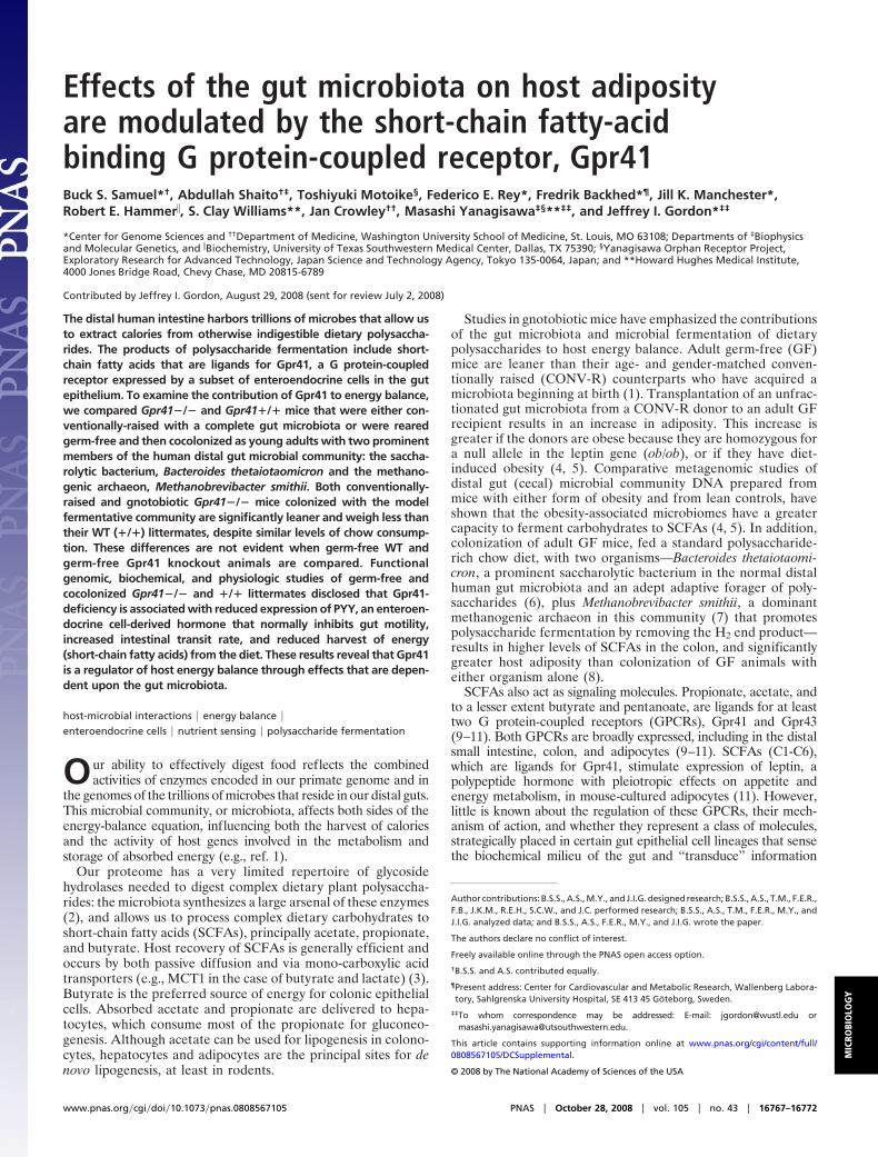
Gpr41 Is Needed for Microbiota-Induced Increases in Host Adiposity.Eight to ten-week-old male GF Gpr41�/� mice, maintained ona standard polysaccharide-rich chow diet, exhibited no signifi-cant differences in their epididymal fat pad or total body weightscompared with �/� littermates (P � 0.05; n � 8–14 per group)(Fig. 1A and data not shown). In contrast, Gpr41�/� micecocolonized with Bt/Ms had significantly lower epididymal fatpad weights than �/� controls (11.4 � 0.6 versus 14.4 � 0.9 mg/gbody weight, respectively; P � 0.05) (see Fig. 1 A), gainedsignificantly less body weight per day (0.08 � 0.03 versus 0.19 �0.02 g/day, respectively; P � 0.05) (Fig. 1B), and weighedsignificantly less at the end of the 28-day colonization period(24 � 0.4g versus 26 � 0.4g; P � 0.05) (n, 13 to 14 animals pergroup, representing two independent experiments).
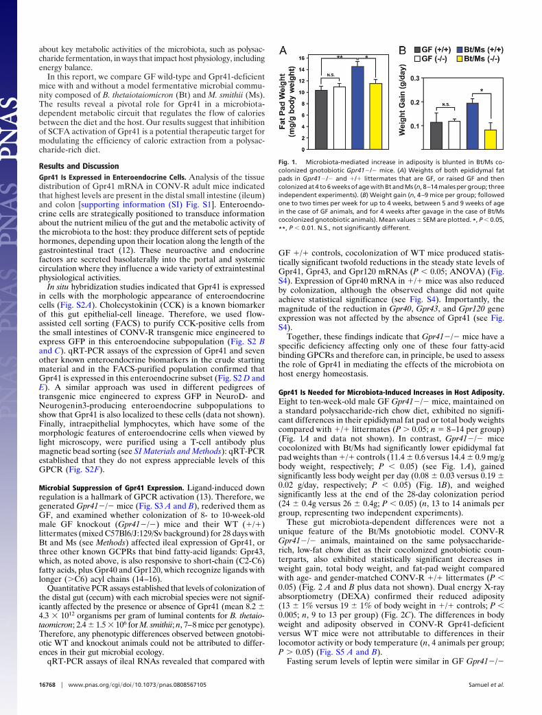
These gut microbiota-dependent differences were not aunique feature of the Bt/Ms gnotobiotic model. CONV-RGpr41�/� animals, maintained on the same polysaccharide-rich, low-fat chow diet as their cocolonized gnotobiotic coun-terparts, also exhibited statistically significant decreases inweight gain, total body weight, and fat-pad weight comparedwith age- and gender-matched CONV-R �/� littermates (P �0.05) (Fig. 2 A and B plus data not shown). Dual energy X-rayabsorptiometry (DEXA) confirmed their reduced adiposity(13 � 1% versus 19 � 1% of body weight in �/� controls; P �0.005; n, 9 to 13 per group) (Fig. 2C). The differences in bodyweight and adiposity observed in CONV-R Gpr41-deficientversus WT mice were not attributable to differences in theirlocomotor activity or body temperature (n, 4 animals per group;P � 0.05) (Fig. S5 A and B).
Fasting serum levels of leptin were similar in GF Gpr41�/�
Fig. 1. Microbiota-mediated increase in adiposity is blunted in Bt/Ms co-colonized gnotobiotic Gpr41�/� mice. (A) Weights of both epididymal fatpads in Gpr41�/� and �/� littermates that are GF, or raised GF and thencolonized at 4 to 6 weeks of age with Bt and Ms (n, 8–14 males per group; threeindependent experiments). (B) Weight gain (n, 4–9 mice per group; followedone to two times per week for up to 4 weeks, between 5 and 9 weeks of agein the case of GF animals, and for 4 weeks after gavage in the case of Bt/Mscocolonized gnotobiotic animals). Mean values � SEM are plotted. *, P � 0.05,
**, P � 0.01. N.S., not significantly different.
16768 � www.pnas.org�cgi�doi�10.1073�pnas.0808567105 Samuel et al.
and �/� littermates, but significantly lower in Bt/Ms cocolo-nized and CONV-R Gpr41�/� animals (P � 0.05; n, 5–7 miceper genotype per treatment group) (Table S1). Moreover, serumleptin levels were significantly lower in CONV-R Gpr41�/�animals than would be expected based solely on the observeddecrease in their adiposity (P � 0.02) (see Fig. 2 D and E).Together, these findings implicate Gpr41 in microbiota-dependent regulation of host adiposity and leptin production.
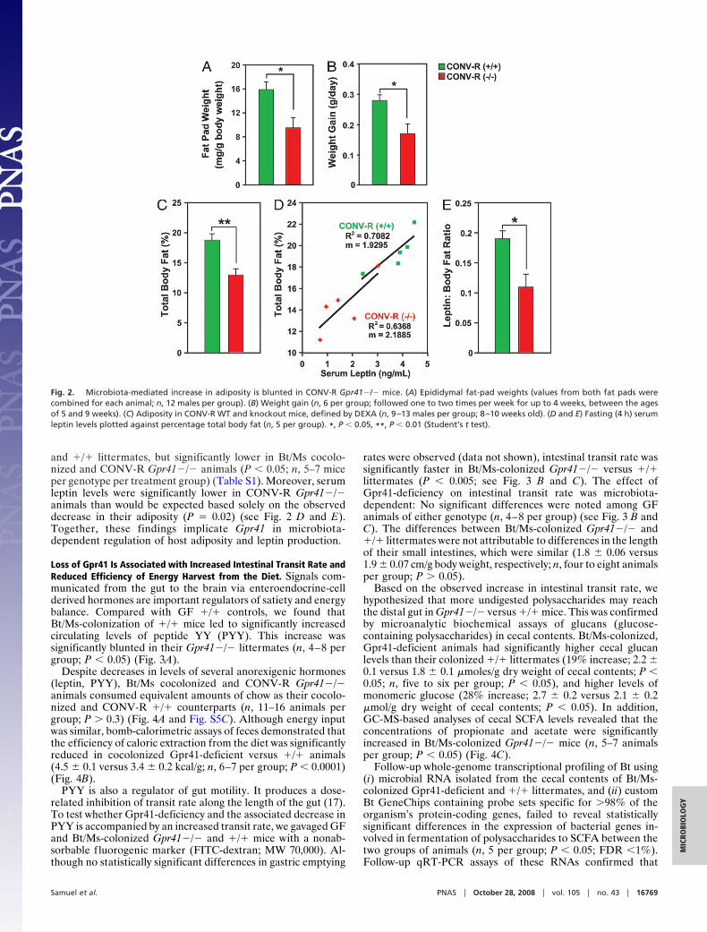
Loss of Gpr41 Is Associated with Increased Intestinal Transit Rate andReduced Efficiency of Energy Harvest from the Diet. Signals com-municated from the gut to the brain via enteroendocrine-cellderived hormones are important regulators of satiety and energybalance. Compared with GF �/� controls, we found thatBt/Ms-colonization of �/� mice led to significantly increasedcirculating levels of peptide YY (PYY). This increase wassignificantly blunted in their Gpr41�/� littermates (n, 4–8 pergroup; P � 0.05) (Fig. 3A).
Despite decreases in levels of several anorexigenic hormones(leptin, PYY), Bt/Ms cocolonized and CONV-R Gpr41�/�animals consumed equivalent amounts of chow as their cocolo-nized and CONV-R �/� counterparts (n, 11–16 animals pergroup; P � 0.3) (Fig. 4A and Fig. S5C). Although energy inputwas similar, bomb-calorimetric assays of feces demonstrated thatthe efficiency of caloric extraction from the diet was significantlyreduced in cocolonized Gpr41-deficient versus �/� animals(4.5 � 0.1 versus 3.4 � 0.2 kcal/g; n, 6–7 per group; P � 0.0001)(Fig. 4B).
PYY is also a regulator of gut motility. It produces a dose-related inhibition of transit rate along the length of the gut (17).To test whether Gpr41-deficiency and the associated decrease inPYY is accompanied by an increased transit rate, we gavaged GFand Bt/Ms-colonized Gpr41�/� and �/� mice with a nonab-sorbable fluorogenic marker (FITC-dextran; MW 70,000). Al-though no statistically significant differences in gastric emptying
rates were observed (data not shown), intestinal transit rate wassignificantly faster in Bt/Ms-colonized Gpr41�/� versus �/�littermates (P � 0.005; see Fig. 3 B and C). The effect ofGpr41-deficiency on intestinal transit rate was microbiota-dependent: No significant differences were noted among GFanimals of either genotype (n, 4–8 per group) (see Fig. 3 B andC). The differences between Bt/Ms-colonized Gpr41�/� and�/� littermates were not attributable to differences in the lengthof their small intestines, which were similar (1.8 � 0.06 versus1.9 � 0.07 cm/g body weight, respectively; n, four to eight animalsper group; P � 0.05).
Based on the observed increase in intestinal transit rate, wehypothesized that more undigested polysaccharides may reachthe distal gut in Gpr41�/� versus �/� mice. This was confirmedby microanalytic biochemical assays of glucans (glucose-containing polysaccharides) in cecal contents. Bt/Ms-colonized,Gpr41-deficient animals had significantly higher cecal glucanlevels than their colonized �/� littermates (19% increase; 2.2 �0.1 versus 1.8 � 0.1 �moles/g dry weight of cecal contents; P �0.05; n, five to six per group; P � 0.05), and higher levels ofmonomeric glucose (28% increase; 2.7 � 0.2 versus 2.1 � 0.2�mol/g dry weight of cecal contents; P � 0.05). In addition,GC-MS-based analyses of cecal SCFA levels revealed that theconcentrations of propionate and acetate were significantlyincreased in Bt/Ms-colonized Gpr41�/� mice (n, 5–7 animalsper group; P � 0.05) (Fig. 4C).
Follow-up whole-genome transcriptional profiling of Bt using(i) microbial RNA isolated from the cecal contents of Bt/Ms-colonized Gpr41-deficient and �/� littermates, and (ii) customBt GeneChips containing probe sets specific for �98% of theorganism’s protein-coding genes, failed to reveal statisticallysignificant differences in the expression of bacterial genes in-volved in fermentation of polysaccharides to SCFA between thetwo groups of animals (n, 5 per group; P � 0.05; FDR �1%).Follow-up qRT-PCR assays of these RNAs confirmed that
Fig. 2. Microbiota-mediated increase in adiposity is blunted in CONV-R Gpr41�/� mice. (A) Epididymal fat-pad weights (values from both fat pads werecombined for each animal; n, 12 males per group). (B) Weight gain (n, 6 per group; followed one to two times per week for up to 4 weeks, between the agesof 5 and 9 weeks). (C) Adiposity in CONV-R WT and knockout mice, defined by DEXA (n, 9–13 males per group; 8–10 weeks old). (D and E) Fasting (4 h) serumleptin levels plotted against percentage total body fat (n, 5 per group). *, P � 0.05, **, P � 0.01 (Student’s t test).
Samuel et al. PNAS � October 28, 2008 � vol. 105 � no. 43 � 16769
MIC
ROBI
OLO
GY
several key genes in the pathway involved in SCFA production,including pyruvate formate lyase (BT4738; EC 2.3.1.54), andacetate kinase (BT3963; EC 2.7.2.1) exhibited no significantdifferences in their expression between Bt/Ms colonizedGrp41�/� and �/� mice (data not shown).
GC-MS analysis of feces indicated that Bt/Ms-colonizedGpr41�/� animals had a statistically significant (37 � 9%) increasein total SCFAs compared with their �/� littermates (n, 5–7 pergroup; P � 0.05) (Fig. 4D). In contrast, fecal levels of free C6-C18fatty acids (FFA) and triglycerides were not significantly differentbetween the two groups of mice [0.6 � 0.1 versus 0.7 � 0.1 mg/gweight of feces (FFA), and 1.9 � 0.2 versus 1.9 � 0.1 mg/g weightof feces (triglycerides); n � seven to eight animals assayed pergroup; P � 0.05]. qRT-PCR assays of distal small intestinal (ileal)RNA (SI Methods and Table S2) indicated that there were nosignificant differences in expression of key host genes involved inthe active uptake or transepithelial transport of lipids [e.g., Mct1(mono-carboxylate transporter), CD36 (lipid-binding glycoprotein)or ApoB (chylomicron-mediated transport); data not shown]. Thesefindings are not surprising, given that the majority of long-chainfatty acids are absorbed in the proximal intestine.
We reasoned that the observed increase in fecal SCFAs reflects,at minimum, reduced host-passive absorption. SCFAs stimulate,and are substrates for, de novo lipogenesis in the liver. Bt/Ms-colonization of GF �/� animals resulted in statistically significantincreases in liver triglyceride levels (n, seven to eight per group; P �0.05) (Fig. S6A). This effect of colonization was not seen inGpr41�/� animals, nor were any differences observed between GFGpr41�/� and �/� mice (n, 7–8 per group; P � 0.05) (see Fig.
S6A). qRT-PCR assays confirmed reduced expression of fatty acidsynthase (Fas) in the livers of Bt/Ms-colonized Gpr41�/� micecompared to their �/� littermates (77 � 12% lower; n, 5–7 pergroup; P � 0.01) (Fig. S6B). In addition, fasting (4 h) serumtriglyceride levels were decreased in Bt/Ms-colonized Gpr41-deficient versus �/� animals (n, 7–8 per group) (see Table S1).These differences were not attributable to alterations in hepaticexpression of genes involved in long-chain fatty-acid transport,trafficking, or fatty-acid reesterification (data not shown). To-gether, our results indicate that Gpr41-deficient mice have reducedhepatic lipogenesis, consistent with reduced intestinal absorptionand delivery of SCFAs.
Prospectus. A complete picture of how digestive physiology isregulated requires an understanding of how products generated bythe microbiota interact with host nutrient sensors to modulatenutrient and energy flux. The data presented above indicate thatGpr41 mediates a key microbial-host communication circuit. Ex-trapolating from the phenotype of Gpr41-deficient mice, we positthat at least one feature of the interaction between SCFA productsof microbial fermentation of dietary polysaccharides and Gpr41 isto increase circulating levels of enteroendocrine cell-derived hor-mones that reduce gut motility and thus increase absorption ofSCFAs, which are used as substrates for lipogenesis in the liver.Analysis of these Gpr41-deficient mice suggests that an inhibitor ofGpr41 activation could result in decreased extraction of energyfrom the diet and leaner individuals.
Materials and MethodsGeneration of Gpr41 Knockout Mice. A 129/SV mouse BAC clone obtained fromChildren’s Hospital Oakland Research Institute was used to construct the
Fig. 3. Increased rate of intestinal transit in Bt/Ms colonized gnotobiotic Gpr41�/� mice. (A) Fasting serum PYY levels in GF and Bt/Ms cocolonized, Gpr41�/�and �/� mice (n � four to eight per group; two independent experiments; all samples assayed in duplicate). (B and C) Gut transit time measured by oral gavageof a fluorescently labeled nonabsorbable substrate (fluorescine isothiocyanate-dextran; 70,000 MW) in GF and cocolonized WT and Gpr41-deficient animals. (Band C) Distribution of fluorescence signal intensity 60 min after gavage along the length of the gut; data are plotted as a fraction of total fluorescence in B. Nosignal was observed in the ceca or colons in any of the animals. Panel (C) presents the calculated geometric center of the fluorescence (n � 4–8 animals analyzedper group). *, P � 0.05, **, P � 0.01, ***, P � 0.005.
16770 � www.pnas.org�cgi�doi�10.1073�pnas.0808567105 Samuel et al.

targeting vector shown in Fig. S3A. SM-1 ES cells (18), cultured on irradiatedLIF-producing STO feeder layers, were electroporated with the linearizedtargeting vector and selected for resistance to G418 (18). Resistant ES cellclones were screened by Southern blotting using a flanking 3� genomicfragment external to the targeting vector (see Fig. S3B). Two of these ES cellclones were microinjected into C57BL/6 blastocysts to produce germline trans-mitting chimeric mice. PCR genotyping used the primer set 5�-CACACTGCTC-GATCCGGAACCCTT and 5�-GAGAACTGTCTGGAAAACGCTCAC to identify themutant Gpr41 allele, and 5�-CGACGCCCAGTGGCTGTGGACTTA and 5�-GTACCACAGTGGATAGGCCACGC to detect the WT allele. This PCR genotypingprotocol was validated by Southern blotting (see Fig. S3B).
Mice were provided with food and water ad libitum and maintained on a strict12-h light–dark cycle. All procedures involving genetically engineered mice usedin this study were approved by the Institutional Review Board for Animal Re-search of the University of Texas Southwestern Medical Center at Dallas.
Husbandry of Gnotobiotic Mice. Gpr41�/� mice and their �/� littermates(mixed C57BL/6J:129/Sv background) were rederived as GF and housed inflexible film plastic gnotobiotic isolators (19), where they were maintainedon a strict 12-h light cycle (lights on at 0600 h) and fed a standardautoclaved polysaccharide-rich chow diet (B and K Universal) ad libitum.Four- to six-week-old male mice were inoculated with a single gavage of108 CFU of the sequenced human gut-derived B. thetaiotaomicron strainVPI-5482 [harvested from overnight culture in TYG medium (6)], and M.
smithii strain PS [cultured at 37°C for 5 d in 125-ml serum bottles containing15 ml of MBC medium (8) supplemented with 3 g/L formate, 3 g/L acetate,and 0.3 ml of a freshly prepared oxygen-free solution of filter-sterilized2.5% Na2S (note that the remaining volume or headspace in the bottlecontained a 4:1 mixture of H2 and CO2, and the headspace was rejuvenatedevery 1 to 2 d) (8)]. All colonized mice were killed 28 d after gavage. Thedensity of colonization was determined in cecal contents using qPCR assaysthat used species-specific primers (8). Age-matched, male CONV-R WT andGpr41-deficient male mice were also fed the same autoclaved polysaccha-ride-rich chow diet ad libitum as the gnotobiotic animals. All experimentsperformed with gnotobiotic mice used protocols approved by the Wash-ington University Animal Studies Committee.
Analysis of Host Adiposity and Energy Harvest. All mice were fasted (4 h) beforebeing killed. Epididymal fat pads, livers, and segments of the distal intestine(ileum) and colon were removed and flash-frozen in liquid nitrogen. Epidid-ymal fat pad and liver weights were recorded before freezing.
Before killing, total body fat content was measured by DEXA (Lunar PIXI-mus Mouse, GE Medical Systems), 5 min after mice had been anesthesized withan intraperitoneal injection of ketamine (10 mg/kg body weight) and xylazine(10 mg/kg). Weight gain and chow consumption were monitored weekly inmice who were individually caged for the duration of the experiment. Theenergy content of fecal samples (freeze-dried immediately after collection)was defined using a bomb-calorimeter (Parr Instruments) and previouslyestablished methods (8).
Measurement of Physiological Parameters. Locomotor activity and body tem-perature were assessed for 5 d using a telemetry device (minimitter PDT-4000;Mini Mitter) beginning 7 d after implantation (20). Locomotor activity datawere processed using VitalView software (Mini Mitter).
Gastric emptying and gastrointestinal transit time was measured in GFand Bt/Ms-colonized Gpr41�/� and �/� littermates using establishedmethods, after an 18-h overnight fast (21, 22). FITC-labeled dextran (70,000MW; Molecular Probes) was administered by gavage (100 �l of a 5-mg/mlsolution prepared in PBS). Sixty minutes later, the entire GI tract fromstomach to rectum was removed and placed in ice-cold PBS for 30 s toinhibit motility. The stomach, small intestine (divided into 10 equal lengthsegments), cecum, and colon (subdivided into two equal-length segments)were each placed in a separate tube containing 1 ml of PBS (5 ml forstomach and cecum). The segments were coarsely chopped with scissors,and luminal contents suspended using a combination of vigorous washingand vortexing. A dilution series was completed for each sample (1:10 to1:1000 in PBS) and the fluorescent signal quantified in a multiwell fluores-cence plate reader (Stratagene Mx3000; excitation at 485 nm; emission at530 nm). A histogram of the fluorescence signal distributed along thegastrointestinal tract was then plotted and the geometric center deter-mined (SUM [% of total fluorescence per segment � segment number])/100) (23). Gastric emptying was calculated based on the amount of FITC-dextran left in the stomach compared with the total amount offluorescence in the intestine.
The methods used for measurement of serum proteins, cecal, and fecalSCFAs, plus triglycerides are described in the SI Materials and Methods.
Statistical Analysis. Unless otherwise noted, the significance of differencesnoted among different groups of mice was defined using ANOVA and Tukey’sposthoc test.
ACKNOWLEDGMENTS. The authors thank Sabrina Wagoner, DavidO’Donnell, Maria Karlsson, Trey Coleman, Shelley Dixon, Randal Floyd, AmberSkach, and Marcus Thornton for technical assistance; Peter Turnbaugh, PeterCrawford, and Robert Heuckeroth for helpful advice; and Laura Kyro forgraphics support. This work was supported in part by Graduate ResearchFellowship DGE-0202737 from the National Science Foundation (to B.S.S.), byGrants DK70977 and DK30292 from the National Institutes of Health, and bygrants from the W.M. Keck Foundation, Exploratory Research for AdvancedTechnology program of the Japan Science and Technology Agency, and TheHoward Hughes Medical Institute.
1. Backhed F, et al. (2004) The gut microbiota as an environmental factor that regulatesfat storage. Proc Natl Acad Sci USA 101:15718–15723.
2. Flint HJ, et al. (2008) Polysaccharide utilization by gut bacteria: Potential for newinsights from genomic analysis. Nature Rev Microb 6:121–131.
3. Ritzhaupt A, Wood IS, Ellis A, Hosie KB, Shirazi-Beechey SP (1998) Identification andcharacterization of a monocarboxylate transporter (MCT1) in pig and humancolon: Its potential to transport L-lactate as well as butyrate. J Physiol 513:719 –732.
4. Turnbaugh PJ, et al. (2006) An obesity-associated gut microbiome with increasedcapacity for energy harvest. Nature 444:1027–1031.
5. Turnbaugh P, Gordon JI (2008) Diet-induced obesity is linked to marked but reversiblealterations in the mouse distal gut microbiome. Cell Host Microbe 17:213–223.
6. Sonnenburg JL, et al. (2005) Glycan foraging in vivo by an intestine-adapted bacterialsymbiont. Science 307:1955–1959.
7. Eckburg PB, et al. (2005) Diversity of the human intestinal microbial flora. Science308:1635–1638.
Fig. 4. Gnotobiotic Gpr41�/� mice extract fewer calories from their po-lysaccharide-rich chow diet and excrete more SCFAs. (A) Dietary energy intakein GF and Bt/Ms cocolonized �/� and Gpr41�/� littermates (calories of chowconsumed per day monitored concurrently with body weight, as in Fig. 1). (B)Bomb calorimetry-based assessment of remaining calories in the feces ofcolonized (Bt/Ms) Gpr41�/� and �/� mice (n � 6–7 per group). (C) GC-MSassay of cecal SCFA levels (n � 5–7 per group; each sample assayed in dupli-cate). (D) GC-MS study of fecal SFCAs from the same mice assayed in (C). *,P �0.05, **, P � 0.01, ***, P � 0.005. (Student’s t test used for comparisons of twogroups.)
Samuel et al. PNAS � October 28, 2008 � vol. 105 � no. 43 � 16771
MIC
ROBI
OLO
GY
8. Samuel BS, Gordon JI (2006) A humanized gnotobiotic mouse model of host-archaeal-bacterial mutualism. Proc Natl Acad Sci USA 103:10011–10016.
9. Brown AJ, et al. (2003) The orphan G protein-coupled receptors GPR41 and GPR43 areactivated by propionate and other short chain carboxylic acids. J Biol Chem 278:11312–11319.
10. Le Poul E, et al (2003) Functional characterization of human receptors for short chainfatty acids and their role in polymorphonuclear cell activation. J Biol Chem 278:25481–25489.
11. Xiong Y, et al. (2004) Short-chain fatty acids stimulate leptin production in adipo-cytes through the G protein-coupled receptor GPR41. Proc Natl Acad Sci USA101:1045–1050.
12. Roth KA, Hertz JW, Gordon JI (1990) Mapping enteroendocrine cell populations intransgenic mice reveals an unexpected degree of complexity in cellular differentiationwithin the gastrointestinal tract. J Cell Biol 110:1791–1801.
13. Bunemann M, Hosey MM (1999) G-protein coupled receptor kinases as modulators ofG-protein signaling. J Physiol 517:5–23.
14. Briscoe CP, et al. (2003) The orphan G protein-coupled receptor GPR40 is activated bymedium and long chain fatty acids. J Biol Chem 278:11303–11311.
15. Hirasawa A, et al. (2005) Free fatty acids regulate gut incretin glucagon-like peptide-1secretion through GPR120. Nat Med 11:90–94.
16. Itoh Y, et al. (2003) Free fatty acids regulate insulin secretion from pancreatic beta cellsthrough GPR40. Nature 422:173–176.
17. Lin HC, Neevel C, Chen JH (2004) Slowing intestinal transit by PYY depends onserotonergic and opioid pathways. Am J Physiol 286:G558–G563.
18. Yanagisawa H, et al. (1998) Dual genetic pathways of endothelin-mediated intercel-lular signaling revealed by targeted disruption of endothelin converting enzyme-1gene. Development 125 825–836.
19. Hooper LV, et al. (2002) Mol Cell Microbiol, eds Sansonetti P, Zychlinsky A. (Academic,San Diego, CA), pp 559–589.
20. Backhed F, Manchester JK, Semenkovich CF, Gordon JI (2007) Mechanisms underlyingthe resistance to diet-induced obesity in germ-free mice. Proc Natl Acad Sci USA104:979–984.
21. Moore BA, Otterbein LE, Turler A, Choi AM, Bauer AJ (2003) Inhaled carbon monoxidesuppresses the development of postoperative ileus in the murine small intestine.Gastroenterol 124:377–391.
22. Wehner S, et al. (2007) Inhibition of macrophage function prevents intestinal inflam-mation and postoperative ileus in rodents. Gut 56:176–185.
23. Miller MS, Galligan JJ, Burks TF (1981) Accurate measurement of intestinal transit in therat. J Pharmacol Methods 6:211–217.
16772 � www.pnas.org�cgi�doi�10.1073�pnas.0808567105 Samuel et al.