Embed Size (px)
Citation preview

Effects of technique variations on kneebiomechanics during the squat and leg press
RAFAEL F. ESCAMILLA, GLENN S. FLEISIG, NAIQUAN ZHENG, JEFFERY E. LANDER,STEVEN W. BARRENTINE, JAMES R. ANDREWS, BRIAN W. BERGEMANN, and CLAUDE T. MOORMAN, III
Michael W. Krzyzewski Human Performance Laboratory, Division of Orthopaedic Surgery and Duke Sports Medicine,Duke University Medical Center, Durham, NC 27710; American Sports Medicine Institute, Birmingham, AL 35205;Department of Sports Health Science, Life University, Marietta, GA 30060; and Department of Exercise Science,Campbell University, Buies Creek, NC 27506
ABSTRACT
ESCAMILLA, R. F., G. S. FLEISIG, N. ZHENG, J. E. LANDER, S. W. BARRENTINE, J. R. ANDREWS, B. W. BERGEMANN,and C. T. MOORMAN, III. Effects of technique variations on knee biomechanics during the squat and leg press. Med. Sci. SportsExerc., Vol. 33, No. 9, 2001, pp. 1552–1566. Purpose: The specific aim of this project was to quantify knee forces and muscle activitywhile performing squat and leg press exercises with technique variations. Methods: Ten experienced male lifters performed the squat,a high foot placement leg press (LPH), and a low foot placement leg press (LPL) employing a wide stance (WS), narrow stance (NS),and two foot angle positions (feet straight and feet turned out 30°). Results: No differences were found in muscle activity or knee forcesbetween foot angle variations. The squat generated greater quadriceps and hamstrings activity than the LPH and LPL, the WS-LPHgenerated greater hamstrings activity than the NS-LPH, whereas the NS squat produced greater gastrocnemius activity than the WSsquat. No ACL forces were produced for any exercise variation. Tibiofemoral (TF) compressive forces, PCL tensile forces, andpatellofemoral (PF) compressive forces were generally greater in the squat than the LPH and LPL, and there were no differences inknee forces between the LPH and LPL. For all exercises, the WS generated greater PCL tensile forces than the NS, the NS producedgreater TF and PF compressive forces than the WS during the LPH and LPL, whereas the WS generated greater TF and PF compressiveforces than the NS during the squat. For all exercises, muscle activity and knee forces were generally greater in the knee extendingphase than the knee flexing phase. Conclusions: The greater muscle activity and knee forces in the squat compared with the LPL andLPH implies the squat may be more effective in muscle development but should be used cautiously in those with PCL and PF disorders,especially at greater knee flexion angles. Because all forces increased with knee flexion, training within the functional 0–50° range maybe efficacious for those whose goal is to minimize knee forces. The lack of ACL forces implies that all exercises may be effective duringACL rehabilitation. Key Words: POWERLIFTING, KINETICS, PATELLOFEMORAL, TIBIOFEMORAL, ACL, PCL, COMPRES-SIVE, SHEAR, REHABILITATION, FORCE, MUSCLE ACTIVITY, EMG
The dynamic squat and leg press (LP) exercises arecommon core exercises that are utilized by athletes toenhance performance in sport. These multi-joint exer-
cises develop the largest and most powerful muscles of thebody and have biomechanical and neuromuscular similaritiesto many athletic movements, such as running and jumping.Because the squat and LP are considered closed kinetic chainexercises (11,34), they are often recommended and utilized inclinical environments, such as during knee rehabilitation afteranterior cruciate ligament (ACL) reconstruction surgery(17,23). Athletes and rehabilitation patients perform the squatand LP exercises with varying techniques according to theirtraining or rehabilitation protocols. An athlete or patient withpatellar chondromalacia, or recovering from ACL reconstruc-tion, may prefer a squat or LP technique that minimizes patel-lofemoral compressive force or tibiofemoral anterior shearforce. Athletes or patients typically choose a squat or LP
technique according to personal preference and effectiveness.Furthermore, athletes often use varying techniques to developspecific muscles. Some prefer training the squat and LP with anarrow stance, whereas others prefer a wide stance. Similarly,some athletes prefer their feet pointing straight ahead, whereasothers prefer their feet slightly turned out. In addition, someathletes prefer a high foot placement on the LP foot plate,whereas others prefer a low foot placement. However, theeffects that these varying stances, foot angles, and foot place-ments have on knee forces and muscle activity is currentlyunknown.
During performance of the dynamic squat exercise,several studies have quantified tibiofemoral compressiveforces (4,9,11,12,21,30,34), tibiofemoral shear forces (3,4,9,11,12,21,30,32,34), patellofemoral compressive forces(9,11,21,25,35), and muscle activity about the knee (9,11,15,19,20,26,27,30,34–37). There are two known studiesthat have quantified tibiofemoral forces, patellofemoralforces, and muscle activity during the dynamic LP (11,34).However, none of these squat or LP studies quantified kneeforces while performing these exercises. Although there area few studies that quantified muscle activity while perform-ing the squat with varying foot positions (7,19,20,26,31),
0195-9131/01/3309-1552/$3.00/0MEDICINE & SCIENCE IN SPORTS & EXERCISE®
Copyright © 2001 by the American College of Sports Medicine
Submitted for publication April 2000.Accepted for publication December 2000.
1552

there are no known studies that have quantified muscleactivity while performing the LP with varying foot posi-tions. Having 10 subjects perform the squat and LP withtheir preferred stance width and foot angle, Escamilla et al.(11) reported a mean stance (distance between medial cal-canei) of 40 � 8 cm for the squat and 34 � 14 cm for theLP, and a mean forefoot abduction of 22 � 11° for the squatand 18 � 12° for the LP. Although these stance and footangle measurements are typical for athletes performing thesquat or LP, many athletes prefer a more narrow or widestance while performing the squat and LP. Therefore, it isimportant to understand how knee forces and muscle activ-ity vary if the squat and LP are performed with a morenarrow or wide stance, or with the feet turned out or in to agreater extent. Knee forces and muscle activity may alsovary during the LP by placing the feet higher or lower on thefoot plate. The specific aim of this project was to quantifytibiofemoral compressive forces, ACL/PCL tensile forces,patellofemoral compressive forces, and muscle activityabout the knee while performing the squat and LP withvarying stances, foot angles, and foot placements. We hy-pothesized that knee force and electromyographic (EMG)measurements would be significantly different among thesquat, LP with high foot placement, and LP with low footplacement while employing these varying foot positions.This information will provide valuable insights to athletes,physicians, therapists, and trainers concerning which exer-cises and technique variations would be most effective forathletic training or knee rehabilitation.
METHODS AND MATERIALS
Subjects. Ten male lifters experienced in performingthe squat and LP served as subjects. All subjects had pre-viously performed the squat and LP regularly in their train-ing regimens and employed varying stances, foot angles,and foot placements throughout a periodization yearly train-ing cycle. The subjects had 10.1 � 7.7 yr experience per-forming the squat and 9.0 � 8.3 yr experience performingthe LP. To accurately measure knee forces while performingsquat and LP variations, it was important to have subjectswho had experience in performing these exercises withvarying techniques. The subjects had a mean height of 177.0� 8.5 cm, a mean mass of 93.5 � 14.0 kg, and a mean ageof 29.6 � 6.5 yr. All subjects had no history of knee injuriesor knee surgery. Before subjects participated in the study,informed consent was obtained.
Data collection. A pretest was given to each subject 1wk before the actual testing session. The experimental pro-tocol was reviewed, and the subjects were given the oppor-tunity to ask questions. During the pretest, the subject’sstance, foot angle, foot placement, and 12-repetition maxi-mum (12 RM) were determined and recorded for the squatand LP. The subjects were first asked to perform the squatand LP with their preferred narrow and wide stances thatthey normally used in training. Because the subjects’ pre-ferred narrow stance for the squat and LP ranged between 24and 31 cm when measured between their medial calcanei,
and the mean distance between their anterior superior iliacspines (ASIS) was 28.6 � 2.5 cm, the distance between eachsubject’s ASIS was used to normalize and define the narrowstance. Because the subjects’ preferred wide stance for thesquat and LP ranged between 53 and 65 cm, and twice theirmean ASIS distance was 57.2 cm, twice the distance be-tween each subject’s ASIS was used to normalize and definethe wide stance. The subjects’ mean foot angles measuredduring their preferred narrow stance squat and LP were 7.7� 7.6° and 7.2 � 7.1°, respectively, whereas their mean footangles measured during their preferred wide stance squatand LP were 36.6 � 8.6° and 32.5 � 6.3°, respectively.Because most of the subjects employed foot angles thatranged between 0 and 30° of forefoot abduction, the twofoot positions defined for all exercises and stances were a)the feet pointing straight ahead, which was defined as 0° offorefoot abduction; and b) 30° of forefoot abduction.
To define high and low foot placements on the LP footplate, each subject was asked to perform the LP with theirpreferred high and low foot placements. For their high footplacement, each subject’s preference was to position theirfeet near the top of the foot plate so that their leg was nearparallel with the back pad and near perpendicular to the footplate at approximately 90–100° knee flexion (Fig. 1, bot-tom). For their low foot placement, the subjects preferencewas to move their feet down on the foot plate a meandistance of 20.1 � 1.4 cm from their high foot placement(Fig. 1, top).
FIGURE 1—Performing the narrow stance leg press with a low footplacement (top) and a high foot placement (bottom).
KNEE BIOMECHANICS DURING THE SQUAT AND LEG PRESS Medicine & Science in Sports & Exercise� 1553

Each subject’s 12 RM was determined for both the squatand LP utilizing the most weight they could lift for 12consecutive repetitions. Because it was predetermined thatthe same 12 RM weight would be employed for all tech-nique variations within an exercise, each subject’s 12 RMwas determined for the squat and LP by using a foot positionhalfway between their defined narrow and wide stances,halfway between their two defined foot angle positions (i.e.,15°) and halfway between their defined low and high footplacements on the LP foot plate. The mean 12 RM loads thatwere employed during testing were 133.4 � 37.0 kg for thesquat and 129.1 � 26.8 kg for the LP.
The subjects reported for testing 1 wk after the pretest.Spherical plastic balls (3.8 cm in diameter) covered with re-flective tape were attached to adhesives and positioned over thefollowing bony landmarks: a) medial and lateral malleoli of theleft foot, b) upper edges of the medial and lateral tibial plateausof the left knee, c) posterior aspect of the greater trochanters ofthe left and right femurs, and d) acromion process of the leftshoulder. In addition, a 1-cm2 piece of reflective tape waspositioned on the third metatarsal head of the left foot. Fourelectronically synchronized high-speed charged couple devicevideo cameras were strategically positioned around each sub-ject, and centroid images from the reflective markers weretransmitted directly into a motion analysis system (MotionAnalysis Corporation, Santa Rosa, CA).
EMG was utilized to quantify muscle activity and helpestimate internal muscle forces (11). EMG data from quadri-ceps, hamstrings, and gastrocnemius musculature were quan-tified with an eight channel, fixed cable, Noraxon Myosystem2000 EMG unit (Noraxon U.S., Inc., Scottsdale, AZ). Theamplifier bandwidth frequency ranged from 15 to 500 Hz, withan input voltage of 12 VDC at 1.5 A. The input impedance ofthe amplifier was 20,000 k�, and the common-mode rejectionratio was 130 Db. The skin was prepared by shaving, abrading,and cleaning. A model 1089 mk II Checktrode electrode tester(UFI, Morro Bay, CA) was used to test the contact impedancebetween the electrodes and the skin, with impedance valuesless than 200 k� considered acceptable (11). Most impedancevalues were less than 10 k� .
Blue Sensor (Medicotest Marketing, Inc., Ballwin, MO)disposable surface electrodes (type N-00-S) were used tocollect EMG data. These oval-shaped electrodes (22 mmwide and 30 mm long) were placed in pairs along thelongitudinal axis of each muscle or muscle group tested,with a center-to-center distance between each electrode ofapproximately 2–3 cm. One electrode pair was placed oneach the following muscles in accordance with proceduresfrom Basmajian and Blumenstein (5): 1) rectus femoris, 2)vastus lateralis, 3) vastus medialis, 4) lateral hamstrings(biceps femoris), 5) medial hamstrings (semimembranosus/semitendinosus), and 6) gastrocnemius.
A standard 20.5-kg Olympic barbell, disks (Standard Bar-bell), and a Continental squat rack were used during thesquat. Each subject squatted with his left foot on an Ad-vanced Mechanical Technologies, Inc. (AMTI) force plat-form (Model OR6–6-2000, Advanced Mechanical Technol-ogies, Inc., Watertown, MA) and his right foot on a solid
block (Fig. 2). A variable-resistance LP machine (ModelMD-117, Body Master, Inc., Rayne, LA) was used duringthe LP. An AMTI force platform for the left foot and a solidblock for the right foot were mounted on a customized LPfoot plate (Figs. 1 and 3). The force platform, solid block,and LP foot plate all remained stationary throughout the lift,while the body moved away from the feet.
EMG, force, and video collection equipment were electron-ically synchronized, with EMG and force data sampled at 960Hz and video data sampled at 60 Hz. Because bilateral sym-metry was assumed, force, video, and EMG data were col-lected and analyzed only on the subject’s left side (11). Eachsubject performed four variations of the squat (Fig. 2): a)narrow stance, 0° forefoot abduction; b) narrow stance, 30°forefoot abduction; c) wide stance, 0° forefoot abduction; andd) wide stance, 30° forefoot abduction. These same four vari-ations were also performed during the LP (Fig. 3), with the feetplaced both high and low on the LP foot plate (Fig. 1). There-fore, each subject performed a total of eight LP variations.
The order of performing the four squat variations and theeight LP variations was randomly assigned for each subject.All subjects performed two to three warm-up sets in prep-aration for testing. For all lifting variations, each subjectused their 12 RM weights previously established for the
FIGURE 2—Performing the wide stance squat with 30° forefoot ab-duction (top) and with 0° forefoot abduction (bottom).
1554 Official Journal of the American College of Sports Medicine http://www.acsm-msse.org

squat and LP. To help the subjects determine their definedstance and foot placement for each exercise variation, anumerical grid was overlaid on the squat and LP forceplatforms (Figs. 2 and 3). A tester used a goniometer to helpthe subjects determine 0° and 30° of forefoot abduction.Once the feet were appropriately positioned for the squatand LP, a tester gave a verbal command to begin the exer-cise. The starting and ending positions for the squat and LPwere with the knees in full extension, which was defined as0° knee angle (KA). From the starting position, the subjectflexed their knees to maximum KA (approximately 90–100°) and then extended their knees back to the startingposition. Each exercise variation was performed in a slowand continuous manner according to a subject’s preference.Due to the consistent cadence the subjects displayed for allexercise variations, cadence was not controlled, which al-lowed a subject to perform each exercise variation as theynormally employed in training. Cadence was also similaramong all subjects.
Each subject performed four repetitions for each exercisevariation. Data collection was initiated at the end of the firstrepetition and continued throughout the final three repetitionsof each set. Therefore, three distinct trials were collected foreach of the 12 sets performed. Between each repetition, thesubjects were instructed to pause approximately 1 s to providea clear separation between repetitions. Each subject rested longenough between exercise variations to completely recover fromthe previous set (approximately 3–4 min). Fatigue was as-sumed to be minimal due to the submaximal weight lifted, the
low lifting intensity, the low number of repetitions performedfor each set, a sufficient rest interval between sets, and the highfitness level of the subjects. All subjects acknowledged thatfatigue did not adversely affect their ability to perform any ofthe exercise variations.
Subsequent to completing all exercise trials, EMG datafrom the quadriceps, hamstrings, and gastrocnemius werecollected during maximum voluntary isometric contractions(MVIC) to normalize the EMG data collected during thesquat and LP variations (1,11). Three 3-s MVIC trials werecollected in a randomized manner for each muscle group.The MVICs for the quadriceps and hamstrings muscles wereperformed in the seated position with approximately 90° hipand knee flexion, whereas the MVICs for the gastrocnemiuswere performed in a position of 0° hip and knee flexion withthe feet halfway between the neutral ankle position andmaximum plantar flexion. The methods and positions usedduring these MVICs have been previously described (11).
Data reduction. Video images for each reflectivemarker were automatically digitized in three-dimensionalspace with Motion Analysis ExpertVision software, utiliz-ing the direct linear transformation method (11). Testing ofthe accuracy of the calibration system resulted in reflectiveballs that could be located in three-dimensional space withan error less than 1.0 cm. The raw position data weresmoothed with a double-pass fourth-order Butterworth low-pass filter with a cut-off frequency of 6 Hz (11). A computerprogram was written to calculate joint angles, linear andangular velocities, and linear and angular accelerations dur-ing the squat and LP.
EMG data for each MVIC trial and each test trial wererectified and averaged in a 0.01-s moving window. Data foreach test trial were then expressed as a percentage of thesubject’s highest corresponding MVIC trial. To compare mus-cle activity among the three exercises, between the narrow andwide stances, between the two foot angles, and between theknee flexing (KF) and knee extending (KE) phases, EMG datawere averaged over both the KF and KE phases. CalculatingEMG values over KF and KE phases is in accordance withprocedures from McCaw and Melrose (19), who also examinedhow stance widths affect EMG during the squat. In addition, todetermine where maximum quadriceps, hamstrings, and gas-trocnemius activity occurred during the squat and LP, peakEMG values were calculated as a function of KA. Peak EMGvalues are important in order to compare peak muscle activitybetween muscles and determine where in the squat and LPrange of motion these peak values occurred.
As previously described (11), resultant joint forces andtorques acting on the foot and leg were calculated usingthree-dimensional rigid link models of the foot and leg andprinciples of inverse dynamics. Resultant forces at the kneewere separated into three orthogonal components. However,due to the small magnitudes of mediolateral forces ob-served, only axial compressive and anteroposterior shearforces were further analyzed. Unfortunately, anterior andposterior shear force definitions are inconsistent amongstudies (11,12,17,21,29,30,34). In the current study, an an-terior shear force was resisted primarily by the ACL,
FIGURE 3—Performing the wide stance leg press with 30° forefootabduction (top) and with 0° forefoot abduction (bottom).
KNEE BIOMECHANICS DURING THE SQUAT AND LEG PRESS Medicine & Science in Sports & Exercise� 1555
whereas a posterior shear force was resisted primarily by thePCL (8). Resultant torque applied by the thigh to the leg wasseparated into three orthogonal components. Due to thesmall magnitudes in valgus, varus, internal rotation, andexternal rotation torques, only flexion and extension torqueswere analyzed. Resultant force, torque, and EMG data werethen expressed as functions of KA. For each squat and LPvariation, data from the three exercise trials were averaged.
To estimate tibiofemoral compressive forces, cruciate tensileforces, and patellofemoral compressive forces, a biomechani-cal model of the sagittal plane of the knee was employed(11,38). Quadriceps, hamstrings, and gastrocnemius muscleforces (Fm(i)) were estimated by the following equation: Fm(i) �cikiAi�m(i)[EMGi/MVICi], where ki was a muscle force-lengthvariable defined as a function of knee and hip flexion angle, Ai
was the physiological cross sectional area (PCSA) of the ithmuscle, �m(i) was MVIC force per unit PCSA for each muscle,EMGi and MVICi were EMG window averages during squat,LP, and MVIC variations, and ci was a weight factor adjustedin a computer optimization program used to minimize errors inmuscle force estimations due to nonlinear relationships be-tween EMG and muscle force (11,38). Linear or near linearrelationships between EMG and muscle force have been shownfor the quadriceps and hamstrings (biceps femoris) during thestatic LP exercise (1). Muscle and ligament moment arms andlines of action angles were represented as polynomial functionsof KA (13), whereas angles between the patellar tendon, quad-riceps tendon, and patellofemoral joint were expressed as func-tions of KA, utilizing a mathematical model of the patel-lofemoral joint (33). All forces were calculated every 2°KAthroughout the KF and KE phases.
Statistical analysis. To determine significant force andEMG differences among the exercise variations, a three-wayrepeated measures analysis of variance (P � 0.05) withplanned comparisons was used, with exercise, foot angle,and stance comprising the three factors. The three exerciseswere the squat, LP with high foot placement (LPH), and LPwith low foot placement (LPL). The two stances were nar-row stance (NS) and wide stance (WS). The two foot angleswere 0° and 30° forefoot abduction. For each of the threeexercises, the NS with 0° forefoot abduction was comparedwith the NS with 30° forefoot abduction, the WS with 0°forefoot abduction was compared with the WS with 30°forefoot abduction, and the NS with 0° forefoot abductionwas compared with the WS with 30° forefoot abduction. Inaddition, the three exercises were compared with each otherfor both the NS with 0° forefoot abduction and the WS with30° forefoot abduction. PCL/ACL tensile force, tibiofemo-ral compressive force, and patellofemoral compressive forcedata were analyzed every 2° of KA during both the KF andKE phases (11). Because multiple comparisons were made,only significant force differences that occurred over fiveconsecutive 2°KA intervals (i.e., a 10°KA interval) werereported in the result tables (11). For graphical presentationof knee forces, data for all subjects performing each type ofexercise were averaged and presented as means and standarddeviations.
RESULTS
Each squat and LP trial took approximately 3–3.5 s tocomplete. Across all squat trials for all subjects, the KF phasetook 1.74 � 0.36 s to complete, whereas the KE phase took1.56 � 0.29 s to complete. Across all LP trials for all subjects,the KF phase took 1.83 � 0.40 s to complete, whereas the KEphase took 1.52 � 0.25 s to complete. During both the KF andKE phases, each subject’s lifting cadence displayed less than10% variation among all exercises. Lifting cadences were alsosimilar among the subjects, with lifting cadence variationsgenerally less than 20%.
There were no significant force or EMG differences ob-served between the two foot angle positions for all exerciseand stance variations. Because during the squat and LPpretest the subjects employed a foot angle near 0° forefootabduction during their preferred NS and near 30° forefootabduction during their preferred WS, all stance comparisonsreported in the tables and figures are with 0° forefoot ab-duction for the NS and 30° forefoot abduction for the WS.
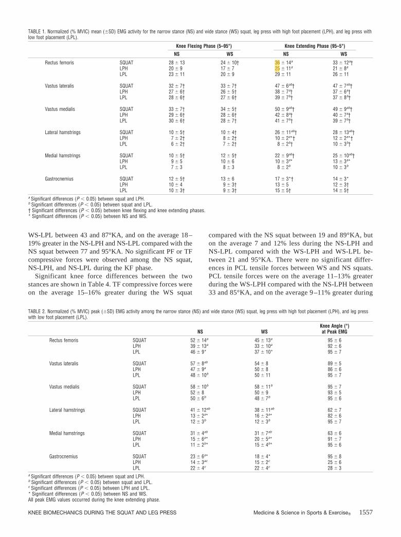
Normalized EMG values are shown in Table 1. No sig-nificant EMG differences were observed during the KFphase among exercise and stance variations. During the KEphase, the squat generated greater rectus femoris activitycompared with the LPH and greater vasti activity comparedwith the LPH and LPL. There were no differences in quad-riceps activity between the NS and WS. Lateral and medialhamstring activity were greater in the squat compared withthe LPH and LPL, and greater in the WS compared with theNS for the LPH. Gastrocnemius activity was greater for theNS squat compared with the WS squat. Quadriceps, ham-strings, and gastrocnemius activity was generally greaterduring the KE phase compared with the KF phase.
Peak EMG activity during the squat and LP exercises(Table 2) occurred during the KE phase. Peak quadricepsactivity occurred near maximum KA for the squat, LPH, andLPL. Peak hamstrings activity occurred at approximately60°KA for the squat and near maximum KA for the LPHand LPL. Peak gastrocnemius activity occurred near maxi-mum KA for the squat and at approximately 25°KA for theLPH and LPL. Peak quadriceps, hamstrings, and gastrocne-mius activity were greater in the squat compared with theLPH and LPL. Peak hamstrings activity during the LPH andLPL were greater in the WS compared with the NS, whereaspeak gastrocnemius activity during the squat was greater inthe NS compared with the WS. Peak gastrocnemius activitywas greater in the LPL compared with the LPH.
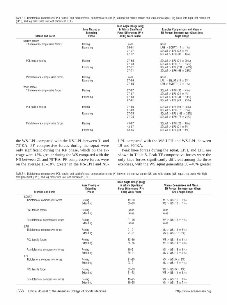
Significant knee force differences among the three exer-cises are shown in Table 3. Tibiofemoral (TF) compressiveforces were on the average 32–43% greater in the squatcompared with the LPH and LPL between 27 and 87°KA,and on the average 17% greater in the LPH compared withthe squat between 79 and 91°KA. PCL tensile forces wereon the average 18–131% greater in the squat compared withthe LPH and LPL between 27 and 89°KA. ACL tensileforces were not produced during any exercise. Patellofemo-ral (PF) compressive forces were on the average 21–39%greater in the WS squat compared with the WS-LPH and
1556 Official Journal of the American College of Sports Medicine http://www.acsm-msse.org
WS-LPL between 43 and 87°KA, and on the average 18–19% greater in the NS-LPH and NS-LPL compared with theNS squat between 77 and 95°KA. No significant PF or TFcompressive forces were observed among the NS squat,NS-LPH, and NS-LPL during the KF phase.
Significant knee force differences between the twostances are shown in Table 4. TF compressive forces wereon the average 15–16% greater during the WS squat
compared with the NS squat between 19 and 89°KA, buton the average 7 and 12% less during the NS-LPH andNS-LPL compared with the WS-LPH and WS-LPL be-tween 21 and 95°KA. There were no significant differ-ences in PCL tensile forces between WS and NS squats.PCL tensile forces were on the average 11–13% greaterduring the WS-LPH compared with the NS-LPH between33 and 85°KA, and on the average 9 –11% greater during
TABLE 2. Normalized (% MVIC) peak (�SD) EMG activity among the narrow stance (NS) and wide stance (WS) squat, leg press with high foot placement (LPH), and leg presswith low foot placement (LPL).
NS WSKnee Angle (°)at Peak EMG
Rectus femoris SQUAT 52 � 14a 45 � 13a 95 � 6LPH 39 � 13a 33 � 10a 92 � 6LPL 46 � 9* 37 � 10* 95 � 7
Vastus lateralis SQUAT 57 � 8ab 54 � 8 89 � 5LPH 47 � 9a 50 � 8 86 � 6LPL 48 � 10b 50 � 11 95 � 7
Vastus medialis SQUAT 58 � 10b 58 � 11b 95 � 7LPH 52 � 8 50 � 9 93 � 5LPL 50 � 6b 48 � 7b 95 � 6
Lateral hamstrings SQUAT 41 � 12ab 38 � 11ab 62 � 7LPH 13 � 2a* 16 � 2a* 82 � 6LPL 12 � 3b 12 � 3b 95 � 7
Medial hamstrings SQUAT 31 � 4ab 31 � 7ab 63 � 6LPH 15 � 6a* 20 � 5a* 91 � 7LPL 11 � 2b* 15 � 4b* 95 � 6
Gastrocnemius SQUAT 23 � 6a* 18 � 4* 95 � 8LPH 14 � 3ac 15 � 2c 25 � 6LPL 22 � 4c 22 � 4c 28 � 3
a Significant differences (P � 0.05) between squat and LPH.b Significant differences (P � 0.05) between squat and LPL.c Significant differences (P � 0.05) between LPH and LPL.* Significant differences (P � 0.05) between NS and WS.All peak EMG values occurred during the knee extending phase.
TABLE 1. Normalized (% MVIC) mean (�SD) EMG activity for the narrow stance (NS) and wide stance (WS) squat, leg press with high foot placement (LPH), and leg press withlow foot placement (LPL).
Knee Flexing Phase (5–95°) Knee Extending Phase (95–5°)
NS WS NS WS
Rectus femoris SQUAT 28 � 13 24 � 10† 36 � 14a 33 � 12a†LPH 20 � 9 17 � 7 25 � 11a 21 � 8a
LPL 23 � 11 20 � 9 29 � 11 26 � 11
Vastus lateralis SQUAT 32 � 7† 33 � 7† 47 � 6ab† 47 � 7ab†LPH 27 � 6† 26 � 5† 38 � 7a† 37 � 6a†LPL 28 � 6† 27 � 6† 39 � 7b† 37 � 8b†
Vastus medialis SQUAT 33 � 7† 34 � 5† 50 � 9ab† 49 � 9ab†LPH 29 � 6† 28 � 6† 42 � 8a† 40 � 7a†LPL 30 � 6† 28 � 7† 41 � 7b† 39 � 7b†
Lateral hamstrings SQUAT 10 � 5† 10 � 4† 26 � 11ab† 28 � 13ab†LPH 7 � 2† 8 � 2† 10 � 2a*† 12 � 2a*†LPL 6 � 2† 7 � 2† 8 � 2b† 10 � 3b†
Medial hamstrings SQUAT 10 � 5† 12 � 5† 22 � 9ab† 25 � 10ab†LPH 9 � 5 10 � 6 10 � 3a* 13 � 3a*LPL 7 � 3 8 � 3 8 � 2b 10 � 3b
Gastrocnemius SQUAT 12 � 5† 13 � 6 17 � 3*† 14 � 3*LPH 10 � 4 9 � 3† 13 � 5 12 � 3†LPL 10 � 3† 9 � 3† 15 � 5† 14 � 5†
a Significant differences (P � 0.05) between squat and LPH.b Significant differences (P � 0.05) between squat and LPL.† Significant differences (P � 0.05) between knee flexing and knee extending phases.* Significant differences (P � 0.05) between NS and WS.
KNEE BIOMECHANICS DURING THE SQUAT AND LEG PRESS Medicine & Science in Sports & Exercise� 1557
the WS-LPL compared with the NS-LPL between 31 and73°KA. PF compressive forces during the squat wereonly significant during the KF phase, which on the av-erage were 15% greater during the WS compared with theNS between 21 and 79°KA. PF compressive forces wereon the average 10 –18% greater in the NS-LPH and NS-
LPL compared with the WS-LPH and WS-LPL between19 and 95°KA.
Peak knee forces during the squat, LPH, and LPL areshown in Table 5. Peak TF compressive forces were theonly knee forces significantly different among the threeexercises, with the WS squat generating 30 – 40% greater
TABLE 4. Tibiofemoral compressive, PCL tensile, and patellofemoral compressive forces (N) between the narrow stance (NS) and wide stance (WS) squat, leg press with highfoot placement (LPH), and leg press with low foot placement (LPL).
Exercise and Force
Knee Flexing orExtending
Phase
Knee Angle Range (deg)in Which Significant
Force Differences (P <0.05) Were Found
Stance Comparison and Mean �SD Percent Increase over Given
Knee Angle Range
SQUATTibiofemoral compressive forces Flexing 19–83 WS � NS (16 � 5%)
Extending 59–89 WS � NS (15 � 1%)
PCL tensile forces Flexing None NoneExtending None None
Patellofemoral compressive forces Flexing 21–79 WS � NS (15 � 4%)Extending None None
LPHTibiofemoral compressive forces Flexing 21–91 NS � WS (11 � 5%)
Extending 71–91 NS � WS (7 � 3%)
PCL tensile forces Flexing 33–69 WS � NS (13 � 5%)Extending 55–85 WS � NS (11 � 2%)
Patellofemoral compressive forces Flexing 19–91 NS � WS (18 � 6%)Extending 39–91 NS � WS (10 � 3%)
LPLTibiofemoral compressive forces Flexing 21–95 NS � WS (9 � 3%)
Extending 23–91 NS � WS (12 � 4%)
PCL tensile forces Flexing 37–69 WS � NS (9 � 4%)Extending 31–73 WS � NS (11 � 5%)
Patellofemoral compressive forces Flexing 19–95 NS � WS (16 � 9%)Extending 19–95 NS � WS (15 � 7%)
TABLE 3. Tibiofemoral compressive, PCL tensile, and patellofemoral compressive forces (N) among the narrow stance and wide stance squat, leg press with high foot placement(LPH), and leg press with low foot placement (LPL).
Stance and Force
Knee Flexing orExtending
Phase
Knee Angle Range (deg)in Which Significant
Force Differences (P <0.05) Were Found
Exercise Comparisons and Mean �SD Percent Increase over Given Knee
Angle Range
Narrow stanceTibiofemoral compressive forces Flexing None None
Extending 79–91 LPH � SQUAT (17 � 1%)27–37 SQUAT � LPL (32 � 3%)27–37 SQUAT � LPH (37 � 6%)
PCL tensile forces Flexing 27–65 SQUAT � LPL (74 � 29%)27–43 SQUAT � LPH (70 � 15%)
Extending 27–71 SQUAT � LPL (131 � 40%)27–71 SQUAT � LPH (93 � 25%)
Patellofemoral compressive forces Flexing None NoneExtending 77–95 LPL � SQUAT (18 � 2%)
77–95 LPH � SQUAT (19 � 1%)Wide stance
Tibiofemoral compressive forces Flexing 27–87 SQUAT � LPH (36 � 4%)27–87 SQUAT � LPL (34 � 4%)
Extending 27–63 SQUAT � LPH (41 � 13%)27–81 SQUAT � LPL (43 � 23%)
PCL tensile forces Flexing 27–89 SQUAT � LPL (49 � 28%)61–89 SQUAT � LPH (18 � 7%)
Extending 27–75 SQUAT � LPL (103 � 28%)27–75 SQUAT � LPH (73 � 21%)
Patellofemoral compressive forces Flexing 43–87 SQUAT � LPH (28 � 6%)43–87 SQUAT � LPL (21 � 5%)
Extending 43–55 SQUAT � LPL (39 � 1%)
1558 Official Journal of the American College of Sports Medicine http://www.acsm-msse.org

peak forces than the WS-LPH and WS-LPL. Peak TFcompressive forces in the current study were approxi-mately 3.75 times body weight (BW) for the squat at65°KA, approximately 3.35 times BW for the LPH at78°KA, and approximately 3.25 times BW for the LPL at81°KA. Significant differences in knee forces betweenthe KF and KE phases are shown in Table 6, with kneeforces generally significantly greater during the KEphase.
DISCUSSION
The aim of this project was to quantify knee forces andmuscle activity about the knee while performing the squat
and LP with varying stances, foot angles, and foot place-ments. Both the KF and KE phases of each exercise wereexamined. Muscle activity and force for all major kneemuscles were quantified over the entire KF and KE rangesof motion. Muscle forces served as input into a biomechani-cal knee model that calculated PF and TF compressiveforces, and ACL/PCL tensile forces.
Exercise intensity was normalized by each subject em-ploying a 12 RM intensity for each exercise variation, whichis approximately equivalent to 70–75% of each subject’s 1RM (11). Performing 8–12 repetitions is a common repeti-tion scheme that many physical therapy, athletic training,and athletic programs utilize for strength development andrehabilitation. Because the same relative weight was used
TABLE 5. Maximum PCL tensile, tibiofemoral compressive, and patellofemoral compressive forces during the narrow stance (NS) and wide stance (WS) squat, leg press withhigh foot placement (LPH), and leg press with low foot placement (LPL); for each parameter, the mean � SD force (N) is shown at the corresponding mean � SD knee angle.
Force
Knee Flexingor Extending
Phase NS SQUAT WS SQUAT NS-LPH WS-LPH NS-LPL WS-LPL
Tibiofemoral compressive forces Flexing 3009 � 741 3413 � 749ab 2705 � 433 2488 � 478a 2778 � 480 2507 � 456b
@71 � 14° @72 � 13° @85 � 6° @80 � 11° @84 � 8° @79 � 9°Extending 2944 � 1005 3428 � 838b 3073 � 457 2821 � 500 2994 � 481 2646 � 470b
@64 � 16° @65 � 16° @78 � 13° @74 � 10° @81 � 10° @81 � 11°
PCL tensile forces Flexing 1469 � 438 1710 � 506 1404 � 261 1376 � 341 1462 � 246 1463 � 299@88 � 14° @81 � 25° @95 � 0° @94 � 2° @95 � 0° @95 � 1°
Extending 2066 � 881 2212 � 801 1703 � 358 1726 � 553 1690 � 303 1726 � 368@77 � 19° @76 � 16° @94 � 3° @88 � 6° @95 � 0° @95 � 0°
Patellofemoral compressive forces Flexing 4246 � 1047 4674 � 1195 4316 � 832 3761 � 880 4541 � 785 4000 � 829@85 � 3° @82 � 4° @87 � 2° @87 � 5° @87 � 2° @86 � 3°
Extending 3958 � 1105 4313 � 1201 4809 � 954 4389 � 1085 4813 � 978 4224 � 950@85 � 10° @80 � 11° @88 � 5° @84 � 4° @88 � 5° @90 � 5°
a Significant differences (P � 0.05) between squat and LPH.b Significant differences (P � 0.05) between squat and LPL.
TABLE 6. Tibiofemoral compressive, PCL tensile, and patellofemoral compressive forces (N) between the knee flexing (KF) and knee extending (KE) phases of the narrow stance(NS) and wide stance (WS) squat, leg press with high foot placement (LPH), and leg press with low foot placement (LPL).
Exercise and Force Stance
Knee Angle Range (deg)in Which Significant
Force Differences (P <0.05) Were Found
Phase Comparison and Mean �SD Percent Increase over Given
Knee Angle Range
SQUATTibiofemoral compressive forces NS 41–61 KE � KF (17 � 6%)
77–95 KF � KE (10 � 2%)WS 19–55 KE � KF (17 � 6%)
71–95 KF � KE (9 � 4%)
PCL tensile forces NS 29–95 KE � KF (66 � 37%)WS 29–95 KE � KF (57 � 30%)
Patellofemoral compressive forces NS 27–63 KE � KF (21 � 7%)WS 37–49 KE � KF (16 � 2%)
79–95 KF � KE (8 � 3%)LPH
Tibiofemoral compressive forces NS 51–95 KE � KF (11 � 1%)WS 37–95 KE � KF (15 � 3%)
PCL tensile forces NS 29–95 KE � KF (36 � 12%)WS 29–95 KE � KF (37 � 5%)
Patellofemoral compressive forces NS 63–95 KE � KF (11 � 1%)WS 47–95 KE � KF (16 � 3%)
LPLTibiofemoral compressive forces NS 81–95 KE � KF (7 � 3%)
WS None None
PCL tensile forces NS 25–95 KE � KF (28 � 8%)WS 27–95 KE � KF (30 � 9%)
Patellofemoral compressive forces NS None NoneWS None None
KNEE BIOMECHANICS DURING THE SQUAT AND LEG PRESS Medicine & Science in Sports & Exercise� 1559

for each exercise variation, knee forces and muscle activitywere able to be compared among the squat, LPH, and LPL.The propensity of the subjects in the current study was toemploy smaller foot angles during their preferred NS squat(7.7 � 7.6°) and NS-LP (7.2 � 7.1°) and larger foot anglesduring their preferred WS squat (36.6 � 8.6°) and WS-LP(32.5 � 6.3°). This implies that forefoot abduction increasesas stance width increases during the squat (10) and LP.
Muscle activity. Because the quadriceps cross the kneeanteriorly and the hamstrings and gastrocnemius cross theknee posteriorly, co-contractions from these muscle groupsare very important in enhancing anteroposterior knee sta-bility. Co-contractions between the hamstrings and quadri-ceps have been shown to be an important factor in mini-mizing stress to the ACL (22). Co-contractions from thequadriceps, hamstrings, and gastrocnemius were observedin the current study, with the largest magnitudes occurringin the squat during the KE phase. The greater quadriceps(20–60%) and hamstrings (90–225%) activity generated inthe squat compared with the LPH and LPL implies that thesquat may be a more effective exercise for quadriceps andhamstring development compared with the LPH and LPL.Moderate hamstring activity has been reported in previousstudies that employed the barbell squat using a 60–75%1RM lifting intensity similar to the current study(11,19,34,37). Similar to other studies (11,34), low ham-string activity occurred during the LP. The comparablegastrocnemius activity between squat and LP exercises is inagreement with Escamilla et al.(11), who reported no sig-nificant differences in gastrocnemius activity between thesquat and LP. Because there were no significant EMGdifferences in any of the muscles tested between the LPHand LPL, except the LPL demonstrated greater peak gas-trocnemius activity compared with the LPH, either exerciseappears equally effective in quadriceps and hamstrings, andgastrocnemius development may be enhanced during theLPL.
A few studies have examined how stance width during thesquat affects knee musculature (2,19,31). Subjects fromMcCaw and Melrose (19) used relative loads between 60and 75% of their 1 RM, which is similar to the relative loadsused in the current study. In addition, the three differentstance widths employed by their subjects were shoulderwidth, 75% shoulder width (NS), and 140% shoulder width(WS). Although the mean stance width distances in McCawand Melrose (19) were not reported in absolute measure-ments, it can be inferred that their defined NS and WS werevery similar to the defined NS and WS in the current study.In the current study, there were no significant differences inquadriceps activity between NS and WS squats, which is inagreement from EMG data from McCaw and Melrose (19)and Anderson et al.(2), and magnetic resonance imaging(MRI) data from Tesch (31).
Similar to several other studies (11,34–36), vasti activitywas 30–90% greater than rectus femoris activity in the squatand LP exercises. This implies that squat and LP exercisesmay be more effective in vasti development compared withrectus femoris development. Within the squat, LPH, and
LPL exercises, the VM and VL produced approximately thesame amount of activity, which is in agreement with squatand LP data from several studies (11,27,34).
Because there were also no differences in quadricepsactivity between the NS-LPH and WS-LPH, and betweenthe NS-LPL and WS-LPL, stance variations during the LPHand LPL do not appear effective in producing differences inquadriceps development during these exercises. There werealso no differences in hamstring activity between the NSsquat and WS squat, which is in agreement with data fromTesch (31) and McCaw and Melrose (19). However, a smallbut significant increase in hamstring activity was observedin the WS-LPH compared with the NS-LPH, which impliesthat the WS-LPH may be slightly more effective in ham-strings development compared with the NS-LPH. In addi-tion, a small but significant increase in gastrocnemius ac-tivity was observed in the NS squat compared with the WSsquat, which implies that the NS squat may be slightly moreeffective in gastrocnemius development compared with theWS squat.
When comparing muscle activity between the KF and KEphases, quadriceps activity was 25–50% greater in the KEphase during the squat, LPH, and LPL. Hamstring activitywas 100–180% greater in the KE phase during the squat, butonly 10–50% greater in the KE phase for the LPH and LPL.Gastrocnemius activity was 5–55% greater in the KE phaseduring the squat, LPH, and LPL. Greater muscle activity inthe KE phase compared with the KF phase has been previ-ously reported during the squat, especially in the hamstrings(11,19,20,30,34). Because the hamstrings are biarticularmuscles, it is difficult to determine if these muscles acteccentrically during the KF phase and concentrically duringthe KE phase, as commonly is believed. They may actuallybe working nearly isometrically during both the KF and KEphases (19), because they are concurrently shortening at theknee and lengthening at the hip during the KF phase andlengthening at the knee and shortening at the hip during theKE phase. If they are indeed working eccentrically duringthe KF phase and concentrically during the KE phase, thendata from the current study would be in accord with datafrom Komi et al. (16), who reported decreased activityduring eccentric work and increased activity during concen-tric work. In any case, the hamstrings probably do notchange length much throughout the squat, LPH, and LPL.Hence, in accordance with the length-force relationship inskeletal muscle, a constant length in the hamstrings willallow them to be more effective in generating force through-out the entire lifting movement. It is interesting that al-though peak quadriceps EMG values during the squat oc-curred near maximum knee flexion at the beginning of theascent, peak hamstrings EMG values during the squat oc-curred at approximately 1/3 of the way up (approximately60°KA) from the beginning of the ascent. The hamstringsmay need to work harder at 60°KA during the ascent tocompensate for attenuated force generation from the gluteusmaximus due to muscle shortening. In contrast, the quadri-ceps (especially the vasti muscles) are most effective ingenerating force at maximum knee flexion, which may
1560 Official Journal of the American College of Sports Medicine http://www.acsm-msse.org
reflect the higher peak EMG values at this point, because asthese muscles shorten, their ability to generate forcediminishes.
The nonsignificant differences in muscle activity between0 and 30° of forefoot abduction imply that employing vary-ing foot angles is not effective in altering muscle recruit-ment patterns during the squat, LPH, and LPL. This is inagreement with data from other squat studies (7,20,26),which demonstrated that employing varying foot anglesduring the squat did not affect quadriceps or hamstringsactivity.
Tibiofemoral (TF) compressive forces. TF com-pressive forces have been demonstrated to be an importantfactor in knee stabilization by resisting shear forces andminimizing tibia translation relative to the femur (18). Com-paring among exercises (Table 3), the greater TF compres-sive forces in the NS squat compared with the NS-LPH andNS-LPL between 27 and 37°KA implies that the NS squatmay provide enhanced knee stability between this smallerKA range. In contrast, the greater TF compressive forces inthe NS-LPH compared with the NS squat between 79 and91°KA implies that the NS-LPH may provide enhancedknee stability between this larger KA range. In addition,knee stability may be greater in the WS squat compared withthe WS-LPH and WS-LPL between 27 and 87°KA. Thegenerally greater TF compressive forces observed duringthe squat compared with the LPH and LPL are primarily dueto the greater quadriceps and hamstrings activity generatedin the squat compared with the LPH and LPL, because thesemuscles generate large TF compressive forces at the knee(11,34). It has been demonstrated that during a maximumvoluntary contraction of the quadriceps the force generated
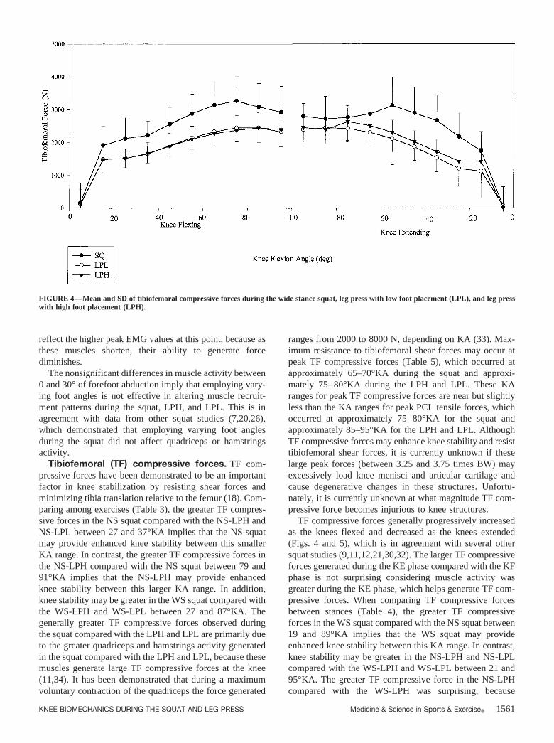
ranges from 2000 to 8000 N, depending on KA (33). Max-imum resistance to tibiofemoral shear forces may occur atpeak TF compressive forces (Table 5), which occurred atapproximately 65–70°KA during the squat and approxi-mately 75–80°KA during the LPH and LPL. These KAranges for peak TF compressive forces are near but slightlyless than the KA ranges for peak PCL tensile forces, whichoccurred at approximately 75–80°KA for the squat andapproximately 85–95°KA for the LPH and LPL. AlthoughTF compressive forces may enhance knee stability and resisttibiofemoral shear forces, it is currently unknown if theselarge peak forces (between 3.25 and 3.75 times BW) mayexcessively load knee menisci and articular cartilage andcause degenerative changes in these structures. Unfortu-nately, it is currently unknown at what magnitude TF com-pressive force becomes injurious to knee structures.
TF compressive forces generally progressively increasedas the knees flexed and decreased as the knees extended(Figs. 4 and 5), which is in agreement with several othersquat studies (9,11,12,21,30,32). The larger TF compressiveforces generated during the KE phase compared with the KFphase is not surprising considering muscle activity wasgreater during the KE phase, which helps generate TF com-pressive forces. When comparing TF compressive forcesbetween stances (Table 4), the greater TF compressiveforces in the WS squat compared with the NS squat between19 and 89°KA implies that the WS squat may provideenhanced knee stability between this KA range. In contrast,knee stability may be greater in the NS-LPH and NS-LPLcompared with the WS-LPH and WS-LPL between 21 and95°KA. The greater TF compressive force in the NS-LPHcompared with the WS-LPH was surprising, because
FIGURE 4—Mean and SD of tibiofemoral compressive forces during the wide stance squat, leg press with low foot placement (LPL), and leg presswith high foot placement (LPH).
KNEE BIOMECHANICS DURING THE SQUAT AND LEG PRESS Medicine & Science in Sports & Exercise� 1561

hamstrings activity was greater in the WS-LPH. However,the difference in hamstring activity between the NS-LPHand WS-LPH is very small, although significant, as were thedifferences in TF compressive forces between these twoexercises.
PCL tensile forces. It has been reported by Butler et al.(8) that the ACL provides 86% of the total restraining force toanterior drawer and the PCL provides 95% of the total restrain-ing force to posterior drawer. An interesting result was thatthere were no ACL forces observed during the squat, LPH, andLPL throughout the KF and KE phases. However, this was notsurprising because several studies have demonstrated PCLtensile forces exclusively during squat and LP exercises(9,11,17,30,34). Additional squat studies have reported mod-erate PCL tensile forces between 50 and 130°KA and mini-mum ACL tensile forces between 0 and 50°KA (12,21,28,32).Small ACL forces during the body weight squat have also beenreported in vivo by Beynnon et al.(6), who inserted straintransducers into the anteromedial bundle of the ACL in eightsubjects immediately after arthroscopic knee meniscectomiesand debridements. Minimal ACL strain (�4%) was observedat less than 70°KA during both the KF and KE phases, withACL strain greatest at full extension and progressively decreas-ing as the knees flexed to 90°. However, because this study wasperformed immediately after surgery, it difficult to extrapolatethese results to the barbell squat as performed by healthyathletes in the current study.
The absence of ACL forces in the current study may inpart be due to force contributions from the hamstrings,which have been shown to unload strain on the ACL (23).Quadriceps activity also affects cruciate ligament strain.Quadriceps force, via the patella tendon, exerts an anteriorshear force on the leg when the knee is flexed less than
50–60°KA, and a posterior shear force when the knee isflexed greater than 50–60°KA (6,13). In addition to muscleforces, inertial forces and the effects of gravity based ontechnique variations also affect ACL and PCL loading.When muscle and inertial forces acting on the leg are greaterin the posterior shear direction than the anterior shear di-rection, the PCL is loaded. Because the ultimate strength ofthe PCL has been estimated up to 4000 N for young activepeople (24), the peak PCL tensile forces of approximately2000 N (Table 5) observed during the squat, LPH, and LPLare probably not of great enough magnitude to be injuriousto the healthy PCL.
During PCL rehabilitation, in which the initial goal is tominimize PCL strain, the LPH and LPL may be preferredover the squat, because the squat generated greater PCLtensile forces than the LPL and LPH over a large KA range(Table 3). In addition, the NS-LPH and NS-LPL may bepreferred over the WS-LPH and WS-LPL, because the NS-LPH and NS-LPL generated smaller forces than the WS-LPH and WS-LPL over a large KA range. Performing thesquat, LPH, and LPL within the functional range of0–50°KA may be preferred for PCL rehabilitation, becausePCL tensile forces generally increased as the knees flexedand decreased as the knees extended (9,11,12,21,30,32),peaking near maximum KA (Figs. 6 and 7).
Patellofemoral (PF) compressive forces. Exces-sive PF compressive forces and stresses, or repetitiveoccurrences of lower magnitude forces and stresses, maycontribute to patellofemoral degeneration and patholo-gies, such as patella chondromalacia, osteoarthritis, andosteochondritis dissecans. There are primarily threeforces acting on the patella during the squat, LPH, andLPL: 1) quadriceps tendon force, 2) patellar tendon force,
FIGURE 5—Mean and SD of tibiofemoral compressive forces during the narrow stance squat, leg press with low foot placement (LPL), and leg presswith high foot placement (LPH).
1562 Official Journal of the American College of Sports Medicine http://www.acsm-msse.org

and 3) PF compressive force. PF compressive forces arisefrom contact between the undersurface of the patella andthe patellar surface of the femur and vary according toKA. Patellofemoral joint contact areas are also affectedby KA. From full extension to full flexion, the patellamoves caudally approximately 7 cm, with femoral con-tact on the patella moving cranially as the knee flexes
(14). Patellofemoral contact has been reported to initiallyoccur between 10 and 20°KA (14), which is when thepatella begins to glide onto the patellar surface of thefemur. The femur makes contact with the medial andlateral inferior facets between approximately 20 –30°KA,with the medial and lateral middle facets between ap-proximately 30 – 60°KA, with the medial and lateral
FIGURE 7—Mean and SD of PCL tensile forces during the narrow stance squat, leg press with low foot placement (LPL), and leg press with highfoot placement (LPH).
FIGURE 6—Mean and SD of PCL tensile forces during the wide stance squat, leg press with low foot placement (LPL), and leg press with high footplacement (LPH).
KNEE BIOMECHANICS DURING THE SQUAT AND LEG PRESS Medicine & Science in Sports & Exercise� 1563

superior facets between approximately 60 –90°KA, andwith the medial vertical “odd” facet and lateral superiorfacet between approximately 90 and 135°KA (14). Atapproximately 90°KA, the “odd” facet for the first timemakes contact with the lateral margin of the medialcondyle (14).
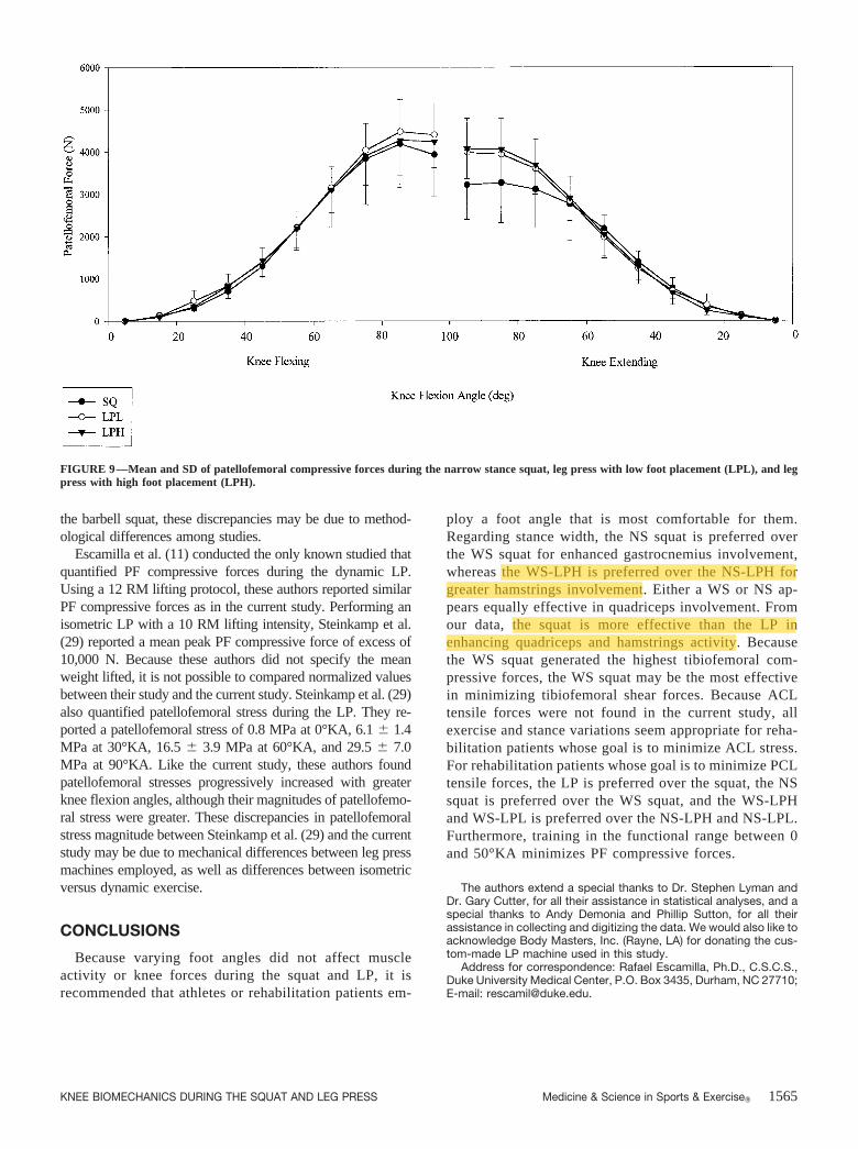
Patellofemoral contact area has been reported to be 2.6 �0.4 cm2 at 20°KA, 3.1 � 0.3 cm2 at 30°KA, 3.9 � 0.6 cm2 at60°KA, 4.1 � 1.2 cm2 at 90°KA, and 4.6 � 0.7 cm2 at 120°KA(14). When PF compressive forces are distributed over patel-lofemoral contact areas, patellofemoral stress is produced.Consider the KF phase in Figure 9, in which there were nopatellofemoral differences among the squat, LPH, and LPL.Mean PF compressive forces collapsed across exercises were238 N at 20°KA, 615 N at 30°KA, 2731 N at 60°KA, and 4186N at 90°KA. By using these PF compressive force data fromthe KF phase of Fig. 9, and the patellofemoral contact areasgiven above, patellofemoral stress at 20°, 30°, 60°, and 90°KAwould be 0.92 MPa, 1.98 MPa, 7.00 MPa, and 10.21 MPa,respectively. Consequently, during the squat, LPH, and LPL,PF stresses increase as knee flexion increases, peaking near80–90°KA. Increasing PF stresses as the knees flex and de-creasing PF stresses as the knees extend is in agreement withseveral other studies (9,11,21,25,35). From these data, it can beinferred that individuals with patellofemoral disorders shouldavoid performing the squat, LPH, and LPL at higher kneeflexion angles. Furthermore, squat and LP data from Escamillaet al. (11) and the current study illustrate that the rate ofincrease in PF stress appears maximum between approximately50 and 80°KA, thus generating proportionately greater PFstress between 50 and 80°KA compared with 0 and 50°KA.Therefore, performing the squat, LPH, and LPL within the
functional range of 0–50°KA may be most effective for ath-letes or patients with patellofemoral pathologies.
Although the loads lifted in the current study (approximately1.5 times BW) are higher than most rehabilitation patients willexperience, they are typical loads for strength and power ath-letes while performing the squat and LP exercises. However,performing the squat, LPH, and LPL at greater knee flexionangles may not be problematic for athletes with healthy knees,as long as heavy loads are not used excessively. Interestingly,PF compressive forces have been shown to remain relativelyconstant or slightly decrease beyond 85–90°KA (11,21) (Figs.8 and 9). Hence, patellofemoral stress may decrease withgreater than 90°KA, because patellofemoral contact area con-tinues to increase as knee flexion increases (14). Nevertheless,training with excessive loads can be a potential problem forpowerlifters and football players, who often train with heavyloads for long periods of time. Unfortunately, it is currentlyunknown how much PF compressive force and stress is detri-mental to the patellofemoral joint while performing squat andLP exercises.
When normalized by body weight and load lifted, and ex-pressed as a percentage, mean peak PF compressive force inthe current study was 210 � 54% BW for the squat. This issimilar to the 180 � 93% BW from Wretenberg et al. (35) andthe nearly 200% BW from Nisell and Ekholm (21), whosesubjects also performed the barbell squat with a similar liftingintensity (65–75% 1 RM) as the current study. Surprisingly, theremaining two studies that quantified PF compressive forcesduring the dynamic squat found normalized values in excess of700% BW (9,25). Because both of these studies examined PFcompressive forces during the body weight squat, which re-quires relatively little effort and muscle activity compared with
FIGURE 8—Mean and SD of patellofemoral compressive forces during the wide stance squat, leg press with low foot placement (LPL), and leg presswith high foot placement (LPH).
1564 Official Journal of the American College of Sports Medicine http://www.acsm-msse.org
the barbell squat, these discrepancies may be due to method-ological differences among studies.
Escamilla et al. (11) conducted the only known studied thatquantified PF compressive forces during the dynamic LP.Using a 12 RM lifting protocol, these authors reported similarPF compressive forces as in the current study. Performing anisometric LP with a 10 RM lifting intensity, Steinkamp et al.(29) reported a mean peak PF compressive force of excess of10,000 N. Because these authors did not specify the meanweight lifted, it is not possible to compared normalized valuesbetween their study and the current study. Steinkamp et al. (29)also quantified patellofemoral stress during the LP. They re-ported a patellofemoral stress of 0.8 MPa at 0°KA, 6.1 � 1.4MPa at 30°KA, 16.5 � 3.9 MPa at 60°KA, and 29.5 � 7.0MPa at 90°KA. Like the current study, these authors foundpatellofemoral stresses progressively increased with greaterknee flexion angles, although their magnitudes of patellofemo-ral stress were greater. These discrepancies in patellofemoralstress magnitude between Steinkamp et al. (29) and the currentstudy may be due to mechanical differences between leg pressmachines employed, as well as differences between isometricversus dynamic exercise.
CONCLUSIONS
Because varying foot angles did not affect muscleactivity or knee forces during the squat and LP, it isrecommended that athletes or rehabilitation patients em-
ploy a foot angle that is most comfortable for them.Regarding stance width, the NS squat is preferred overthe WS squat for enhanced gastrocnemius involvement,whereas the WS-LPH is preferred over the NS-LPH forgreater hamstrings involvement. Either a WS or NS ap-pears equally effective in quadriceps involvement. Fromour data, the squat is more effective than the LP inenhancing quadriceps and hamstrings activity. Becausethe WS squat generated the highest tibiofemoral com-pressive forces, the WS squat may be the most effectivein minimizing tibiofemoral shear forces. Because ACLtensile forces were not found in the current study, allexercise and stance variations seem appropriate for reha-bilitation patients whose goal is to minimize ACL stress.For rehabilitation patients whose goal is to minimize PCLtensile forces, the LP is preferred over the squat, the NSsquat is preferred over the WS squat, and the WS-LPHand WS-LPL is preferred over the NS-LPH and NS-LPL.Furthermore, training in the functional range between 0and 50°KA minimizes PF compressive forces.
The authors extend a special thanks to Dr. Stephen Lyman andDr. Gary Cutter, for all their assistance in statistical analyses, and aspecial thanks to Andy Demonia and Phillip Sutton, for all theirassistance in collecting and digitizing the data. We would also like toacknowledge Body Masters, Inc. (Rayne, LA) for donating the cus-tom-made LP machine used in this study.
Address for correspondence: Rafael Escamilla, Ph.D., C.S.C.S.,Duke University Medical Center, P.O. Box 3435, Durham, NC 27710;E-mail: [email protected].
FIGURE 9—Mean and SD of patellofemoral compressive forces during the narrow stance squat, leg press with low foot placement (LPL), and legpress with high foot placement (LPH).
KNEE BIOMECHANICS DURING THE SQUAT AND LEG PRESS Medicine & Science in Sports & Exercise� 1565
REFERENCES
1. ALKNER, B. A., P. A. TESCH, and H. E. BERG. Quadriceps EMG/force relationship in knee extension and leg press. Med. Sci. SportsExerc. 32:459–463, 2000.
2. ANDERSON, R., C. COURTNEY, and E. CARMELI. EMG analysis of thevastus medialis/vastus lateralis muscles utilizing the unloadednarrow-and wide-stance squats. J. Sport Rehabil. 7:236–247,1998.
3. ANDREWS, J. G., J. G. HAY, and C. L. VAUGHAN. Knee shear forcesduring a squat exercise using a barbell and a weight machine. In:Biomechanics VIII-B, H. Matsui and K. Kobayashi (Eds.). Cham-paign: Human Kinetics, 1983, pp. 923–927.
4. ARIEL, B. G. Biomechanical analysis of the knee joint during deepknee bends with heavy loads. In: Biomechanics IV, R. Nelson andC. Morehouse (Eds.). Baltimore: University Park Press, 1974, pp.44–52.
5. BASMAJIAN, J. V., and R. BLUMENSTEIN. Electrode Placement inEMG Biofeedback. Baltimore: Williams and Wilkins, 1980, pp.79–86.
6. BEYNNON, B. D., R. J. JOHNSON, B. C. FLEMING, C. J. STANKEWICH,P. A. RENSTROM, and C. E. NICHOLS. The strain behavior of theanterior cruciate ligament during squatting and active flexion-extension: a comparison of an open and a closed kinetic chainexercise. Am. J. Sports Med. 25:823–829, 1997.
7. BOYDEN, G., J. KINGMAN, and R. DYSON. A comparison of quadricepselectromyographic activity with the position of the foot during theparallel squat. J. Strength Cond. Res. 14:379–382, 2000.
8. BUTLER, D. L., F. R. NOYES, and E. S. GROOD. Ligamentousrestraints to anterior-posterior drawer in the human knee: a bio-mechanical study. J. Bone Joint Surg. Am. 62:259–270, 1980.
9. DAHLKVIST, N. J., P. MAYO, and B. B. SEEDHOM. Forces duringsquatting and rising from a deep squat. Eng. Med. 11:69–76, 1982.
10. ESCAMILLA, R. F., G. S. FLEISIG, T. M. LOWRY, S. W. BARRENTINE,and J. R. ANDREWS. A three-dimensional biomechanical analysis ofthe squat during varying stance widths. Med. Sci. Sports Exerc.33:984–998, 2001.
11. ESCAMILLA, R. F., G. S. FLEISIG, N. ZHENG, S. W. BARRENTINE, K. E.WILK, and J. R. ANDREWS. Biomechanics of the knee during closedkinetic chain and open kinetic chain exercises. Med. Sci. SportsExerc. 30:556–569, 1998.
12. HATTIN, H. C., M. R. PIERRYNOWSKI, and K. A. BALL. Effect of load,cadence, and fatigue on tibiofemoral joint force during a halfsquat. Med. Sci. Sports Exerc. 21:613–618, 1989.
13. HERZOG, W., and L. J. READ. Lines of action and moment arms ofthe major force-carrying structures crossing the human knee joint.J. Anat. 182:213–230, 1993.
14. HUBERTI, H. H., and W. C. HAYES. Patellofemoral contact pres-sures: the influence of q-angle and tendofemoral contact. J. BoneJoint Surg. Am. 66:715–724, 1984.
15. ISEAR, J. A., JR., J. C. ERICKSON, and T. W. WORRELL. EMGanalysis of lower extremity muscle recruitment patterns during anunloaded squat. Med. Sci. Sports Exerc. 29:532–539, 1997.
16. KOMI, P. V., M. KANEKO, and O. AURA. EMG activity of the legextensor muscles with special reference to mechanical efficiencyin concentric and eccentric exercise. Int J Sports Med. 8(Suppl.1):22–29, 1987.
17. LUTZ, G. E., R. A. PALMITIER, K. N. AN, and E. Y. CHAO. Comparisonof tibiofemoral joint forces during open-kinetic-chain and closed-kinetic-chain exercises. J. Bone Joint Surg. Am. 75:732–739, 1993.
18. MARKOLF, K. L., W. L. BARGAR, S. C. SHOEMAKER, and H. C.AMSTUTZ. The role of joint load in knee stability. J. Bone JointSurg. Am. 63:570–585, 1981.
19. MCCAW, S. T., and D. R. MELROSE. Stance width and bar loadeffects on leg muscle activity during the parallel squat. Med. Sci.Sports Exerc. 31:428–436, 1999.
20. NINOS, J. C., J. J. IRRGANG, R. BURDETT, and J. R. WEISS. Electro-myographic analysis of the squat performed in self-selected lowerextremity neutral rotation and 30 degrees of lower extremityturn-out from the self-selected neutral position. J. Orthop. SportsPhys. Ther. 25:307–315, 1997.
21. NISELL, R., and J. EKHOLM. Joint load during the parallel squat inpowerlifting and force analysis of in vivo bilateral quadricepstendon rupture. Scand. J. Sports Sci. 8:63–70, 1986.
22. O’CONNOR, J. J. Can muscle co-contraction protect knee ligamentsafter injury or repair? J. Bone Joint Surg. Br. 75:41–48, 1993.
23. OHKOSHI, Y., K. YASUDA, K. KANEDA, T. WADA, and M. YA-MANAKA. Biomechanical analysis of rehabilitation in the standingposition. Am. J. Sports Med. 19:605–611, 1991.
24. RACE, A., and A. A. AMIS. The mechanical properties of the twobundles of the human posterior cruciate ligament. J. Biomech.27:13–24, 1994.
25. REILLY, D. T., and M. MARTENS. Experimental analysis of thequadriceps muscle force and patello-femoral joint reaction forcefor various activities. Acta Orthop. Scand. 43:126–137, 1972.
26. SIGNORILE, J. F., K. KWIATKOWSKI, J.F. CARUSO, and B. ROBERTSON.Effect of foot position on the electromyographical activity of thesuperficial quadriceps muscles during the parallel squat and kneeextension. J. Strength Cond. Res. 9:182–187, 1995.
27. SIGNORILE, J. F., B. WEBER, B. ROLL, J. F. CARUSO, I. LOWENSTEYN, andA. C. PERRY. An electromyographical comparison of the squat andknee extension exercises. J. Strength Cond. Res. 8:178–183, 1994.
28. SINGERMAN, R., J. BERILLA, M. ARCHDEACON, and A. PEYSER. Invitro forces in the normal and cruciate-deficient knee duringsimulated squatting motion. J. Biomech Eng. 121:234–242, 1999.
29. STEINKAMP, L. A., M. F. DILLINGHAM, M. D. MARKEL, J. A. HILL, andK. R. KAUFMAN. Biomechanical considerations in patellofemoral jointrehabilitation. Am. J. Sports Med. 21:438–444, 1993.
30. STUART, M. J., D. A. MEGLAN, G. E. LUTZ, E. S. GROWNEY, andK. N. AN. Comparison of intersegmental tibiofemoral joint forcesand muscle activity during various closed kinetic chain exercises.Am. J. Sports Med. 24:792–799, 1996.
31. TESCH, P. A. Muscle Meets Magnet. Stockholm: PA Tesch AB,1993, pp. 79.
32. TOUTOUNGI, D. E., T. W. LU, A. LEARDINI, F. CATANI, and J. J.O’CONNOR. Cruciate ligament forces in the human knee duringrehabilitation exercises. Clin. Biomech. 15:176–187, 2000.
33. VAN EIJDEN, T. M., W. A. WEIJS, E. KOUWENHOVEN, and J. VERBURG.Forces acting on the patella during maximal voluntary contractionof the quadriceps femoris muscle at different knee flexion/exten-sion angles. Acta Anat. 129:310–314, 1987.
34. WILK, K. E., R. F. ESCAMILLA, G. S. FLEISIG, S. W. BARRENTINE,J. R. ANDREWS, and M. L. BOYD. A comparison of tibiofemoraljoint forces and electromyographic activity during open and closedkinetic chain exercises. Am. J. Sports Med. 24:518–527, 1996.
35. WRETENBERG, P., Y. FENG, and U. P. ARBORELIUS. High-and low-bar squatting techniques during weight-training. Med. Sci. SportsExerc. 28:218–224, 1996.
36. WRETENBERG, P., Y. FENG, F. LINDBERG, and U. P. ARBORELIUS.Joint moments of force and quadriceps activity during squattingexercise. Scand. J. Med. Sci. Sports. 3:244–250, 1993.
37. WRIGHT, G. A., T. H. DELONG, and G. GEHLSEN. Electromyographicactivity of the hamstrings during performance of the leg curls,stiff-leg deadlift, and back squat movements. J. Strength Cond.Res. 13:168–174, 1999.
38. ZHENG, N., G. S. FLEISIG, R. F. ESCAMILLA, and S. W. BARRENTINE.An analytical model of the knee for estimation of internal forcesduring exercise. J. Biomech. 31:963–967, 1998.
1566 Official Journal of the American College of Sports Medicine http://www.acsm-msse.org