Embed Size (px)
Citation preview
ORIGINAL RESEARCHpublished: 17 October 2016
doi: 10.3389/fcimb.2016.00128
Frontiers in Cellular and Infection Microbiology | www.frontiersin.org 1 October 2016 | Volume 6 | Article 128
Edited by:
David Dockrell,
University of Sheffield, UK
Reviewed by:
J. Christopher Fenno,
University of Michigan, USA
Justin A. Thornton,
Mississippi State University, USA
*Correspondence:
Yuan Yuan
†These authors have contributed
equally to this work and also senior
authors.
Received: 11 July 2016
Accepted: 27 September 2016
Published: 17 October 2016
Citation:
Zhang S, Wang J, Chen S, Yin J,
Pan Z, Liu K, Li L, Zheng Y, Yuan Y
and Jiang Y (2016) Effects of Suilysin
on Streptococcus suis-Induced
Platelet Aggregation.
Front. Cell. Infect. Microbiol. 6:128.
doi: 10.3389/fcimb.2016.00128
Effects of Suilysin on Streptococcussuis-Induced Platelet Aggregation
Shengwei Zhang 1, 2, Junping Wang 3, Shaolong Chen 1, Jiye Yin 4, Zhiyuan Pan 1, Keke Liu 1,
Lin Li 1, Yuling Zheng 1, Yuan Yuan 1*† and Yongqiang Jiang 1 †
1 State Key Laboratory of Pathogen and Biosecurity, Beijing Institute of Microbiology and Epidemiology, Beijing, China,2Department of Clinical Laboratory, Dongfang Hospital, Beijing University of Chinese Medicine, Beijing, China, 3Urumqi
Ethnic Cadres’ College, Urumqi, China, 4 State Key Laboratory of Toxicology and Medical Countermeasures, Institute of
Pharmacology and Toxicology, Academy of Military Medical Sciences, Beijing, China
Blood platelets play important roles during pathological thrombocytopenia in
streptococcal toxic shock syndrome (STSS). Streptococcus suis (S. suis) an emerging
human pathogen, can cause STSS similarly to S. pyogenes. However, S. suis
interactions with platelets are poorly understood. Here, we found that suilysin (SLY),
different from other bacterial cholesterol-dependent cytolysins (CDCs), was the sole
stimulus that induced platelet aggregation. Furthermore, the inside-out activation of
GPIIb/IIIa of platelets mediated SLY-induced platelet aggregation. This process was
triggered by Ca2+ influx that depend on the pore forming on platelets by SLY.
Additionally, although SLY induced α-granule release occurred via the MLCK-dependent
pathway, PLC-β-IP3/DAG-MLCK and Rho-ROCK-MLCK signaling were not involved in
SLY-induced platelet aggregation. Interestingly, the pore dependent Ca2+ influx was also
found to participate in the induction of platelet aggregation with pneumolysin (PLY) and
streptolysin O (SLO), two other CDCs. It is possible that the CDC-mediated platelet
aggregation we observed in S. suis is a similar response mechanism to that used by a
wide range of bacteria. These findings might lead to the discovery of potential therapeutic
targets for S. suis-associated STSS.
Keywords: Streptococcus suis (S. suis), suilysin (SLY), platelet aggregation, Ca2+ influx, streptococcal toxic shock
syndrome (STSS)
INTRODUCTION
Streptococcal toxic shock syndrome (STSS) is a severe, invasive streptococcal infection associatedwith the sudden onset of shock, acute respiratory distress syndrome, renal failure, bacteremia,and disseminated intravascular coagulation (DIC). The Gram-positive bacterium Streptococcus suisserotype 2 (S. suis 2) is an emerging human pathogen. As well as causing disease in pigs, S. suis 2 canalso cause serious enzootic infections in humans, which are associated with septicemia, meningitis,and endocarditis (Tang et al., 2006; Wangkaew et al., 2006). In 2005, China reported over 200human cases of STSS that had an unusual clinical disease presentation and a mortality rate of up to20% (Sriskandan and Slater, 2006). Thrombocytopenia and multisystem dysfunction, such as liverfailure, heart failure, and disseminated intravascular coagulation (DIC were found in more thanhalf of the STSS patients. Moreover, purpura and gangrenous changes are found to be the typicalskin manifestations in STSS patients (Tang et al., 2006; Yu et al., 2006).

Zhang et al. Suilysin-Induced Platelet Aggregation
Platelet–bacterium interactions are known to be involvedin bacterial-associated diseases, such as infective endocarditis(IE), DIC, and purpura gangrenosa. Lactococcus lactis thatexpresses either ClfA or FnbpA was shown to be 100 timesmore infective than the wild-type L. lactis strain in an animalmodel of IE (Que et al., 2001). Helicobacter pylori infectionscause platelet activation in patients (Davi et al., 2005), andP-selectin-dependent platelet aggregation might contribute toH. pylori-associated thrombocytopenia in patients (Yeh et al.,2010). Mouse infection models suggest that S. pneumoniae-induced thrombocytopenia and DIC are caused by thisbacterium’s neuraminidase, which removes sialic acid fromplatelet proteins and is involved in platelet clearance fromthe circulation (Grewal et al., 2008). However, the interactionsbetween platelets and S. suis and the underlying molecularmechanism(s) involved in these interactions remain poorlyunderstood.
Bacteria interact with platelets through a variety ofmechanisms (Cox et al., 2011). Previous studies have shown thatbacteria interact with platelets by direct binding to adhesionfactors such as GPIIb/IIIa (Miajlovic et al., 2010), GPIbα(Plummer et al., 2005), and Toll-like receptor 4 (TLR4) (Stahlet al., 2006), or by indirect binding to these adhesion factorsvia plasma proteins such as fibrinogen (Walsh et al., 2008),IgG (Fitzgerald et al., 2006), von Willebrand factor (O’seaghdhaet al., 2006), or complement 1q (Ford et al., 1996). In addition,bacteria secrete toxins that activate platelets and cause plateletaggregation (Lourbakos et al., 2001). However, our knowledgeof the molecular events by which bacterial toxins controlplatelet–bacteria interactions is rudimentary.
Suilysin (SLY) is a 497 amino-acid protein belonging to thecholesterol-dependent cytolysin (CDC) family, which has morethan 20 members, including pneumolysin (PLY) and streptolysinO (SLO), which are expressed by S. pneumoniae and S. pyogenes,respectively. Like other CDC family members produced byGram-positive bacteria, a classical feature of these toxins is theirability to create transmembrane pores in cholesterol-containingmembranes, thereby causing cell lysis (Giddings et al., 2004;Xu et al., 2010). In this study, different from other bacterialcholesterol-dependent cytolysins (CDCs), we found that SLYwas the sole stimulus responsible for platelet activation andaggregation induced by S. suis. We also found that the SLY-induced pore dependent Ca2+ influx triggered “inside-out”signaling to activate GPIIb/IIIa, which mediated SLY-inducedplatelet aggregation. Our findings extend the similar observationsrelated to PLY- and SLO-induced platelet aggregation, which istriggered by pore dependent Ca2+ influx.
EXPERIMENTAL PROCEDURE
Ethics StatementThe healthy donors who supplied blood for this study providedwritten informed consent in accordance with the Declarationof Helsinki. Approval was obtained from the InstitutionalMedical Ethics Committee of the Academy of Military MedicalSciences. This research was approved by Ethics Committee onAnimal Experimentation of the Chinese Association for the
Accreditation of Laboratory Animals Care (CAALAC), and itincluded the relevant local animal welfare bodies in China. Thepermit number for all the animal work [SCXK-(JUN) 2013-018]was obtained from the Institutional Medical Ethics Committeeof the Academy of Military Medical Sciences, China. All effortswere made to minimize suffering in the animals employed in thisstudy.
ReagentsMonoclonal mouse anti-human antibodies, including fluoresceinisothiocyanate (FITC) conjugated anti-CD42b (clone HIP1),FITC conjugated anti-CD41a (clone HIP8), and the isotypecontrol antibodies were from BD Bioscience (USA). FITCconjugated mouse anti-fibrinogen antibody was from Abcam(USA). U73122, ML-7, Y27632, and eptifibatide acetate, whichare inhibitors for phospholipase C (PLC), myosin light chainkinase (MLCK), rho-associated, coiled-coil-containing proteinkinase (ROCK), and CD41a, respectively, were from SigmaAldrich (USA). Cholesterol and EGTA were also purchased fromSigma Aldrich. Adenosine diphosphate (ADP) was from MPBiomedicals (USA). Quest Fluo-8 calcium fluorescence probewas from AAT Bioquest R©, Inc. (USA). Wright’s stain was fromBeijing CellChip Biotechnology Co., Ltd., China.
Strains and Culture SupernatantChinese virulent S. suis strains (05ZYH33 and 4) were isolatedoriginally from an STSS patient (Pian et al., 2012). The slyor mrp isogenic mutants of 05ZYH33 (1sly and 1mrp) wereconstructed in our previous studies (He et al., 2014; Pian et al.,2015). The 1mrp mutant was used as an irrelevant controlfor 1sly in this study. The Canadian avirulent strain 1330,which does not express SLY, was donated by Prof. MarceloGottschalk (Université de Montréal, Montreal Quebec, Canada).The European SLY+-associated virulent strains (4005 and s735)were donated by Dr. Henk J. Wisselink. Group A streptococcus(GAS) was the M1 type, E477. The above-mentioned bacterialstrains were stored in our laboratory. S. suis strains were culturedin Todd-Hewitt broth (THB, BD Biosciences) at 37◦C for 4 h(OD600 = 0.4, exponential growth phase) or 6 h (OD600 =
1.0, stationary growth phase) and then harvested for the nextexperiments. Bacterial culture supernatants were obtained bycentrifugation of the cultures at 8000 rpm for 5 min, afterwhich they was filtered through a 0.22µm bacterial filter. Thefiltered supernatants were stored in −80◦C for future use. In theexperiment where bacterial cells were used as the stimulus, S.suis strains and GAS were washed three times with phosphate-buffered saline (PBS) by centrifugation at 8000 rpm for 5 min andwere then re-suspended in PBS at a density of 2 × 10⁹ CFU/mlfor use. Strains are listed in Table S1.
Preparation of Recombinant Suilysin(rSLY), Pneumolysin (rPLY), Streptolysin O(rSLO), and Factor H-Binding Protein (rFhb)To express recombinant proteins rPLY and rSLO in E. coli,the ORFs encoding PLY and SLO proteins (removed the signalsequence) were amplified by PCR and were cloned into theexpression vector pTrcHis and transformed into E. coli strain
Frontiers in Cellular and Infection Microbiology | www.frontiersin.org 2 October 2016 | Volume 6 | Article 128

Zhang et al. Suilysin-Induced Platelet Aggregation
BL21(DE3). The cloned gene sequences were confirmed by DNAsequencing. For protein expression, E. coli were cultured in 1 lof LB media containing 50µg/ml of ampicillin at 37◦C until theculture reached mid-log growth phase (OD600 = 0.6–0.8), afterwhich 1 mM isopropyl β-D-1-thiogalactopyranoside (IPTG) wasadded to the culture to induce expression of the recombinantprotein. Subsequently, the bacteria were cultured continuouslyat 16◦C overnight. The bacterial culture was then collected bycentrifugation (HITICH, Japan) at 8000 rpm for 10 min andthe cell pellet was sonicated for 30 min on ice to release therecombinant protein. Recombinant proteins were purified usingNi2+ affinity chromatography columns (GE Healthcare). Therecombinant proteins rSLY (Lv et al., 2011) and the irrelevantcontrol protein rFhb (Pian et al., 2012) used in this study werepurified as reported previously. The bacterial strains, primers,and plasmids used in this study are listed in Table S1.
Human PlateletsWhole blood from the healthy donors, who had not used anti-platelet drugs within the previous 15 days, was withdrawn atthe 307 Hospital, and the blood from each person was collectedin a tube containing sodium citrate (final concentration, 3.2%)(Shannon et al., 2007). A total of 6 ml of whole blood wascentrifuged at 900 rpm for 10 min in a horizontal centrifuge(Sigma Aldrich) to prepare platelet-rich plasma (PRP, 3×10⁸/ml).The platelets in the PRP were marked by FITC conjugated anti-CD41a (clone HIP8) and the purity was determined by flowcytometry analysis. Whole blood was recentrifuged at 3000 rpmfor 5 min to obtain platelet-poor plasma (PPP), which was usedas a control for the aggregation assays.
Platelet Aggregation in PRPBefore the platelet aggregation detection assay was performed,the purity of the platelets in the PRP was analyzed by flowcytometry. Contaminating leukocytes were not seen in the PRPisolated, and the purity of the platelets in the PRP was >99%(Figure S1). Platelet aggregation was determined in a 4-channelplatelet aggregometer SE-2000 (Succeeder, China) as describedpreviously (Keane et al., 2010). Aggregation is represented bychanges in light transmission, and the light transmission of PPPwas used as the baseline. PRP (270µl) was added to a glasscuvette and incubated at 37◦C for 1 min, and then one or otherof 30µl of SLY (final concentration was 1µg/ml), S. suis cells(2×10⁸ CFU/ml), 05ZYH33 supernatant (Sup), 1sly Sup, ADP(20µM, as positive control), or PBS/THB (negative control)was added to the cuvette. The rSLY (1µg/ml) possesses equalhemolytic activity to the 05ZYH33 supernatant (10%) used inthis study (Figure 3A). However, The final concentration of SLYin 05ZYH33 supernatant, as deduced from our previous study(He et al., 2014), was≈0.15µg/ml. This might be due to the rSLYused in this study was not added β-ME to keep its high hemolyticactivity for avoiding the negative effects of β-ME on platelets.Hemolytic activity detecting assay was performed as reportedpreviously (Hao et al., 2013). The cuvettes were incubated at37◦C for 10 min. To block platelet aggregation stimulation, PRP(270µl) was pre-incubated with 2.7µl of EGTA (3mM), U73122(20µM), eptifibatide (10µM), or anti-CD62P antibody (15µl)
at 37◦C for 10 min, or with 2.7µl ML-7 (100µM), or Y27632(100µM) for 60 min before being exposed to the stimulus. Totest the effect of cholesterol, SLY (990 µl) was pre-incubatedwith of 10µl cholesterol (100µg/ml) at 37◦C for 10 min beforeaddition to the platelets. Data are expressed as the mean ± SD.The experiment was repeated three times independently, andblood from a different donor was used in each experiment.
Observation of Platelet Aggregation inWhole Blood by MicroscopyWhole blood (100µl) was incubated with 10 µl of SLY (1µg/ml)or other stimuli at 37◦C for 10 min after which 5µl of the bloodsample was used to prepare a blood smear on a glass slide. Afterthe blood smear had dried completely, Wright’s stain was addedto cover the blood smear and an equal volume of PBS (pH 6.4–6.8) was added 1 min later. Wright’s stain and PBS were gentlymixed and the blood smear was incubated with the mixture for5 min. The slide was then washed gently with distilled waterand subjected to microscopic analysis using an oil-immersionobjective (Olympus, Japan).
Measurement of Platelet LacticDehydrogenase (LDH) ReleaseLDH release was measured to evaluate the cytotoxicity ofrecombinant SLY, PLY, and SLO to platelets. PRP was incubatedwith the either one of the recombinant proteins and thesupernatant was collected by centrifugation at 3000 rpm for5 min. LDH in the supernatant was measured by a CytoTox96 R© Non-Radioactive Cytotoxicity Assay (Promega, USA). Celllysis buffer and PBS were used as the positive and the negativecontrols, respectively. The relative cytotoxicity (%) = [(OD490
sample − OD490 PBS) ÷ (OD490 positive control − OD490
PBS)] × 100%. Data are expressed as the mean ± SD of threeindependent experiments, with each experiment using bloodfrom a different donor.
Flow Cytometry Analysis of PlateletActivationThe surface GPIIb/IIIa was detected by Flow cytometry withFITC conjugated anti-CD41a (CD41a often referring to GPIIb,clone HIP8), as per a previous description (Parimon et al.,2013). Briefly, 100µl of heparinized human blood was incubatedwith 15µl of FITC conjugated anti-human CD41a at 37◦C for10 min. Next, rSLY or a different stimulus was added andthe blood was incubated for another 15 min. One mL of redcell lysis buffer containing formalin was then added to theblood to lyse the red cells and to fix the platelets at roomtemperature for 8 min. After complete lysis of the red bloodcells, the blood was centrifuged and the cell pellet was washedwith PBS. The cell pellet was re-suspended in 300µl of PBSand analyzed on an Accuri C6 flow cytometer (BD Biosciences,USA). The surface CD41a was determined by measuring themean fluorescence intensity (MFI) of 10,000 events for eachsample. To test the effect of cholesterol, SLY was pre-incubatedwith cholesterol (100µg/ml) for 10 min and the blood wasincubated with the mixture. To test the effect of EGTA, blood
Frontiers in Cellular and Infection Microbiology | www.frontiersin.org 3 October 2016 | Volume 6 | Article 128

Zhang et al. Suilysin-Induced Platelet Aggregation
was pre-incubated with EGTA (3mM) for 10 min and thenincubated with rSLY or other stimuli. Fg binding to platelets wasalso analyzed by flow cytometry. Briefly, 100µl of whole bloodwas incubated with 1µl of FITC-conjugated anti-fibrinogenpolyclonal antibody at 37◦C for 10 min. The blood sample wasthen analyzed by flow cytometry. Representative histograms ofthe MFI for CD41a and fibrinogen binding are displayed. Dataare expressed as the mean MFI ± SD of three independentexperiments, with each experiment using blood from a differentdonor.
Measurement of Dense Granule (or ATP)SecretionATP secretion was determined to evaluate platelet activation afterstimulation (Arman et al., 2014). Briefly, 270µl of PRP wasincubated with 30µl of rSLY or 05ZYH33 supernatant or otherstimuli at 37◦C for 10 min. The supernatant was obtained bycentrifugation at 5000 rpm for 5 min. ATP in the supernatantwas measured using a luciferin-luciferase kit (CellTiter-Glo R© 2.0Assay, Promega, USA), according to the manufacturer’s manual.Data are expressed as the mean ± SD. The experiment wasrepeated three times and blood from a different donor was usedin each experiment.
Calcium Influx to PlateletsA total of 200µl of PRP was added to each well of a 96-wellmicroplate. The plate was centrifuged at 2000 rpm for 10 minand then washed twice with PBS. Next, 100µl of calcium probefluo-8 (5µM) was added to the wells and the plate was incubatedat 37◦C for 30 min. The plate was centrifuged at 2000 rpm for10 min and then washed twice with 200µl of PBS to removeany calcium probe remaining in the solution. The platelets werere-suspended in 180µl of HBSS (containing 2mM Ca2+) orD-HBSS (without Ca2+), and then either 20µl rSLY (1µg/ml),rSLY (0.1µg/ml), rSLYP353V (1µg/ml), cholesterol (100µg/ml),or other control reagents were added. Ca2+ influx to the plateletswas measured in a Varioskan Flash Multiplate Reader (Thermo,USA).
In vivo Infection05ZYH33 and mutant 1sly were cultured at 37◦C in 5% CO2 forabout 6 h to stationary phase (OD600 = 1.0, ∼1×10⁹ CFU/ml)and then centrifuged at 8000 rpm for 5 min. PBS was used towash the bacteria and the dose was adjusted to∼1×10⁹ CFU/ml.Female BALB/c mice (6–8 weeks old) were challenged with100µl (∼1×10⁸ CFU) of 05ZYH33 or the mutant 1sly bacteriathrough the caudal vein. Pathological changes were observedby hematoxylin and eosin (H&E) pathological staining for 48 hpost-inoculation.
Statistical AnalysisUnless otherwise specified, all data are expressed as the mean ±
standard deviation. All the platelet aggregation test assays wereperformed in PRP/human blood from three individual donorsin independent experiments. Differences between two groupswere assessed using an unpaired two-tailed Student’s t-test whereLevene’s-test did not show statistical significance (P > 0.05);
otherwise, non-parametric tests were used. For all tests, a valueof P < 0.05 was considered as the threshold for significance. Allstatistical analyses were carried out using SPSS 15.0 (SPSS Inc.,Chicago, IL, USA).
RESULTS
SLY Is the Sole Stimulus Responsible forPlatelet Aggregation Induced by S. suisTo elucidate the biological basis for stimulus-induced plateletaggregation, bacterial cells and stationary phase culturesupernatants from S. suis strains were tested for plateletaggregation in human PRP. The 05ZYH33 stationary phase(OD600 = 1.0) supernatant significantly induced plateletaggregation, whereas the bacterial supernatant at exponentialgrowth phase (OD600 = 0.4) and 05ZYH33 bacterial cells didnot (Figures 1A,B), suggesting that there are secreted stimulithat induce platelet aggregation. SLY is an important secretedtoxin of S. suis and high levels of it are produced at the end ofthe exponential growth phase (Gottschalk et al., 1995); therefore,we used it to test the aggregation-inducing activity of platelets.Interestingly, different from other bacteria, SLY seemed to be thesole stimulus responsible for platelet aggregation induced by S.suis. Because neither1sly bacterial cells nor the supernatant wereable to induce platelet aggregation (Figures 1A,B). Moreover,the supernatant from the SLY-negative Canadian strain 1330culture and the heat-inactivated 05ZYH33 supernatant alsofailed to induce platelet aggregation (Figure 1A). Additionally,recombinant SLY (rSLY) stimulated platelet aggregation, whereasthe non-hemolytic recombinant mutant SLYP353V (rSLYP353V)and the irrelevant control protein rFhb did not (Figure 1A).Obvious platelet aggregation in the presence of the 05ZYH33supernatant and rSLY was observed in aggregometer cuvettes(Figure 1C), and was also evident on blood smears withWright’s stain (Figure 1D). In SLY+ S. suis-associated Chineseand European strains, S. suis 1940 displayed poor hemolyticactivity (He et al., 2014). Interestingly, besides S. suis 1940, thestationary phase (OD600 = 1.0) from other SLY+ S. suis strainssignificantly induced platelet aggregation, whereas the bacterialcells failed (Figure S2). Taken together, these results indicatethat SLY is the sole protein involved in S. suis-induced plateletaggregation.
SLY Induced Platelet AggregationOccurred Via GPIIb/IIIaDuring platelet activation, the agonist induces a change in plateletshape, granular secretion, and inside–out signaling resultingin a conformational change in the extracellular domains ofGPIIb/IIIa, thus allowing fibrinogen binding, while “outside-in” signaling is activated and regulates the extent of plateletaggregation (Zarbock et al., 2007).
ATP exists in dense granules and is often used as a measureof dense granule release. Under resting conditions, plateletsdo not release ATP from their dense granules. 05ZYH33supernatant, rSLY protein, or positive control ADP significantlystimulated ATP release from platelets (Figure 2A). GPIIb/IIIa
Frontiers in Cellular and Infection Microbiology | www.frontiersin.org 4 October 2016 | Volume 6 | Article 128

Zhang et al. Suilysin-Induced Platelet Aggregation
FIGURE 1 | SLY is the sole stimulus of S. suis-induced platelet aggregation. (A) The culture supernatant of S. suis and recombinant proteins or (B) the washed
bacteria cells were added to PRP in a stirred cuvette. Platelet aggregation was expressed as a final percentage of light transmission detected by Platelet
Aggregometer se-2000. ADP (20µM) and GAS bacterial cells were used as the positive controls. Unpaired two-tailed Student’s t-tests were used and the threshold
for significance was P< 0.05; ns, not significant. Data are expressed as the mean ± SD of three independent experiments, with each experiment using blood from a
different donor. (C) Platelet aggregations were observed when detecting the light transmission of PRP stimulated by bacterial supernatant (at OD600 = 1.0) or rSLY.
(D) Wright’s staining of cytospin preparations of S. suis culture supernatant or rSLY protein-treated whole blood samples. 05ZYH33, wild type strain; 1sly, isogenic
mutant of sly; 1mrp, isogenic mutant of mrp (which was used as an irrelevant control for 1sly); 1330, SLY-negative Canadian strain; H-05ZYH33, heat-inactive
05ZYH33; Sup, supernatant; rSLY, recombinant SLY; rSLYP353V, recombinant non-hemolytic mutant of SLYP353V; rFhb, recombinant Factor H-binding protein, which
was used as an irrelevant protein and was purified by the same procedure as that used for rSLY.
is the most abundant platelet surface membrane glycoprotein(∼70%), but there are additional pools of GPIIb/IIIa (∼30%)in α-granule membranes (Phillips et al., 1988; Bennett,
2005), and the increased surface GPIIb/IIIa from the secretedcontents of α-granules (when platelets activating) has also beendetected by FITC conjugated anti-CD41a (GPIIb). Similarly
Frontiers in Cellular and Infection Microbiology | www.frontiersin.org 5 October 2016 | Volume 6 | Article 128

Zhang et al. Suilysin-Induced Platelet Aggregation
FIGURE 2 | Platelet activation by SLY, and GPIIb/IIIa (CD41a) mediated the SLY-induced platelet aggregation. (A) SLY induces dense granule release from
platelets. PRP was incubated with S. suis culture supernatant or SLY protein (1µg/ml). The Mann–Whitney U-test was used for statistical analysis. (B,C) S. suis
culture supernatant or rSLY protein (1µg/ml)-induced surface GPIIb/IIIa (CD41a) increase and fibrinogen binding to platelets in human blood was assessed by flow
cytometry (Methods Section). Representative histograms for the MFI of CD41a/fibrinogen binding are shown in (B, left panel). Unpaired two-tailed Student’s t-test was
used for (B) statistical analysis. Mann–Whitney U-test was used for (C) statistical analysis. (D) PRP was preincubated with eptifibatide (10µM) for 15 min prior to
addition of S. suis supernatant or SLY protein. Platelet aggregation was expressed as a final percentage of light transmission. Unpaired two-tailed Student’s t-test was
used for statistical analysis. ADP (20µM) was used as the positive control for platelet activation. THB and PBS were the negative controls for the culture supernatant
and proteins, respectively. Data in panels (A–D) are expressed as the mean ± SD of three independent experiments, with each experiment using blood from a
different donor. P < 0.05 is considered to be the threshold for statistical significance; ns, not significant; 05ZYH33, wild type strain; 1sly, isogenic mutant of sly; Sup,
supernatant; rSLY, recombinant SLY.
to dense granules, 05ZYH33 supernatant and rSLY, as wellas positive control ADP, increased the surface GPIIb/IIIa(CD41a, Figure 2B). However, the GPIIb/IIIa (CD41a) levelstimulated by the supernatant from the 1sly group wasalmost equal to that of the THB/PBS control. Moreover,binding of the ligand of GPIIb/IIIa fibrinogen to the plateletsurface also increased significantly in the presence of either05ZYH33 supernatant, rSLY, or ADP (positive control);however, the level of fibrinogen upon platelets exposed to thesupernatant from 1sly was as low as those of the backgroundcontrols THB and PBS (Figure 2C). These results indicatethat SLY is the sole S. suis protein that activates humanplatelets.
GPIIb/IIIa is the prominent adhesion receptor on plateletsby virtue of its role in mediating platelet aggregation (Zarbocket al., 2007). Therefore, eptifibatide, a specific inhibitor offibrinogen binding to the GPIIb/IIIa receptor, was used toexplore the role of GPIIb/IIIa in the SLY-induced platelet
aggregation. Pre-incubation of platelets with eptifibatide(10µM) significantly decreased the platelet aggregationinduced by the supernatant of 05ZYH33 or rSLY (Figure 2D).Besides GPIIb/IIIa, P-selectin is another important adhesionmolecule that mediates neutrophil–platelet, platelet–platelet,and monocyte–platelet interactions (Gottschalk et al., 1995).Although SLY induced P-selectin release in α-granules (datanot shown), in contrast to eptifibatide, the platelet aggregationinduced by SLY remained unaffected by pretreatment with ananti-P-selectin antibody (Figure S3).
SLY-Induced Platelet Activation andAggregation Depend on Pore Formation onPlateletsCDCs can bind to membrane cholesterol to create large pores(350–450 Å in diameter) and consequently lyse the target cells(Rossjohn et al., 1997; Gilbert et al., 1999). Free cholesterol can
Frontiers in Cellular and Infection Microbiology | www.frontiersin.org 6 October 2016 | Volume 6 | Article 128

Zhang et al. Suilysin-Induced Platelet Aggregation
inhibit pore formation by CDCs (Giddings et al., 2004; Xu et al.,2010). To determine whether SLY-induced platelet aggregationis caused by pore formation, the cholesterol inhibition assaywas used in this study. One µg/ml rSLY exhibited equalhemolytic activity to the supernatant of 05ZYH33 used inthis study (Figure 3A). Substantial platelet aggregation occurredwhen rSLY was 1µg/ml, whereas 0.1µg/ml rSLY with lowesthemolytic activity did not (Figure 3B). Interestingly, cholesterol(100µg/ml) that inhibited the hemolytic activity of rSLY (Biet al., 2015) abrogated platelet aggregation (Figures 3B). ADP(20µM) and rSLY (10µg/ml) induced 93.9% platelet aggregationwithout a lag time, whereas at a low dose of rSLY (1µg/ml),there was an approximate 3-min lag time prior to massivestimulation (Figure S4), possibly because of SLY (10µg/ml)-induced platelet lysis leading to the highest light transmission.As expected, cholesterol (100µg/ml) significantly inhibited rSLY(1µg/ml)-induced increased surface GPIIb/IIIa (CD41a) andfibrinogen binding (Figures 3C,D). Cholesterol subsequentlyinhibited 05ZYH33 supernatant-induced platelet aggregation(Figure 3E). These results suggest that SLY-induced platelet
activation and aggregation depend on pore formation onplatelets.
SLY-Induced Platelet Activation andAggregation Required PoreFormation-Dependent Ca2+ InfluxA previous study reported that Staphylococcus aureus α-toxinpromotes assembly of the prothrombinase complex, and that thisprocess was dependent on Ca2+ but not on platelet lysis (Arvandet al., 1990). However, α-toxin belongs to the small β-barrelpore-forming group of toxins with different structures to otherCDCs (Meesters et al., 2009; Xu et al., 2010). The mechanismsunderlying SLY-induced Ca2+ influx were investigated in thepresent study. In Ca2+-free HBSS, rSLY (1µg/ml) caused a mildincrease in the intracellular Ca2+ levels in platelets (data notshown). In contrast, in HBSS containing 2mM Ca2+, 1µg/mlof SLY increased platelet intracellular Ca2+ levels dramatically,whereas 0.1µg per mL SLY and the non-hemolytic recombinantmutant SLYP353V did not (Figure 4A). In addition, cholesterol
FIGURE 3 | SLY-induced platelet activation and aggregation dependent on pore formation on platelets. (A) The hemolytic activity of 05ZYH33 supernatant
and rSLY. Culture supernatants of S. suis 05ZYH33 and rSLY were tested the hemolytic activity as described by Materials and Methods. One hemolytic unit is defined
as the reciprocal of the suilysin titer, which was calculated as the highest dilution of the supernatant/rSLY which caused at least 50% hemolysis. (B) Dose response of
rSLY-induced platelet aggregation and the cholesterol inhibiting effect. (C,D) The cholesterol inhibiting effect of rSLY-induced surface GPIIb/IIIa (CD41a) increase and
fibrinogen binding to platelets in human blood were assessed by flow cytometry (Methods Section). A total of 1µg/ml of rSLY and 100µg/ml of cholesterol were used.
(E) The cholesterol (100µg/ml) effect on S. suis supernatant-induced platelet aggregation was detected using a platelet aggregometer. Unpaired two-tailed Student’s
t-test was used for (C) statistical analysis. Unpaired t-test with Welch’s correction was used for (B,D,E) statistical analysis. THB and PBS were the negative controls
for culture supernatant and proteins, respectively. Cholesterol was dissolved in ethanol. rSLY, recombinant SLY; Cho, cholesterol; 1+Cho, 1µg/ml of rSLY added to
cholesterol. Data in panels (A–E) are expressed as the mean ± SD of three independent experiments, with each experiment using blood from a different donor. P <
0.05 is considered to be the threshold for statistical significance; ns, not significant; 05ZYH33, wild type strain; 1sly, isogenic mutant of sly; Sup, supernatant.
Frontiers in Cellular and Infection Microbiology | www.frontiersin.org 7 October 2016 | Volume 6 | Article 128

Zhang et al. Suilysin-Induced Platelet Aggregation
FIGURE 4 | Pore dependent Ca2+ influx by SLY triggers platelet activation and aggregation. (A) rSLY induces Ca2+ influx in human platelets. The purified
platelets marked with fluo-8 were resuspended in HBSS (with 2 mM Ca2+), and rSLY (1µg/ml), rSLY (0.1µg/ml), rSLYP353V (1µg/ml), cholesterol (10µg/ml), or other
control reagents were added. Ca2+ influx to platelets was detected using a Varioskan Flash Multiplate Reader. (B,C) The EGTA effect on rSLY-induced surface
GPIIb/IIIa (CD41a) increase and fibrinogen binding to platelets in human blood was assessed by flow cytometry (Methods Section). Human blood was preincubated
with EGTA (3 mM) for 10 min prior to addition of rSLY (1µg/ml). Unpaired two-tailed Student’s t-test was used for (B) statistical analysis. (D) The EGTA (3 mM) effect
on S. suis supernatant-induced platelet aggregation was detected using a platelet aggregometer (Methods Section). Unpaired t-test with Welch’s correction was used
for (C,D) statistical analysis. THB and PBS were the negative controls for culture supernatant and proteins, respectively. EGTA was dissolved in H2O. Data in panels
(A–D) are expressed as the mean ± SD of three independent experiments, with each experiment using blood from a different donor. P < 0.05 is considered to be the
threshold for statistical significance; ns, not significant; Cho, cholesterol; rSLY, recombinant SLY; rSLYP353V, recombinant non-hemolytic mutant of SLYP353V;
05ZYH33, wild type strain; 1sly, isogenic mutant of sly; Sup, supernatant.
(100µg/ml) strongly diminished rSLY-induced Ca2+ influx(Figure 4A). These results indicate that rSLY-induced Ca2+
influx may depend on rSLY cytotoxicity.Agonists that stimulate platelet aggregation (e.g., ADP and
platelet-activating factor, PAF) commonly cause platelets tomobilize Ca2+ stores and take up extracellular Ca2+, therebyincreasing the concentration of cytosolic Ca2+ (Bird et al., 2004).The roles of Ca2+ influx in platelet activation and aggregationwere studied further by using EGTA to chelate the extracellularCa2+. Blockage of Ca2+ influx by EGTA significantly reducedrSLY-induced platelet surface GPIIb/IIIa (CD41a, Figure 4B)and fibrinogen binding (Figure 4C). Consequently, EGTA alsoinhibited SLY-induced platelet aggregation (Figure 4D). Withthe ADP positive control, ADP-induced platelet aggregation wasalso blocked by EGTA (Figure 4D). Taken together, these resultsindicate that pore dependent Ca2+ influx plays essential roles inplatelet activation and aggregation induced by SLY.
Platelet Signaling in Response to SLYPLC-β-IP3/DAG-MLCK (Gottschalk et al., 1995; Rebecchi andPentyala, 2000; Rhee, 2001) and Rho-ROCK-MLCK signaling(Klages et al., 1999) are usually involved in platelet activation byagonists such as ADP and PAF. Signaling by PLC-β-IP3/DAG-MLCK is often accompanied by an increase in intracellular Ca2+
binding to calmodulin (Bird et al., 2004). The final signalingevents for the pathways described above involve phosphorylationof MLC by MLCK and this leads to actin–myosin interactions,the consequence of which is platelet degranulation (Gottschalket al., 1995).
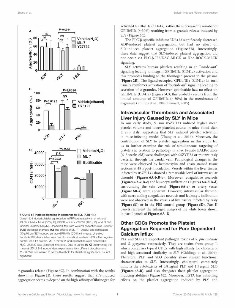
To investigate the possible signaling involved in plateletactivation induced by SLY, specific inhibitors were used.Both the MLCK-specific inhibitor ML-7 and ROCK-specificinhibitor Y27632 had no effect on SLY or ADP-inducedplatelet aggregation (Figure 5A). However, ML-7 had someeffects on the increased surface GPIIb/IIIa (CD41a) that from
Frontiers in Cellular and Infection Microbiology | www.frontiersin.org 8 October 2016 | Volume 6 | Article 128
Zhang et al. Suilysin-Induced Platelet Aggregation
FIGURE 5 | Platelet signaling in response to SLY. (A,B) rSLY
(1µg/mL)-induced platelet aggregation in PRP pretreated with or without
MLCK inhibitor ML-7 (100µM), ROCK inhibitor Y27632 (100 µM), and PLC-β
inhibitor U73122 (20µM). Unpaired t-test with Welch’s correction was used for
(A,B) statistical analyses. (C) The effects of ML-7 (100µM) and eptifibatide
(10µM) on rSLY-induced surface GPIIb/IIIa (CD41a) increase. Unpaired
two-tailed Student’s t-test was used for statistical analysis. PBS is the negative
control for rSLY protein. ML-7, Y27632, and eptifibatide were dissolved in
H2O. U73122 was dissolved in ethanol. Data in panels (A–C) are given as the
mean ± SD of 3–8 independent experiments from different blood donors.
P < 0.05 is considered to be the threshold for statistical significance; ns, not
significant.
α-granules release (Figure 5C). In combination with the resultsshown in Figure 2D, these results suggest that SLY-inducedaggregation seems to depend on the high-affinity of fibrinogen for
activated GPIIb/IIIa (CD41a), rather than increase the number ofGPIIb/IIIa (∼30%) resulting from α-granule release induced bySLY (Figure 3C).
The PLC-β-specific inhibitor U73122 significantly decreasedADP-induced platelet aggregation, but had no effect onSLY-induced platelet aggregation (Figure 5B). Interestingly,these data suggest that SLY-induced platelet aggregation didnot occur via PLC-β-IP3/DAG-MLCK or Rho-ROCK-MLCKsignaling.
SLY activates human platelets resulting in an “inside-out”signaling leading to integrin GPIIb/IIIa (CD41a) activation andthis promotes binding to the fibrinogen present in the plasma(Figure 2B). The ligand-occupied GPIIb/IIIa (CD41a) in turnusually reinforces activation of “outside-in” signaling leading tosecretion of α-granules. However, eptifibatide had no effect onGPIIb/IIIa (CD41a) (Figure 5C); this probably results from thelimited amounts of GPIIb/IIIa (∼30%) in the membranes ofα-granule (Phillips et al., 1988; Bennett, 2005).
Intravascular Thrombosis and AssociatedLiver Injury Caused by SLY in MiceIn our early study, S. suis 05ZYH33 induced higher meanplatelet volume and lower platelets counts in mice blood thanS. suis 1sly, suggesting that SLY induced platelet activationin mice-infecting model (Zhang et al., 2016). Moreover, thecontribution of SLY to platelet aggregation in this study ledus to further examine the role of simultaneous targeting ofplatelets in relation to pathology in vivo. Female BALB/c mice(6–8 weeks old) were challenged with 05ZYH33 or mutant 1slybacteria, through the caudal vein. Pathological changes in themice were observed by hematoxylin and eosin stained tissuesections at 48 h post-inoculation. Vessels within the liver tissuesinfected by 05ZYH33 showed a remarkable level of intravascularthrombi (Figures 6A-b,B-b). Moreover, coagulative necrosis(Figures 6A-c,B-c) and leukocyte infiltration (Figures 6A-d,B-d)surrounding the vein vessel (Figure 6A-a) or artery vessel(Figure 6B-a) were apparent. However, intravascular thrombiwith surrounding coagulative necrosis and leukocyte infiltrationwere not observed in the vessels of live tissues infected by 1sly(Figure 6C) or in the PBS control group (Figure 6D). Part IIpanels represent the enlarged images of the white boxes shownin part I panels of Figures 6A–D.
Other CDCs Promote the PlateletAggregation Required for Pore DependentCalcium InfluxPLY and SLO are important pathogen toxins of S. pneumoniaeand S. pyogenes, respectively. They are toxins from group I,which comprises typical CDCs with high affinity for cholesteroland high structural similarity to SLY (Giddings et al., 2004).Therefore, PLY and SLO possibly share similar functionalcharacteristics to SLY. Interestingly, cholesterol completelyinhibits the cytotoxicity of 0.8µg/ml PLY and 1.5µg/ml SLO(Figures 7A,B), and also abrogates their platelet aggregationinducing abilities (Figure 7C). Moreover, EGTA has inhibitingeffects on the platelet aggregation induced by PLY and
Frontiers in Cellular and Infection Microbiology | www.frontiersin.org 9 October 2016 | Volume 6 | Article 128

Zhang et al. Suilysin-Induced Platelet Aggregation
FIGURE 6 | SLY contributes to the intravascular thrombosis and its associated liver injury caused by S. suis. Female BALB/c mice (6–8 weeks old) were
challenged with (A,B) 05ZYH33; (C) mutant 1sly (∼1×10⁸ CFU); (D) PBS control through the caudal vein. Pathological changes were observed by hematoxylin and
eosin pathological staining of tissue sections at 48 h post-inoculation. Panels numbered II are the enlarged images from the white boxes in panels numbered I. a, b, c,
and d in the panels represent blood vessels, intravascular thrombosis, coagulative necrosis, and leukocyte infiltration, respectively.
Frontiers in Cellular and Infection Microbiology | www.frontiersin.org 10 October 2016 | Volume 6 | Article 128

Zhang et al. Suilysin-Induced Platelet Aggregation
FIGURE 7 | rPLY and rSLO induce platelet aggregation via pore dependent calcium influx. The cytotoxicity of rPLY (A) and rSLO (B) against platelets and the
cholesterol inhibiting effect were assessed by an LDH assay (Methods Section). (C) The cholesterol (100µg/mL) effect on rPLY (0.8µg/mL)- and rSLO
(1.5µg/mL)-induced platelet aggregation. (D) The EGTA (3 mM) effect on rPLY (0.8µg/mL)- and rSLO (1.5µg/mL)-induced PNA formation. PBS acted as the negative
control for the recombinant proteins. Cholesterol and EGTA were dissolved in ethanol and H2O, respectively. Unpaired t-test with Welch’s correction was used for
(A,B,D) statistical analyses. Unpaired two-tailed Student’s t-test was used for (C) statistical analysis. Data in panels (A–D) are given as the mean ± SD of three
independent experiments, with each experiment using blood from a different donor. P < 0.05 is considered to be the threshold for statistical significance; ns, not
significant; rPLY, recombinant pneumolysin; rSLO, recombinant streptolysin O; Cho, cholesterol; 0.8+Cho, 0.8µg/mL of rPLY added to cholesterol; 1.5+Cho,
1.5µg/mL of rSLO added to cholesterol.
SLO (Figure 7D), although the difference was not statisticallysignificant in the SLO group. It is possible that CDC-mediatedplatelet aggregation is a common mechanism used by a widerange of bacteria, and is triggered by pore dependent calciuminflux.
DISCUSSION
We have identified the cytolysin SLY is the main stimulusrequired for human platelet activation in S. suis 05ZYH33, whichis a sequenced strain belonging to sequence type 7 (ST-7) strainsthat caused the 2005 S. suis outbreak and STSS in China (Yeet al., 2009). Our systematic analysis of SLY found that it inducesplatelet activation and aggregation triggered by pore dependentCa2+ influx.
Different from S. aureus, S. pyogenes, and S. pneumoniae,SLY was found to be the sole factor inducing human platelet
activation and aggregation. For example, the fibrinogen-bindingproteins, ClfA, ClfB, and SdrE of S. aureus can all interact withGPIIb/IIIa, generating an outside-in signal to trigger plateletaggregation (O’brien et al., 2002; Liu et al., 2007). S. suis isan emerging human pathogen that causes STSS. ST-7 strainscaused the outbreak in humans in China in 2005 and were moretoxic to human peripheral blood mononuclear cells than ST-1strains (mainly referring to the European virulent strains) (Yeet al., 2006). In particular, we have previously shown that theST-7 strains produce more SLY protein than the non-epidemicstrains, and this contributes to invasive infections (He et al.,2014). Additionally, similar to STSS patients infected by S. suis,05ZYH33 induced intravascular thrombosis and associated-liverinjury in our mouse infection model (Figure 6). Therefore, SLYmay be a potential therapeutic target for preventing S. suis-mediated platelet activation, thrombocytopenia related DIC, andpurpura gangrenosa.
Frontiers in Cellular and Infection Microbiology | www.frontiersin.org 11 October 2016 | Volume 6 | Article 128

Zhang et al. Suilysin-Induced Platelet Aggregation
Although it has been reported that there are 20 types ofbacterial cytolysin belonging to Group I CDCs with high affinityto cholesterol (Tabata et al., 2014), there is scant evidenceshowing associations existing betweenGroup I CDCs and plateletactivation. Ohkuni et al. has reported that recombinant SLY,PLY, and Sm-hPAF were stimuli capable of inducing plateletaggregation (Ohkuni et al., 2012). However, the mechanismsunderlying how these bacterial toxins induce platelet activationare not clearly understood. In a comprehensive review, Cox et al.proposed that pore-forming toxins activate platelets in a mannersimilar to α-toxin (Cox et al., 2011), but the data in the articlescited in this review did not clearly support this viewpoint. Ourcurrent study presents direct evidence showing that SLY induceda pore dependent Ca2+ influx in platelets, which was required forSLY-induced platelet aggregation; the other two CDCs, PLY, andSLO, also induced platelet aggregation in a similar manner.
The present study also found that SLY-induced plateletGPIIb/IIIa (CD41a) activation was Ca2+ influx dependent,suggesting that stimulation of this adhesion factor appearsto be downstream of SLY-induced Ca2+ influx. Contrastingly,MLCK may not be involved in platelet aggregation becauseML-7 failed to suppress SLY-induced platelet aggregationalthough it inhibited GPIIb/IIIa (CD41a) release in α-granules(Figures 5A,C). SLY-induced Ca2+ influx may directly activate“inside-out” signaling to enhance the affinity of GPIIb/IIIa tofibrinogen, which subsequently induces platelet aggregation.
We also found that the PLC-β inhibitor U73122 dramaticallydecreased ADP-induced platelet aggregation but had no impacton SLY-induced platelet aggregation. The data imply that thesignaling events involved in SLY-induced platelet activationmay differ from typical platelet agonists such as ADP andthromboxane A2 (TXA2) because these are dependent on PLC-β associated signaling (Zarbock et al., 2007). Furthermore, Bornaggregometry revealed that SLY-induced platelet aggregationshowed a 3-min lag time prior to massive aggregation occurring(Figure S4). In contrast, most platelet agonists such as ADP causerapid activation (Cox et al., 2011). The differential dynamicsfor SLY-induced platelet aggregation and ADP-induced plateletaggregation also suggest that distinct signaling might be inducedby SLY.
Overall, we found that SLY, which was secreted by S. suis05ZYH33 at the stationary growth phase, stimulated plateletactivation and aggregation. This stimulation required SLY-induced Ca2+ influx and subsequent “inside-out” signaling toactivate GPIIb/IIIa (CD41a) (as proposed in Figure 8).Moreover,PLY, SLO, and SLY, which are all Group I CDCs, seem to sharesimilar mechanisms for inducing platelet aggregation.We foreseethat this similar mode of activation identifies Group I CDCsas potential therapeutic targets for preventing bacterial-inducedplatelet activation and thrombotic-related disorders.
AUTHOR CONTRIBUTIONS
YY, SZ, and YJ conceived and designed the experiments. SZ, SC,and JW performed the experiments. SZ, JY, ZP, and YY analyzedthe data. KL, LL, and YZ contributed reagents/materials/analysis
FIGURE 8 | Schematic representation of platelet activation and
aggregation induced by S. suis. Ca2+ influx across transmembrane pores
created by SLY, the CDC of S. suis, can trigger inside-out signaling leading to
integrin GPIIb/IIIa activation and α-granule (GPIIb/IIIa) or dense granule (ATP)
secretion. Subsequently, GPIIb/IIIa activation leads to platelet aggregation.
Additionally, Ca2+-MLCK signaling is involved in α-granule release induced by
SLY.
tools. YY wrote the paper. All authors contributed to theinterpretation of the data and writing of the manuscript and readand approved the final version.
FUNDING
This work was supported by grants from the National BasicResearch Program (973) of China (2012CB518804) and theNational Natural Science Foundation of China (81371766).
ACKNOWLEDGMENTS
We are grateful to Prof. Marcelo Gottschalk (Université deMontréal, Montreal Quebec, Canada) for providing the avirulentstrain 1330.We thanks for Dr. Henk J.Wisselink donating strains4005 and s735. We also thank Dr. Jiye Yin and Dr. Zhiyuan Panfor analysis of the pathological changes.
SUPPLEMENTARY MATERIAL
The Supplementary Material for this article can be foundonline at: http://journal.frontiersin.org/article/10.3389/fcimb.2016.00128Figure S1 | The purity of human platelets in PRP. (A) Leukocytes in human
blood were analyzed by flow cytometry as forward- and side-scatter. (B) PRP was
analyzed by flow cytometry as forward- and side-scatter. (C) The percent of
CD41a positive cells in panel (B) was shown as representative histograms. The
Frontiers in Cellular and Infection Microbiology | www.frontiersin.org 12 October 2016 | Volume 6 | Article 128

Zhang et al. Suilysin-Induced Platelet Aggregation
platelets in PRP were detected by flow cytometry analysis using FITC conjugated
anti-CD41a (clone HIP8) antibody.
Figure S2 | The activity of S. suis-induced platelet aggregation. (A) The
culture supernatant and the washed bacteria cells of S. suis were added to PRP in
a stirred cuvette. Platelet aggregation was expressed as a final percentage of light
transmission detected by Platelet Aggregometer se-2000. ADP (20µM) and THB
were used as the positive controls and negative controls, respectively. Data are
expressed as the mean ± SD of three independent experiments, with each
experiment using blood from a different donor. (B) The platelet aggregation curves
shown in panel B are from one representative experiment of three independent
experiments. S. suis 4 (SLY+, 89K+) isolated from human patient in China (2005);
S. suis 4005 (SLY+, 89K−) and s735 (SLY+, 89K−) isolated from diseased piglets
in Netherlands; S. suis 1940 (SLY+, 89K−) isolated from diseased piglets in China
(1980); Sup, supernatant; SLY, suilysin; 89K−, 89 kb pathogenicity island.
Figure S3 | S. suis supernatant and rSLY-induced platelet aggregation in
PRP was assessed in the presence of 15µL anti-CD62P blocking antibody
or an isotype-matched control antibody (BD Bioscience). Unpaired
two-tailed Student’s t-test was used for statistical analysis. Data in are expressed
as the mean ± SD for three independent experiments, with each experiment using
blood from a different donor. P < 0.05 is considered as the threshold for
significance; ns, no significance; 05ZYH33, wild type strain; 1sly, The isogenic
mutants of sly; Sup, supernatant.
Figure S4 | Dose response of rSLY-induced platelet aggregation. Serial
concentrations of SLY were added to platelet-rich plasma (PRP) in a stirred
cuvette. Platelet aggregation was expressed as a final percentage of light
transmission.
Table S1 | Bacterial strains and plasmids used in this study.
REFERENCES
Arman, M., Krauel, K., Tilley, D. O., Weber, C., Cox, D., Greinacher, A., et al.(2014). Amplification of bacteria-induced platelet activation is triggered byFcgammaRIIA, integrin aIIbb3, and platelet factor 4. Blood 123, 3166–3174. doi:10.1182/blood-2013-11-540526
Arvand, M., Bhakdi, S., Dahlbäck, B., and Preissner, K. T. (1990). Staphylococcusaureus alpha-toxin attack on human platelets promotes assembly of theprothrombinase complex. J. Biol. Chem. 265, 14377–14381.
Bennett, J. S. (2005). Structure and function of the platelet integrin αIIbβ3. J. Clin.Invest. 115, 3363–3369. doi: 10.1172/JCI26989
Bi, L., Pian, Y., Chen, S., Ren, Z., Liu, P., Lv, Q., et al. (2015). Toll-like receptor4 confers inflammatory response to suilysin. Front. Microbiol. 6:644. doi:10.3389/fmicb.2015.00644
Bird, G. S., Aziz, O., Lievremont, J. P., Wedel, B. J., Trebak, M., Vazquez, G., et al.(2004). Mechanisms of phospholipase C-regulated calcium entry. Curr. Mol.
Med. 4, 291–301. doi: 10.2174/1566524043360681Cox, D., Kerrigan, S.W., andWatson, S. P. (2011). Platelets and the innate immune
system: mechanisms of bacterial-induced platelet activation. J. Thromb.
Haemost. 9, 1097–1107. doi: 10.1111/j.1538-7836.2011.04264.xDavì, G., Neri, M., Falco, A., Festi, D., Taraborelli, T., Ciabattoni, G., et al. (2005).
Helicobacter pylori infection causes persistent platelet activation in vivo throughenhanced lipid peroxidation. Arterioscler. Thromb. Vasc. Biol. 25, 246–251. doi:10.1161/01.ATV.0000147128.10278.99
Fitzgerald, J. R., Loughman, A., Keane, F., Brennan, M., Knobel, M., Higgins, J.,et al. (2006). Fibronectin-binding proteins of Staphylococcus aureus mediateactivation of human platelets via fibrinogen and fibronectin bridges to integrinGPIIb/IIIa and IgG binding to the FcgammaRIIa receptor. Mol. Microbiol. 59,212–230. doi: 10.1111/j.1365-2958.2005.04922.x
Ford, I., Douglas, C. W., Heath, J., Rees, C., and Preston, F. E. (1996).Evidence for the involvement of complement proteins in platelet aggregationby Streptococcus sanguis NCTC 7863. Br. J. Haematol. 94, 729–739. doi:10.1046/j.1365-2141.1996.d01-1857.x
Giddings, K. S., Zhao, J., Sims, P. J., and Tweten, R. K. (2004). Human CD59 isa receptor for the cholesterol-dependent cytolysin intermedilysin. Nat. Struct.Mol. Biol. 11, 1173–1178. doi: 10.1038/nsmb862
Gilbert, R. J., Jiménez, J. L., Chen, S., Tickle, I. J., Rossjohn, J., Parker, M.,et al. (1999). Two structural transitions in membrane pore formation bypneumolysin, the pore-forming toxin of Streptococcus pneumoniae. Cell 97,647–655. doi: 10.1016/S0092-8674(00)80775-8
Gottschalk, M. G., Lacouture, S., and Dubreuil, J. D. (1995). Characterization ofStreptococcus suis capsular type 2 haemolysin.Microbiology 141(Pt 1), 189–195.doi: 10.1099/00221287-141-1-189
Grewal, P. K., Uchiyama, S., Ditto, D., Varki, N., Le, D. T., Nizet, V., et al. (2008).The Ashwell receptor mitigates the lethal coagulopathy of sepsis. Nat. Med. 14,648–655. doi: 10.1038/nm1760
Hao, H., Hui, W., Liu, P., Lv, Q., Zeng, X., Jiang, H., et al. (2013). Effect oflicochalcone A on growth and properties of Streptococcus suis. PLoS ONE
8:e67728. doi: 10.1371/journal.pone.0067728
He, Z., Pian, Y., Ren, Z., Bi, L., Yuan, Y., Zheng, Y., et al. (2014). Increasedproduction of suilysin contributes to invasive infection of the Streptococcus suisstrain 05ZYH33.Mol. Med. Rep. 10, 2819–2826. doi: 10.3892/mmr.2014.2586
Keane, C., Tilley, D., Cunningham, A., Smolenski, A., Kadioglu, A., Cox, D.,et al. (2010). Invasive Streptococcus pneumoniae trigger platelet activation viaToll-like receptor 2. J. Thromb. Haemost. 8, 2757–2765. doi: 10.1111/j.1538-7836.2010.04093.x
Klages, B., Brandt, U., Simon, M. I., Schultz, G., and Offermanns, S. (1999).Activation of G12/G13 results in shape change and Rho/Rho-kinase-mediatedmyosin light chain phosphorylation in mouse platelets. J. Cell Biol. 144,745–754. doi: 10.1083/jcb.144.4.745
Liu, C. Z., Huang, T. F., Tsai, P. J., Tsai, P. J., Chang, L. Y., and Chang, M. C. (2007).A segment of Staphylococcus aureus clumping factor A with fibrinogen-bindingactivity (ClfA221-550) inhibits platelet-plug formation in mice. Thromb. Res.
121, 183–191. doi: 10.1016/j.thromres.2007.03.019Lourbakos, A., Yuan, Y. P., Jenkins, A. L., Travis, J., Andrade-Gordon, P., Santulli,
R., et al. (2001). Activation of protease-activated receptors by gingipains fromPorphyromonas gingivalis leads to platelet aggregation: a new trait in microbialpathogenicity. Blood 97, 3790–3797. doi: 10.1182/blood.V97.12.3790
Lv, Q. Y., Hao, H. J., Bi, L. L., Zheng, Y. L., Jiang, Y. Q., and Lv, S. X. (2011).Purification and biological activities analysis of Streptococcus suis Serotype 2suilysin. Xi Bao Yu Fen Zi Mian Yi Xue Za Zhi 27, 374–376.
Meesters, C., Brack, A., Hellmann, N., and Decker, H. (2009). Structuralcharacterization of the alpha-hemolysin monomer from Staphylococcus aureus.Proteins 75, 118–126. doi: 10.1002/prot.22227
Miajlovic, H., Zapotoczna, M., Geoghegan, J. A., Kerrigan, S. W., Speziale, P., andFoster, T. J. (2010). Direct interaction of iron-regulated surface determinantIsdB of Staphylococcus aureus with the GPIIb/IIIa receptor on platelets.Microbiology (Reading Engl). 156, 920–928. doi: 10.1099/mic.0.036673-0
O’brien, L., Kerrigan, S. W., Kaw, G., Hogan, M., Penadés, J., Litt, D., et al. (2002).Multiple mechanisms for the activation of human platelet aggregation byStaphylococcus aureus: roles for the clumping factors ClfA and ClfB, the serine-aspartate repeat protein SdrE and protein A. Mol. Microbiol. 44, 1033–1044.doi: 10.1046/j.1365-2958.2002.02935.x
Ohkuni, H., Nagamune, H., Ozaki, N., Tabata, A., Todome, Y., Watanabe, Y.,et al. (2012). Characterization of recombinant Streptococcus mitis-derivedhuman platelet aggregation factor. APMIS 120, 56–71. doi: 10.1111/j.1600-0463.2011.02813.x
O’seaghdha, M., van Schooten, C. J., Kerrigan, S. W., Emsley, J., Silverman, G.J., Cox, D., et al. (2006). Staphylococcus aureus protein A binding to vonWillebrand factor A1 domain is mediated by conserved IgG binding regions.FEBS J. 273, 4831–4841. doi: 10.1111/j.1742-4658.2006.05482.x
Parimon, T., Li, Z., Bolz, D. D., McIndoo, E. R., Bayer, C. R., Stevens, D. L., et al.(2013). Staphylococcus aureus alpha-hemolysin promotes platelet-neutrophilaggregate formation. J. Infect. Dis. 208, 761–770. doi: 10.1093/infdis/jit235
Phillips, D. R., Charo, I. F., Parise, L. V., and Fitzgerald, L. A. (1988). The plateletmembrane glycoprotein IIb-IIIa complex. Blood 71, 831–843.
Pian, Y., Gan, S., Wang, S., Guo, J., Wang, P., Zheng, Y., et al. (2012). Fhb,a novel factor H-binding surface protein, contributes to the antiphagocytic
Frontiers in Cellular and Infection Microbiology | www.frontiersin.org 13 October 2016 | Volume 6 | Article 128

Zhang et al. Suilysin-Induced Platelet Aggregation
ability and virulence of Streptococcus suis. Infect. Immun. 80, 2402–2413. doi:10.1128/IAI.06294-11
Pian, Y., Wang, P., Liu, P., Zheng, Y., Zhu, L., Wang, H., et al. (2015).Proteomics identification of novel fibrinogen-binding proteins of Streptococcussuis contributing to antiphagocytosis. Front. Cell. Infect. Microbiol. 5:19. doi:10.3389/fcimb.2015.00019
Plummer, C., Wu, H., Kerrigan, S. W., Meade, G., Cox, D., and Ian Douglas, C. W.(2005). A serine-rich glycoprotein of Streptococcus sanguis mediates adhesionto platelets via GPIb. Br. J. Haematol. 129, 101–109. doi: 10.1111/j.1365-2141.2005.05421.x
Que, Y. A., François, P., Haefliger, J. A., Entenza, J. M., Vaudaux, P., andMoreillon,P. (2001). Reassessing the role of Staphylococcus aureus clumping factor andfibronectin-binding protein by expression in Lactococcus lactis. Infect. Immun.
69, 6296–6302. doi: 10.1128/IAI.69.10.6296-6302.2001Rebecchi, M. J., and Pentyala, S. N. (2000). Structure, function, and control of
phosphoinositide-specific phospholipase C. Physiol. Rev. 80, 1291–1335.Rhee, S. G. (2001). Regulation of phosphoinositide-specific phospholipase
C. Annu. Rev. Biochem. 70, 281–312. doi: 10.1146/annurev.biochem.70.1.281
Rossjohn, J., Feil, S. C., McKinstry, W. J., Tweten, R. K., and Parker, M. W.(1997). Structure of a cholesterol-binding, thiol-activated cytolysin and amodel of its membrane form. Cell 89, 685–692. doi: 10.1016/S0092-8674(00)80251-2
Shannon, O., Hertzén, E., Norrby-Teglund, A., Morgelin, M., Sjöbring, U., andBjörck, L. (2007). Severe streptococcal infection is associated with M protein-induced platelet activation and thrombus formation. Mol. Microbiol. 65,1147–1157. doi: 10.1111/j.1365-2958.2007.05841.x
Sriskandan, S., and Slater, J. D. (2006). Invasive disease and toxic shock due tozoonotic Streptococcus suis: an emerging infection in the East? PLoS Med.
3:e187. doi: 10.1371/journal.pmed.0030187Ståhl, A. L., Svensson, M., Morgelin, M., Svanborg, C., Tarr, P. I., Mooney, J.
C., et al. (2006). Lipopolysaccharide from enterohemorrhagic Escherichia coli
binds to platelets through TLR4 and CD62 and is detected on circulatingplatelets in patients with hemolytic uremic syndrome. Blood 108, 167–176. doi:10.1182/blood-2005-08-3219
Tabata, A., Ohkura, K., Ohkubo, Y., Tomoyasu, T., Ohkuni, H., Whiley, R. A.,et al. (2014). The diversity of receptor recognition in cholesterol-dependentcytolysins. Microbiol. Immunol. 58, 155–171. doi: 10.1111/1348-0421.12131
Tang, J., Wang, C., Feng, Y., Yang, W., Song, H., Chen, Z., et al. (2006).Streptococcal toxic shock syndrome caused by Streptococcus suis serotype 2.PLoS Med. 3:e151. doi: 10.1371/journal.pmed.0030151
Walsh, E. J., Miajlovic, H., Gorkun, O. V., and Foster, T. J. (2008). Identificationof the Staphylococcus aureusMSCRAMM clumping factor B (ClfB) binding sitein the alphaC-domain of human fibrinogen. Microbiology (Reading Engl). 154,550–558. doi: 10.1099/mic.0.2007/010868-0
Wangkaew, S., Chaiwarith, R., Tharavichitkul, P., and Supparatpinyo, K. (2006).Streptococcus suis infection: a series of 41 cases from Chiang Mai UniversityHospital. J. Infect. 52, 455–460. doi: 10.1016/j.jinf.2005.02.012
Xu, L., Huang, B., Du, H., Zhang, X. C., Xu, J., Li, X., et al. (2010). Crystal structureof cytotoxin protein suilysin from Streptococcus suis. Protein Cell 1, 96–105. doi:10.1007/s13238-010-0012-3
Ye, C., Zheng, H., Zhang, J., Jing, H., Wang, L., Xiong, Y., et al. (2009). Clinical,experimental, and genomic differences between intermediately pathogenic,highly pathogenic, and epidemic Streptococcus suis. J. Infect. Dis. 199, 97–107.doi: 10.1086/594370
Ye, C., Zhu, X., Jing, H., Du, H., Segura, M., Zheng, H., et al. (2006). Streptococcussuis sequence type 7 outbreak, Sichuan, China. Emerging Infect. Dis. 12,1203–1208. doi: 10.3201/eid1208.060232
Yeh, J. J., Tsai, S., Wu, D. C., Wu, J. Y., Liu, T. C., and Chen, A. (2010). P-selectin-dependent platelet aggregation and apoptosis may explain the decreasein platelet count during Helicobacter pylori infection. Blood 115, 4247–4253.doi: 10.1182/blood-2009-09-241166
Yu, H. J., Jing, H. Q., Chen, Z. H., Zheng, H., Zhu, X. P., Wang, H., et al. (2006).Human Streptococcus suis outbreak, Sichuan, China. Emerging Infect. Dis. 12,914–920. doi: 10.3201/eid1206.051194
Zarbock, A., Polanowska-Grabowska, R. K., and Ley, K. (2007). Platelet–neutrophil-interactions: linking hemostasis and inflammation. Blood Rev. 21,99–111. doi: 10.1016/j.blre.2006.06.001
Zhang, S., Liu, P., Xu, M., Sang, X., Zheng, Y., Yuan, Y., et al. (2016). Activityidentification of Streptococcus suis suilysin inducing platelets aggregation. Mil.
Med. Sci. 40, 1–5. doi: 10.1016/j.socscimed.2015.12.035
Conflict of Interest Statement: The authors declare that the research wasconducted in the absence of any commercial or financial relationships that couldbe construed as a potential conflict of interest.
Copyright © 2016 Zhang, Wang, Chen, Yin, Pan, Liu, Li, Zheng, Yuan and Jiang.
This is an open-access article distributed under the terms of the Creative Commons
Attribution License (CC BY). The use, distribution or reproduction in other forums
is permitted, provided the original author(s) or licensor are credited and that the
original publication in this journal is cited, in accordance with accepted academic
practice. No use, distribution or reproduction is permitted which does not comply
with these terms.
Frontiers in Cellular and Infection Microbiology | www.frontiersin.org 14 October 2016 | Volume 6 | Article 128


![Dec. 1869.] Veazie Bank v. Fenno](https://img.pdfslide.us/doc/110x75/6286913eb27a7175ba52f4e5/dec-1869-veazie-bank-v-fenno.jpg)