Embed Size (px)
Citation preview
EFFECTS OF STINGLESS BEE PROPOLIS ON
OXIDATIVE STRESS AND STRUCTURAL
INTEGRITY OF HEART IN STREPTOZOTOCIN-
INDUCED DIABETIC RATS
LIM OON ZHI
UNIVERSITI SAINS MALAYSIA
2020
EFFECTS OF STINGLESS BEE PROPOLIS ON
OXIDATIVE STRESS AND STRUCTURAL
INTEGRITY OF HEART IN STREPTOZOTOCIN-
INDUCED DIABETIC RATS
by
LIM OON ZHI
Thesis submitted in fulfilment of the requirements
for the degree of
Master of Science
April 2020
ACKNOWLEDGEMENT
First and foremost, I would like to express highest gratitude to my main supervisor,
Dr Norsuhana Omar for her support throughout my Master of Science (MSc)
research study. “To achieve your dream, you must be diligent in your field” are the
words of encouragement she told her sons in front of me, and indirectly sparked my
will to gather knowledge as much as possible and be very hardworking to achieve
what I wanted. Next, I felt very thankful to have an excellent co-researcher, Dr Yeoh
Boon Seng who would strengthen my study framework and his ability to construct
powerful sentences to link scientific evidence in a short and sweet manner. I
appreciate all the robust comments from my co-supervisors, Associate Professor Dr
Mahaneem Binti Mohamed, Dr Anani Aila Mat Zin. I am grateful to Dr Rozaziana
Ahmad as contributive co-researcher. I credit laboratory staff and students especially
Madam Aminah Che Romli, Madam Siti Nor Zuraini, Madam Che Nurrul Hidayu
Che Ismail and Mr Khairul Hizman B. Mohd Sobri for their assistance in handling
advanced lab equipments. I thank the wonderful service by librarians, Madam Nurul
Azurah Mohd Roni and Mr Shaari Nora Bin Md Yusof. I honour School of Medical
Sciences (PPSP), School of Health Sciences (PPSK), School of Dental Sciences
(PPSG), Institute of Postgraduate Studies and Hamdan Tahir Library for organising
workshops essential for postgraduate students including myself in research study. I
acknowledge the financial support from Universiti Sains Malaysia, School of
Medical Sciences Postgraduate Conference Fund in presentation of my research
findings in National Heart Association Malaysia Congress 2019. I am grateful to my
parents, Dr Lim Kok Ewe and Madam Wong Hie Nien for subsidising payment of
tuition fees, cost of living and participation in workshops and conferences
ii
TABLE OF CONTENT
ACKNOWLEDGEMENT.........................................................................................ii
TABLE OF CONTENT............................................................................................iii
LIST OF TABLES....................................................................................................xii
LIST OF FIGURES.................................................................................................xiii
LIST OF PLATES....................................................................................................xv
LIST OF ACRONYMS, ABBREVIATIONS AND SYMBOLS.........................xvi
ABSTRAK...............................................................................................................xxii
ABSTRACT............................................................................................................xxiv
CHAPTER 1 INTRODUCTION...............................................................................1
1.1 A Concerning Global Health Threat: Non-communicable Diseases...................1
1.2 A Silent Killer: Diabetes Mellitus.......................................................................1
1.3 Landscape of Diabetes Mellitus in the world and Malaysia................................2
1.4 Cardiovascular Disease as a Major Complication of Diabetes Mellitus.............3
1.5 Cardiovascular Disease in Type 1 Diabetes Mellitus..........................................4
1.6 Epidemiological Evidence of Diabetic Cardiomyopathy....................................6
1.7 Oxidative Hypothesis of Diabetic Cardiomyopathy............................................7
1.8 Justification of Study...........................................................................................9
1.9 Research Objectives..........................................................................................10
1.9.1 General Objective................................................................................10
1.9.2 Specific Objectives..............................................................................10
1.10 Research Hypothesis.........................................................................................10
CHAPTER 2 LITERATURE REVIEW.................................................................11
iii
2.1 Meliponiculture or Apiculture: The Battle of Sustainability in
Malaysian Beekeeping Industry........................................................................11
2.2 Biology of Stingless Bee...................................................................................12
2.3 History of Propolis............................................................................................13
2.4 Bioactivity and Chemical Composition of Stingless Bee Propolis...................15
2.4.1 Stingless Bee Propolis as Natural Antioxidant...................................15
2.4.2 Antimicrobial activity of Stingless Bee Propolis................................16
2.4.3 Anticancer and Antiinflammatory Property of Stingless
Bee Propolis........................................................................................17
2.5 Therapeutic Potential of Stingless Bee Propolis in Diabetic
Cardiomyopathy................................................................................................17
2.6 Experimental Model of Type 1 Diabetes Mellitus............................................18
2.6.1 Methods of Type 1 Diabetes Mellitus Induction in Animal
Models.................................................................................................19
2.6.2 Chemical Induction of Type 1 Diabetes Mellitus...............................20
2.6.3 Mechanism of Streptozotocin Action..................................................21
2.6.4 Practical Application of Streptozotocin..............................................21
2.6.5 Diabetic Cardiomyopathy in Streptozotocin-induced
Diabetic Rat Model.............................................................................22
2.7 Structural and Functional Phenotype in Diabetic Cardiomyopathy..................22
2.7.1 Cardiac Hypertrophy...........................................................................23
2.7.2 Interstitial and Perivascular Fibrosis...................................................23
2.7.3 Diastolic Dysfunction..........................................................................25
2.8 Oxidative Stress in Diabetic Cardiomyopathy..................................................25
iv
2.8.1 AGE-RAGE Signaling and Oxidative Stress in Diabetic
Cardiomyopathy..................................................................................26
2.8.2 Endogenous Secretory Receptor for Advanced Glycation
End Products: A Protective Scavenger................................................28
2.8.3 Glycaemic Variability in Production of Oxidative Stress...................29
2.8.4 Restoring Antioxidative as Therapeutic Target..................................30
2.9 Therapeutic Effect of Metformin in Type 1 Diabetes Mellitus.........................30
2.9.1 Clinical and Preclinical Data Supporting Cardioprotective
Effect of Metformin............................................................................31
CHAPTER 3 MATERIALS AND METHODS......................................................33
3.1 Materials............................................................................................................33
3.1.1 Stingless bee propolis sample.............................................................33
3.1.2 Chemicals and reagents.......................................................................36
3.1.3 Commercial kits and consumables......................................................36
3.1.4 Instruments..........................................................................................36
3.2 Methods.............................................................................................................40
3.2.1 Preparation of ethanol extract of Malaysian propolis.........................40
3.2.2 Animals...............................................................................................40
3.2.3 Experimental design............................................................................41
3.2.4 Calculation of sample size...................................................................43
3.2.5 Induction and assessment of type 1 diabetes mellitus.........................43
3.2.5 (a) Animal grouping...............................................................43
3.2.5 (b) Streptozotocin solution preparation and
induction of T1DM...........................................................43
3.2.5 (c) Induction of T1DM..........................................................44
v
3.2.5 (d) Post-induction assessment of DM....................................44
3.2.6 Administration of treatment................................................................44
3.2.7 Assessment of body weight (BW), food intake (FI) and
water intake (WI)................................................................................44
3.2.8 Determination of fasting blood glucose (FBG)...................................45
3.2.9 Animal sacrifice..................................................................................45
3.2.9 (a) Preparation of serum samples...........................................45
3.2.9 (b) Preparation of heart homogenate samples........................45
3.2.9 (c) Preparation of phosphate buffer saline.............................46
3.2.9 (d) Preparation of histological samples..................................46
3.2.10 Assessment of oxidative stress and antioxidant biomarkers...............46
3.2.11 Determination of total protein concentration using BCA
method.................................................................................................47
3.2.11 (a) Preparation of standard working solution and
sample dilution.................................................................47
3.2.11 (b) Procedures using 96-wells microplate..............................47
3.2.11 (c) Calculation of total protein concentration in
heart homogenate.............................................................48
3.2.12 Determination of homogenate malondialdehyde (MDA)
activity.................................................................................................50
3.2.12 (a) Preparation of standard working solution and
sample dilution.................................................................50
3.2.12 (b) Preparation of biotinylated detection antibody
working solution...............................................................50
3.2.12 (c) Preparation of wash buffer...............................................50
vi
3.2.12 (d) Preparation of avidian to horseradish peroxidase
(HRP) conjugate working solution...................................51
3.2.12 (e) Assay procedure using 96-well microplate......................51
3.2.12 (f) Calculation of homogenate malondialdehyde
(MDA) activity.................................................................51
3.2.13 Determination of homogenate glutathione peroxidase 1
(GPx-1) activity...................................................................................54
3.2.13 (a) Preparation of standard working solution and
sample dilution.................................................................54
3.2.13 (b) Preparation of biotinylated detection antibody
working solution...............................................................54
3.2.13 (c) Preparation of wash buffer...............................................54
3.2.13 (d) Preparation of avidian to horseradish peroxidase
(HRP) conjugate working solution...................................54
3.2.13 (e) Assay procedure using 96-well microplate......................55
3.2.13 (f) Calculation of homogenate glutathione
peroxidase 1 (GPx-1) activity...........................................55
3.2.14 Determination of homogenate superoxide dismutase (SOD)
activity.................................................................................................57
3.2.14 (a) Preparation of substrate applied solution.........................57
3.2.14 (b) Preparation of enzyme working solution..........................57
3.2.14 (c) Preparation of diluted samples.........................................57
3.2.14 (d) Assay procedure using 96-well microplate......................57
3.2.14 (e) Calculation of homogenate SOD activity.........................57
3.2.15 Determination of homogenate catalase (CAT) activity.......................60
vii
3.2.15 (a) Preparations of reagents...................................................60
3.2.15 (b) Assay procedure plastic test tube and cuvette..................60
3.2.15 (c) Calculation of homogenate CAT activity.........................60
3.2.16 Determination of homogenate advanced glycation end
product (AGE) concentration..............................................................62
3.2.16 (a) Preparation of AGE conjugate.........................................62
3.2.16 (b) Coating of 96-wells microplate with AGE
conjugate overnight..........................................................62
3.2.16 (c) Preparation of AGE-BSA standard and sample
dilution..............................................................................62
3.2.16 (d) Preparation of wash buffer...............................................62
3.2.16 (e) Preparation of primary anti-AGE antibody and
secondary antibody...........................................................63
3.2.16 (f) Assay procedure with 96-well microplate........................63
3.2.16 (g) Calculation of AGE concentration...................................63
3.2.17 Determination of serum and heart endogenous secretory
advanced glycosylation end product specific receptor
(esRAGE) concentration.....................................................................66
3.2.17 (a) Preparation of standard working solution and
sample dilution.................................................................66
3.2.17 (b) Preparation of wash buffer...............................................66
3.2.17 (c) Preparation of biotinylated detection antibody
working solution...............................................................66
3.2.17 (d) Preparation of HRP conjugate working solution..............66
3.2.17 (e) Assay procedure using 96-well microplate......................67
viii
3.2.17 (f) Calculation of esRAGE activity.......................................67
3.2.18 Histomorphology assessment..............................................................69
3.2.18 (a) Tissue fixation..................................................................69
3.2.18 (b) Tissue processing.............................................................69
3.2.18 (c) Tissue embedding.............................................................69
3.2.18 (d) Tissue sectioning..............................................................70
3.2.18 (e) Tissue staining..................................................................70
3.2.18 (e)(i) Haematoxylin and Eosin staining................70
3.2.18 (e)(ii) Masson trichrome staining...........................73
3.2.19 Histomorphological analysis...............................................................75
3.2.19 (a) Qualitative analysis of H&E slides..................................75
3.2.19 (b) Quantitative analysis for cardiomyocyte size...................75
3.2.19 (c) Quantitative analysis for perivascular collagen
area to luminal area ratio (PVCA/LA).............................75
3.2.19 (d) Quantitative analysis for interstitial collagen
volume fraction (CVF).....................................................76
3.2.20 Statistical analysis...............................................................................77
CHAPTER 4 RESULTS...........................................................................................79
4.1 Body weight, food intake, water intake and fasting blood glucose
among rats.........................................................................................................79
4.2 Oxidative stress biomarker and antioxidative defense in right ventricle..........86
4.3 Histology of left ventricle..................................................................................93
CHAPTER 5 DISCUSSION...................................................................................102
CHAPTER 6 SUMMARY AND CONCLUSION................................................110
6.1 Summary and Conclusion of Study.................................................................110
ix
6.2 Strengths and Limitations................................................................................114
6.3 Recommendation for Future Research............................................................114
REFERENCES........................................................................................................114
APPENDICES
Appendix A: Participation In National Heart Association Of Malaysia Congress
2019 As Oral Presenter
Appendix B: The Abstract For Young Investigator Award Oral Presentation
At National Heart Association Congress 2019
Appendix C: The Photograph Taken When Receiving Prize From Chairman At
Main Stage Of National Heart Association Of Malaysia Congress 2019
Appendix D: The Certificate Of Merit As Consolation Prize Winner Of Young
Investigator Award At National Heart Association Congress 2019
Appendix E: Participation As Oral Presenter At Clinical Research Malaysia Research
Track In Conjunction With National Heart Association Malaysia
Congress 2019
Appendix F: The Abstract For Oral Presentation At National Heart Association
Malaysia - Clinical Research Malaysia Research Track 2019
Appendix G: The Photograph Taken When Receiving Prize Ceremony From Ceo
Of Clinical Research Malaysia
Appendix H: Certificate Of Merit For Winning 2nd Runner Up At National Heart
Association Malaysia- Clinical Research Malaysia Research Track
2019
Appendix I: Participation Of 3rd International Conference On Medical And Health
Sciences & 24th National Conference On Medical And Health Sciences
As Oral Presenter
x
Appendix J: The Abstract For Oral Presentation At 3rd International Conference On
Medical And Health Sciences & 24th National Conference On Medical
And Health Sciences
Appendix K: Certificate Of Presentation For 3rd International Conference On Medical
And Health Sciences & 24th National Conference On Medical And
Health Sciences
Appendix L: Participation Of Seminar Perubatan Integratif As Poster Presenter
Appendix M: The Abstract For Poster Presentation At Seminar Perubatan Integratif
Appendix N: Certificate Of Acknowledgement For Poster Presentation At Seminar
Perubatan Integratif
Appendix O: Proof Of Accepted Manuscript For Publication In Brazilian Journal Of
Pharmaceutical Sciences
LIST OF PRESENTATION AND PUBLICATIONS
xi
LIST OF TABLES
Page
Table 2.1 Methods to induce type 1 diabetes mellitus in Animal models 19
Table 3.1 List of chemical and reagents 37
Table 3.2 List of commercial kits and consumables 38
Table 3.3 List of instruments 39
Table 3.4 Dilution of BSA reference standard 49
Table 3.5 Serial dilution of the MDA and GPx-1 reference standard 52
Table 3.6 Protocol of competitive enzyme-linked immunosorbent assay
(ELISA) 53
Table 3.7 Assay procedure for sandwich-ELISA 56
Table 3.8 Operating procedure for SOD 59
Table 3.9 Operational procedures of CAT activity 61
Table 3.10 Serial dilution of AGE-BSA standard 64
Table 3.11 Operational steps for AGE 65
Table 3.12 Serial dilution of esRAGE reference standard 68
Table 3.13 Haematoxylin and Eosin (H&E) staining protocol 72
Table 3.14 Masson trichrome staining protocol 74
Table 4.1 Heart oxidative stress biomarker, (n=8) 86
xii
LIST OF FIGURES
Page
Figure 3.1 Flow chart of study 42
Figure 4.1 Food intake of rats across experimental period, n=8 per group 80
Figure 4.2 Water intake of rats across experimental period, n=8 per group 82
Figure 4.3 Body weight of rats across experimental period, n=8 per group 83
Figure 4.4 Fasting blood glucose of rats across experimental period, n=8 8585
Figure 4.5 Heart Glutathione Peroxidase 1 (GPx-1) concentration among
study groups in rats, n=8 88
Figure 4.6 Heart SOD activity among study groups in rats, n=8 89
Figure 4.7 Heart CAT activity among experimental groups, n=8 90
Figure 4.8 Heart esRAGE concentration among experimental groups,
n=8 91
Figure 4.9 Serum esRAGE concentration among study groups in rats,
n=8 92
Figure 4.10 Photomicrograph of representative transverse section of rat
myocardium in left ventricle viewed under 400x
magnification with Haematoxylin and eosin stain 95
Figure 4.11 Photomicrograph of representative of longitudinal section of
rat myocardium in left ventricle viewed under 200x magnification
with Haematoxylin and eosin stain 96
Figure 1.12 Photomicrograph of representative longitudinal orientation
xiii
of rat myocardium in left ventricle viewed under 200x
magnification with Masson Trichrome stain 97
Figure 4.13 Cardiomyocyte size of rat heart, (n=8) 98
Figure 4.14 Perivascular collagen area to luminal area ratio of rat heart,
(n=8) 99
Figure 4.15 Interstitial collagen volume fraction of rat heart, (n=8) 100
Figure 4.16 Heart weight corrected for body weight, n=8) 101
Figure 5.1 Concept diagram of study of oxidative stress origin of diabetic
cardiomyopathy in causal sequence 111
xiv

LIST OF PLATES
Page
Plate 3.1 Raw soft stingless bee propolis 34
Plate 3.2 Raw hard stingless bee propolis 35
xv
LIST OF ACRONYMS AND ABBREVIATIONS
β-MHC beta myosin heavy chain
AGE advanced glycation end products
AgRP agouti gene-related peptide
ANOVA analysis of variance
ARASC animal research and service centre
ANF atrial natriuretic factor
BCA bicinchoninic acid
BMI body mass index
BSA bovine serum albumin
BW body weight
CAT catalase
CEL N-ε-carboxy-ethyl-lysine
CI colour index
CML N-ε-carboxy-methyl-lysine
CVD cardiovascular diseases
CVF collagen volume fraction
cRAGE cleaved receptor for advanced glycation end products
DCCT diabetes control and complication trial
DiaMond Diabetes Mondiale
DiCARE diabetes in children and adolescent registry
DM diabetes mellitus
DNA deoxyribonucleic acid
DPPH 1, 1-diphenyl-2-picrylhydrazyl
EEP ethanolic extract of propolis
xvi
ELISA enzyme-linked immunosorbent assay
esRAGE endogenous secretory receptor for advanced glycation end
products
EURODIAB European Diabetes
FBG fasting blood glucose
FFI 2-(2-Furoyl)-4(5)-(2-furanyl)-1H-imidazole
FI food intake
HbA1c haemoglobin A1c
IDF International Diabetes Federation
IHME Institute for Health Metrics and Evaluation
GLUT 2 glucose transporter 2
GOLD glyoxal-lysine dimmer
GPRD General Practice Research Database
GPx glutathione peroxidase
GPx-1 glutathione peroxidase 1
H&E hematoxylin and eosin
HDL high density lipoprotein
HRP horseradish peroxidase
HSV-1 herpes simplex virus 1
LDL low density lipoprotein
MAPK mitogen-activated protein kinase
MET-REMODEL metformin and its effects on left ventricular hypertrophy in
normotensive patients with coronary artery disease
MARDI Malaysian Agricultural Research and Development Institute
MDA malondialdehyde
xvii
MOLD methylglyoxal-lysine dimer
MRSA Methicillin-resistant Staphylococcus aureus
MT Masson’s trichrome
NF-κBB Nuclear Factor kappa light chain enhancer of activated B cells
NKEA National Key Economic Area
NKRA National Key Result Areas
NPY neuropeptide Y
O2- superoxide anion
OD optical density
PBS phosphate buffer saline
PEPCK phosphoenolpyruvate carboxykinase
PKC protein kinase C
POMC preproopiomelanocortin
PVCA/LA perivascular collagen area to luminal area ratio
RCS reactive chlorine species
RHQ reinventing honey quality
RNS reactive nitrogen species
ROS reactive oxygen species
SEARCH SEARCH for Diabetes in Youth
SOD superoxide dismutase
SPSS statistical package of social science
SRLS Scottish Registry Linkage Study
sRAGE soluble form of receptor for advanced glycation end products
STZ streptozotocin
T&CM traditional and complementary medicine
xviii
TBARS thiobarbituric acid reactive substances
TGF-β transforming growth factor beta
TGF-β1 transforming growth factor beta 1
UPS ubiquitin-proteasome system
WI water intake
WST-1 (2-(4-iodophenyl)-3-(4-nitrophenyl)-5-(2,4-disulfophenyl)-2H-
tetrazolium
XO xanthine oxidase
xix

LIST OF SYMBOLS
χ2 chi-square
μg/mLg/mL microgram per milliliter
μg/mLg/mgprot microgram per milligram of protein
μg/mLL microliter
μg/mLm micrometer
μg/mLmol micromole
% percent
C̊ C� degree Celsius
® registered trademark
™ trademark
dH2O distilled water
g gram
G gauge
mg milligram
mg/dL milligram per deciliter
mg/kg milligram per kilogram
mg/kg/day milligram per kilogram per day
mgprot/mL milligram of protein per milliliter
mL milliliter
mm millimeter
mmol/L millimole per liter
mL milliliter
nm nanometer
ng/mgprot nanogram per milligram of protein
xx

ng/mL nanogram per milliliter
pg/mgprot picogram per milligram of protein
pg/mL picogram per milliliter
rpm revolutions per minute
U unit
U/mgprot unit per milligram of protein
UV ultroviolet
xxi
KESAN PROPOLIS KELULUT KEPADA TEKANAN OKSIDATIF DAN
INTEGRITI STRUKTUR JANTUNG KE ATAS TIKUS DIABETES
TERARUH STREPTOZOTOSIN
ABSTRAK
Penyakit Diabetes mellitus (DM) merupakan penyakit tidak berjangkit yang
sangat membimbangkan disebabkan impak sosio-ekonomi yang besar ke atas negara
terutamanya Malaysia di mana prevalens mengatasi angka global. T1DM adalah
sejenis penyakit metabolik kronik yang bercirikan hiperglisemia yang berterusan dan
penghasilan tekanan oksidatif yang berlebihan yang menyebabkan diabetik
kardiomiopati. Propolis kelulut dihasilkan daripada sebatian resin daripada getah
pokok dan saliva kelutut yang kaya dengan kompaun fenolik. Ia mempunyai potensi
sebagai antihiperglisemia, antioksida dan antiiskemia. Namun demikian, tiada
penyelidikan terdahulu yang melaporkan kesan pengambilan propolis kelulut ke atas
jantung pesakit DM. Jadi, kajian ini bertujuan untuk mengkaji kesan rawatan
propolis kelulut terhadap stres oksidatif dan histopatologi jantung tikus DM aruhan
streptozotocin. Sebatian polar diekstrak daripada propolis kelulut mentah melalui
kaedah pengekstrakan etanol. Tikus jantan Sprague Dawley dibahagikan kepada lima
kumpulan (n=8) iaitu normoglisemia (non-DM), diabetes tanpa rawatan (DM),
diabetik dirawat dengan 300 mg/kg/day metformin (DM+Metformin), diabetik
dirawat dengan 300 mg/kg/day propolis (DM+Propolis) dan diabetik dirawat dengan
300 mg/kg/day metformin dan 300 mg/kg/day propolis (DM+Combined) dan
rawatan diberi sekali sehari. Satu dos mengandungi 60mg/kg streptozotocin
diberikan melalui suntikan intraperitoneal untuk mengaruh DM jenis satu. Rawatan
xxii
diberi melalui gavaj oral selepas aruhan DM berjaya. Berat badan, glukosa darah
semasa puasa, pengambilan air dan pengambilan makanan diambil setiap minggu.
Selepas empat minggu, tikus akan dimatikan dengan 300 mg/kg sodium
pentobarbital. Serum darah dan jantung dikumpul untuk menganalisis asai
kalorimetri (penanda stres oksidatif dan enzim antioksidan) dan histopatologi. Tikus
DM mengalami polidipsia, polifagia dan penyusutan berat badan disebabkan
hiperglisemia. Jantung tikus DM menunjukkan perubahan ciri-ciri diabetik
kardiomiopati seperti hipertrofi kardiomiosit, fibrosis dan fibrosis perivaskular.
Pengambilan metformin atau propolis melindungi histopatologi dan perubahan
biokimia yang berlaku semasa diabetik kardiomiopati. Gabungan metformin dan
propolis juga menampakkan perubahan yang baik berbanding dengan metformin
sahaja. Natijahnya, kajian mengenai propolis kelulut ini boleh memberikan hasil
positif ke atas tikus diabetik kardiomiopati melalui sifat antihiperglisemia dan
antioksidatif yang dikandungnya.
xxiii
EFFECTS OF STINGLESS BEE PROPOLIS ON OXIDATIVE STRESS AND
STRUCTURAL INTEGRITY OF HEART IN STREPTOZOTOCIN-
INDUCED DIABETIC RATS
ABSTRACT
Diabetes mellitus is a concerning non-communicable disease worldwide that
has great socio-economic impact especially in Malaysia where the prevalence beats
global figure. Type 1 diabetes mellitus is a chronic metabolic disorder characterised
by persistent hyperglycaemia leading to overproduction of oxidative stress that
causes diabetic cardiomyopathy. Stingless bee propolis is rich in phenolic
compounds that is made of resins from plant exudates and stingless bee’s saliva. It
has antihyperglycaemia, antioxidative and antiischemic potential. Nevertheless, no
previous study reported the effect of stingless bee propolis on diabetic heart. Thus,
this study aims to determine the effect of supplementation of stingless bee propolis
on oxidative stress and histopathology of heart in streptozotocin-induced diabetic
rats. The polar antioxidative compounds was extracted from raw stingless bee
propolis using ethanolic extract. Adult male Sprague Dawley rats was divided into
five groups (n=8): normoglycaemia (non-DM), untreated diabetes mellitus (DM),
diabetic treated with 300 mg/kg/day metformin (DM+Metformin), diabetic treated
with 300 mg/kg/day propolis (DM+Propolis), diabetic treated with both 300
mg/kg/day metformin and 300 mg/kg/day propolis (DM+Combined) and treatment
was given on daily basis. Single dose of 60mg/kg streptozotocin was administered
intraperitoneally to induce type 1 diabetes mellitus. Treatment was given for four
weeks duration following successful induction of diabetes mellitus via oral gavage.
xxiv
Body weight, fasting blood glucose, water intake and food intake were taken every
week. The rats were sacrificed after four weeks using 300 mg/kg of sodium
pentobarbital. Serum and heart were collected for determination of colourimetric
assays (oxidative stress markers and antioxidative enzymes) and histopathology.
Diabetic rats experienced manifestation of hyperglycaemia such as polydipsia,
polyphagia and weight loss. Their heart contains higher oxidative stress markers and
alteration in antioxidative enzymes. Heart of diabetes mellitus rats showed features
of diabetic cardiomyopathy including cardiomyocyte hypertrophy, cardiac fibrosis
and perivascular fibrosis. Metformin or propolis supplementation reversed the
clinical manifestation of diabetic mellitus but propolis alleviated histopathology and
biochemical alteration of diabetic cardiomyopathy better than metformin. However,
combination of metformin and propolis supplementation observed better
improvement than metformin alone. In a nutshell, this study of stingless bee propolis
managed to produce positive data on diabetic cardiomyopathy in rats through its
antihyperglycaemic and antioxidative properties.
xxv
CHAPTER 1
INTRODUCTION
1.1 A Concerning Global Health Threat: Non-communicable Diseases
Amid era of globalisation, the life expectancy of population increased worldwide.
From 1950 to 2017, life expectancy of men increased from 48.1 years to 70.5 years,
whereas life expectancy of women gained from 52.9 years to 75.6 years. The
improvement of life expectancy means greater emphasis on health care sector to
provide sustainable health care goals. Researchers from the Institute for Health
Metrics and Evaluation (IHME) came out with a systematic analysis for the global
burden of disease study 2017. The statistic was very concerning. Non-communicable
diseases occupied the base of the pyramid in worldwide mortality, accounting for
73.4% death in 2017. For just 10 years (2007-2017), they saw an increased of 22.7%
deaths from non-communicable diseases, which translated to an additional 7.61
million deaths. The predicted percentage of mortality from non-communicable
diseases was bound to climb in the future as the other causes of mortality such as
communicable, maternal, neonatal and nutritional causes and accidental death
decrease over time (Roth et al., 2018)
1.2 A Silent Killer: Diabetes Mellitus
According to World Health Statistic 2018, the top four contributors to non-
communicable disease mortality include cardiovascular disease with 17.9 million
deaths (44%), cancer with 9.0 million deaths (22%), chronic respiratory disease with
3.8 million deaths (9%) and diabetes mellitus with 1.6 million deaths (4%) (World
Health Organization, 2018). Although diabetes mellitus accounts for only 4% of non-
1
communicable diseases mortality, it should not be overlooked. In Framingham study,
diabetes mellitus was implicated as precursor of mortality and morbidity in
cardiovascular diseases, subsequently listed as major risk factor for cardiovascular
diseases (Kannel and McGee, 1979). Furthermore, a series of meta-analyses show
that diabetes mellitus increased risk of cancers such as colorectal cancer, breast
cancer, liver cancer, pancreatic cancer, bladder cancer and non-Hodgkin lymphoma
(Vigneri et al., 2009).
1.3 Landscape of Diabetes Mellitus in the World and Malaysia
Diabetes mellitus is a complex metabolic disorder characterised by presence of
persistent hyperglycemia due to defects in insulin secretion, defective insulin
activity, insulin resistance or both (American Diabetes Association, 2017). The
diagnosis of diabetes mellitus is by the presence of clinical symptoms such as
polyuria, polydipsia or thirst, together with laboratory tests such as fasting plasma
glucose ≥7.0 mmol/L or 2-hour plasma glucose ≥11.1 mmol/L or glycated
haemoglobin A1c (HbA1c) >6.5 %. In the absence of clinical symptoms, repeated
abnormal laboratory result is required to warrant diagnosis of diabetes mellitus
(Clinical Practice Guideline Malaysia, 2015; Goldenberg and Punthakee, 2013).
Diabetes mellitus is a heterogenous disease and pathogenesis is not fully understood.
However, studies identified several risk factors to diabetes mellitus. The risk factors
for diabetes mellitus include modifiable and non-modifiable risk factors. The non-
modifiable factors are family history of diabetes mellitus and ethnicity such as Asian,
Hispanics and African American (Meigs et al., 2000; Shai et al., 2006). Modifiable
2
factors are obesity, lack of exercise, smoking and unhealthy diet (Menke et al., 2014;
Reis et al., 2011).
Diabetes mellitus poses a major health threat worldwide, despite numerous measures
to tackle this problem. The data from International Diabetes Federation (IDF) on
global prevalence of diabetes mellitus is alarming, with estimated prevalence to rise
in the coming years. The prevalence of diabetes mellitus is 151 million in 2000, 285
million in 2009 and 451 million in 2017 (1 in 11 adults)(Cho et al., 2018). This will
lead to a huge global economic burden, with expenditure of USD 1.3 trillion in 2015
and expected a substantial increase to USD 2.1 trillion in 2030 (Bommer et al.,
2018). The situation of diabetes mellitus in Malaysia is worse compared to global
figure, one in five Malaysian adults has diabetes mellitus. Therefore, the health
expenditure to manage diabetes and it’s complication will be costlier to manage
(Kadir Abu Bakar et al., 2015).
1.4 Cardiovascular Disease as a Major Complication of Diabetes Mellitus
Complications that arise from diabetes mellitus are common and responsible for
significant morbidity and mortality. It is broadly categorised as microvascular and
macrovascular complications with microvascular complication predominating the
picture. Microvascular diseases are nephropathy, neuropathy and retinopathy,
whereas macrovascular diseases include cardiovascular diseases (Papatheodorou et
al., 2018).
Cardiovascular diseases (CVD) are the number one killer worldwide and Malaysia is
not spared. Since early 1980s, Malaysia has been plagued with health issue from
3
cardiovascular diseases. The trend kept increasing over time and caught the attention
of ministry of health of Malaysia. In 2010, National Strategic Plan was initiated to
combat non-communicable diseases especially cardiovascular diseases. Integrated
approach is the missing puzzle. The collaboration from dietitian to clinicians is
required to tackle this seemingly uncontrollable situation (Clinical Practice
Guidelines, 2017).
Cardiovascular diseases include two major categories which are vascular and heart
diseases. Examples of vascular diseases are coronary artery disease, cerebrovascular
disease, peripheral vascular disease and disease of aorta. Congenital heart disease,
rheumatic heart disease, cardiomyopathy and cardiac arrhythmia are heart diseases
(Mendis et al., 2011).
1.5 Cardiovascular Disease in Type 1 Diabetes Mellitus
Type 1 diabetes mellitus is an autoimmune disease mediated by T-lymphocytes,
causing destruction in β-cell in pancreatic islets of Langerhans leading to
hyperglycemia (American Diabetes Association., 2014). The global prevalence,
incidence and trend of type 1 diabetes mellitus varies in geographical location
worldwide, rendering small epidemiological studies inaccurate. Several large youth
registry studies of type 1 diabetes mellitus such as World Health Organization
Multinational Project for Childhood Diabetes (DIAMOND project) (Karvonen et al.,
2000), EURODIAB study (Eurodiab Ace Study Group, 2000) and SEARCH for
Diabetes in United States Youth study (SEARCH) (Liese et al., 2006). Both
DIAMOND project and SEARCH study found out that highest prevalence of type 1
diabetes mellitus are younger age groups especially 10-14 years old. Regarding
4
temporal trends, all three studies reflected a rise in type 1 diabetes mellitus.
DIAMOND, EURODIAB and SEARCH studies reported 2.8%, 2.3% and 3.4%
annual rise in type 1 diabetes mellitus respectively. The cause is still unknown to
researchers. The sparse data on adult type 1 diabetes mellitus impedes further
research. However, type 1 diabetes mellitus is a lifelong condition, the rising number
of youths living with diabetes mellitus will grow into adulthood as the treatment
improves.
When compared to type 2 diabetes mellitus, type 1 diabetes mellitus has less data on
cardiovascular disease. The increment of cardiovascular disease risk in diabetes
mellitus is largely derived from cohorts of type 2 diabetes mellitus or
undistinguished diabetes mellitus subtypes. Thus, the relation of type 1 diabetes
mellitus and cardiovascular disease needs further clarity (De Ferranti et al., 2014).
There are two large observational studies which are Scottish Registry Linkage Study
(SRLS) and UK General Practice Research Database (GPRD) that demonstrated
higher rates of cardiovascular diseases in type 1 diabetes mellitus as compared to
general population (Livingstone et al., 2012; Soedamah-Muthu et al., 2006). Both
studies reported large proportion of cardiovascular disease that derived from older
age group when compared to younger age group. When the glucose control is strictly
monitored and adhered in type 1 diabetes mellitus, the risk of cardiovascular disease
falls 42% as shown in a landmark Diabetes Control and Complications Trial (DCCT)
(Nathan et al., 2005).
In Malaysia, Diabetes in Children and Adolescent Registry (DiCARE) reported
69.2% of type 1 diabetes occurred in children and adolescents as opposed to 90%
5
worldwide. Majority of type 1 diabetes mellitus patients presented with diabetic
ketoacidosis, with 64.7% recorded in Malaysia, 19.4% in Finland, 26.3% in
Germany. The proportion of diabetic ketoacidosis in Malaysia is increasing in trend,
implying poor awareness of glucose monitoring among public (Hong et al., 2015).
Indeed, the glycaemic control of patients with type 1 diabetes mellitus in Malaysia
recorded HbA1c of 9.46%, far from the optimal target of 7.5% (Lim et al., 2016).
Poor glycaemic control, HbA1c > 9% is associated with increased total cholesterol,
LDL cholesterol, triglyceride, reduced HDL cholesterol. Therefore, good glycaemic
control is important to minimise risk of cardiovascular disease (Dobrovolskiene et
al., 2013).
Only a few studies have reported risk of cardiac failure in type 1 diabetes mellitus.
Swidish national diabetes registry found that incidence of cardiac failure is inversely
related to glycaemic control. Both type 1 and 2 diabetes mellitus both adversely
affect heart structure and function but the mechanism and underlying
pathophysiology differs (Hölscher et al., 2016). Diastolic dysfunction is more
common than systolic dysfunction in type 1 diabetes mellitus (Patil et al., 2011).
However, several small-scale studies found no increased risk of heart failure in type
1 diabetes mellitus. It is noteworthy that patients in those studies are treated with
insulin and will normalise blood glucose that attenuate the detrimental effects of
metabolic derangement on heart (Hölscher et al., 2016).
1.6 Epidemiological Evidence of Diabetic Cardiomyopathy
Diabetes Mellitus is an atherogenic state leading to myocardial infarction and stroke.
It was strongly related to microvascular disease and macrovascular disease. Before
6
1972, the increased cardiovascular mortality and morbidity in diabetic patients was
thought to be only due to vasculature disease. In 1972, Rubler did a postmortem on
four diabetic patients with congestive cardiac failure in the absence of coronary,
hypertensive or valvular heart disease. The pathology findings of heart such as
myocardial hypertrophy, fibrosis and perivascular fibrosis were noted. Rubler
proposed a type of cardiomyopathy specific to diabetes mellitus. However, his
opinion was refuted on few confounding factors surrounding study subjects. For
instance, mitral regurgitation, anemia and renal insufficiency (Fein and Sonnenblick,
1985).
In 1974, Framingham study over 18 years involving 5209 people to determine the
incidence of cardiac failure in relation to diabetes mellitus was published.
Interestingly, after adjustment for all the confounding variables such as age, blood
pressure, weight and serum cholesterol, diabetes mellitus patients have fourfold to
fivefold increased risk of congestive cardiac failure (Kannel et al., 1974). Since then,
multiple epidemiological studies support the concept of diabetic cardiomyopathy.
Diabetic cardiomyopathy can cause cardiac failure alone or accelerate cardiac failure
in the presence of additional cardiac complications such as hypertension and
coronary heart disease. A study done using ultrasound to screen diabetic patients for
diabetic cardiomyopathy found that 40-60% of patients suffered some degree of
diastolic dysfunction (Sharma and McNeill, 2006). Despite the high mortality and
morbidity caused by diabetes-associated heart failure, the pathophysiology remains
understudied (Russo and Frangogiannis, 2016).
7
1.7 Oxidative Hypothesis of Diabetic Cardiomyopathy
Prolonged hyperglycaemia promotes endogenous non-enzymatic formation of
advanced glycation end products (AGE) known as Millard reaction. Receptor for
AGE (RAGE) is a multiligand membrane bound receptor from immunoglobulin
superfamily exhibited by numerous cell types. AGE can cross link membrane bound
RAGE and causes activation of wide array of pro-oxidative and pro-inflammatory
cascade, leading to generation of thiobarbituric acid reactive substances (TBARS)
and activation of oxidative stress-sensitive NF-κBB. (Goldin et al., 2006). There are
two soluble forms of RAGE (sRAGE) which are endogenous secretory RAGE
(esRAGE) and cleaved RAGE (cRAGE). esRAGE is formed by alternative splicing
of RAGE gene within a cell and secreted into plasma. Whereas cRAGE is produced
when metalloproteinases cleave the membrane bound RAGE. Both soluble variant of
RAGE can act as decoy receptor to RAGE ligand such as AGE, reducing RAGE
signaling (Daffu et al., 2013; Heier et al., 2015a; Tan et al., 2007).
Malondialdehyde (MDA) is formed from peroxidation of lipids (arachidonic,
eicosapentaenoic and docosahexaenoic acid) due to oxidants or oxidative stress and
accumulation of MDA in tissue and is associated with complications in diabetes
mellitus (Negre-Salvayre et al., 2008). MDA classically reacts reversibly and
irreversibly to protein and phospholipid causing profound damaging effect in
cardiovascular system. In diabetes mellitus, the collagen is first modified by
glycation process leading to series of lipid peroxidation to MDA. MDA and AGE
both can form intermolecular cross link within protein and lipid. The damaging effect
can be prevented through antioxidant therapy (Slatter et al., 2000).
8
Superoxide dismutase (SOD), catalase (CAT) and glutathione peroxidase (GPx) are
first line antioxidative defense system in every cell that convert free radicals to stable
and harmless compounds. Superoxide ions and singlet oxygen radical produced in
cells will be converted to hydroxyl radical by SOD and then to water and oxygen by
both CAT and GPx. Decrease in antioxidative defense system will lead to buildup of
oxidative stress reflected by increased lipid peroxidation (MDA) (Ighodaro and
Akinloye, 2018). In conclusion, disruption in metabolic and antioxidative function in
diabetes mellitus will increase oxidative stress, causing diabetic cardiomyopathy (Jia
et al., 2018).
1.8 Justification of Study
Natural products are rich in antioxidants such as polyphenols and flavonoids which
can protect against cardiovascular diseases (Jia et al., 2018). Phytochemical analysis
of Malaysian stingless bee propolis reveals flavonoids, polyphenols, terpenoids,
resins, tannins, saponins and xanthoproteins (Nurhamizah Ibrahim et al., 2016 ;
Usman et al., 2016). In addition, Malaysian stingless bee propolis confer
antihyperglycaemic, antiinflammatory, antioxidative and cardioprotective benefits. A
study of stingless bee propolis supplementation on myocardial infarcted rats was
observed to improve antioxidant enzymes in cardiac tissue (Ahmed et al., 2017).
However, the cardioprotective effect of Malaysian stingless bee propolis on diabetic
heart has not been reported. Thus, this study is aims to determine the bioactivity of
Malaysian stingless bee propolis specifically on diabetic cardiomyopathy.
Meliponiculture is seen as a potential area for Malaysia to achieve high income
nation. Stingless beekeeping can generate huge agricultural income and relatively
safe (Ismail. M & Ismail. W, 2018). World Health Organisation reported 50-80%
9
population of developing and developed countries utilised traditional and
complementary medicine (T&CM). In line with WHO traditional medicine strategy
2014-2023 (Qi, 2013) and Malaysia’s T&CM blueprint 2018-2027, the safety and
efficacy of any T&CM product should be proven by research. In addition, National
Key Result Areas (NKRA) and National Key Economic Area (NKEA) also
emphasised on self-sustainable agro-food industry together with food security and
food safety policy (Bakar et al., 2012). Therefore, current study conforms to the
national policies and represent medical evidence for further development in
meliponiculture industry.
1.9 Research Objectives
1.9.1 General Objective
To determine the effects of stingless bee propolis on oxidative stress and structural
integrity of heart in streptozotocin-induced diabetic rats.
1.9.2 Specific Objectives
1) To determine the effect of stingless bee propolis on body weight, food and
water intake of diabetic rats.
2) To determine the effect of stingless bee propolis on fasting blood glucose of
diabetic rats.
3) To investigate the effect of stingless bee propolis on oxidative stress marker
(MDA and AGE) and antioxidative defences (SOD, GPx, CAT, esRAGE) in
heart homogenate and serum esRAGE.
10

4) To evaluate the effect of stingless bee propolis on histomorphological
changes in heart of diabetic rats.
1.10 Research Hypothesis
Stingless bee propolis protects diabetic cardiomyopathy of streptozotocin-induced
diabetic rats by its antioxidative properties.
11
CHAPTER 2
LITERATURE REVIEW
2.1 Meliponiculture or Apiculture: The Battle of Sustainability in Malaysian
Beekeeping Industry
Meliponiculture refers to beekeeping with stingless bee comprising of tribe
Meliponini (genus Melipona and Trigona), whereas apiculture is the beekeeping with
stinger honeybee (genus Apis) (Razali et al., 2018). Beekeeping industry will
continue to bloom globally, with honey as the main production and majority of
honey originates from well-established apiculture. The honey from honey bee was
traditionally regarded as better quality than honey from stingless bee, the reason
being stingless bee honey contains higher water content favorable for fermentation
(Chidi and Odo, 2017).
However, Malaysia apiculture was badly hit by colony collapsed disorder due to the
Varroa destructor parasite mite outbreak in 1996. The imported Apis Mellifera bee
was infected and spread to other honey bee. Malaysia had observed plunging honey
production from 1996 to 2010 and it took nearly 14 years for the beekeeping industry
to recover. In contrast, the meliponiculture has expanded over the years, without
major disease outbreak within the stingless bee community (Ismail, 2016). This
sparked researches on the ecology and behavior of stingless bees.
According to Malaysian Agricultural Research and Development Institute (MARDI),
stingless bees with small and dimunitive figure than honeybee, can pollinate
numerous crops including small-sized flowers, therefore environment conservation
can be achieved. In addition, stingless bees are not chosey in forming a colony hive,
12
giving huge space for manipulation of artificial hive without jeopardising their
ecology. Also, as the name implies, the stingless bees do not sting, so they poses no
danger to the surroundings and make extraction of honey, pollen and propolis
relatively easier and safer. Besides, honeybees often lost their way back to their
colony when foraging as compared to stingless bees. Furthermore, stingless bees are
more resistant to diseases from pests and parasites than honeybees (Chidi and Odo,
2017; Jalil et al., 2017).
Despite numerous benefits of meliponiculture, there are setbacks to it. In Malaysia,
the production of stingless bee product is smaller per bee colony compared to honey
bee product. This can be attributed to limited knowledge of beekeeper about stingless
bees (Jaffé et al., 2015). The lack of knowledge also resulted in meliponiculture
being less popular choice in beekeeping industry, stingless bee products with shorter
shelf life and lower quality (Jalil et al., 2017). In response, Reinventing Honey
Quality (RHQ) project was launched in 2012 to improve stingless beekeeping, with a
vision of achieving world class stingless bee honey industry by addressing all issues
surrounding meliponiculture (Mustafa et al., 2018). In sum, stingless beekeeping
industry is a holistic approach to beekeeping in Malaysia which will also benefit
socio-economies, survival of stingless bee species and long-term ecological
conservation and preservation.
2.2 Biology of Stingless Bee
Stingless bees originate from Africa, then migrated to the north to Europe and North
America and subsequently to Asia. They adapt well to tropical countries. There are
nearly 500 species of stingless bees known around the globe. In Malaysia,
13
approximately 38 species of stingless bees identified, but only four species are
involved in beekeeping industry including Heterotrigona itama, Geniotrigona
thoracica, Lepidotrigona terminata and Tetragonula leviceps (Mustafa et al., 2018).
Out of the four species, only two species (Heterotrigona itama, Geniotrigona
thoracica) are most notable in Malaysia beekeeping industry (Hassan et al., 2018).
Stingless bees had undergone evolutionary changes such as reduction in wing size
and sting. Their flight range is shorter than honeybees, around 0.3 to 2 km from
beehive depending on their size (Van Veen, 2014).
The caste of stingless bee consists of male and female bees. The male bees are called
drones and their job is to mate with queen bees of other beehives. While female bees
can differentiate into nurse bees and then worker bees or queen bees. In a beehive,
the most nourished female bee will grow into queen bee. Others will differentiate
into worker bees. Nurse bee are young worker bees and they are involved in
provisioning, construction and cleaning of nests as well as feeding larvae and queen
bee. The worker bees are responsible to search for pollen, nectar and plant resin.
Pollen, nectar and plant resin can be processed by worker bees to honey, bee bread
and propolis respectively. The foraged items by worker bees will be brought into nest
to be reorganised into nest cavity. Water is collected more in hot environment to cool
the nest cavity and for liquifying honey. Propolis or geopropolis or cerumen is a
mixture of beewax and plant-based resins with the addition of processing by
mandibular secretion to construct comb and lining of nest cavity (Van Veen, 2014;
Wille, 2003).
14
2.3 History of Propolis
The term “propolis” is derived from ancient Greek writing: pro refers to “in front of”
or “at the entrance of” and polis means “city” or “community”, in other words,
propolis means substance for hive defense. Indeed, the bees know that there are
diseases within their community will spread very fast as their hive is small. Propolis
is their antibiotic, preventing bacteria, virus or parasite to lurk in beehive. It is used
extensively, from lining of internal layer of nest cavity and reparative works in
beehive to embalming dead intruders which are too big to be transported. Also,
propolis functions to keep the beehive humid and cool (Bankova et al., 2000;
Kuropatnicki et al., 2013; Toreti et al., 2013).
During ancient times, propolis was sold together with honey and became a
commodity amongst Greeks, Romans, Persians and Jews due to the strong aroma.
People often used propolis externally and consumption. Since then, propolis was
found to have multiple medicinal benefit. Propolis can cure bruises, wounds,
suppurative sores, ulcers, pain, tumor, eczema, myalgia, rheumatism. During
medieval times, propolis disappeared from mainstream medicine. Fortunately, the
knowledge of propolis survived in traditional folk medicine and was resurrected in
European regions. Propolis was then called “penicillin” and made its way to herbal
medicine. In 19th century, the modern research era, alcoholic extract of propolis was
established. The development of research was extensive especially in chemistry to
identify chemical composition of propolis (Bankova et al., 2000; Kuropatnicki et al.,
2013).
15
A glamourous name “Dr Propolis” emerged in Demark in 1970 named Dr Karl Lund
Aagaard who is a Danish biologist. He spent 20 years in propolis collection and
research and found even broader benefit of propolis in addition to existing literature.
The benefit of propolis extends to cancer, urinary tract infection, gout, sinus
congestion, influenza, bronchitis, gastritis, ear diseases, intestinal infection, lung
infection, headache, biliary infection, warts and conjunctivitis (Kuropatnicki et al.,
2013). Such exhausting list sparks more researches until today. However, most of the
researches done were from honey bee propolis. Stingless bee propolis is poorly
studied and obviously less data available in the literature as compared to honey bee
propolis (Sanches et al., 2017).
2.4 Bioactivity and Chemical Composition of Stingless Bee Propolis
Stingless bee propolis exhibits pharmacological potential such as antioxidant
antimicrobial, anticancer, antiinflammatory properties (Sanches et al., 2017).
Stingless bees explore different plants to yield propolis and the plants are located
within their flight range. It is worth nothing that different species will prefer different
plants for harvesting resin and secretion. The plants they foraged are still poorly
understood. Therefore, the phytochemical compositions of propolis is largely
influenced by bee species, location, vegetation and seasonal factors (Aleixo et al.,
2017; Farnesi et al., 2009; Rushdi et al., 2014). When ethanolic extract of propolis
from two main stingless bee species in Malaysian meliponiculture are compared,
Heterotrigona itama propolis contains more chemical constituent than G. thoracica
propolis. H. itama propolis consists of phenolic compounds, terpenoids, saponins,
steroids, coumarins and essential oils but G. thoracica propolis lacks saponins,
steroids and coumarins (Ismail et al., 2016; Nazir et al., 2018).
16
2.4.1 Stingless Bee Propolis as Natural Antioxidant
The antioxidant nature of stingless bee propolis can help in preventing diseases
caused by oxidative stress. The free radical is converted to harmless compound by
both enzymatic and non-enzymatic antioxidants. The presence of antioxidant in
propolis is attributed to phenolic compounds and flavonoids (Lavinas et al., 2018).
Most utilised laboratory methods to determine antioxidant property from propolis
extract is free radical capturing DPPH (2,2-diphenyl-1-picrylhydrazyl). H. itama
propolis showed higher antioxidative activity than G. thoracica propolis using DPPH
scavenging method (Ismail et al., 2016).
2.4.2 Antimicrobial activity of Stingless Bee Propolis
Studies have reported antibacterial, antiviral and antifungi nature of stingless bee
propolis and is a promising natural agent for infectious diseases. Stingless bee
propolis possess bacteriostatic and bactericidal action against wide range of Gram
positive and Gram negative bacterias. The bacterias implicated are Pseudomonas
aeruginosa, Staphylococcus aureus, and methicillin-resistant S. aureus (MRSA),
Escherichia coli, Enterococcus faecalis, Proteus mirabilis and Klebsiella pneumonia
(Lavinas et al., 2018). The antibacterial effect of stingless bee propolis was stronger
in Gram positive than Gram negative bacteria. H. itama propolis in Malaysia showed
better antibacterial property than G. thoracica propolis in all 7 types of bacteria
tested such as Staphylococcus aureus, Bacillus subtilis, E. faecalis, Listeria
monocytogen, Acinetobacter baumannii, Salmonella typhi and E. coli (N. Ibrahim et
al., 2016).
17
Stingless bee propolis can inhibit the growth of Candida albicans at a lower
concentration and kill them at a higher concentration (Campos et al., 2014). The
bioactive substance giving antifungi effect is unknown. Also, stingless bee propolis
managed to act against herpes simplex virus 1 (HSV-1) in infected cell cultures. The
concentration of virus that was reflected by the number of viral copies significantly
fall by about 98% after treatment. C-glycosylflavones, catechin-3-O-gallate, and 3,4-
dicaffeoylquinic acid are the main phenolic compounds that offer antiviral activity
(Coelho et al., 2015).
2.4.3 Anticancer and Antiinflammatory Property of Stingless Bee Propolis
The anticancer activity of stingless bee propolis has been reported. Some of the
cancer cell studied are glioblastoma, breast adenocarcinoma, oral squamous cell
carcinoma and osteosarcoma. Stingless bee propolis managed to stop the progression
cancer cells (Sanches et al., 2017). Furthermore, flavonoids in stingless bee propolis
contains antiinflammatory property both in vitro and in vivo by immunomodulation
of inflammatory mediators. For instance, induction of antiinflammatory cytokines
and inhibition of proinflammatory cytokines (Campos et al., 2015).
2.5 Therapeutic Potential of Stingless Bee Propolis in Diabetic
Cardiomyopathy
Regarding the bioactivity of stingless bee propolis that exhibited antidiabetic effect,
six studies reported glucose lowering effect (Ismail et al., 2016; Mahani et al., 2013;
Nna et al., 2018; Usman et al., 2017; Vongsak et al., 2015) and one study advocated
its cardioprotective activity through antiischemic property in myocardial infarction
(Ahmed et al., 2017). Stingless bee propolis from Indonesia managed to lower blood
18
glucose in mice with type 1 diabetes mellitus and comparable to insulin after two
weeks of supplementation (Mahani et al., 2013). Besides, Malaysian stingless bee
propolis showed antihyperglycaemic effect by increasing plasma insulin, reducing
plasma glucagon and insulin resistance and restoring degenerated pancreatic beta
cells (Nna et al., 2018; Usman et al., 2017). Synergistic glucose lowering effect of
metformin and Malaysian stingless bee propolis was observed (Nna et al., 2018).
Also, Malaysian and Thailand stingless bee propolis can inhibit α-glucosidase
activity (Ismail et al., 2016; Nna et al., 2018; Vongsak et al., 2015). The hypothesis
of insulin-like action was reported when reduction of blood glucose was observed
after one hour of H. Itama propolis supplementation (Nna et al., 2018).
In myocardial infarcted rat model, stingless bee propolis was able to reduce oxidative
stress, increase antioxidative enzymes, improve lipid profile and histopathological
changes in heart tissue (Ahmed et al., 2017). Whereas diabetic cardiomyopathy is
due to the join action of inflammation and oxidative stress. The antioxidative and
antiinflammatory activity of stingless bee propolis provide a sturdy concept to study
on the cardioprotective effect of stingless bee propolis through the combined
antioxidative and antihyperglycaemic activity in diabetic rats.
2.6 Experimental Model of Type 1 Diabetes Mellitus
In 1880s, the first animal model of diabetes was a byproduct of experimental
pancreatectomy procedure on a dog to study malabsorption of fat in gastrointestinal
system done by Minkowski. The animal developed overt clinical diabetes with
polyuria, polydipsia and weight loss (Rees and Alcolado, 2005). In 1920s, Banting
and Macleod named the only dog that survived in their experiment “Marjorie” and
19
subsequently involved in the discovery of insulin. This discovery was a huge leap in
physiology and medicine because before that, patients diagnosed with type 1 diabetes
mellitus were only awaiting death. Banting and Macleod were awarded nobel prize
for physiology or medicine in 1923 (Rees and Alcolado, 2005; Vecchio et al., 2018).
Over the years, several animal models were established with the aim getting to the
root of diabetes mellitus. In accordance to the animal ethics principle, animal at the
lowest rank on phylogenic scale should be used (Johnson and Besselsen, 2002). For
instance, rodents should be used to replace dogs in any experimental procedures
unless there is suitable reason to do otherwise. Besides that, there are a few methods
to induce diabetes mellitus in animal models such as chemical, genetic alterations
and viral induction of diabetes mellitus. Each method has been revised to cater for
different experiments. The advantages and disadvantages of each method are
simplified in the table below.
2.6.1 Methods of Type 1 Diabetes Mellitus Induction in Animal Models
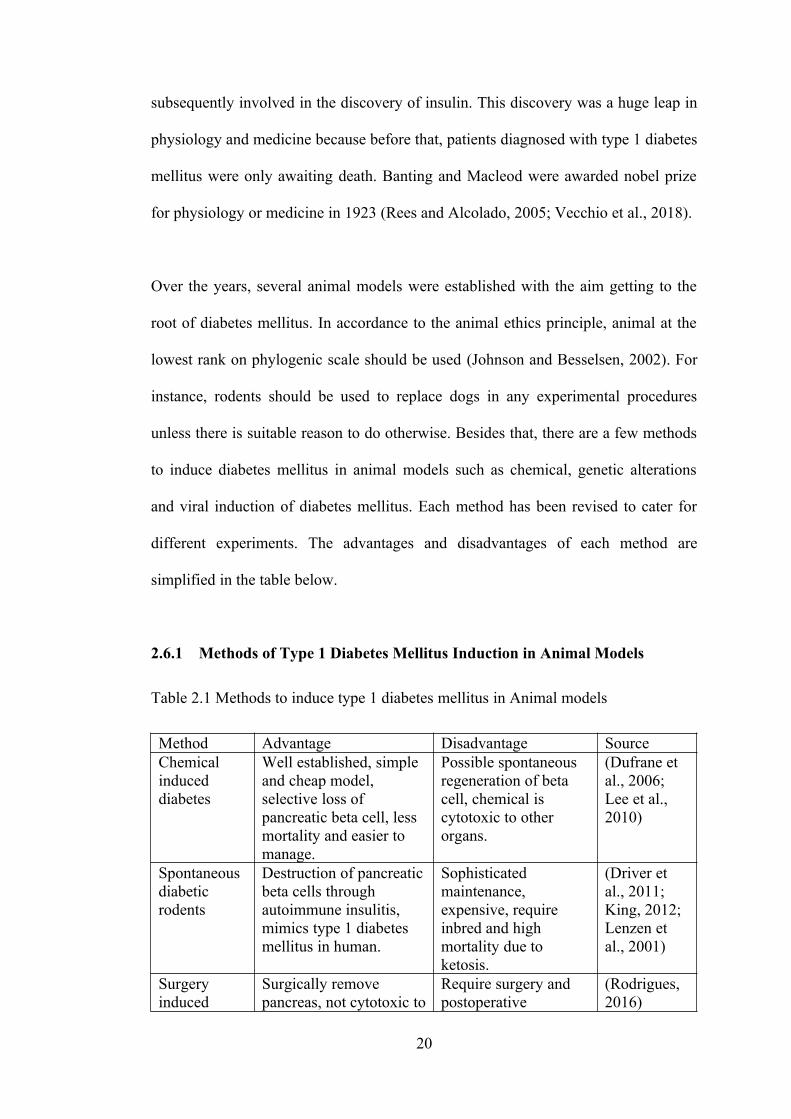
Table 2.1 Methods to induce type 1 diabetes mellitus in Animal models
Method Advantage Disadvantage SourceChemical induced diabetes
Well established, simple and cheap model, selective loss of pancreatic beta cell, less mortality and easier to manage.
Possible spontaneous regeneration of beta cell, chemical is cytotoxic to other organs.
(Dufrane et al., 2006; Lee et al., 2010)
Spontaneous diabetic rodents
Destruction of pancreaticbeta cells through autoimmune insulitis, mimics type 1 diabetes mellitus in human.
Sophisticated maintenance, expensive, require inbred and high mortality due to ketosis.
(Driver et al., 2011; King, 2012; Lenzen et al., 2001)
Surgery induced
Surgically remove pancreas, not cytotoxic to
Require surgery and postoperative
(Rodrigues, 2016)
20
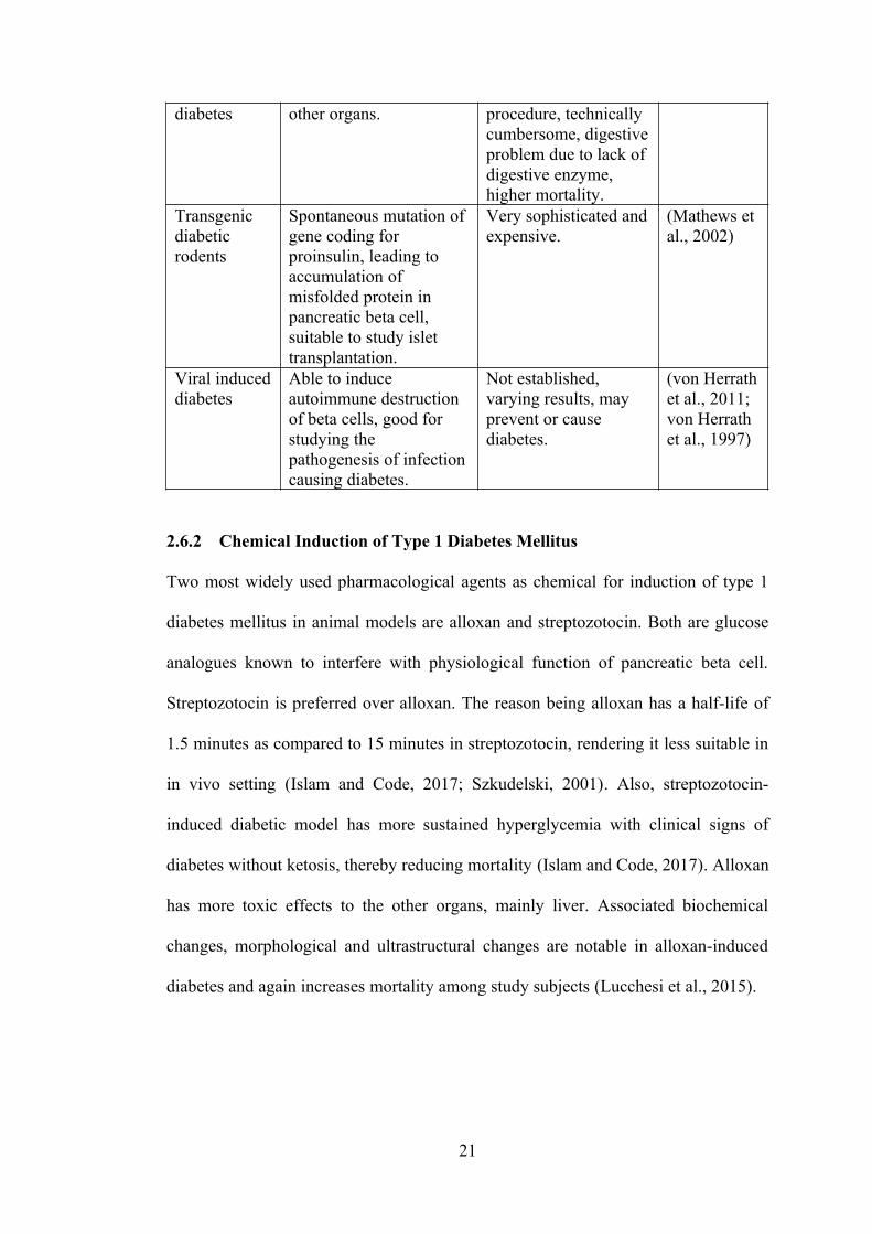
diabetes other organs. procedure, technically cumbersome, digestiveproblem due to lack of digestive enzyme, higher mortality.
Transgenic diabetic rodents
Spontaneous mutation of gene coding for proinsulin, leading to accumulation of misfolded protein in pancreatic beta cell, suitable to study islet transplantation.
Very sophisticated andexpensive.
(Mathews et al., 2002)
Viral induceddiabetes
Able to induce autoimmune destruction of beta cells, good for studying the pathogenesis of infectioncausing diabetes.
Not established, varying results, may prevent or cause diabetes.
(von Herrathet al., 2011; von Herrath et al., 1997)
2.6.2 Chemical Induction of Type 1 Diabetes Mellitus
Two most widely used pharmacological agents as chemical for induction of type 1
diabetes mellitus in animal models are alloxan and streptozotocin. Both are glucose
analogues known to interfere with physiological function of pancreatic beta cell.
Streptozotocin is preferred over alloxan. The reason being alloxan has a half-life of
1.5 minutes as compared to 15 minutes in streptozotocin, rendering it less suitable in
in vivo setting (Islam and Code, 2017; Szkudelski, 2001). Also, streptozotocin-
induced diabetic model has more sustained hyperglycemia with clinical signs of
diabetes without ketosis, thereby reducing mortality (Islam and Code, 2017). Alloxan
has more toxic effects to the other organs, mainly liver. Associated biochemical
changes, morphological and ultrastructural changes are notable in alloxan-induced
diabetes and again increases mortality among study subjects (Lucchesi et al., 2015).
21
2.6.3 Mechanism of Streptozotocin Action
Streptozotocin, 2-deoxy-2-(3-(methyl-3- nitrosoureido)-D-glucopyranose, is a
derivative of Streptomycetes achromogenes and used as antimicrobial agent. The
deoxy group within the chemical structure attached to glucose molecule represents
highly reactive deoxyglucose and methylnitrosurea moieties, acting as powerful
cytotoxic agent directed specifically towards pancreatic beta cells (Szkudelski,
2001). Streptozotocin recognises glucose transporter 2 (GLUT 2) receptors and
pancreatic beta cells expresses high amount of GLUT 2 in contrast to liver and
kidney that express lower amount of GLUT 2. Therefore, mild acute kidney and liver
injury is highly reversible. For this reason, streptozotocin-induced diabetes offers
good animal model for studying the chronic complications of hyperglycemia in many
organs such as liver, kidney, brain, heart and muscle (Wu and Yan, 2015). Inside
pancreatic beta cells, streptozotocin’s main mechanism of induction of diabetes is in
DNA alkylation. The transfer of methyl group from methylnitrosurea to DNA
molecule initiate chain reaction of DNA fragmentation and destruction, leading to
beta cell necrosis and lack of insulin production (Radenkovic et al., 2016).
2.6.4 Practical Application of Streptozotocin
Single dose with 50-60 mg/kg streptozotocin to induce type 1 diabetes mellitus in
adult male Wistar rats resulted in clinical overt diabetic symptoms. Dosage more and
less than the range of dosage resulted in death and inadequate hyperglycemia
respectively. The mortality rate was 12.5-25% due to the acute hypoglycemia in
within 6-24 hours of induction with streptozotocin. So, glucose solution in the first
day is recommended to reduce mortality. Streptozotocin-induced type 1 diabetes
mellitus in rats achieved stable hyperglycemia even after 17 weeks of induction
22
(Gajdosik et al., 1999). Therefore, streptozotocin provides a feasible framework for
studying chronic complications of type 1 diabetes mellitus including heart.
2.6.5 Diabetic Cardiomyopathy in Streptozotocin-induced Diabetic Rat Model
Rodents are useful model in studying diabetic cardiomyopathy due to the resistive
nature to atherosclerosis, effectively rule out coronary heart disease as confounding
factor among study subjects. The first scientist who pioneered the method of using
rodents in proving the existence of diabetic cardiomyopathy in type 1 diabetes
mellitus was Dr. John McNeill. The comprehensive evidence covered decrease in
contractile performance of working heart and isolated cardiomyocyte, associated
biochemical changes and adrenergic dysregulation (Severson, 2004). Multiple
studies managed to replicate the findings using type 1 and even type 2 diabetic
rodent models (Boudina and Abel, 2007). Four weeks after induction of type 1
diabetes mellitus with streptozotocin lead to the features of diabetic cardiomyopathy
such as cardiac hypertrophy, apoptosis, fibrosis and perivascular fibrosis in rats
(Fiordaliso et al., 2000; Miric et al., 2001). Treatment given within two to four weeks
after induction of type 1 diabetes mellitus can reverse pathological changes in heart
such as cardiac and perivascular fibrosis and manifested as improvement in cardiac
function (Miric et al., 2001).
2.7 Structural and Functional Phenotype in Diabetic Cardiomyopathy
There is no universally accepted definition of diabetic cardiomyopathy due to the
challenges faced by scientists when dealing with complex pathophysiology. The
general definition of diabetic cardiomyopathy is structural and functional
abnormalities of myocardium in diabetic patients in the absence of hypertension or
23
coronary artery disease (Miki et al., 2013). Diabetic cardiomyopathy includes
structural abnormalities such as cardiac hypertrophy, interstitial fibrosis and
perivascular fibrosis and functional changes such as diastolic dysfunction and in late
stage, systolic dysfunction. The most striking feature of diabetic cardiomyopathy is
cardiac fibrosis including cardiac fibrosis and perivascular fibrosis. The histological
changes such as cardiac hypertrophy and interstitial fibrosis can stiffen the heart
ventricles and reduce compliance. In addition, cross-linking of collagen due to
uncontrolled hyperglycemia will further impair cardiac elasticity, leading to diastolic
dysfunction. (Sharma and McNeill, 2006).
2.7.1 Cardiac Hypertrophy
Cardiac hypertrophy is a non-specific phenomenon in reaction to stress. For instance,
cardiac hypertrophy seen in athletes is the initial stage of physiological response to
stress. On the other hand, pathological hypertrophy occurs when cell death, fibrosis,
altered cardiomyocyte, mitochondrial dysfunction and inadequate angiogenesis in
response to stress (Nakamura and Sadoshima, 2018). There is a link between
progression of cardiac hypertrophy in diabetes mellitus and heart failure which is
pathological. This can be attributed to cardiomyocyte hypertrophy, death and loss of
function (Feng et al., 2010; Samak et al., 2016).
2.7.2 Interstitial and Perivascular Fibrosis
The architecture of myocardium is a complex network of tightly and well-arranged
cardiomyocytes and extracellular matrix protein. The main component of
extracellular matrix is collagen, with 85% of type 1 collagen offering greatest tensile
strength and 11% of type III collagen giving elasticity of cardiac tissue. The
24


![Propolis: A Complex Natural Product with a Plethora of ... · a Plethora of Biological Activities That Can Be ... [41]. Propolis of Australian stingless bees (Tetragonula carbonaria)](https://img.pdfslide.us/doc/110x75/5ac7a2027f8b9a5d718befa2/propolis-a-complex-natural-product-with-a-plethora-of-plethora-of-biological.jpg)