Embed Size (px)
Citation preview
EFFECTS OF PARA SAGITTAL CARBON DIOXIDE LASER APPLICATION ON
ERYTHROCYTE DEFORMABILITY
Bekta~ A<;IKGOZ, M.D., Oguz. K. BA~KURT.M.D .. Tuncalp OZGEN. M.D.
Hacettepe University. school of Medicine. Department of Neurosurgery (BA TO), Department of Physiology (OKB)Ankara, TURKEY
Turkish Neurosurgery 2 : 7·9 1991
SUMMARY:
In this study we investigated the influence oflaser energy on the rheological properties of blood passing throughthe superior sagittal sinus. Twenty-two guinea-pigs were used. In the control group craniectomies were doneand only the superior sagittal sinuses were exposed. This procedure had no significant influence on haematologica1factors and erythrocyte deformability. In the experimental group a carbon dioxide laser application was applied to the parasagittal brain tissue. In this group MCR. MCHC values and erythrocyte deformability werefound to be changed singmcantly.
KEY WORDS:
Lasers. Erythrocyte Deformability
INTRODUCTION
Laser energy found its way into neurosurgicalpractice after the initial report of Rosomoff and Carrollin 1965 (11).
Many laboratory trials were carried out in the latesixties (3, 15, 19).In these studies the morphologicaleffects of laser energy was investigated mostly (3, 15,19).
Operating theatres of many centres were equipped with laser machines in the 1980's and data concerning the clinical experience began appearing in theliterature (1.2.12,21).
Laser beams are used for the treatment of intracranial mass lesions. vascular lesions and even for
performing microvascular anastomosis (2.12). Thebest indications are benign tumours such as meningiomas. However in recent years, interstitial tissuetherapy has been designed for deeply situated malignant glial tumours.
After wide clinical usage. papers appeared in theliterature mentioning the unexpected effects of laserenergy espedally severe cerebral oedema aftervaporization or shrinkage of the tumour (6.17).In recent years investigators began to search for the influence of laser energy on dynamic factors such ascerebral blood flow, intracranial pressure, EEG andcerebral water content (8,9.17.19).
In this study the influence of carbon dioxide laserenergy on the rheological properties of blood passing through the superior sagittal sinus is investigated.
A review of the literature failed to find any otherreport dealing with this effect.
MATERIALS AND METHODS:
Twenty-two guinea-pigs each weighing 400-500gm·s. were used in the study. They were divided into two groups of eleven. The first group was thesurgical control group and the second the experimental group. The subjects were anaesthetized with nembuthal sodium (25 mglKg) and midline scalp andtransverse indsions were made in anterior cervical
region. In all animals the internal jugular veins andcarotid arteries were exposed and prepared for obtaining blood spedmens. In the parasagittal areacraniectomies were done to expose the superior sagittal sinus. To compare the effect of laser energy onred blood cells. samples were taken from the carotidartery and from the jugular vein, because one is thepre-laser area where the latter is postlaser area. Bloodspedmens were obtained from one jugular vein afterthe contralateral one was ligated.
In the control group blood samples were obtained from the carotid artery and 30 minutes later fromthe jugular verno (Waiting for 30 minutes in order toequalize the interval in both groups. because in theexperimental group the laser application took nearly 30 minutes,) In the experimental group after bloodsamples were taken from the carotid artery. the carbon dioxide laser application was done to theparasagittal brain tissue (Tissue beside superior sagittal sinus).
7
In a preliminary study the necessary doses werecalibrated. Power was 6-8 Watts. exposure time was0.5 seconds and IO to 12 shouts were done in each
animal (Coherent System 451). Care was taken notto the irradiate superior sagittal sinus. During surgicalintervention and laser application the corticaltemperature was recorded with the aid of a YSITelethermometer and fine needle probe(Yellow SpringsInstrument, Kings KM 51-01fine needle probe). Afterlaser application blood samples were obtained fromthe jugular vein. This procedure was completed in30 minutes. In both groups the sagittal sinuses remained open for 30 minutes. Red cell count (RBe).haemoglobin. haematocrit, mean corpuscular volume(MCV),mean corpuscular haemoglobin (MCH).meancorpuscular haemoglobin concentration (MCHe) anderythrocyte filtrability were measured in heparinized blood samples (15 IU m1). Haematologicalparameters were determined with an haematologyanalyser (Colter Counter, Model 5 plus VI).
Erythrocyte filtrability was measured by the constant pressure filtration technique (7). Heparinizedblood was washed 3 times with particle free tris NaC1buffer (PH 7.4). while the buffy coat was eliminatedcarefully at each step, Packed red cells wereresuspended with the same buffer to give ahaematocrit of IO %. 1 ml of suspension was filteredthrough policarbonate filters (Pore size 4.7 urn.Nucleopore-Hemafil, Lot: 54B6A7) under 5 em H20pressure and filtration time was determined electronically.
Measurements were perfomed at roomtemperature and within thirty minutes after thecollection of blood spedmens. Filtration times of redcell suspensions were divided by the filtrationtimes of the same amount of buffer through the samefilter. The index calculated from these measurements
was inversely related to the deformability oferythrocytes.
The results obtained before and after surgery werecompared with Wilcoxon Paired T-test.
RESULTS
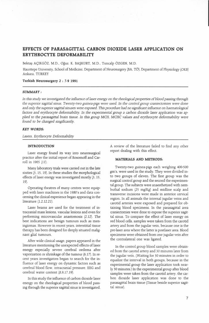
The haematological parameters and erythrocytedeformability indexes are presented in Table 1. Inthe control group cortex temperatures wererecorded as 32-34degrees centigrade (Mean 33.3+0.9).During laser application cortical temperatureswere recorded as 36-38 degrees centigrade (Mean37.6+0.7).
8
Table 1 : Results obtained from the
control group and from theexperiment group.carotid
jugulerWilcolon P
RBC
4.32 + 1.134.48 + 0.53p> 0,05
Hb
12.90 + 2.2112.92 + 1.18p> 0.05
Het
37.43 + 6.9636.72 + 3.82p> 0.05
MCV
81.67 + 4.6082.09 + 2,98p> 0.05
MCH
27.94 + 1.4128.98 + 2.46p> 0.05
MCHC
33.91 + 2.1035.30 + 3.11p> 0.05Control
DEF.lND.
1.51 + 0.201.62 + 0.20p> 0.05Group
RBC
4.39 + 0.724.00 + 0,56p> 0.05
Hb
12.40 + 2.3612.25 + 1.67p> 0,05
Het
36.09 + 6.4133.17 + 3.97p> 0.05
MCV
82.00 + 1.7383.22 + 6.38p> 0.05
MCH
28.17 + 1.4130.90 + 4.60p < 0.05
MCHC
34.34 + 3.1137.00 + 4.62p < 0.05 Experiment
DEF.lND.
1.55 + 0,202.16 + 0.50p < 0.05 Group
DISCUSSION
Erythrocyte deformability is mainly affected bythe volume, haemoglobin concentration and shapeof the test erythrocytes (16). With currently usedtechniques, it is possible to detect minimal changes(16), In the present study, the effects oflaser energyon erythrocyte rheology at the parasagittal area wastested. In order to detect the local changes. one mustknow the rheological properties of the blood entering and coming out from the test area. In this studyspedmens were obtained from the carotid artery andjugular vein. In order not to dismiss the blood fromthe sagittal sinus. blood was obtained from onejuguler vein after the contralateral one has beenligated. No attempt was made to obtain blood fromthe sagittal sinus direct because of the relatively smallcalibre of the sinus in guinea-pigs which may be influenced by veni-puncture. The results showed thatthe rheological properties of the whole blood beforeand after laser energy was not influenced.
The intrinsic factors influendng erythrocyte deformability are the cell geometry. cytoplasmic viscosityand membrane viscoelastidty (16).Mean corpuscularhemoglobin and mean corpuscular hemoglobin concentrations were influenced significantly. These findings indicate the intrinsic changes in erythrocyteswhich are consistent with those of Glassberg (4)whoshowed a dose-dependent lysis of haemoglobin inerythrocytes. Because MCHC is an important deter-
minant of cytoplasmic viscosity. there must be otherfactors which influenced this as well as the cell shape.
The first factor may be effect of laser energy oncerebral microvasculature. Carbon dioxide laser application of 4 Watts for 2 seconds causes wedgeshaped lesions composed of charred layer and aroundthis an oedematous layer appears (9). It was clearlydemonstrated by Kuroiwa et a1.that (9).in more than80 percent of animals. there was evidence of extravasation of red blood cells and sometimes massive
haemorrhagies. When the dosage was increased to8 Watts they observed dilatation and sometimesthrombosis of the vessels (9).
Tiznado et al (17). also found a lesion of 15
millimetres on the cortical surface. Using a power of40 Watts for a total of 4 seconds. they found thatthese lesions. composed of haemorrhagies andnecrosis. extended to a depth 5 millimetres (17).
As seen from this data the influence of carbon
dioxide energy of 6-8 Watts for a total of 5 to 6seconds on the cerebral microcirculation. may be oneof the factors which caused changes in hematologicalfactors.
The second factor may be the rise in temperature.The heat effect of carbon dioxide laser energy initiallyproposed by Stellar (14). has also been observed byclinidans (10,18) and written by others (2.13).
It is a well known fact that heat treatment causes
red blood cells to change their morphological andmechanical properties (5,14,20), Snabre (14) indicatesthe critical thermal denaturation range as 46 degreesCentigrade and 51 degrees Centigrade. At highertemperatures. red cells are transformed intospherocytes. they exhibit budding and they undergofragmentation (14.20). Although there may be changesin erythrocyte deformability between 24 to 37 degreesCentigrade. the deformability decreases dramatically between 48 to 50 degrees Centigrade. In the initialstudy although increases in cortical temperature wererecorded, these were found relatively low. so heatcould not be accused of being the only factor causing the changes. This study was designed to searchfor the oedematous effects of laser energy.
In summary the study showed the influence ofcarbon dioxide energy on erythrocyte rheology. Butas a preliminary conclusion we can state that inclinical practice, in order to avoid damage. low dosesof laser energy must be utilized and care must betaken near the vital structures. Disturbance of
microdrculation and erythrocyte deform ability maybe one factor causing 'unexpected' cerebral oedemaduring laser application.
For Correspondence: Dr. Bekta~ A~lkgbzHacettepe UniversitesiNbro~in:irji Anabilim Dati
06100 Ankara- TiirkiyeTel (4) 310 35 45
REFBRBNCBS
I. Beck OJ. Frank F: The use of Nd: Yag laser in neurosurgery.Lasers Surg Med. 5:354-356. 1985
2. Boggan JE. Powers SK:Use of lasers in neurological surgery. in
Youmans JR (eds) Neurological Surgery 3rd ed. WB Saunders.Philadelphia 1990. pp 992-1004
3. Fox JL. Stein MN. Hayes JR et al.:Effects of laser irradiation onthe CNS. J Neurol Neurosug Psychiat 31:43-49. 1968
4. Glassberg E. Lask GP. Tam EM:Cellular effects of the pulsedtunable dye laser at 577 nanometers on human endothelial cells.
fibroblasts and erythrocytes:an in vitro study. Lasers Surg Med8:567-572. 1988
5. Hanss M. Kautsouris D:Thermal transitions of red blood cell
deformability. Correlation with membrane rheological proper
ties. Biochimica et Biophysica Acta 769:461-470. 19846. Hudgins WR:Comment after Tiznado et al.·s article.
Neurosurgery 16:8. 1985
7. International committee for standardisation in hematology(ICSH) Guidelines for measurement of blood viscosity anderythrocytee deformability (Expert panel on blood rheology).Clinical Hemorheology 6:439-453. 1986
8. Kiessling M. Herchenhan E. Eggert HR:Cerebrovascular andmetabolic effects on the rat brain of focal Nd-YAGlaser irradia
tion. J Neurosurg 73:909-917. 19909. Kuroiwa T. Tsuyumu M. Takei H et al.:Effects of Nd:YAG and
CO 2 lasers on cerebral microvasculature. J Neurosurg 64:128-133.1986
10. Pendl G. Vorkapic P:Microsurgery of midbrain lesions. Actaneurochirurgica 42:130-136. 1988.
11. Rosomoff HL. Carroll F:Effect of laser on brain and neoplasm.Surg Forum 16:431. 1965
12. Roux FX. Merienne L. Cioloca C et al.:Neurosurgicallasers fortumour removal. Lasers in Medical Science 5:241-244. 1990
13. Saunders ML. Young HF. Becker DP et al.:The use of laser in
neurological surgery. Surg NeuroI14:1-1O. 198014. Snabre P. Baumler H. Mills P:Aggregation of human red blood
cells after moderata heat treatment. Biorheology 22:185-195,198515. Stellar S. Polanyi TG. Bredemeier HC:Experimental studies with
the carbon dioxide laser as a neurosurgical instrument. Med BioiEngng 8:549-558, 1970
16. Stuart J:Erythrocyte rheology. Review article. J Clin Pathol38:965-977. 1985
17. Tiznado E. James HE. Kemper C:Experimental carbon dioxide
laser brain lesions and intracranial dynamics. Neurosurgery1+:5-8. 1985
18. Tobler WD. Sawaya R. Tew MJ:Successfullaser-assisted excision of a metastatic midbrain tumour. Neurosurgery 18:795-797.1986
19. Walter GF. Ascher PW. Ingolitsch E:The effects of carbon dioxirde and neodynium YAG lasers on the central and peripheralnervous systems and cerebral blood vessels. J Neurol and Psyhiat4x:745-749. 1984
20. Wirth FB. Downing EF. Cannon CL et al.:Experience with theneodynium-yttrium aluminum garnet laser in forty-two cases.Neurosurgery 21:867-871. 1987
9