Embed Size (px)
Citation preview
Brit. 7. Ophthal. (I 973) 57, 910
Effects of papaverine on the retinalmicrocirculation
T. J. FFYTCHE, C. J. BULPITT, D. ARCHER, E. MI. KOHNER, ANDC. T. DOLLERY
M.R.C. Clinical Pharmacology Research Group, Royal Postgraduate Medical School, London
Many pathological changes in the retina which give rise to visual deterioration are causedby circulatory disturbances. A number of vaso-active substances have been suggestedfor the treatment of these disorders in an attempt to increase the flow of blood throughthe eye. One such substance is papaverine which has been used in the management ofacute occlusions of the central retinal artery (Varley, Holt-Wilson, and Watson, I968,Gombos, 1970) and in chronic ischaemic conditions of the choroid and retina, when it hasbeen given by arterial, venous, oral, and retrobulbar routes. Little effort, however, hasbeen made to investigate the action of papaverine on the retinal blood vessels, and, apartfrom observations by several authors on its dilating effects (Frayser and Hichkam, I965;de Rivas Cherif, I967; Zahn, i966), no quantitative assessment or analysis of the responscof the retinal circulation to papaverine infusions has yet been reported.The present experiments were designed to measure the effect of both local and systemic
administration of papaverine on the retinal blood flow, so that an appraisal of its possiblevalue in the management of retinal vascular disease can be made.
Method
Studies were carried out on seventeen young pigs aged between 6 and I 2 weeks with weights varyingfrom I2 to 30 kg. and on three mature rhesus monkeys. The pig was chosen as the main experimentalanimal for several reasons. The eye is similar in size and form to that of man; the fundus lacks atapetum and is moderately pigmented and this allows photographs of good contrast to be taken of theretina, and considerable experience has been gained in using this animal for experimental work onthe retina (Ashton, Dollery, Henkind, Hill, Paterson, Ramalho, and Shakib, I966). The animalsare easy to obtain and their management is not difficult. There is, however, no central retinalartery, the retinal vessels arising from the main ciliary arteries which pierce the sclera around theoptic disc. Small amounts of smooth muscle are present in the walls of the larger arteries and thereis a trilaminar capillary network together with a well-marked radial peripapillary plexus arisingfrom the intraretinal arterioles (Henkind, I967; Rootman, 1971).
Anaesthesia was induced in both types of animal by intravenous 5 per cent. sodium thiopentoneand continued after tracheal intubation by further thiopentone injections through a leg vein catheter.Ventilation was maintained using a Palmer pump adjusted to keep the blood gases at normallevels and, to control this, serial measurements of the blood PO2, pCO2, and pH were made with aRadiometer BMS 3 analyser.The systemic blood pressure and pulse rate were recorded by means of a strain-gauge transducer
and Devices M 4 recorder connected to a P 6o catheter inserted into the aorta via the femoral artery.
Received for publicatioii May i6, 1973Address for reprints: T. J. ffTtche, F.R.t:.S., MoorfieldIs Eye Hospital, Higis Holbosrn, Lond(loin, W.C.i
copyright. on M
arch 4, 2020 by guest. Protected by
http://bjo.bmj.com
/B
r J Ophthalm
ol: first published as 10.1136/bjo.57.12.910 on 1 Decem
ber 1973. Dow
nloaded from
Effects of papaverine on the retinal microcirculation 9I1
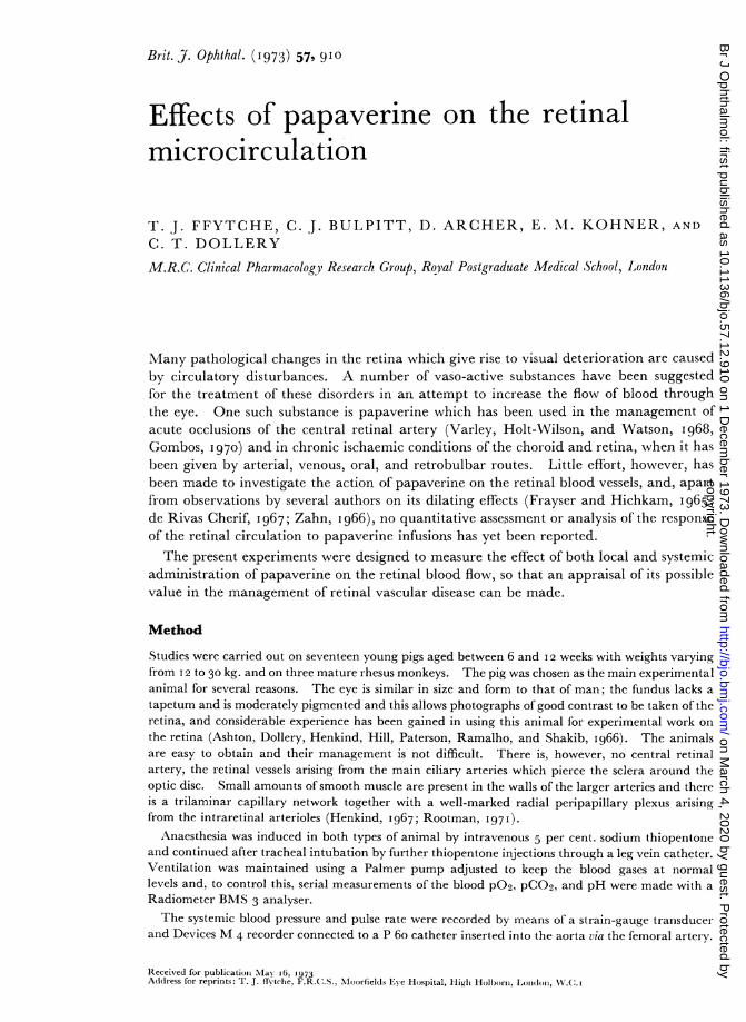
Eye PreparationThe eye was prepared for photography by dilating the pupil with Mydrilate (Cyclopentolate Iper cent.) eye drops. 'rhe position of the eye was controlled by using a modified Flieringa ringsutured to the sclera at the limbus (Fig. I). Four stay sutures were attached to the projecting parts ofthe ring and these served to anchor the eye to a ring of four micromanipulators. By this method itwas possible to adjust the position of the eye to allow different areas to be studied without causingdistortion of the globe or elevations of intraocular pressure. Clarity of the cornea was maintainedby continuous saline drip.
FI G. IExternal view ofpig eye, showingznodified Flieringa ring attached to 4 micro-inanipulators
In the experiments on the rhesus monkeys, the position of the eye was controlled by moving theanimal's head on a special head-rest, the lids being kept open by a speculum.
Pressure recordingIn six of the pig experiments, a continuous recording of the intraocular pressure was made in orderto have an accurate measurement of the perfusing pressure of the retinal circulation. A 25 gaugeneedle was inserted into the anterior chamber at the limbus and connected by a catheter and three-way tap to a strain-gauge transducer and Devices M 4 recorder on the one hand and to an adjustablesaline reservoir on the other. By this method a constant intraocular pressure could be maintainedand recorded. In the remaining eleven animals an intraocular pressure of I6 mm. Hg was assumed.
Pal5averine admiiinistrationPapaverine hydrochloride 2-5 per cent. was infused intra-arterially through a 25 gauge needleinserted into the exposed common carotid artery in the neck, or intravenously through a P 6ocatheter placed in the femoral vein. Retrobulbar injections of papaverine were also given using astandard retrobulbar needle.
Photography
Circulation studies were made using fluorescein angiography and colour photography. Fluoresceinand papaverine form an insoluble yellow precipitate when mixed together since fluorescein pre-cipitates out in an acid solution; it was therefore necessary to inject fluorescein by an alternativearterial route to the acid papaverine. Injections were made through a catheter threaded via an earartery into the common carotid well above the level of the papaverine infusion. About o 5 ml. ofIO per cent. fluorescein sodium solution was injected rapidly for each angiogram.
Retinal photography was performed using a vertically mounted Zeiss fundus camera. Colourphotographs were taken with 35 mm. Kodachrome II film, and for the fluorescein angiography35 mm. Kodak TriX film was used after the insertion of a Baird Atomic B 4 exciting filter and a Zeiss
copyright. on M
arch 4, 2020 by guest. Protected by
http://bjo.bmj.com
/B
r J Ophthalm
ol: first published as 10.1136/bjo.57.12.910 on 1 Decem
ber 1973. Dow
nloaded from
T. j. ffytche, C. J. Bulpiit, D. Archer, E. M. Kohner, and C. T. Dollery
300976 barrier filter in the light pathway of the camera. The TriX film was force processed inundiluted D76 at room temperature for I41 min. Cine-angiography was performed on Ilford MarkV film, using a Bolex H I6 mm. camera adapted for use on the vertical fundus camera mounting.The frame speed was in the region of 30 frames per second, and the film was forced processed inundiluted Kodak D I9 developer.
Data measurements
(I) VESSEL DIAMETERS
Arteries and veins of approximately IOO pt in diameter were studied; the area chosen was usually thesuperior temporal region, this being the area of the fundus most accessible to examination, and thearea adjacent to the optic disc. Retinal vessel diameters were measured from the colour photographsor from the cine film negatives, using a microscope with a micrometer scale in the ocular head.The total internal diameter of the vessels was taken to be the width of fluorescein within them whenthe vessel was full (Bulpitt, Dollery, and Kohner, 1970) and, using the micrometer, the standarddeviation of a single measurement of a vessel of IOOo± was found to be I-6 L for the arteries and0o5 ,u for the veins when measured on 35 mm. film, whereas it was 2-4 ,u for the arteries and I ,u
for the veins on the cine film. Despite the loss in accuracy when compared with measurementsfrom the 35 mm. film, most calculations were made from the cine angiograms. There were tworeasons for this;
(i) It was not always possible to be sure that a vessel being measured was completely filled withfluorescein when a single photograph was taken, whereas on the cine film the point of maximum filling(and therefore the true internal diameter) could be observed by frame-to-frame analysis.
(ii) Measurement of the vessel diameters from cine films meant that the vessel width could bedetermined at the same time as the velocity of the dye front passing along it, whereas there was a gapin time when 35 mm. film was used, caused by having to change the cameras.
These advantages more than compensated for the less accurate measurements from cine film.
(2) DYE-FRONT VELOCITY
The velocity of the fluorescein dye front along the artery under observation was measured by frame-to-frame analysis using a Vanguard Motion Analyser. A section of an artery was chosen which wasstraight and which had no major branches, and the high-contrast photography enabled the progressionof the fluorescein bolus along the vessel to be easily visualized (Bulpitt and Dollery, 1970). Diametermeasurements were taken at both ends of the section under observation. The absolute value fordye-front velocity has to be adjusted for the individual magnification of the optical system of theeye under observation. The dye-front velocity is therefore presented in arbitrary units and changesin velocity are recorded as a percentage change of the normal in each experimental animal.
(3) VOLUME FLOW
Originally it was assumed that Poiseiulle flow existed in retinal arteries of the size that have beenexamined, so that the volume of blood flowing along these vessels (Volume Flow) could be expressedby the formula:
F °r2vk,2
where k is a magnification constant, r is the radius in arbitrary units, and v is the dye front velocityin arbitrary units per minute.Volume flow therefore varies directly with the square of the radius (r) and the dye front velocity
(v). Studies by Bulpitt, Kohner, and Dollery (I973) suggest that Poseuille flow does not exist inthese small vessels, but rather plug flow or some intermediate configuration. It was assumed thatthe flow profile showed little alteration throughout the experiment, and volume flow was expressedin arbitrary volume units per minute, and where comparative studies were made in the same animalvariations in volume flow were expressed as percentage changes.
912
copyright. on M
arch 4, 2020 by guest. Protected by
http://bjo.bmj.com
/B
r J Ophthalm
ol: first published as 10.1136/bjo.57.12.910 on 1 Decem
ber 1973. Dow
nloaded from
Effects ofpapaverine on the retinal microcirculation 913
(4) PERFUSION PRESSURE
For the purpose of these experiments the perfusion pressure was taken as the mean blood pressure(Diastolic + 1/3 systolic-diastolic difference) minus the intraocular pressure. No allowance wasmade for the difference between the pressures measured in the aorta and those in the central retinalartery, since the technique of ophthalmodynamometry cannot be applied to the pig to measureophthalmic artery pressure because of the different origin of the retinal vessels. It was assumed thatthe drop in pressure between the aorta and the central retinal artery was constant during the experi-ment provided that the circulation remained healthy.
Results
The following parameters were measured during intra-arterial and intravenous infusionof papaverine: retinal arterial and venous diameters, arterial blood flow velocity, andvolume flow. In addition, separate experiments were undertaken to determine the dose/response curve for papaverine and to measure the cardiac output during the infusion.
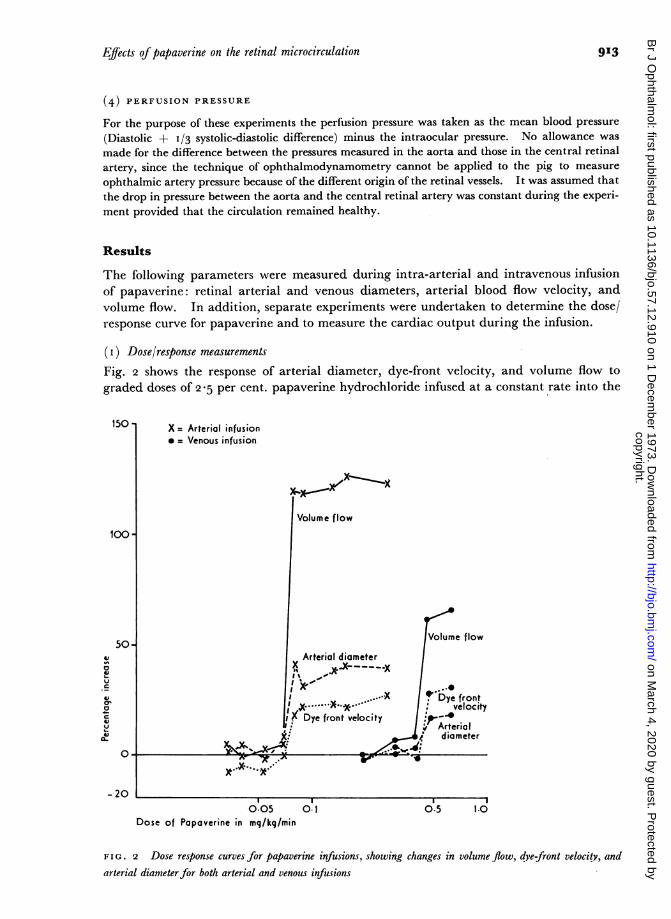
(i) Dose/response measurementsFig. 2 shows the response of arterial diameter, dye-front velocity, and volume flow tograded doses of 2-5 per cent. papaverine hydrochloride infused at a constant rate into the
150 - X = Arterial infusion* = Venous infusion
Volume flow
100 -
50o -
Vume flow
Arterial diameter
A' 0
velocity |+ Dye fronto ~~~~~~~~~~~~~~~~~~~~velocity_AeDy front velocity
:,Arterial,f diameter
-20
Dose of Papaverine in mg/kq/min
FIG. 2 Dose response curves for papaverine infusions, showing changes in volume flow, dye-front velocity, and
arterial diameterfor both arterial and venous infusions
copyright. on M
arch 4, 2020 by guest. Protected by
http://bjo.bmj.com
/B
r J Ophthalm
ol: first published as 10.1136/bjo.57.12.910 on 1 Decem
ber 1973. Dow
nloaded from
914 T. 5. ifytche, C. 5. Bulpitt, D. Archer, E. M. Kohner, and C. T. Dollery
carotid artery of the pig. All three parameters showed a steep increase when the amountof papaverine infused exceeded o o8 mg./kg./min., and at doses above this value there wasno further increase in arterial diameter and velocity. A similar curve is shown duringintravenous infusions of graded doses of papaverine with a threshold level of o045 mg./kg./min. The arterial diameter was found to be an accurate index of the volume flow and thesteep slope of the curve suggested that the vasodilator effect of papaverine occurred whenan active intraluminal concentration of the drug was reached. Dose/response measure-ments were not obtained in the monkeys.
For the purpose of the experiments papaverine was infused at about twice the thresholddose and at this level no cardiac irregularity or change in the systemic circulation wasobserved. At very high doses (ten times threshold) the systemic blood pressure fell,leading to circulatory collapse.
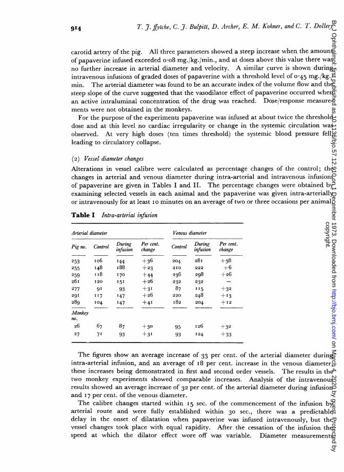
(2) Vessel diameter changesAlterations in vessel calibre were calculated as percentage changes of the control; thechanges in arterial and venous diameter during intra-arterial and intravenous infusionsof papaverine are given in Tables I and II. The percentage changes were obtained byexamining selected vessels in each animal and the papaverine was given intra-arteriallyor intravenously for at least I o minutes on an average of two or three occasions per animal.
Table I Intra-arterial infusion
Arterial diameter Venous diameter
Pig no. Control During Per cent. Control During Per cent.infusion change infusion change
253 io6 144 +36 204 28I +38255 I48 i88 +23 210 222 +6
259 II8 170 +44 236 298 +2626I 120 151 +26 232 232 -
277 9I 93 +3' 87 I'5 +32291 117 147 +26 220 248 +13289 104 147 +4' I82 204 +12
Monkeyno.26 67 87 +30 95 I26 +32
27 71 93 +3' 93 124 +33
The figures show an average increase of 33 per cent. of the arterial diameter duringintra-arterial infusion, and an average of I8 per cent. increase in the venous diameter,these increases being demonstrated in first and second order vessels. The results in thetwo monkey experiments showed comparable increases. Analysis of the intravenousresults showed an average increase of 32 per cent. of the arterial diameter during infusionand I 7 per cent. of the venous diameter.The calibre changes started within I5 sec. of the commencement of the infusion by
arterial route and were fully established within 30 sec., there was a predictabledelay in the onset of dilatation when papaverine was infused intravenously, but thevessel changes took place with equal rapidity. After the cessation of the infusion thespeed at which the dilator effect wore off was variable. Diameter measurements
copyright. on M
arch 4, 2020 by guest. Protected by
http://bjo.bmj.com
/B
r J Ophthalm
ol: first published as 10.1136/bjo.57.12.910 on 1 Decem
ber 1973. Dow
nloaded from
Effects ofpapaverine on the retinal microcirculation 915
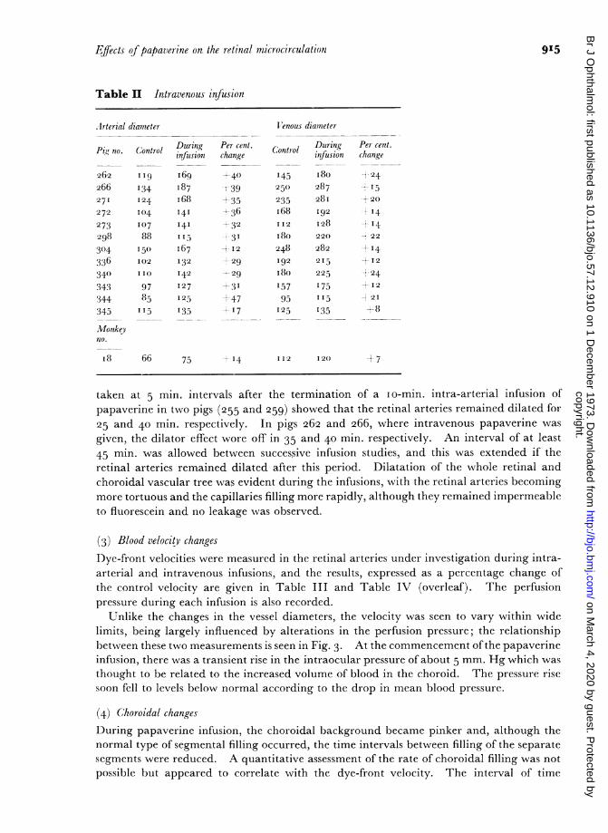
Table II Intravenous infusion
.lrterial diameter X enous diameter
no. Control During Per cent. n During Pet cent.P "I no. Control Controlinfusion change infusion change
262 II9 I69 +40 I45 i8o --24
266 I34 87 i39 250 287 15
271 124 i68 +35 235 28I +20
272 I04 14I i36 I68 192 I4
273 I07 I41 +32 112 128 +I4298 88 11-) -31 i8o 220 ,22
304 I50 I67 +I2 248 282 +-I4
336 I02 132 --29 I92 2I5 +12
340 II0 I42 -29 i8o 225 -+24343 97 I27 +3I 157 175 +12
344 85 125 ±47 95 I15 -t 2I
345 I15 I35 +17 125 135 +8
Monikeyno.
i8 66 75 4-4 II2 I20 +7
taken at 5 min. intervals after the termination of a io-min. intra-arterial infusion ofpapaverine in two pigs (255 and 259) showed that the retinal arteries remained dilated for25 and 40 min. respectively. In pigs 262 and 266, where intravenous papaverine wasgiven, the dilator effect wore off in 35 and 40 min. respectively. An interval of at least45 min. was allowed between successive infusion studies, and this was extended if theretinal arteries remained dilated after this period. Dilatation of the whole retinal andchoroidal vascular tree was evident during the infusions, with the retinal arteries becomingmore tortuous and the capillaries filling more rapidly, although they remained impermeableto fluorescein and no leakage was observed.
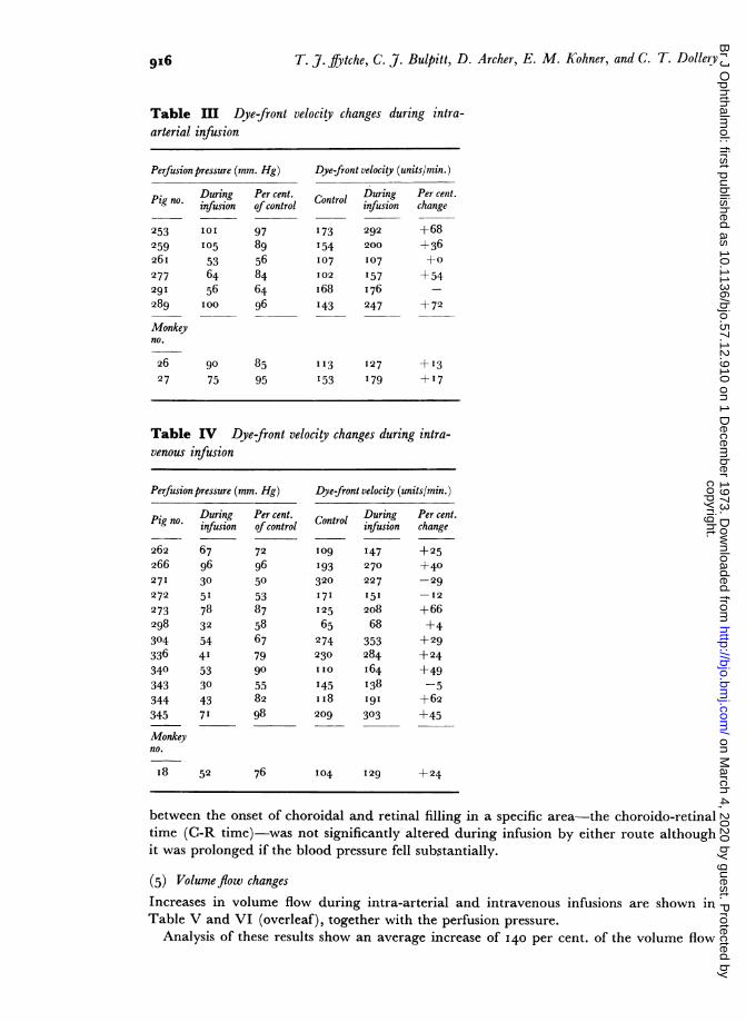
(3) Blood velocity changes
Dye-front velocities were measured in the retinal arteries under investigation during intra-arterial and intravenous infusions, and the results, expressed as a percentage change ofthe control velocity are given in Table III and Table IV (overleaf). The perfusionpressure during each infusion is also recorded.
Unlike the changes in the vessel diameters, the velocity was seen to vary within widelimits, being largely influenced by alterations in the perfusion pressure; the relationshipbetween these two measurements is seen in Fig. 3. At the commencement ofthe papaverineinfusion, there was a transient rise in the intraocular pressure of about 5 mm. Hg which wasthought to be related to the increased volume of blood in the choroid. The pressure risesoon fell to levels below normal according to the drop in mean blood pressure.
(4) Choroidal changesDuring papaverine infusion, the choroidal background became pinker and, although thenormal type of segmental filling occurred, the time intervals between filling of the separatesegments were reduced. A quantitative assessment of the rate of choroidal filling was notpossible but appeared to correlate with the dye-front velocity. The interval of time
copyright. on M
arch 4, 2020 by guest. Protected by
http://bjo.bmj.com
/B
r J Ophthalm
ol: first published as 10.1136/bjo.57.12.910 on 1 Decem
ber 1973. Dow
nloaded from
T. J. frytche, C. J. Bulpitt, D. Archer, E. M. Kohner, and C. T. Dollery
Table HI Dye-front velocity changes during intra-arterial infusion
Perfusion pressure (mn
Pig no.
253
259
26I277
291
289
Monkeyno.
2627
Duringinfusion
IOI
I05
536456IOO
n. Hg) Dye-front velocity (units/min.)
Per cent. Control During Per cent.ofcontrol infusion change
97 173 292 +68
89 154 200 +36
56 I07 107 +0
84 I02 157 +54
64 i68 I76 -
96 I43 247 +72
90 8575 95
I I3 127 +13
153 179 + I7
Table IV Dye-front velocity changes during intra-venous infusion
Perfusion pressure (mn
Pig no. Duringinfusion
262 67266 9627I 30
272 51
273 78298 32
304 54
336 4I340 53343 30344 43345 7'
Monkeyno.
I8 52
n. Hg) Dye-front velocity (units/min.)
Per cent. Control During Per cent.ofcontrol infusion change
72 l09 I47 +25
96 193 270 +4050 320 227 -29
53 171 151 -I2
87 I25 208 +6658 65 68 +467 274 353 +29
79 230 284 +2490 iio I64 +49
55 145 138 -582 ii8 I9I +6298 209 303 +45
76 104 I29 +24
between the onset of choroidal and retinal filling in a specific area-the choroido-retinaltime (C-R time)-was not significantly altered during infusion by either route althoughit was prolonged if the blood pressure fell substantially.
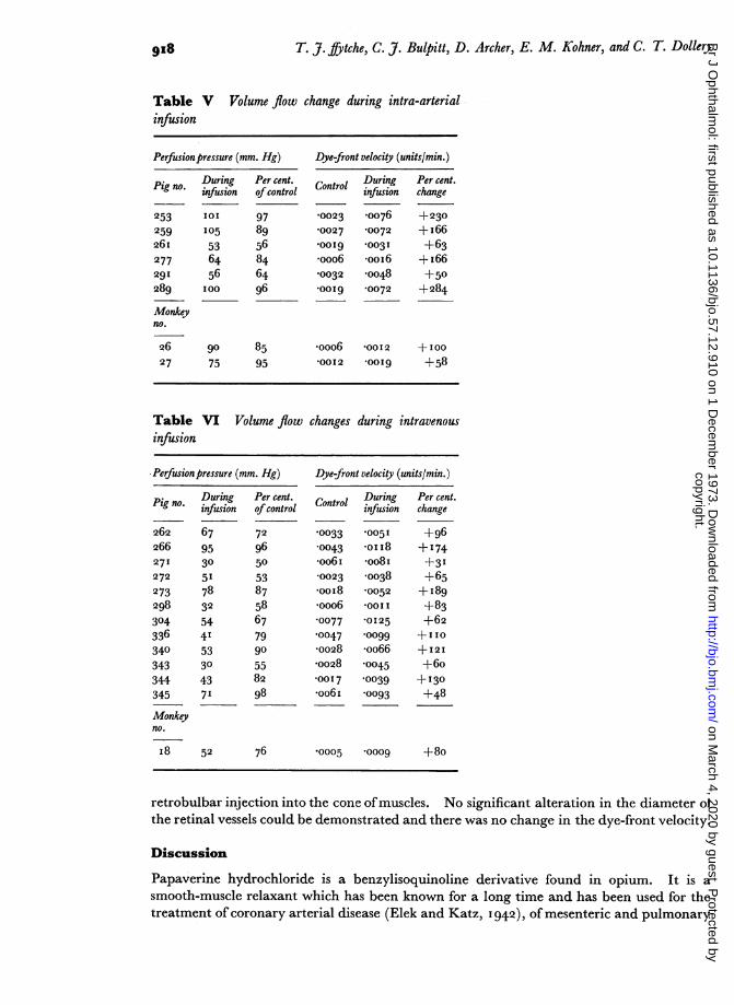
(5) Volume flow changesIncreases in volume flow during intra-arterial and intravenous infusions are shown inTable V and VI (overleaf), together with the perfusion pressure.
Analysis of these results show an average increase of I40 per cent. of the volume flow
9I6
I -,
copyright. on M
arch 4, 2020 by guest. Protected by
http://bjo.bmj.com
/B
r J Ophthalm
ol: first published as 10.1136/bjo.57.12.910 on 1 Decem
ber 1973. Dow
nloaded from
Efects ofpapaverine on the retinal microcirculation 917
+100 X = Arterial infusion*=Venous infusion 300 X=Arterial infusion
*=Venous infusion x
+50-
41 1 s 1-O * C Xu 2o_ -
41 41
-0 . 0,5 100
x~~~~~~~~~~~~0~~~~~~~~~~~
60 0 _ _ _ _ _ _ _ _ _ _
9- 100- ~ 5010
Percentage of control perfusion pre ssure Percentage of control perfusion pressure
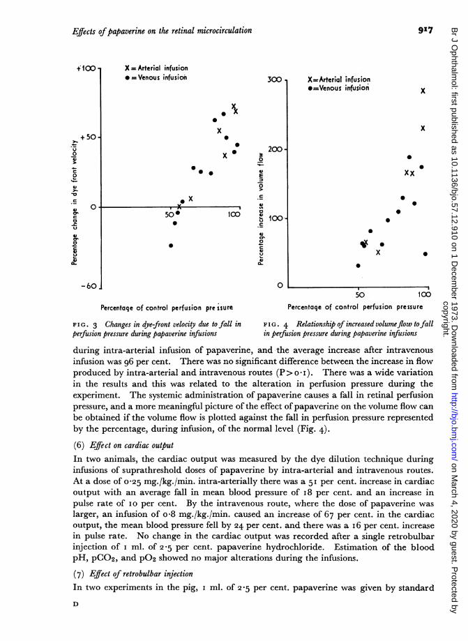
FIG. 3 Changes in dye-front velocity due to fall in FIG. 4 Relationship ofincreasedvolumeflow tofallperfusion pressure during papaverine infusions in perfusion pressure during papaverine infusions
during intra-arterial infusion of papaverine, and the average increase after intravenousinfusion was 96 per cent. There was no significant diffierence between the increase in flowproduced by intra-arterial and intravenous routes (P> o *I ). There was a wide variationin the results and this was related to the alteration in perfusion pressure during theexperiment. The systemic administration of papaverine causes a fall in retinal perfusionpressure, and a more meaningful picture of the effect of papaverine on the volume flow canbe obtained if the volume flow is plotted against the fall in perfusion pressure representedby the percentage, during infusion, of the normal level (Fig. 4).
(6) Eff7ect on cardiac outputIn two animals, the cardiac output was measured by the dye dilution technique duringinfusions of suprathreshold doses of papaverine by intra-arterial and intravenous routes.At a dose of 0o25 mg./kg./min. intra-arterially there was a 51 per cent. increase in cardiacoutput with an average fall in mean blood pressure of I8 per cent. and an increase inpulse rate of IO per cent. By the intravenous route, where the dose of papaverine waslarger, an infusion of o*8 mg./kg./min. caused an increase of 67 per cent. in the cardiacoutput, the mean blood pressure fell by 24 per cent. and there was a I6 per cent. increasein pulse rate. No change in the cardiac output was recorded after a single retrobulbarinjection of I ml. of 2*5 per cent. papaverine hydrochloride. Estimation of the bloodpH, pCO2, and p°2 showed no major alterations during the infusions.
(7) Effiect of retrobulbar injectionIn two experiments in the pig, I ml. of 25 per cent. papaverine was given by standard
D
copyright. on M
arch 4, 2020 by guest. Protected by
http://bjo.bmj.com
/B
r J Ophthalm
ol: first published as 10.1136/bjo.57.12.910 on 1 Decem
ber 1973. Dow
nloaded from
9I8 T. J. ffytche, C. J. Bulpitt, D. Archer, E. M. Kohner, and C. T. Dollery
Table V Volume flow change during intra-arterialinfusion
Perfusion pressure (mm. Hg)
Pig no.
25325926I
277291
289
Monkeyno.
2627
Duringinfusion
105
536456IOO
Per cent.ofcontrol
978956846496
go 8575 95
Dye-front velocity (units/min.)
Control During Per cent.infusion change
'0023 '0076 +230
'0027 '0072 +I66
*0019 '0031 +63
*ooo6 'ooi6 + I66'0032 '0048 +50
*00I9 '0072 +284
*ooo6 '0012 +100'0012 0OOI9 +58
Table VI Volume flow changes during intravenousinfusion
Perfusionpressure (mm. Hg) Dye-front velocity (units/min.)
During Per cent. Control During Per cent.* infusion ofcontrol infusion change
262 67 72 '0033 '005 I +96266 95 96 '0043 *oiI8 +174271 30 50 *oo6i *oo8i +31272 51 53 0023 '0038 +65273 78 87 *ooi8 '0052 +I8g298 32 58 *ooo6 *0011 +83304 54 67 '0077 *0125 +62
336 41 79 '0047 0oo99 +110
340 53 go '0028 *oo66 +I2I343 30 55 'oo28 '0045 +6o
344 43 82 I0017 '0039 +130345 71 98 *oo6i '0093 +48
Monkeyno.
I 8 52 76 '0005 '0009 +80
retrobulbar injection into the cone ofmuscles. No significant alteration in the diameter ofthe retinal vessels could be demonstrated and there was no change in the dye-front velocity.
Discussion
Papaverine hydrochloride is a benzylisoquinoline derivative found in opium. It is asmooth-muscle relaxant which has been known for a long time and has been used for thetreatment of coronary arterial disease (Elek and Katz, I 942), of mesenteric and pulmonary
copyright. on M
arch 4, 2020 by guest. Protected by
http://bjo.bmj.com
/B
r J Ophthalm
ol: first published as 10.1136/bjo.57.12.910 on 1 Decem
ber 1973. Dow
nloaded from
Effects ofpapaverine on the retinal microcirculation
emboli, and of peripheral arterial occlusions, where it is said to be effective in openingup anastomotic channels and overcoming reflex spasm (de Takats, I936). It has alsobeen used in the treatment of cerebral ischaemia and hypertensive encephalopathy(McHenry, Jaffe, Kawamura, and Goldberg, I970). It has been found to be effectiveon cerebral vessels in humans (Jayne, Scheinberg, Rich, Belle, and Blackburn, I952;Morello, Bartecek, Stellar, and Cooper, I956), and dilatation of the retinal vessels hasbeen described by Lende and Ellis (I964) in cats, and by Gombos (I97I, and de RivasCherif (i 967) in man. Retinal artery dilatation was not observed in experiments in manby Frayser and Hickam (I 965) when papaverine was given by single intravenous injections.The present studies show that papaverine produces a marked dilatation of the retinal
vessels when infused by arterial or venous routes with a significant increase in the volumeflow through the retinal circulation. Figs 3 and 4 show that these increases diminishas the perfusion pressure falls in response to the systemic effect of the vasodilator, so thatthe dye-front velocity becomes less than normal at a perfusion pressure of 50 per cent.of the control (Fig. 3), although in no experimental situation did the volume flow fallbelow control levels (Fig. 4). This continued increase in volume flow despite low perfusionpressures was the result of sustained retinal artery dilatation.The explanation for this effect of systemically infused papaverine on the retinal circu-
lation may depend on the lack of innervation of the retinal arteries. The general fall inperipheral resistance that accompanies the infusion of this vasodilator leads to a reflexstimulation of the sympathetic system with an associated increase in sympathetic tone in theperipheral vessels overcoming the effect of the drug. Massive doses of papaverine mayoverride this sympathetic action and a profound fall in blood pressure follows. The retinalcirculation does not have a sympathetic supply and therefore no reflex constriction occurs;consequently it remains a dilated vascular bed associated with an increased cardiac outputduring the infusion, and a greater retinal volume flow is produced.The failure of retrobulbar papaverine to influence either modality of retinal blood
flow can be explained by consideration of its direct action on blood vessesl. Lende andEllis (i 964) showed, in their experiments on exposed retinal vessels in cats, that papaverinewas the most effective topical vasodilator, overcoming spasm when applied to the artery.This dilatation was localized, did not spread distally or proximally along the vessel, andlasted for only a few minutes. Even assuming that a retrobulbar injection deliverssufficient quantities of papaverine into the immediate vicinity of the ophthalmic artery-and most retrobulbar injections lack this degree of accuracy-, localized dilatation of theartery would be produced without propagation along the vessel into the eye. Such alocal dilatation would have a minimal haemodynamic effect on the distal circulation andno improvement of volume flow would be seen.
Papaverine was shown to be a potent retinal vasodilator when delivered in sufficientconcentration into the systemic circulation. Above a threshold concentration, an increasein volume flow in the retinal circulation was maintained, although it diminished if thesystemic dose of papaverine reduced the retinal perfusion pressure.
Retinal arteries may become occluded through a variety of factors. They may undergospasm, they may be blocked by various sorts of emboli, or they may become obstructedas the result of disease in the vessel wall leading to a severe narrowing of their lumen.The results of these experiments suggest that papaverine may play a role in the managementof occlusions caused by spasm or emboli. The spasmolytic action of the drug has beendiscussed and in the case of emboli the improved perfusion caused by the drug may helpto maintain oxygenation by increasing the flow of blood through the choroidal circulation
9I19copyright.
on March 4, 2020 by guest. P
rotected byhttp://bjo.bm
j.com/
Br J O
phthalmol: first published as 10.1136/bjo.57.12.910 on 1 D
ecember 1973. D
ownloaded from
T. J. ifytche, C. J. Bulpitt, D. Archer, E. M. Kohner, and C. T. Dollery
and by opening up collateral -channels in the retina. With emboli, dilatation of the arteryaround the embolus may encourage it to move more peripherally in the vessel.The most effective method of delivery would be into the obstructed artery itself; by this
means the systemic response to the drug would be minimized. Varley and others (i 968)have shown that infusions of papaverine into the ophthalmic artery via the supra-orbitalartery can be successful in overcoming retinal artery spasm and dilatation of the wholeof the carotid system has been observed after carotid injection of papaverine (Morelloand others, I956). The risks and difficulties of arterial infusions may, however, outweighthe theoretical advantages. Intravenous infusion produces a comparable retinal arterydilatation and the improvement in volume flow is related inversely to the fall in meanblood pressure. An intravenous infusion, therefore, in which the mean blood pressurewas not allowed to fall by more than 20 per cent. would produce an increase in volumeflow of more than IOO per cent. (see Fig. 4). Retrobulbar injections of papaverine werenot found to have an effect.
SummaryPapaverine hydrochloride was infused intra-arterially and intravenously in pigs andmonkeys. The haemodynamic changes in the retinal circulation were measured. Therewas an average dilatation of the retinal arteries of 33 per cent. when given by either route.The volume flow through the retina was increased by an average of I I8 per cent. whenpapaverine was given intra-arterially or intravenously, the increase depending on theretinal perfusion pressure which fell by an average of 2I per cent. during the infusions.Retrobulbar injections of papaverine did not have any effect on the retinal circulation.The possible value of papaverine in the management of acute occlusions of the retinal
arteries is discussed.
We wish to acknowledge the help given in these experiments by Mr. M. Tudball and by the technical staffof the Department of Clinical Pharmacology, Royal Postgraduate Medical School, Hammersmith Hospital.We are indebted to Mr. T. Tarrant and the Audiovisual Department, Institute of Ophthalmology, for theillustrations, and we are grateful for the secretarial assistance of Miss Josephine Lace.
This work was supported by grants from the Wellcome Trust and from the Tobacco Research Council.
ReferencesASHTON, N., DOLLERY, C. T., HENKIND, P., HILL, D. W., PATERSON, J. w., RAMALHO, P. S., and SHAKIB, M.
(I966) Brit. J. Ophthal., 50, 283BULPITT, C. j., and DOLLERY, C. T. (1970) Brit. Kinematography Sound and Television, 52, 14
and KOHNER, E. M. (I970) Cardiovasc. Res., 4, 207
KOHNER, E. M., and DOLLERY, C. T. (1973) Bibl, anat, (Basel), II, 448DE TAKATS, G. (1936) J. Amer. med. Ass., Io6, 1003
ELEK, S. R., and KATZ, L. N. (I942) Ibid., 120, 434
FRAYSER, R., and HICKAM, J. B. (I965) Arch. Ophthal. (Chicago), 73, 640GOMBOS, G. M. (1970) Ann. Ophthal., 2, 893HENKIND, P. (I967) Invest. Ophthal., 6, 103JAYNE, H. W., SCHEINBERG, P., RICH, M., BELLE, M. S., and BLACKBURN, I. (1952) J. clin. Invest., 31, I I I
LENDE, R. A., and ELLIS, P. P. (i964) Arch. Ophthal. (Chicago), 71, 701
MCHENRY, L. C., JAFFE, M. E., KAWAMURA, j., and GOLDBERG, H. (1970) New. Engl. J. Med., 282, II67MORELLO, A., BARTECEK. A., STELLAR, S., and COOPER, I. (1956) Angiology, 7, I6RIVAS CHERIF, M. DE (I967) An. Soc. mex. Oftal., 40, 50
ROOTMAN, J. (I97I) Brit. J. Ophthal., 55, 8W8VARLEY, E. W. B., HOLT-WILSON, A. D., and WATSON, P. G. (I968) Brit. J. oral Surg., 6, 31ZAHN, K. (I966) Trans. ophthal. Soc. U.K., 869 529
920copyright.
on March 4, 2020 by guest. P
rotected byhttp://bjo.bm
j.com/
Br J O
phthalmol: first published as 10.1136/bjo.57.12.910 on 1 D
ecember 1973. D
ownloaded from