Embed Size (px)
Citation preview
J.Cell.Mol.Med. Vol 6, No 3, 2002 pp. 377-382
Diverted colorectal segments are prone to develop
trophic and inflammatory changes [1]. Diversion
colitis is related to the absence of fecal stream and
nutritional elements usually present in colon lumen
[2,3]. Although reversible with the restoring of
intestinal continuity, these changes acquire special
importance in patients whose colostomy becomes
permanent for clinical reasons, as well as in the
differential diagnosis with other inflammatory
diseases [4]. Several studies have shown the ability
of glutamine to avoid and restore epithelial changes
on small bowel wall in patients maintained in total
parenteral nutrition for long periods [5–10]. This
Effects of oral supplement of L-glutamine
on diverted colon wall
F. L. Paulo *
Colorectal Surgery Division, Department of Surgery, State University of Rio de Janeiro,
Rio de Janeiro, Brazil
Received: March 15, 2002; Accepted: August 15, 2002
Abstract
Diverted colorectal segments can present trophic and inflammatory changes. These alterations are of special importance
in the patients whose colostomy becomes permanent, as well as in the differential diagnosis with other inflammatory
diseases. This study was accomplished to quantify these alterations and to determine if oral supplement of L-glutamine
would avoid them. Twenty-six adult male Wistar rats were distributed in three groups: control, colostomized and
colostomized+L-glutamine. The colostomized group received a loop colostomy. The colostomized+L-glutamine group
received a colostomy similar to the previous group and oral supplement of L-glutamine. Partial volumes of all layers of
the colonic wall were measured by image analysis stereology. The diversion caused a decrease of partial volumes of the
mucosa and the epithelium as well, and also of the height of the intestinal crypts (p<0.05). There was an increase of
partial volumes of the lamina propria, of the submucosa and of the muscularis mucosae vs controls (p<0.05). The partial
volume of the muscularis propria didn’t show significant alteration. The supplementation of L-glutamine was effective
in preventing the atrophy of mucosa and epithelium (p<0.05), also avoiding the increase of partial volumes of the
submucosa and lamina propria (p<0.05). This supplement didn’t change significantly the muscular layers. In
conclusion, colostomy causes the atrophy of the colon wall, mainly due to the atrophy of the epithelium. The supple-
mentation of L-glutamine is able to avoid these changes.
Keywords: diversion colitis • colon atrophy • glutamine • stereology
* Correspondence to: Francisco Lopes PAULO, M.D., Ph.D.Rua Ferreira Pontes, 430, bloco 1, apt 404,20541-280 - Rio de Janeiro, Brasil.E-mail: [email protected]
Introduction
study was accomplished in order to quantify the
changes in the colon wall due to diversion and to
check if oral supplement of glutamine was able to
avoid trophic changes on the colon wall in a
diverted segment.
Materials and Methods
Animals
Twenty-six adult male Wistar rats, with an average weight
of 379.37 g, were divided in three groups. There was no
weight difference between the groups (p = NS). Group I
(n=10, control) was used to determine the normal
parameters of the colon wall. Group II (n=7, CST)
received a loop colostomy 8 cm from the anal verge, and
was sacrificed after four weeks to determine the changes
on the colon wall due to diversion of fecal stream. Group
III (n=9, CST–Gln) received a colostomy like group II,
and L-glutamine oral supplement (Glutamin®, Support
Produtos Nutricionais, RJ, Brazil), 1 g / kg body weight /
day, for four weeks, and then sacrificed to verify the effect
of glutamine on the colon wall.
All the experimental protocols were approved by
Ethic Commitee and fulfilled the requirements of “Care
Use of Laboratory Animals” published by the US
National Institutes of Health (NIH Publication No. 85-
23; revised 1985).
Surgery
A 2 cm long midline incision was performed under
general anesthesia (pentobarbital, 50 mg /kg body weight
intra-peritoneal). An 8 cm long peridural catheter was
inserted in the anus and a loop colostomy done at its
distal end. The support to the stoma was done by a non-
absorbent suture (Prolene® 5-0, Ethicon, SP, Brazil)
passed through an avascular area of the mesocolon
joining the recto abdominalis muscles. The colon was
then transversally opened 2/3 of its circumference and
sutured to the skin by absorbent sutures (Dexon® 5-0,
Davis+Geck, SP, Brazil). All animals had free access to
normal pellet food (Nuvilab CR1® , PR, Brazil) and
water during the postoperative period until sacrifice. The
segments of colon used for analysis were 2 cm long and
located between 5 and 7 cm from the anal verge. Each
segment was cut in four 5 mm long rings, opened along
their longitudinal axis, flattened on moist filter paper and
fixed in Bouin solution for 12 hours. After dehydration
the material was included in paraffin for histological
section. Sections were 4 m thick and stained by the
Gomori trichromic method. Two random fields from
each colon ring, meaning a total of 8 fields for each
animal, were studied.
Stereology
Vertical sections stereology was used in this study
[11,12]. Under x250 magnification, the colon crypts
height (Ch) and partial volumes of mucosa (Vvm),
muscularis mucosae (Vvmm), submucosa (Vvs) and
muscularis propria (Vvmp) were studied. Under x500
magnification, the partial volumes of the epithelium
(Vvepi) and lamina propria (Vvlp) were studied. The
crypts length was directly measured on the images by the
Image Pro-Plus for Windows software (Media
Cybernetics, 1994, v.1.2). This software was also used to
superpose a stereological grid to the images, in order to
determine the partial volumes of the intestinal wall
layers. The stereological grid was composed of 35
cicloid arcs and 70 test points. The partial volumes (Vv)
are a ratio between the points of the grid touching the
element in the study (Pp) and the total number of points
lying over the structure where that element is contained
(Pt). Then Vv = Pp / Pt · 100 [13–17]. Statistical analysis
was done by the Mann-Whitney test.
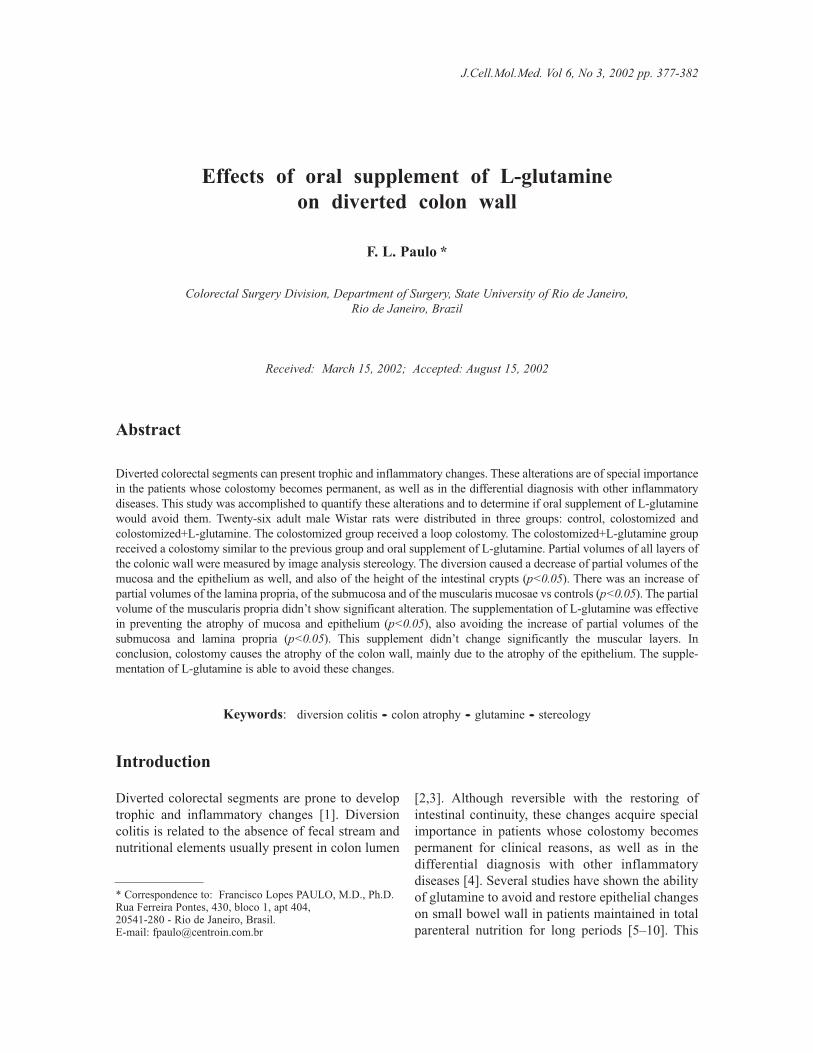
Results
Colostomy and associated diversion colitis change
the structure of the colon wall in terms of partial
volumes of each colon layer. These changes are
diminished by glutamine supplement. The height of
crypts (Fig. 1a) was significantly diminished on
CST group as compared to control (p<0.001) and
the supplement of glutamine was able to diminish
this change (p<0.05). The thickness of mucosa
(Vvm) was diminished (Fig. 1b) in CST group when
compared to control (p<0.001) and showed an
intermediate value in CST–Gln group (p<0.05). The
muscularis mucosae partial volume (Vvmm) raised
(p<0.01) in CST group and CST–Gln group without
statistical difference between them (Fig. 1c). The
submucosa partial volume (Vvs) increased in CST
group (p<0.001) but not in CST–Gln group when
compared to control (Fig. 1d). The muscularis
propria partial volume (Vvmp) was diminished both
in CST group and CST–Gln group when compared
to control (Fig. 1e). There was no statistical difference
between CST and CST–Gln groups (p=NS). An
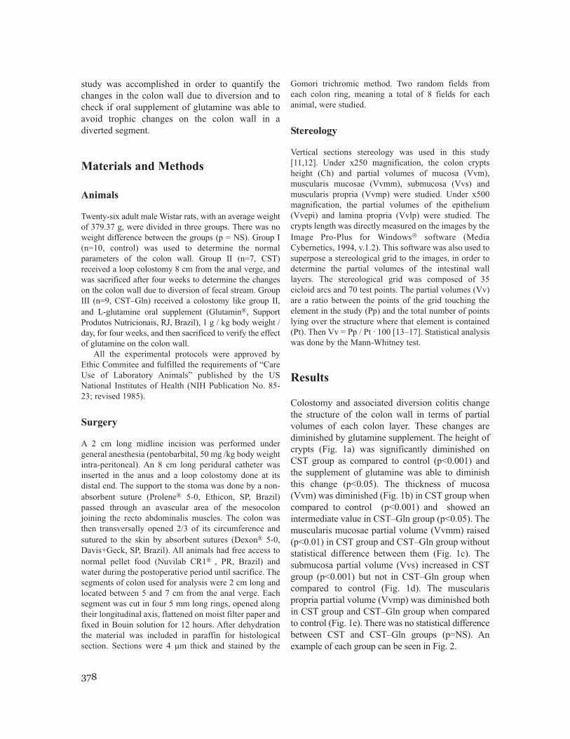
example of each group can be seen in Fig. 2.
378
379
J.Cell.Mol.Med. Vol 6, No 3, 2002
Fig. 1 Variation of crypts height (a) and partial
volumes of mucosa (b), muscularis mucosae (c),
submucosa (d), muscularis propria (e),
epithelium (f) and lamina propria (g) between
control, CST and CST–Gln groups. Significance
indices were evaluated by Mann-Whitney test.
p=NS
Cripts height Partial volumes of submucosa
Partial volumes of muscularis propria
Partial volumes of muscularis mucosae
Partial volumes of mucosa
Partial volumes of epithelium
Partial volumes of lamina propria
Control Diversion Diversion+glutamine
Control Diversion Diversion+glutamine
Control Diversion Diversion+glutamineControl Diversion Diversion+glutamine
Control Diversion Diversion+glutamine
Control Diversion Diversion+glutamineControl Diversion Diversion+glutamine
p<0.05p<0.001
p<0.05
p<0.001
p<0.05
p<0.01
p<0.001
p<0.001 p<0.01
p<0.001
p<0.001
p<0.05
p<0.05
p<0.001
p<0.001
p<0.001
a d
e
f
g
b
c
micrometers
On a detailed study of the mucosal layer the
epithelium (Vvepi) diminished in CST group when
compared to control. Under glutamine supplement,
the Vvepi raised when compared to control and CST
group (Fig. 1f). The partial volume of lamina
propria raised in CST group when compared with
control. In CST–Gln group that volume diminished
(p<0.001) when compared to the other groups (Fig.
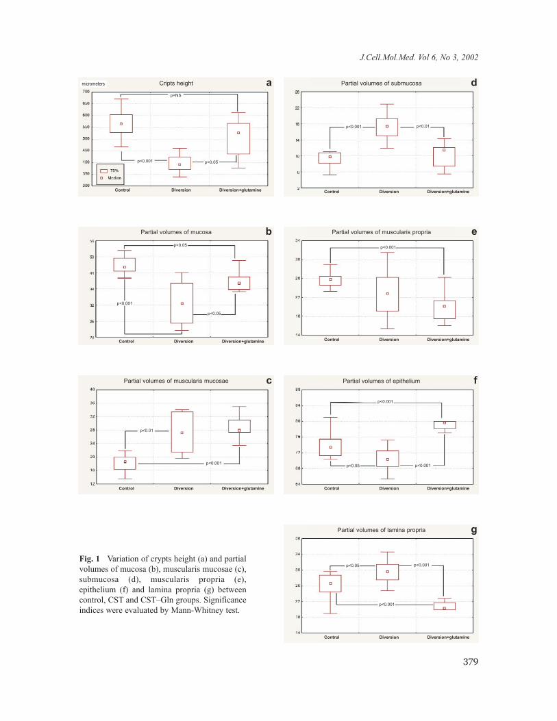
1g). An example of each group can be seen in Fig. 3.
Discussion
The colorectal mucosa epithelial cells use short
chain fatty acids as a primary energetic fuel,
especially acetic, propionic and butyric acids.
These are formed by bacterial degradation of
alimentary fibers contained on fecal stream [18]. In
colorectal segments downwards colostomy, the
absence of dietary fibers on the bowel lumen
380
Fig. 2 An example of the colon wall on groups:
A - control; B - colostomy; C - colostomy+glutamine.
Magnification 250 X. Gömöry staining.
Fig. 3 Effect of glutamine supplement upon colon wall
epithelium: A - control; B - colostomy; C - colo-
stomy+glutamine. Magnification 500 X. Gömöry staining.
precludes fatty acid formation and their absorption
and utilization by the epithelial cells. Glutamine is
usually a secondary fuel source for those cells, but
in diverted segments it may become the first
energetic source [19].
Glutamine is usually absorbed in jejunum and
ileum. In this experiment, the small bowel segments
were maintained intact, ensuring the absorption of
the oral supplement of glutamine given to the
animals. This amino acid is equally metabolized if
absorbed from the intestinal lumen or from the
capillary bed, as it is expected to occur in colon
after colostomy [20–22].
Colostomy caused a significant (p<0.001) re-
duction of the crypts length and the supplement of
glutamine was able to avoid this change. Other
authors found a similar effect of deviation of fecal
stream on the crypts length [23]. Glutamine was
partially effective to avoid the reduction of the
partial volume of mucosa in CST–Gln group. The
mucosa volume remained at an intermediate value
between CST and control groups (p<0.05). A
previous article showed a reduction in partial
volume and mucosa weight in Wistar rats four
weeks after colostomy [12].
Diversion caused a significant (p<0.001)
increase of partial volume of muscularis mucosae
and the supplement of glutamine was not able to
avoid that change. Muscular tissue is rich on
glutamine-synthetase being able to provide large
amounts of glutamine and therefore being less
sensitive to variations of demand and delivery of
this aminoacid [24,25].
Partial volume of submucosa raised in CST
group when compared to control. Other authors
have studied the pathological changes of this layer
in diversion colitis in man and described an
increase in the conjunctive tissue and blood vessels
[26]. These changes can explain the increase in
volume. The use of glutamine was able to avoid this
change in CST–Gln group.
Partial volume of muscularis propria was dimi-
nished in both colostomized groups, and glutamine
was not able to avoid it, probably for the same reasons
discussed above on muscularis mucosae.
A high magnification study of mucosa showed a
fall on the partial volume of epithelium and a
raising of the partial volume of lamina propria
caused by colostomy. The supplement of L-glu-
tamine was able to avoid these changes and
promote a further raising on partial volume of the
epithelium, showing an important trophic effect of
this amino acid in epithelial cells and being in
agreement with findings of other authors studying
small bowel epithelium [8,9,27].
In conclusion, the oral supplement of L-glu-
tamine is able to avoid trophic changes on colon
mucosa and submucosa downwards colostomy.
These effects were not observed on muscular
layers. These findings can be useful either in the
prevention or in the treatment of patients suffering
from diversion colitis.
References
1. Haas P.A., Diversion colitis. In:. Mazier W.P., ed.,
Surgery of the colon, rectum and anus. W.B.
Saunders Company, Philadelphia, 1995, pp. 1011-15
2. Agarwal V.P., Schimmel E.M., Diversion colitis: a
nutritional deficiency syndrome? Nutr. Ver. 47:257,
1989
3. Kissmeyer-Nielsen P., Mortensen F.V., Laurberg
S., and Hessov I., Transmural trophic effect of short
chain fatty acid infusions on atrophic, defunctioned
rat colon, Dis. Colon Rectum, 38: 946, 1995
4. Giardello F.M., Lazenby A.J., and Bayless T.M.,
The new colitides, collagenous, lymphocytic, and
diversion colitis, Gastroenterol. Clin. North. Am.,
24: 717, 1995
5. Burke D.J., Alverdy J.C., Aoys E., and Moss G.S.,
Glutamine-supplemented total parenteral nutrition
improves gut immune function, Arch. Surg.,
124:1396, 1989
6. Harald T., Kienle B., Weilemann L.S., Stehle P.,
and Fürst P., Glutamine dipeptide-supplemented
parenteral nutrition maintains intestinal function in
the critically ill, Gastroenterology, 107: 1595, 1994
7. Okuma T., Kaneko H., Chen K., Ogawa N.,
Torigoe Y., Miyauchi Y., and Tosaka M., Total
parenteral nutrition supplemented with L-alanyl-L-
glutamine and gut structure and protein metabolism
in septic rays, Nutrition, 10: 241, 1993
8. Li I.S., Li J.S., Jiang J.W., Liu F.N., Li N., Qin
W.S., and Zhu H., Glycyl-glutamine-enriched long-
term total parenteral nutrition attenuates bacterial
translocation following small bowel transplantation
in the pig, J. Surg. Res., 85: 106, 1999
9. Khan J., Iiboshi Y., Cui L., Wasa M., Sando K.,
Takagi Y., and Okada A., Alanyl-glutamine-
supplemented parenteral nutrition increases luminal
mucus gel and decreases permeability in the rat small
intestine, J. Parenter. Enter. Nutr., 35: 24, 1999
381
J.Cell.Mol.Med. Vol 6, No 3, 2002
10. Buchman A.L., Glutamine for the gut: mystical
properties or an ordinary amino acid?, Curr.
Gastroenterol. Rep., 1: 417, 1999
11. Gundersen H.J.G., Bendtsen T.F., Korbo L.,
Marcussen N., Moller A., Nielsen K., Nyengaard
J.R., Pakkenberg B., Sorensen F.B., Vesterby A.,
and West M.J., Some new, simple and efficient
stereological methods and their use in
pathological research and diagnosis, APMIS , 96:
379, 1988
12. Kissmeyer-Nielsen P., Christensen H., and
Laurberg S., Diverting colostomy induces
mucosal and muscular atrophy in rat distal colon,
Gut, 35: 1275, 1994
13. Chalkley H.W., Cornfield I., and Park K., A
method for estimating volume-surface ratios,
Science, 110: 295, 1949
14. Weibel E.R., Stereological methods, vol.1,
Practical methods for biological morphometry,
Academic Press, London, 1979
15. Elias H., Hennig A., and Schwartz D.E.,
Stereological applications to biomedical research,
Phisiol.Rev., 51: 158, 1971
16. Elias H., Dallas M.H., An elementary
introduction to stereology (quantitative
microscopy), Am.J.Anat., 159: 411, 1980
17. Elias H., Hyde D.M., A Guide to Practical
Stereology, Karger, Basileia, 1983
18. Roedger W.E.W., Utilization of nutrients by
isolated epithelial cells of the rat colon,
Gastroenterology, 83: 424, 1982
19. Windmueller H.G., Glutamine utilization by the
small intestine, Adv.Enzymol., 53: 202, 1982
20. Windmueller H.G., Spaeth A.E., Uptake and
metabolism of plasma glutamine by the small
intestine, J.Biol.Chem., 249: 5070, 1974
21. Windmueller H.G., Spaeth A.E., Intestinal
metabolism of glutamine and glutamate from the
lumen as compared to glutamine from blood,
Arch.Biophys.Biochem., 171: 662, 1975
22. Windmueller H.G., Spaeth A.E., Respiratory fuels
and nitrogen metabolism in vivo in small intestine of
fed rats, J.Biol.Chem., 255: 107, 1980
23. Keli E., Bouchoucha M., Devroede G., Carnot F.,
Ohrant T., Cugnenc P.H., Diversion-related experi-
mental colitis in rats, Dis.Colon Rectum, 40: 222, 1997
24. Souba W.W., Smith R.J., Wilmore D.W.,
Glutamine metabolism by the intestinal tract,
J.Parenter.Enter.Nutr., 9: 608, 1985
25. Souba W.W., Klimberg V.S., Plumley D.A.,
Salloum R.M., Flynn T.C., et al., The role of
glutamine in maintaining a healthy gut and
supporting the metabolic response to injury and
infection, J.Surg.Res., 48: 383, 1990
26. Ma C.K., Gottlieb C., Haas P.A., Diversion colitis:
A clinicopathologic study of 21 cases. Human
Pathol., 21: 429, 1990
27. Murnin M., Kumar A., Li G.D., Brown M.,
Sumpio B.E., Basson M.D., Effects of glutamine
isomers on human (Caco-2) intestinal epithelial
proliferation, strain-responsiveness, and differen-
tiation, J. Gastrointest. Surg., 4: 435, 2000
382


![The Roles of Glutamine in the Intestine and Its ...€¦ · utilize large amounts of glutamine, exceeding the endogenous glutamine production [12,13], and that plasma and muscle glutamine](https://img.pdfslide.us/doc/110x75/5fd64d48c22ac35b4b7b6b55/the-roles-of-glutamine-in-the-intestine-and-its-utilize-large-amounts-of-glutamine.jpg)