Embed Size (px)
Citation preview
BearWorks BearWorks
MSU Graduate Theses
Summer 2018
Effects of Neem on Plasmatocyte Counts in Galleria Mellonella Effects of Neem on Plasmatocyte Counts in Galleria Mellonella
Larvae Larvae
Michael Anthony Fenske Missouri State University, [email protected]
As with any intellectual project, the content and views expressed in this thesis may be
considered objectionable by some readers. However, this student-scholar’s work has been
judged to have academic value by the student’s thesis committee members trained in the
discipline. The content and views expressed in this thesis are those of the student-scholar and
are not endorsed by Missouri State University, its Graduate College, or its employees.
Follow this and additional works at: https://bearworks.missouristate.edu/theses
Part of the Entomology Commons
Recommended Citation Recommended Citation Fenske, Michael Anthony, "Effects of Neem on Plasmatocyte Counts in Galleria Mellonella Larvae" (2018). MSU Graduate Theses. 3297. https://bearworks.missouristate.edu/theses/3297
This article or document was made available through BearWorks, the institutional repository of Missouri State University. The work contained in it may be protected by copyright and require permission of the copyright holder for reuse or redistribution. For more information, please contact [email protected].
EFFECTS OF NEEM ON PLASMATOCYTE COUNTS IN GALLERIA
MELLONELLA LARVAE
A Master’s Thesis
Presented to
The Graduate College of
Missouri State University
TEMPLATE
In Partial Fulfillment
Of the Requirements for the Degree
Master of Science, Agriculture
By
Michael Fenske
August 2018
ii
EFFECTS OF NEEM ON PLASMATOCYTE COUNTS IN GALLERIA
MELLONELLA LARVAE
Agriculture
Missouri State University, August 2018
Master of Science
Michael Fenske
ABSTRACT
The neem tree (Azadirachta indica) has been called the tree of the 21st century, due to its
many beneficial properties. This research studies the effects of neem products on
plasmatocytes of Galleria mellonella larvae challenged by injections of Sumi ink.
Counts of circulating plasmatocytes increased in response to the ink within 4 hours.
Concurrent injections of neem essential oil reversed this process in a dose-dependent
manner. Methanolic extracts of neem essential oil had similar effects on counts of
circulating plasmatocytes mobilized by ink injections. Aqueous extracts from neem bark
did not have effects on plasmatocyte counts. Comparative high-performance thin layer
chromatography (HTPLC) showed a band of difference between chromatographic
profiles of biologically active methanolic extracts of neem essential oil and biologically
inactive aqueous extracts from neem bark. This band was stigmasterol. Stigmasterol
alone reversed effects of ink injections on plasmatocyte counts with a dynamic like that
exhibited by methanolic extracts of neem.
KEYWORDS: neem, Galleria mellonella, Sumi ink, plasmatocytes, HPTLC,
stigmasterol
This abstract is approved as to form and content
_______________________________
Dr. Maciej Pszczolkowski
Chairperson, Advisory Committee
Missouri State University
iii
EFFECTS OF NEEM ON PLASMATOCYTE COUNTS IN GALLERIA
MELLONELLA LARVAE
By
Michael Fenske
A Master’s Thesis
Submitted to the Graduate College
Of Missouri State University
In Partial Fulfillment of the Requirements
Master of Science, Agriculture
August 2018
Approved:
_______________________________________
Maciej Pszczolkowski, PhD
_______________________________________
Chin-Feng Hwang, PhD
_______________________________________
Martin Kaps, PhD
_______________________________________
Julie Masterson, PhD: Dean, Graduate College
In the interest of academic freedom and the principle of free speech, approval of this thesis indicates the
format is acceptable and meets the academic criteria for the discipline as determined by the faculty that
constitute the thesis committee. The content and views expressed in this thesis are those of the student-
scholar and are not endorsed by Missouri State University, its Graduate College, or its employees.
iv
ACKNOWLEDGEMENTS
I wish to express my deep appreciation to Dr. Maciej Pszczolkowski for his
passion for entomology, and for the conveyance of his passion to me. He has been my
mentor, and he has opened my eyes to a new world. Without his help, I would not have
accomplished my research and this dissertation. He gave a nontraditional student a
chance, and for that I am eternally grateful.
I wish to thank my committee members Dr. Chin-Feng Hwang and Dr. Martin
Kaps for their time spent on my advisory committee.
Finally, I want to thank my lab partners, Westley Peterson and Rachel Veenstra
“The Queen of the Lab.” Together, we accomplished our work and had fun doing it.
I dedicate this thesis to my wife Mary and my dad Wilbert. Words cannot fully
express my appreciation. Their constant support and encouragement made my college
endeavor possible. I was able to attain my educational goals through their sacrifices of
time and expense, and for this I say, Thank You.
v
TABLE OF CONTENTS
Introduction ......................................................................................................................... 1
Methods............................................................................................................................. 10
Insects ................................................................................................................... 10 Chemicals .............................................................................................................. 11 Validation of the Results from Previous Studies; Response to Ink ...................... 11 Validation of the Results of Previous Studies; Effects of Crude Neem Essential
Oil on Plasmatocyte Mobilization by Ink Injections ............................................ 13
Extraction of Crude Neem Essential Oil ............................................................... 13 Effects of Methanolic Extract from Neem Essential Oil on Counts of
Plasmatocyte Mobilization by Ink Injections ....................................................... 14 Effects of Aqueous Extract from Neem Bark on Plasmatocyte Mobilization by Ink
Injections ............................................................................................................... 15 Comparative Chromatography of Neem Essential Oil Extract and Neem Bark
Extract ................................................................................................................... 15 Confirmation of Stigmasterol Presence in Methanolic Extract of Neem Essential
Oil ......................................................................................................................... 16
Effects of Stigmasterol Acetate on Plasmatocyte Mobilization by Ink Injections 16 Statistical Analysis ................................................................................................ 16
Results ............................................................................................................................... 18 Validation of the Results from Previous Studies; Response to Ink ...................... 18 Validation of the Results from Previous Studies; Effects of Crude Neem Essential
Oil on Plasmatocyte Mobilization by Ink Injections ............................................ 19
Extraction of Crude Neem Essential Oil ............................................................... 19 Effects of Methanolic Extract from Neem Essential Oil on Plasmatocyte
Mobilization by Ink Injections .............................................................................. 20
Effects of Aqueous Extract from Neem Bark on Plasmatocyte Mobilization by Ink
Injections ............................................................................................................... 21
Comparative Chromatography of Neem Essential Oil Extract and Neem Bark
Extract ................................................................................................................... 23 Confirmation of Stigmasterol Presence in Methanolic Extract from Neem
Essential Oil .......................................................................................................... 23 Effects of Stigmasterol Acetate on Plasmatocyte Mobilization by Ink Injections 25
Discussion ......................................................................................................................... 27
References ......................................................................................................................... 32
vi
LIST OF TABLES
Table 1. Effects of various Sumi ink concentrations on plasmatocyte counts in Galleria
mellonella larvae ............................................................................................................... 18
Table 2. Time dependent effects of Sumi ink on plasmatocyte counts in Galleria
mellonella larvae ............................................................................................................... 19
Table 3. Dose-dependent effects of neem essential oil on plasmatocyte counts in Galleria
mellonella larvae challenged with Sumi ink ..................................................................... 20
Table 4. Summary of the high-performance thin layer chromatogram of three extracts
from neem essential oil ..................................................................................................... 20
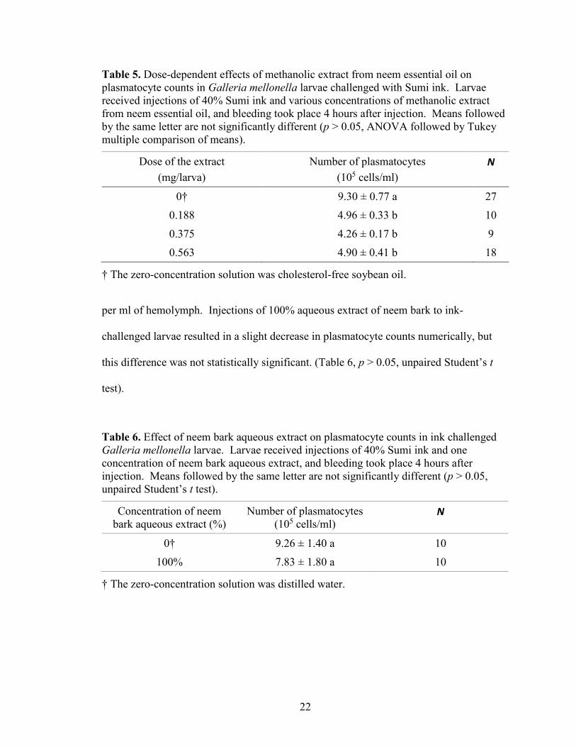
Table 5. Dose-dependent effects of methanolic extract from neem essential oil on
plasmatocyte counts in Galleria mellonella larvae challenged with Sumi ink ................. 22
Table 6. Effect of neem bark aqueous extract on plasmatocyte counts in ink challenged
Galleria mellonella larvae ................................................................................................ 22
Table 7. Summary of comparative high-performance thin layer chromatograms of
methanolic extract from neem essential oil and aqueous extract from neem bark ........... 23
Table 8. Summary of comparative high-performance thin layer chromatograms of
methanolic extract from neem essential oil and methanolic solutions of stigmasterol
acetate ............................................................................................................................... 24
Table 9. Dose-dependent effects of stigmasterol acetate on plasmatocyte counts in
Galleria mellonella larvae challenged with Sumi ink ...................................................... 26
vii
LIST OF FIGURES
Figure 1. High-performance thin layer chromatogram of three extracts from neem
essential oil........................................................................................................................ 21
Figure 2. Comparative high-performance thin layer chromatogram of methanolic extracts
from neem essential oil (Lanes A-D) and aqueous extract of neem bark (Lanes E-H) .... 24
Figure 3. Comparative high-performance thin layer chromatogram of stigmasterol acetate
(Lanes 1-3), methanolic extract from neem essential oil (Lane 4), and stigmasterol acetate
again (Lanes 5-7) .............................................................................................................. 25
1
INTRODUCTION
The global population is likely to grow at a rate of 70 million per year, with a
projected 9.2 billion people by 2050. Food production will also have to escalate at a
projected rate of 70%. Currently, a percentage of agriculture production focuses on
biofuel and fiber (Popp, Pető, & Nagy, 2013). Combined with the limited and decreasing
amount of agricultural land available, this is a monumental task.
Synthetic pesticide development after World War II has dramatically increased
(Popp et al., 2013). This, with other practices, has led to the growth of agricultural
productivity to all new highs. To accelerate the food production to new levels, pesticides
are part of the answer. Global pesticide use is already at 3 million tons annually, at a cost
of over 40 billion dollars. There is a continued dependence on the use of chemical
pesticides, which entomologists have called the “pesticide treadmill” (Popp et al., 2103,
p. 246). The treadmill, also called a trap, is when agriculture producers must use larger
amounts of pesticides and/or ones that are increasingly stronger due to pest resistance.
There are two determining factors involved. The first is to use less effective chemicals
that need higher treatment rates coupled with a higher frequency of applications. The
second is to develop new pesticides that are effective until the resistance of the target is
met (Popp et al., 2013).
Continued use of chemical pesticides is having detrimental effects on the
environment. Pesticides can enter bodies of water through infiltration or point-source
inputs. Infiltration is the leaching of chemicals into ground water from land applications.
Point-source inputs may come from several factors, including surface runoff, erosion,
2
spray drift, tile drains, and volatilization (Reichenberger, Bach, Skitschak, & Frede,
2007).
Surface runoff can occur in practically every arable field, even ones without a
measurable slope. During heavy rains or prolonged periods of moisture saturation,
excess runoff occurs, and it is called Hortonian flow. This happens when the water table
rises to the surface, causing any rainfall to run off (Reichenberger et al., 2007). The U.S.
Geological Society estimated that over 90% of water and fish samples obtained from all
streams contained one or more pesticides. In ground water, it has found over 143
different pesticides (Aktar, Sengupta, & Chowdhury, 2009).
Spray drift and volatilization also are problems with pesticides. Spray drift may
account for up to 25% loss of chemicals, which can spread from short distances to
hundreds of miles. Volatilization can account for up to a 90% loss of applied pesticides.
Almost every pesticide has been detected in the atmosphere and precipitation, throughout
the United States at various times of the year (Aktar et al., 2009).
Human exposure is especially problematic for agricultural laborers and chemical
manufacturing. Globally, chemical pesticides are responsible for nearly one million
chronic diseases and deaths annually. It is possible for organochlorine insecticides to
contaminate the atmosphere, water, and tissues of all life forms on the planet (Aktar et
al., 2009).
Pesticides affect every life form, from plants to soil microorganisms to wildlife.
Many plants use mycorrhizal fungi, found on their roots, to increase nutrient uptake.
Laboratory studies have shown that Roundup is toxic to this beneficial organism, even in
slight amounts. Dolphins’ high trophic level combined with their low ability to
3
metabolize chemicals is causing global poisoning. The Indus river dolphin (Platanista
minor), found in China, is facing extinction due to pesticides (Aktar et al., 2009). In the
United States alone, pesticides account for over 9.6 billion dollars annually in losses from
public health, natural pollinators, pest resistance, wildlife, pets, and water contamination
(Popp et al., 2013).
Biopesticides and integrated pest management (IPM) are essential for our future.
Biopesticides come from natural substances that are ecofriendly, and from control pests
with nontoxic processes. Biopesticides are less toxic than chemical pesticides, are
effective in minute amounts, decompose quickly, and usually only target the desired pest
or related organisms (Dutta, 2015).
Accordingly, the Environmental Protection Agency established the Biopesticides
and Pollution Prevention Division in 1994. It promotes safer pesticides in conjunction
with IPM (Dutta, 2015). IPM is using practices that have a negligible impact on the
environment, while maintaining pests below the economic threshold. This is when the
cost of the pest’s damage exceeds the cost of control.
It usually takes a minimum of 3 years to register a conventional pesticide.
Biopesticides normally require less than a year, due to the lower risks involved. As of
2014, there were over 430 registered active ingredients for biopesticides, and 1,320
registered products. The market for biopesticides was at 1.3 billion dollars globally in
2011, and it may soon top 3.2 billion dollars. The increased demand for organically
produced foods is a key factor in this increased usage of biopesticides, along with faster
certification by the Environmental Protection Agency (Dutta, 2015).
4
Many consider neem the most effective biopesticide, and it is ecofriendly (Dutta,
2015). The international scientific community includes neem in the top 10 list for the
most promising plants for the “sustainable development of the planet and the health of
living beings” (Nicoletti, Maccioni, Coccioletti, Mariana, & Vitali, 2012, p. 414). The
World Health Organization and the United Nations Environment Programme, in their
1989 report, stated that neem is “one of the most promising trees of the 21st century for its
enormous potential in pest management, environmental protection, and medicine”
(Nicoletti et al., 2012, p. 414).
Neem (Azadirachta indica A. Juss) is a tropical evergreen tree, which can be
deciduous in drier zones (Girish & Shankara, 2008). It is native to the Indo-Pakistan
subcontinent, and it grows well in semiarid climates (Ahmed & Grainge, 1986). The
neem tree is tolerant of elevated temperatures, but it cannot survive cold or frost. This
tree grows well in nutrient-deficient soils, and it can extract any necessary nutrients from
its deep root system (Koul, Isman, & Ketkar, 1990). The neem tree bark, leaves, fruit,
and seeds have many uses (Ahmed & Grainge, 1986). Ayurvedic medicine has used
neem for its properties for over 4,000 years, leading to the native Indian name “Village
pharmacy” (Girish & Shankara, 2008, p. 108). For centuries, Indo-Pakistani farmers
have soaked their grain storage sacks in a mixture of dried neem leaves and water
overnight. They then dry these sacks and use them to prevent pest damage in their stored
grain (Ahmed & Grainge, 1986).
Segregation and identification of neem constituents in India began in 1942. The
first compounds researchers found were nimbin and nimbinin, with another bitter
substance called nimbidin (Koul et al., 1990). Not until 1959 was the neem tree globally
5
recognized. It was then that noted entomologist, Dr. Heinrich Schmutterer observed that
swarming locusts defoliated almost all plant life in Sudan, except for a few neem trees
that others had introduced to the area (Ahmed & Grainge, 1986). Since the mid-20th
century, neem research has intensified, with it being the most promising natural substance
for biological control of pests (Biswas, Chattopadhyay, Banerjee, & Bandyopadhyay,
2002).
Research in subsequent years with neem extracts has shown that they disrupt
insect reproduction and development. Researchers have isolated over 135 compounds
from the neem tree (Biswas et al., 2002). In the leaves, bark, fruit, and seeds there are
many biologically active compounds. “These include protolimonoids, limonoids, or
tetranortriterpenoids, pentanortriterpenoids, hexanortriterpenoids, and nontriterpenoidal
constituents” (Koul et al., 1990, p. 2). The nontriterpenoidal compounds include
“hydrocarbons, fatty acids, diterpenoids, sterols, phenols, flavonoids, and glycosides”
(Koul et al., 1990, p. 2).
The most studied are the triterpenoids, which include azadirachtin, salanin, and
meliantriol. These triterpenoids control more than 100 species of insects, mites, and
nematodes (Ahmed & Grainge, 1986). Studies of azadirachtin have shown that it is an
effective antifeedant and growth disruptor in insects (Sinha et al., 1999). Recent research
with biopesticides has targeted the larval stage, due to the chance of resistance and
adaptation by adults (Nicoletti et al., 2012).
Another aspect of pest control is to target the immune response. Multicellular
entities have a two-system response to infectious organisms, innate and acquired (Lavine
& Strand, 2002). An innate immune system is an evolutionary type of nonspecific
6
defense, whereas an acquired defense is a learned way of attacking a specific antigen that
develops an immunological memory (Lavine & Strand, 2002). Insects only have an
innate immune system, but it is well developed. Initial defenses include the integument
and gut, clotting reaction, and cytotoxic molecules produced at the wound site (Lavine &
Strand, 2002). Researchers discovered numerous hemolymph proteins whose function is
innate immune response first in Lepidopteran insects (Jiang, Vilcinskas, & Kanost,
2010).
Insects also have humoral and cellular defense responses. Humoral defenses deal
with antimicrobial peptide production, responsive intermediates of either oxygen or
nitrogen, and multifaceted enzymatic cascades that control coagulation or melanization of
the insect’s hemolymph (Lavine & Strand, 2002). Hemolymph contains the insect’s
blood cells, or hemocytes (Jones, 1962). Cellular defenses represent a hemocyte-
facilitated immune response, such as phagocytosis or encapsulation.
Phagocytosis is a primary response of hemocytes to small particles, such as
bacteria, and it is a form of receptor-mediated endocytosis (Gillespie, Kanost, &
Trenczek, 1997). It is a defense response, in that the target attaches to the receptor and
stimulates the immune cell to create a phagosome (Strand, 2008). Hemocytes
phagocytose many biotic targets, including bacteria and yeast, or abiotic elements like
India ink (Lavine & Strand, 2002). Active polymerization-dependent processes engulf
the target, and the phagosome matures into a phagolysosome, through a succession of
fission and fusion actions with endosomes and lysosomes (Strand, 2008).
Unlike phagocytosis, which involves a single cell, encapsulation is the result of
multiple hemocytes attaching to larger particles, involving multiple cells (Strand, 2008).
7
Multilayer sheaths surround the material that undergoes encapsulation, which is why
scholars refer to them as capsules. Encapsulation occurs within 1 to 3 days (Ottaviani,
2005). After formation, the capsule may melanize, and it can undergo digestion or
degradation. Sometimes when describing the attachment of multiple hemocytes to
masses of bacteria, it is referred to as nodulation (Strand, 2008).
Researchers have categorized hemocytes by morphological characteristics,
determined by light or electron microscopy (Gillespie et al., 1997). Hemocytes increase
in number and segregate into distinct types due to stress, infection, and injury responses
(Strand, 2008). The most common types of hemocytes are granulocytes, plasmatocytes,
spherulocytes, and oenocytoids. Scholars have identified these cell types in species from
diverse orders such as “Lepidoptera, Diptera other than Drosophila, Orthoptera, Blattaria,
Coleoptera, Hymenoptera, Hemiptera, and Collembola” (Strand, 2008, p. 2).
Granulocytes occur in the hemolymph as rounded cells, which firmly adhere to foreign
substances and spread symmetrically. Plasmatocytes also occur as rounded cells, but
they spread asymmetrically after attachment to a foreign substance. In the larval stage of
Lepidoptera, both granulocytes and plasmatocytes make up over 50% of circulating
hemocytes. Oenocytoids are non-adhesive hemocytes that contain phenoloxidase cascade
elements like prophenoloxidase 1, which leads to melanin production. Spherule cells or
spherulocytes are possible sources of cuticle constituents (Strand, 2008). In Lepidoptera,
there are also small numbers of prohemocytes, which are progenitor or stem cells. These
prohemocytes may differentiate into other hemocytes, especially plasmatocytes and
granulocytes, during times of stress or injury (Strand, 2008). Plasmatocytes are the main
capsule-forming hemocytes in Lepidoptera, but granulocytes are also present. In some
8
species like Pseudoplusia includens, granulocytes first attach to the foreign object, then
plasmatocytes adhere in large numbers to the target (Strand, 2008). Granulocytes break
down to sticky granular cells after attachment to a foreign substance, and they may
release a chemoattractant that activates the plasmatocytes to form the capsule (Ottaviani,
2005).
A previous Missouri State University graduate student, Katherine Haszcz, showed
that Sumi ink injections increased numbers of circulating plasmatocytes in Galleria
mellonella larvae, and that crude neem essential oil reversed this process (Haszcz, 2016).
However, the bright light microscopy Haszcz (2016) used made classification of
hemocytes problematic. Therefore, further work was necessary to validate Haszcz’s
results.
Also, Haszcz (2016) performed experiments aiming to delineate which
compounds of neem could be responsible for the reversing effects of Sumi ink on
circulating plasmatocytes. Haszcz tested azadirachtin, one of the most studied
triterpenoids of neem oil. Among many effects, it disrupts insect growth by suppressing
the process of molting (Sinha et al., 1999). However, Haszcz’s research showed that
azadirachtin (at doses that inhibited molting) had no effect on numbers of circulating
plasmatocytes in Galleria mellonella larvae. While crude neem essential oil effectively
impaired the immune response in insects by lowering the numbers of circulating
plasmatocytes, it was still unclear what neem constituent was responsible for these
outcomes.
The aim of this work was to validate the findings of Haszcz (2016) using a
microscope with differential interference contrast (also known as Nomarski contrast), and
9
a new type of microscopy, which is currently under development by my lab partner
Westley Peterson, called pseudo-Nomarski contrast.
In addition, I have extended the work of Haszcz (2016) by isolation and
identification of the chemical responsible for reversal of Sumi ink effects on Galleria
mellonella plasmatocytes (Haszcz, 2016). To that end, I extracted the crude neem
essential oil with methanol, and tested the extract in bioassays for its ability to reverse
increases of plasmatocyte numbers in response to Sumi ink injections. Moreover, using
high-performance thin layer chromatography (HPTLC) and bioassays, I identified a
specific neem oil component, stigmasterol, as the neem-derived substance that reverses
the process of plasmatocyte mobilization by Sumi ink in Galleria mellonella larvae.
10
METHODS
Insects
The experiments used greater wax moth (Galleria mellonella) larvae. This moth
belongs to the order Lepidoptera and the family Pyralidae. Galleria mellonella live in
beehives and nests, consuming wax, honey, and pollen (Wojda, 2017). Galleria
mellonella has a life cycle of 7-8 weeks, and it undergoes seven larval instars. This takes
approximately 5-6 weeks at 25-28°C. Two more weeks are necessary to form adult
moths from the pupae (Wojda, 2017). The advantages of this insect are simple
procedures for injections, and it is an ethically suitable model for experimentation
(Mukherjee et al., 2010). Rearing of Galleria mellonella is also cost effective (Wojda,
2017). Researchers have used Galleria mellonella extensively for studies in physiology,
toxicology, biochemistry, and pathology, among other disciplines. For experimentation
involving the rearing of parasitic and predatory insects, Galleria mellonella larvae are
also useful hosts or prey (Mohamed & Coppel, 1983).
I obtained the larvae from Knutson’s Live Bait (Brooklyn, MI) in their 7th instar
stage. I separated them into one-pint glass mason jars with vented lids at ~ 60 larvae per
jar. I kept them in a light-free incubator (VWR Scientific Products, Model 2005) at 30°C
and 80% relative humidity to maintain their normal life cycle. I fed them a diet
formulated by combining 37ml of glycerin USP 99.5% (Humco), 25ml of pure granulated
cane sugar (C&H), and 25ml of purified water in a plastic bowl. I then heated the
mixture ~ 10 to 20 seconds in a low-power microwave to dissolve the sugar, and stirred it
thoroughly. I placed this mixture and 400 ml of Gerber® multigrain cereal (Fremont, MI)
in a gallon Ziploc bag, and I hand mixed it thoroughly, until a spongy texture formed.
11
The Gerber® multigrain cereal contains essential vitamins and minerals to maintain the
health of the larvae. Excess diet mixture may be stored for up to 2 months at 4°C.
Chemicals
I formulated a stock solution of anticoagulant buffer (0.157g NaOH, 0.435g NaCl,
0.315g citric acid, 0.253g Na2EDTA, pH 4.58) and neutral red stain (2mg of bacto-
neutral red to 1ml of anticoagulant buffer). I obtained NaCl and NaOH from Sigma-
Aldrich® (St. Louis, MO); I bought Na2EDTA and citric acid from Thermo Fisher
Scientific (Pittsburg, PA); I acquired bacto-neutral red from Difco Laboratories (Detroit,
MI). I specify the other chemicals and reagents and their respective vendors in the
following part of the Methods.
Validation of the Results from Previous Studies; Response to Ink
In the hemolymph of Galleria mellonella, the plasmatocytes remain in a rounded
shape, but they spread asymmetrically when they come into contact with plastic or glass.
Therefore, it is easy to distinguish them from other classes of hemocytes.
I used black Sumi ink (Yasutomo, San Francisco, CA) as an artificial pathogen. I
pipetted an aliquot of 200μl of the ink into a 2ml microcentrifuge tube, and centrifuged it
at 2,000g for 10 minutes to eliminate large particles. I pipetted the supernatant into
another microcentrifuge tube and diluted it to the desired concentration with distilled
water.
I anaesthetized the 7th instar larvae of Galleria mellonella by immersion in water
for 10 minutes, blotted them, and injected them with 5μl of ink solution through the
proleg with a Hamilton® syringe equipped with 24G needle (Reno, NV). Control larvae
12
received 5μl of distilled water. I then placed the larvae in glass jars, provided them with
food, and stored them in an incubator at 30°C.
I prepared microcentrifuge tubes for each larva with 40μl of anticoagulant buffer
and 4μl of neutral red stain. After incubation, I collected hemolymph by cautiously
cutting off a proleg with micro iris scissors and bleeding the larva onto a piece of
parafilm. I immediately pipetted 5μl of the hemolymph into the prepared tube with the
buffer and stain, vortexed, and allowed it to set for 5 minutes at room temperature to stain
the cells.
When staining was complete, I then vortexed the prepared tube, pipetted 10μl of
the solution with cells to the upper and lower chamber of an improved Neubauer
hemocytometer and covered it with a Corning cover glass (thickness 1, 22 x 22mm). The
chamber of the hemocytometer filled thanks to capillary action. I then inspected the
hemocytometer using a Galen III microscope (Cambridge Instruments, Cambridge),
equipped with phase contrast or a custom-made oblique illumination filter (W. Peterson,
personal communication, September 26, 2017). I counted the plasmatocytes
boustrophedonically, using the outside four corner quadrants.
I used this procedure for two different series of experiments. First, I tested
several different concentrations of Sumi ink: 0, 10, 20, 40, and 80%. In this experiment,
the incubation time equaled 4 hours, based on the work of Haszcz (2016). In the second
experiment, I injected the ink at only one concentration of 40%, but I bled the larvae and
counted plasmatocytes after various times of larvae incubation: 30 minutes, 2, 3, 4, 6, and
8 hours. I obtained each data point using 6-14 larvae.
13
Validation of the Results of Previous Studies; Effects of Crude Neem Essential Oil
on Plasmatocyte Mobilization by Ink Injections
Haszcz (2016) used neem essential oil from NOW Foods, Bloomingdale, IL for
previous experiments in 2015. For this work, I purchased a new batch of neem essential
oil from the same vendor in 2017. I sealed the newly acquired oil with parafilm
(American National Can, Chicago, IL) to prevent degradation, and stored it in a
refrigerator at 4°C. I measured it precisely in a 2ml microcentrifuge tube by weight, and
then I diluted it to a 30% concentration of neem oil using cholesterol-free soybean oil (as
solvent), purchased from Cal Western Packaging Corporation, Memphis, TN.
I used water-anaesthetized, 7th instar larvae of Galleria mellonella. I injected 10
experimental larvae with 5μl of 40% Sumi ink as described before, and additionally, they
received 5μl of neem essential oil dissolved to 0.188, 0.375, and 1.125 mg/larva, in
cholesterol-free soybean oil. I injected 10 control larvae with 5μl of 40% Sumi ink and
with 5μl of cholesterol-free soybean oil. I provided the injected larvae with food, and I
placed them in an incubator at 30°C and bled them after 4 hours. I obtained each data
point using 5-9 larvae.
Extraction of Crude Neem Essential Oil
I placed 200μg of neem essential oil in a 2ml microcentrifuge tube, and then I
added 1ml of methanol, vortexed the tube, and left it for 10 minutes at room temperature.
Next, I centrifuged the tube at 2,000g for 10 minutes. I removed the supernatant to a
precisely weighed 2ml microcentrifuge tube. I then rotary evaporated the supernatant
(Savant Instruments Inc, Holbrook, NY) for approximately 35 minutes to remove any
methanol. I reweighted the tube to determine the final mass of the residue, which I
14
subsequently resuspended with appropriate solvent to the desired concentration. (For thin
layer chromatography, I used methanol, whereas for bioassaying, I dissolved the residue
in cholesterol-free soybean oil). I used this procedure for two more extractions, one
using ethanol, and another using distilled water instead of methanol.
I visualized the extracted compounds using HPTLC. Briefly, I applied
methanolic, ethanolic, and aqueous extracts onto Merck 10 x 10 cm silica gel F254 glass
plates using a Camag Nanomat 4 HPTLC spotter. I then developed the plates for 6
minutes in a horizontal Camag developing chamber. The mobile phase comprised
chloroform and ethyl acetate at an 8:2 volume to volume ratio. I added acetic acid to this
mixture at 2% concentration. I air dried the developed plates and viewed and
photographed them in ultraviolet light at 254nm. I calculated retention factors (Rfs) for
each band. I only used methanolic extract for further experiments.
Effects of Methanolic Extract from Neem Essential Oil on Counts of Plasmatocyte
Mobilization by Ink Injections
I prepared the methanolic extract as described above, rotary evaporated it, and
suspended it in cholesterol-free soybean oil at 0.188, 0.375, and 0.563 mg/larva
concentrations. I water anaesthetized Galleria mellonella larvae. I injected 10 larvae
with 5μl of 10% methanol-extracted neem and 5μl of 40% Sumi ink. I repeated this
procedure with 20% methanol-extracted neem (10 larvae) and 30% methanol-extracted
neem (another 10 larvae). Each of 10 control larvae received 5μl of pure cholesterol-free
soybean oil. I placed the injected larvae in an incubator at 30°C and bled them after 4
hours. I obtained each data point using 9-27 larvae.
15
Effects of Aqueous Extract from Neem Bark on Plasmatocyte Mobilization by Ink
Injections
I tested aqueous neem bark extract (Sigma, St. Louis, MO) at 100% concentration
using the aforementioned procedures. Briefly, I anaesthetized Galleria mellonella larvae
in water for 10 minutes. Ten experimental larvae received 5μl injections of 40% Sumi
ink, combined with 5μl injections of 100% aqueous neem bark extract. Ten control
larvae received a combination of ink injections and distilled water injections. I then
placed the injected larvae in an incubator at 30°C, and I bled them after 4 hours. I
obtained the data points with 10 larvae.
Comparative Chromatography of Neem Essential Oil Extract and Neem Bark
Extract
To delineate the biologically active substances in methanolic neem essential oil
extract, I used comparative HPTLC. To that end, I applied methanolic extracts at four
concentrations (3.75%, 7.5%, 15%, and 30%) onto silica gel F254 glass plate using a
Camag Nanomat 4 HPTLC spotter. I then developed the plate for 6 minutes in a
horizontal Camag developing chamber. The mobile phase comprised chloroform and
ethyl acetate at an 8:2 volume to volume ratio. I added acetic acid to this mixture at 2%
concentration. I air dried the developed plate. I repeated this procedure with another
plate to which I had applied aqueous extract from neem bark at four concentrations
(3.75%, 7.5%, 15%, and 30%). I then assembled the plates (one with methanolic extract
of neem essential oil and one with aqueous extract of neem bark) side by side and
photographed them in ultraviolet light at 254 nm. I calculated Rfs for each band.
16
Confirmation of Stigmasterol Presence in Methanolic Extract of Neem Essential Oil
I extracted neem essential oil with methanol as previously described, rotary
evaporated it, and diluted it to 15% concentration using pure methanol. I diluted
stigmasterol acetate (Chem Cruz, Dallas, TX) with pure methanol to 10%, 20%, and 30%
concentrations. I then applied the chemicals to a silica gel F254 glass plate in the
following pattern: 10%, 20%, and 30% stigmasterol, 15% methanolic extract of neem
essential oil, and again 10%, 20%, and 30% stigmasterol. I then developed the plate as
described above, air dried it, and photographed it in ultraviolet light at 254 nm. I
calculated Rfs for each band.
Effects of Stigmasterol Acetate on Plasmatocyte Mobilization by Ink Injections
I diluted stigmasterol acetate, which I purchased from Chem Cruz, Dallas, TX, to
0.071, 0.141, and 0.282 mg/larva concentrations in cholesterol-free soybean oil. I then
kept all concentrations on ice for the injections. I water anaesthetized the larvae for 10
minutes. I then subjected the experimental larvae to a 5μl injection of 40% Sumi ink
concurrently with various concentrations of stigmasterol acetate in 5μl of cholesterol-free
soybean oil. The control larvae received 5μl of 40% ink and 5μl of cholesterol-free
soybean oil. I placed the injected larvae in an incubator at 30°C, and I bled them after 4
hours. I obtained the data points with 10-16 larvae.
Statistical Analysis
I analyzed the data I obtained for the effects of aqueous extract from neem bark
on plasmatocyte mobilization by ink injections using Student’s t test. In all remaining
experiments, I used ANOVA, followed by Tukey multiple comparison of means. I tested

17
the data using GraphPad InStat, (GraphPad Software, San Diego, CA). I set the statistical
significance level at p < 0.05.
18
RESULTS
Validation of the Results from Previous Studies; Response to Ink
Haszcz (2016) showed that numbers of plasmatocytes circulating in the
hemolymph of Galleria mellonella larvae increased together with increasing doses of
injected Sumi ink, and together with post injection time of insect incubation.
This work confirms the findings of Haszcz (2016). Sumi ink injections increased
plasmatocyte counts from about 16 x 105 cells per ml of hemolymph to about 33 x 105
cell per ml of hemolymph, in a dose-dependent manner (Table 1). At 40% and 80% ink
concentrations, the increase of plasmatocyte numbers was statistically significant (p <
0.05, ANOVA followed by Tukey Multiple Comparison of Means Test). Also, I showed
that Galleria mellonella larvae respond to Sumi ink injections in a time-dependent
manner (Table 2). Plasmatocyte numbers increased significantly at 3 hours after ink
injection, and they stayed significantly elevated for the next 5 hours (p < 0.05, ANOVA
followed by Tukey multiple comparison of means).
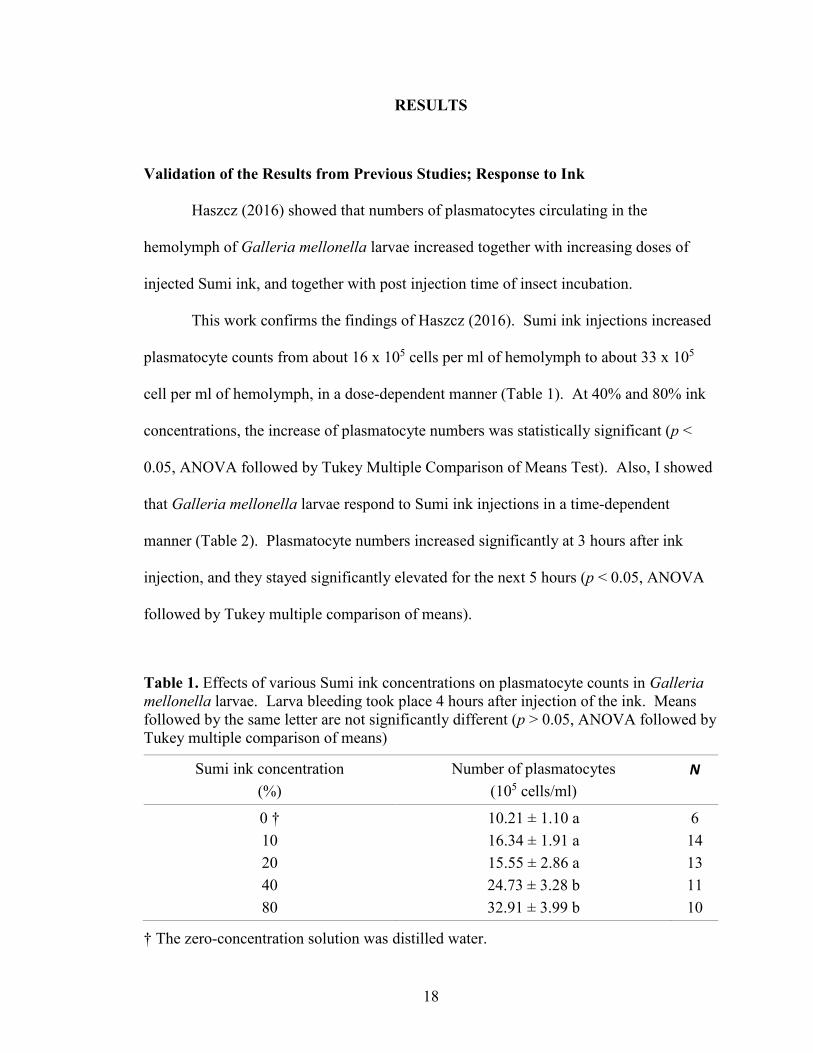
Table 1. Effects of various Sumi ink concentrations on plasmatocyte counts in Galleria
mellonella larvae. Larva bleeding took place 4 hours after injection of the ink. Means
followed by the same letter are not significantly different (p > 0.05, ANOVA followed by
Tukey multiple comparison of means)
Sumi ink concentration
(%)
Number of plasmatocytes
(105 cells/ml)
N
0 †
10
20
40
80
10.21 ± 1.10 a
16.34 ± 1.91 a
15.55 ± 2.86 a
24.73 ± 3.28 b
32.91 ± 3.99 b
6
14
13
11
10
† The zero-concentration solution was distilled water.
19
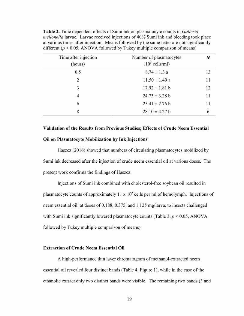
Table 2. Time dependent effects of Sumi ink on plasmatocyte counts in Galleria
mellonella larvae. Larvae received injections of 40% Sumi ink and bleeding took place
at various times after injection. Means followed by the same letter are not significantly
different (p > 0.05, ANOVA followed by Tukey multiple comparison of means)
Time after injection
(hours)
Number of plasmatocytes
(105 cells/ml)
N
0.5 8.74 ± 1.3 a 13
2 11.50 ± 1.49 a 11
3 17.92 ± 1.81 b 12
4 24.73 ± 3.28 b 11
6 25.41 ± 2.76 b 11
8 28.10 ± 4.27 b 6
Validation of the Results from Previous Studies; Effects of Crude Neem Essential
Oil on Plasmatocyte Mobilization by Ink Injections
Haszcz (2016) showed that numbers of circulating plasmatocytes mobilized by
Sumi ink decreased after the injection of crude neem essential oil at various doses. The
present work confirms the findings of Haszcz.
Injections of Sumi ink combined with cholesterol-free soybean oil resulted in
plasmatocyte counts of approximately 11 x 105 cells per ml of hemolymph. Injections of
neem essential oil, at doses of 0.188, 0.375, and 1.125 mg/larva, to insects challenged
with Sumi ink significantly lowered plasmatocyte counts (Table 3, p < 0.05, ANOVA
followed by Tukey multiple comparison of means).
Extraction of Crude Neem Essential Oil
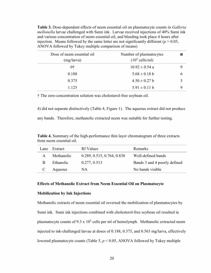
A high-performance thin layer chromatogram of methanol-extracted neem
essential oil revealed four distinct bands (Table 4, Figure 1), while in the case of the
ethanolic extract only two distinct bands were visible. The remaining two bands (3 and
20
Table 3. Dose-dependent effects of neem essential oil on plasmatocyte counts in Galleria
mellonella larvae challenged with Sumi ink. Larvae received injections of 40% Sumi ink
and various concentration of neem essential oil, and bleeding took place 4 hours after
injection. Means followed by the same letter are not significantly different (p > 0.05,
ANOVA followed by Tukey multiple comparison of means)
Dose of neem essential oil
(mg/larva)
Number of plasmatocytes
(105 cells/ml)
N
0† 10.92 ± 0.54 a 9
0.188 5.68 ± 0.18 b 6
0.375 4.50 ± 0.27 b 5
1.125 5.91 ± 0.11 b 9
† The zero-concentration solution was cholesterol-free soybean oil.
4) did not separate distinctively (Table 4, Figure 1). The aqueous extract did not produce
any bands. Therefore, methanolic extracted neem was suitable for further testing.
Table 4. Summary of the high-performance thin layer chromatogram of three extracts
from neem essential oil.
Lane Extract Rf Values Remarks
A Methanolic 0.289, 0.515, 0.764, 0.838 Well-defined bands
B Ethanolic 0.277, 0.513 Bands 3 and 4 poorly defined
C Aqueous NA No bands visible
Effects of Methanolic Extract from Neem Essential Oil on Plasmatocyte
Mobilization by Ink Injections
Methanolic extracts of neem essential oil reversed the mobilization of plasmatocytes by
Sumi ink. Sumi ink injections combined with cholesterol-free soybean oil resulted in
plasmatocyte counts of 9.3 x 10⁵ cells per ml of hemolymph. Methanolic extracted neem
injected to ink-challenged larvae at doses of 0.188, 0.375, and 0.563 mg/larva, effectively
lowered plasmatocyte counts (Table 5, p < 0.05, ANOVA followed by Tukey multiple
21
Figure 1. High-performance thin layer chromatogram of three extracts from neem
essential oil. (A) methanolic extract, (B) ethanolic extract, (C) aqueous extract. All the
extracts had a 30% weight/volume concentration.
comparison of means).
Effects of Aqueous Extract from Neem Bark on Plasmatocyte Mobilization by Ink
Injections
Neem bark aqueous extract had no effect on the numbers of plasmatocytes Sumi
ink mobilized. Sumi ink injections combined with injections of distilled water resulted in
plasmatocyte counts similar to those in previous tests, with approximately 9.3 x 10⁵ cells
A B C
4
3
2
1
22
Table 5. Dose-dependent effects of methanolic extract from neem essential oil on
plasmatocyte counts in Galleria mellonella larvae challenged with Sumi ink. Larvae
received injections of 40% Sumi ink and various concentrations of methanolic extract
from neem essential oil, and bleeding took place 4 hours after injection. Means followed
by the same letter are not significantly different (p > 0.05, ANOVA followed by Tukey
multiple comparison of means).
Dose of the extract
(mg/larva)
Number of plasmatocytes
(105 cells/ml)
N
0† 9.30 ± 0.77 a 27
0.188 4.96 ± 0.33 b 10
0.375 4.26 ± 0.17 b 9
0.563 4.90 ± 0.41 b 18
† The zero-concentration solution was cholesterol-free soybean oil.
per ml of hemolymph. Injections of 100% aqueous extract of neem bark to ink-
challenged larvae resulted in a slight decrease in plasmatocyte counts numerically, but
this difference was not statistically significant. (Table 6, p > 0.05, unpaired Student’s t
test).
Table 6. Effect of neem bark aqueous extract on plasmatocyte counts in ink challenged
Galleria mellonella larvae. Larvae received injections of 40% Sumi ink and one
concentration of neem bark aqueous extract, and bleeding took place 4 hours after
injection. Means followed by the same letter are not significantly different (p > 0.05,
unpaired Student’s t test).
Concentration of neem
bark aqueous extract (%)
Number of plasmatocytes
(105 cells/ml) N
0† 9.26 ± 1.40 a 10
100% 7.83 ± 1.80 a 10
† The zero-concentration solution was distilled water.
23
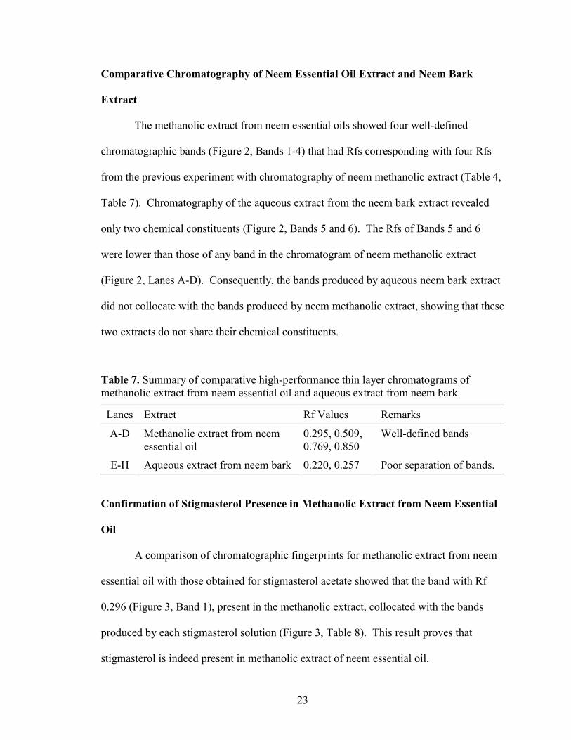
Comparative Chromatography of Neem Essential Oil Extract and Neem Bark
Extract
The methanolic extract from neem essential oils showed four well-defined
chromatographic bands (Figure 2, Bands 1-4) that had Rfs corresponding with four Rfs
from the previous experiment with chromatography of neem methanolic extract (Table 4,
Table 7). Chromatography of the aqueous extract from the neem bark extract revealed
only two chemical constituents (Figure 2, Bands 5 and 6). The Rfs of Bands 5 and 6
were lower than those of any band in the chromatogram of neem methanolic extract
(Figure 2, Lanes A-D). Consequently, the bands produced by aqueous neem bark extract
did not collocate with the bands produced by neem methanolic extract, showing that these
two extracts do not share their chemical constituents.
Table 7. Summary of comparative high-performance thin layer chromatograms of
methanolic extract from neem essential oil and aqueous extract from neem bark
Lanes Extract Rf Values Remarks
A-D Methanolic extract from neem
essential oil
0.295, 0.509,
0.769, 0.850
Well-defined bands
E-H Aqueous extract from neem bark 0.220, 0.257 Poor separation of bands.
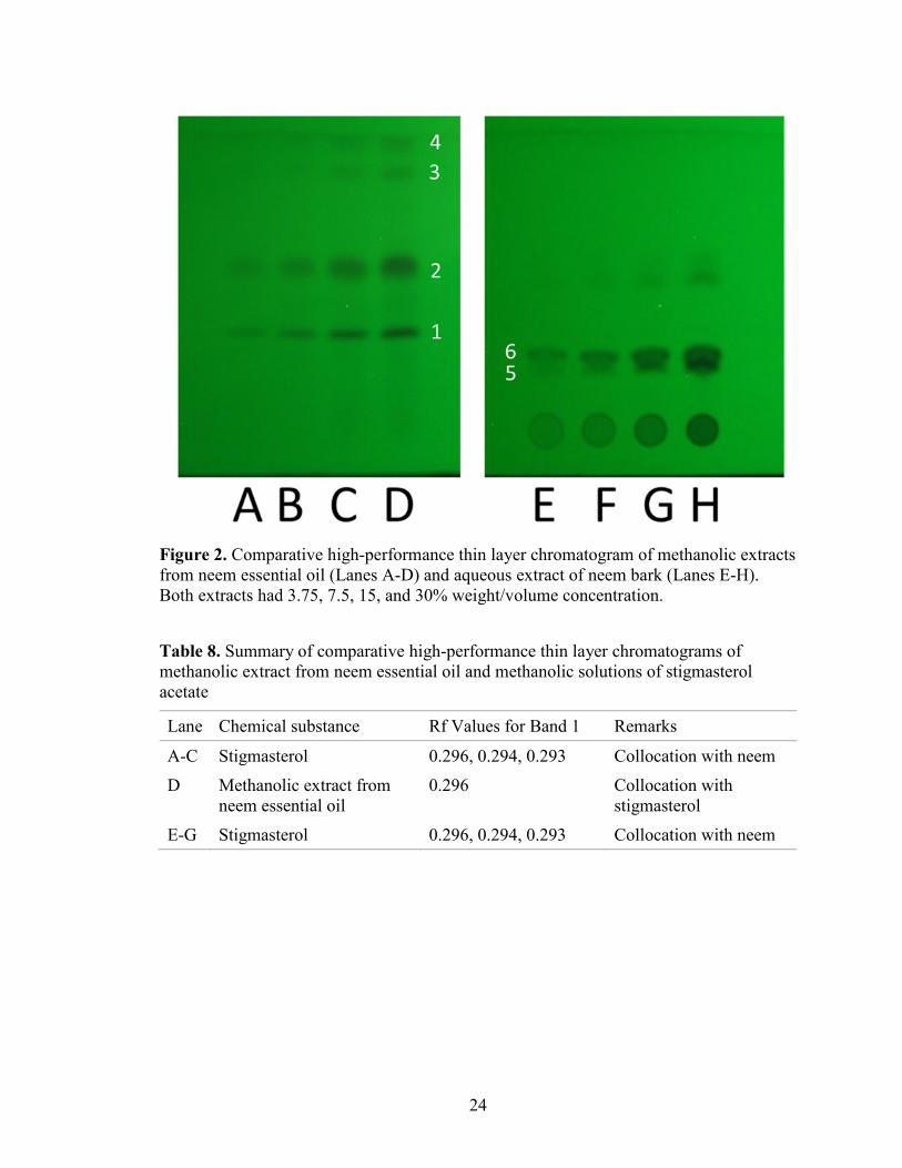
Confirmation of Stigmasterol Presence in Methanolic Extract from Neem Essential
Oil
A comparison of chromatographic fingerprints for methanolic extract from neem
essential oil with those obtained for stigmasterol acetate showed that the band with Rf
0.296 (Figure 3, Band 1), present in the methanolic extract, collocated with the bands
produced by each stigmasterol solution (Figure 3, Table 8). This result proves that
stigmasterol is indeed present in methanolic extract of neem essential oil.
24
Figure 2. Comparative high-performance thin layer chromatogram of methanolic extracts
from neem essential oil (Lanes A-D) and aqueous extract of neem bark (Lanes E-H).
Both extracts had 3.75, 7.5, 15, and 30% weight/volume concentration.
Table 8. Summary of comparative high-performance thin layer chromatograms of
methanolic extract from neem essential oil and methanolic solutions of stigmasterol
acetate
Lane Chemical substance Rf Values for Band 1 Remarks
A-C Stigmasterol 0.296, 0.294, 0.293 Collocation with neem
D Methanolic extract from
neem essential oil
0.296 Collocation with
stigmasterol
E-G Stigmasterol 0.296, 0.294, 0.293 Collocation with neem
25
Figure 3. Comparative high-performance thin layer chromatogram of stigmasterol
acetate (Lanes 1-3), methanolic extract from neem essential oil (Lane 4), and stigmasterol
acetate again (Lanes 5-7). The stigmasterol had 10, 20, and 30% weight/volume
concentration. The methanolic extract from neem essential oil had 15% weight/volume
concentration.
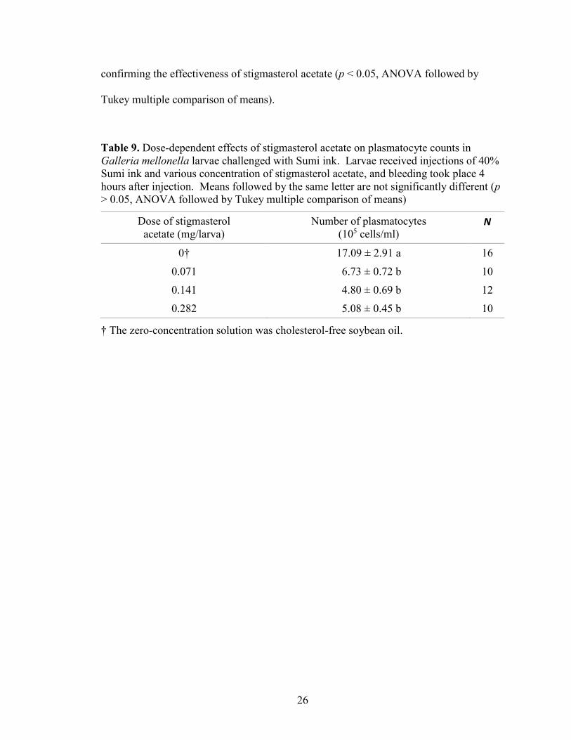
Effects of Stigmasterol Acetate on Plasmatocyte Mobilization by Ink Injections
Stigmasterol acetate lowered plasmatocyte counts in Galleria mellonella larvae
challenged with Sumi ink. Together with increasing doses of stigmasterol, plasmatocyte
counts dropped from about 17 x 105 to less than 6 x 10⁵ cells per ml of hemolymph in a
dose-dependent manner (Table 9). Plasmatocyte counts dramatically lowered,
A B C D E F G
1 1
26
confirming the effectiveness of stigmasterol acetate (p < 0.05, ANOVA followed by
Tukey multiple comparison of means).
Table 9. Dose-dependent effects of stigmasterol acetate on plasmatocyte counts in
Galleria mellonella larvae challenged with Sumi ink. Larvae received injections of 40%
Sumi ink and various concentration of stigmasterol acetate, and bleeding took place 4
hours after injection. Means followed by the same letter are not significantly different (p
> 0.05, ANOVA followed by Tukey multiple comparison of means)
Dose of stigmasterol
acetate (mg/larva)
Number of plasmatocytes
(105 cells/ml) N
0† 17.09 ± 2.91 a 16
0.071 6.73 ± 0.72 b 10
0.141 4.80 ± 0.69 b 12
0.282 5.08 ± 0.45 b 10
† The zero-concentration solution was cholesterol-free soybean oil.
27
DISCUSSION
This research examined neem and its constituents for their potential of impairing
insect immune systems. Former Missouri State graduate student Katherine Haszcz found
that Sumi ink increased the number of circulating plasmatocytes in the 7th instar larvae of
Galleria mellonella. Plasmatocytes are primary hemocytes involved in encapsulation in
the lepidopteran order of insects. Haszcz showed that a 40% concentration of Sumi ink at
4 hours post injection was optimal for the increase in plasmatocytes and their
mobilization (Haszcz, 2016). However, she used bright light microscopy in her research,
which did not allow for accurate differentiation of particular classes of hemocytes. The
results of microscopy based on differential interference contrast and oblique illumination
filters, and shown in the present dissertation, confirmed the previous findings of Haszcz
(2016).
Sumi ink indeed elevates numbers of circulating plasmatocytes in a dose-
dependent manner (Table 1), and in a time-dependent manner (Table 2). Moreover,
optimal concentration of the ink and optimal time of incubation in my experiments were
the same as in the work of Haszcz (2016). Neem essential oil reversed the effects of
Sumi ink, as in Haszcz (2016).
The experimentation presented in the current thesis expanded the findings of
Haszcz (2016) by extracting neem biologically active substances and identifying
stigmasterol as the chemical that reverses plasmatocyte mobilization by Sumi ink.
For chromatography with neem, I tried three extraction solvents: methanol,
ethanol, and water. I subjected all three extracts to comparative HPTLC (Figure 1). Of
the three solvents, methanol worked the best: all the bands were visible and well defined.
28
Ethanol had the first two bands well defined, with the other bands obscure and not
distinguishable. Water failed to work in this experiment, as there were no visible bands.
These findings are in accordance with previous work by Sharma, Dua, and
Srivastva (2014), who investigated the antibacterial activity of extracts made from neem
seed, leaf, bark, and root. In their experiments, methanolic extracts had the highest
antibacterial activity against both Escherichia coli and Bacillus amyloliquefaciens,
whereas ethanolic extracts were not as effective. Interestingly, aqueous extracts of neem
seed and root also had some antibacterial activity in the experiments performed by
Sharma et al. (2014). The fact that water was ineffective in the extraction experiments
reported in the present thesis might have been the result of different methods of
extraction. Sharma et al. made their aqueous extract by boiling plant tissues in water for
30 minutes at 100°C, whereas in the experiments reported herein, extraction took place at
room temperature for 10 minutes.
There are other reports showing that methanol is a good solvent for extraction of
biologically active substances from neem. For instance, Stark and Walter (1995) used
methanolic extracts of neem to enhance the activity of several insecticides against pea
aphids.
De and Ifeoma (2002) conducted their study with extracts of Azadirachta indica
to compare its antimicrobial spectrum with that of conventional antibiotics used against
pathogenic fungi and bacteria. They extracted their samples of powdered neem bark and
leaves separately with water, acetone, or methanol. The aqueous extracts failed to show
antimicrobial activity at any concentration on the test organisms, whereas the methanolic
29
extracts of had comparable results to antibiotics generally used to treat infections caused
by the test organisms (De & Ifeoma, 2002).
This research shows that aqueous neem bark extract had no effect on
plasmatocyte counts in G. mellonella larvae, whereas methanol-extracted neem lowered
plasmatocyte counts, which is in accordance with the works of Sharma et al. (2014), De
and Ifeoma (2002), and Stark and Walter (1995).
I also used HPTLC to delineate substances of interest in neem essential oil.
Nicoletti et al. (2012) used HPTLC to analyze several commercially available neem
products. In their research, HPTLC displayed significant variations in chemical
composition of particular neem products; however, methanol consistently extracted the
principal active metabolites of neem: nimbin, salanin, azadirachtin A, and azadirachtin B.
It is difficult to compare my comparative HPTLC of neem (Figure 2) with the
chromatograms of Nicoletti et al., because these authors used a mobile phase for
chromatography, which differed from that in my experiments.
It is clear, however, that methanolic extract of neem essential oil (which lowers
plasmatocyte counts in ink-challenged Galleria mellonella larvae) contains chemicals
that are not present in the aqueous extract of neem bark (which does not affect
plasmatocyte counts). In particular, it is the band of Rf equaling 0.295 (Figure 2, Band 1)
that collocates with the stigmasterol standard (Figure 3).
Bioassays confirmed that stigmasterol (Figure 4) lowers plasmatocyte counts in
ink-challenged larvae of Galleria mellonella (Table 9). This finding is novel, and
therefore difficult to assess.
30
However, there is a body of research showing that this plant secondary metabolite
exhibits biological activity in animal systems. For instance, stigmasterol has been
successful in docking studies against Human Epidermal Growth Factor Receptor-2
(Sugappriya, Sudarsanam, Bhaskaran, Joseph, & Suresh, 2017). Stigmasterol is also the
most likely inhibitory resource against the envelope protein VP28, which is a major
factor in white spot syndrome virus, a disastrous disease of shrimp (Sahu, Kathiresan,
Singh, & Senthilraja, 2012). In another study, a mixture of β-sitosterol and stigmasterol
extracted from the roots of P. indica proved to be effective as an antidote to snake venom
(Gomes Saha, Chatterjee, & Chakravarty, 2007). Behmer and Grebenok (1998) tested
various sterols in the diet of a lepidopteran, Plutella xylostella, and they found that
stigmasterol was the only one to lower the survival rate of the moths.
More important, stigmasterol showed its potential as an anti-inflammatory agent
in studies of asthma (Antwi, Obiri, & Osafo, 2017), and it has antipyretic effects on mice
(Antwi, Obiri, Osafo, Forkuo, & Essel, 2017). Known compounds that have anti-
inflammatory and antipyretic effects have also been successful in insect immune
response. Büyükgüzel, Tunaz, Stanley, and Büyükgüzel (2007) researched cellular
immune responses to viruses in Galleria mellonella larvae, and they found that
indomethacin, an anti-inflammatory drug, inhibited that response.
This dissertation lays the groundwork for further testing of stigmasterol. This
plant-derived compound impaired the immune defense systems in G. mellonella larvae.
In previous studies with neem oil, it appeared that azadirachtin was the essential
compound of interest. Azadirachtin has proven its effectiveness as an antifeedant in
31
insects, but it failed to show promising results when tested for impairment in the insect
immune system (Haszcz, 2016).
Stigmasterol lowered circulating plasmatocyte counts to < 50% in ink-challenged
larvae. This significant finding allows for larvicidal treatment, where the immune system
of insects is most vulnerable, and allows less chance for adaptation. Future bioassays
with the lepidopteran order of insects, as well as others, are necessary to show the
potential of stigmasterol as a possible plant-based insecticide.
32
REFERENCES
Ahmed, S., & Grainge, M. (1986, April-June). Potential of the neem tree (Azadirachta
indica) for pest control and rural development. Economic Botany, 40(2), 201-209.
Aktar, W., Sengupta, D., & Chowdhury, A. (2009). Impact of pesticides use in
agriculture: Their benefits and hazards. Interdisciplinary Toxicology, 2(1), 1-12.
Antwi, A. O., Obiri, D. D., & Osafo, N. (2017). Stigmasterol modulates allergic airway
inflammation in guinea pig model of ovalbumin-induced asthma. Mediators of
Inflammation, 2017. Article ID 295390.
Antwi, A. O., Obiri, D. D., Osafo, N., Forkuo, A. D., & Essel, L. B. (2017) Stigmasterol
inhibits lipopolysaccharide-induced innate immune responses in murine models.
International Immunopharmacology, 53, 105-113.
Behmer, S. T., & Grebenok, R. J. (1998). Impact of dietary sterols on life-history traits of
a caterpillar. Physiological Entomology, 23(2), 165-175.
Biswas, K., Chattopadhyay, I., Banerjee, R., & Bandyopadhyay, U. (2002). Biological
activities and medicinal properties of neem (Azadirachta indica). Current
Science, 82, 1336-1345.
Büyükgüzel, E., Tunaz, H., Stanley, D., & Büyükgüzel, K. (2007). Eicosanoids mediate
Galleria mellonella cellular immune response to viral infection. Journal of Insect
Physiology, 53(1), 99-105.
De N. B., & Ifeoma, E. (2002). Antimicrobial effects of components of the bark of neem
(Azadirachta indica A. Juss). Technology and Development, 8, 23-28.
Dutta, S. (2015). Biopesticides: An ecofriendly approach for pest control. World Journal
of Pharmacy and Pharmaceutical Sciences, 4(6), 250-265.
Gillespie, J. P., Kanost, M. R., & Trenczek, T. (1997). Biological mediators of insect
immunity. Annual Review of Entomology, 42(1), 611-643.
Girish, K., & Shankara, B. S. (2008). Neem – A green treasure. Electronic Journal of
Biology, 4(3), 102-111.
Gomes, A., Saha, A., Chatterjee, I., & Chakravarty, A. K. (2007). Viper and cobra venom
neutralization by β-sitosterol and stigmasterol isolated from the root extract of
Pluchea indica Less (Asteraceae). Phytomedicine, 14, 637-643.
Haszcz, K. (2016). Impairing the insect immune system with plant-derived substances
(Master’s thesis), Missouri State University, Springfield, MO.
33
Jiang, H., Vilcinskas, A., & Kanost, M. R. (2010). Immunity in lepidopteran insects. In
Invertebrate immunity (pp. 181-204). Boston, MA: Springer.
Jones, J. C. (1962). Current concepts concerning insect hemocytes. American Zoologist,
2(2), 209-246.
Koul, O., Isman, M., & Ketkar, C. (1990). Properties and uses of neem, Azadirachta
indica. Canadian Journal of Botany, 68(1), 1-11.
Lavine, M. D., & Strand, M. R. (2002). Insect hemocytes and their role in immunity.
Insect Biochemistry and Molecular Biology, 32, 1295-1309.
Mohamed, M. A., & Coppel, H. C. (1983). Mass rearing of the greater wax moth,
Galleria mellonella (Lepidoptera: Pyralidae), for small-scale laboratory studies.
Great Lakes Entomologist, 16(4), 139-141.
Mukherjee, K., Altincicek, B., Hain, T., Domann, E., Vilcinskas, A., & Chakraborty, T.
(2010). Galleria mellonella as a model system for studying Listeria pathogenesis.
Applied and Environmental Microbiology, 76(1), 310-317.
Nicoletti, M., Maccioni, O., Coccioletti, T., Mariana, S., & Vitali, F. (2012). Neem tree
(Azadirachta indica A. Juss) as source of bioinsecticides. In Insecticides –
advances in Integrated Pest Management (pp. 411-428). ISBN: 978-953-307-
780-2, InTech.
Ottaviani, E. (2005). Insect immunorecognition. Invertebrate Survival Journal, 2, 142-
151.
Popp, J., Pető, K., & Nagy, J. (2013). Pesticide productivity and food security. A review.
Agronomy for Sustainable Development, 33(1), 243-255.
Reichenberger, S., Bach, M., Skitschak, A., & Frede, H-G. (2007). Mitigation strategies
to reduce pesticide inputs into ground and surface water and their effectiveness; a
review. Science of the Total Environment, 384, 1-35.
Sahu, S. K., Kathiresan, K., Singh, R., & Senthilraja, P. (2012). Molecular docking
analyses of Avicennia marina-derived phytochemicals against white spot
syndrome virus (WSSV) envelope protein-VP28. Bioinformation, 8, 897-900.
Sharma, Y., Dua, D., & Srivastva, S. N. (2014). Comparative study of different parts of
Azadirachta indica (neem) plant on the basis of antibacterial activity,
phytochemical screening and its effect on rat pc-12 (pheochromocytoma) cell
line. International Journal of Biotechnology and Allied Fields, 2(7), 144-154.
Sinha, S., Murthy, P., Rao, C., Ramaprasad, G., Sitaramaiah, S., Kumar, D., & Savant, S.
(1999). Simple method for enrichment of azadirachtin from neem seeds. Journal
of Scientific and Industrial Research, 58, 990-994.
34
Stark, J. D., & Walter, J. F. (1995). Neem oil and neem oil components affect efficacy of
commercial neem insecticides. Journal of Agriculture and Food Chemistry, 43,
507-512.
Strand, M. R. (2008). The insect cellular response. Insect Science, 15(1), 1-14.
Sugappriya, M., Sudarsanam, D., Bhaskaran, R., Joseph, J., & Suresh, A. (2017).
Druggability and binding site interaction studies of potential metabolites isolated
from marine sponge Aurora globostellata against human epidermal growth factor
receptor-2. Bioinformation, 13(8), 261-268.
Wojda, I. (2017). Immunity of the greater wax moth Galleria mellonella. Insect Science,
24(3), 342-357.

![NEEM BASED PESTICIDE - KRISHNAkrishna.nic.in/PDFfiles/MSME/Chemical/NEEM BASED PESTICIDE[1].pdf · 1 neem based pesticide contents section i product characteristics section ii product](https://img.pdfslide.us/doc/110x75/5a9f50e47f8b9a84178cab86/neem-based-pesticide-based-pesticide1pdf1-neem-based-pesticide-contents-section.jpg)