Embed Size (px)
Citation preview

Anesthesiology 2006; 105:902–10 Copyright © 2006, the American Society of Anesthesiologists, Inc. Lippincott Williams & Wilkins, Inc.
Effects of Low and High Plasma Concentrations ofDexmedetomidine on Myocardial Perfusion and CardiacFunction in Healthy Male SubjectsAmir Snapir, M.D., Ph.D.,* Jussi Posti, B.M.,* Erkki Kentala, M.D., Ph.D.,† Juha Koskenvuo, M.D., Ph.D.,‡Jan Sundell, M.D., Ph.D.,§ Helena Tuunanen, M.D.,� Kristo Hakala, M.Sc.,* Harry Scheinin, M.D., Ph.D.,#Juhani Knuuti, M.D., Ph.D.,** Mika Scheinin, M.D., Ph.D.††
This article has been selected for the AnesthesiologyCME Program. After reading the article, go to http://www.asahq.org/journal-cme to take the test and apply forCategory 1 credit. Complete instructions may be found inthe CME section at the back of this issue.
Background: Dexmedetomidine, a selective �2-adrenoceptoragonist, has counteracting effects on the cardiovascular system.It mediates sympatholysis by activating �2 adrenoceptors in thecentral and peripheral nervous system, and vasoconstrictionand vasorelaxation by activating postsynaptic �2 adrenoceptorsin blood vessels. The goal of this study was to determine theeffects of therapeutic and high concentrations of dexmedeto-midine on myocardial perfusion and cardiac function inhealthy subjects.
Methods: The authors studied 12 healthy young men. Myo-cardial blood flow (assessed with positron emission tomogra-phy), myocardial function (by echocardiography), and hemo-dynamic data were collected before and during low (measuredmean plasma concentration, 0.5 ng/ml) and high (5 ng/ml)plasma concentrations of dexmedetomidine.
Results: The low concentration of dexmedetomidine reducedmyocardial perfusion (mean difference, �27% from baseline[95% confidence interval, �31 to �23%], P < 0.001) in parallelwith a reduction in myocardial oxygen demand (estimated bythe rate–pressure product (�23% [�28 to �18%], P < 0.001).The high dexmedetomidine plasma concentration did not fur-ther attenuate myocardial perfusion (�3% [�12 to �6%] fromlow dexmedetomidine, P > 0.05; �29% [�39 to �18%] frombaseline, P < 0.001) or statistically significantly affect the rate–pressure product (�5% [0 to �10%], P > 0.05). Systolic myocar-dial function was attenuated by sympatholysis during the lowinfusion rate and was further attenuated by a combination ofthe sustained sympatholysis and increased afterload during thehigh infusion rate.
Conclusions: In healthy subjects, plasma concentrations ofdexmedetomidine that significantly exceed the recommendedtherapeutic level do not seriously attenuate myocardial perfu-
sion below the level that is observed with usual therapeuticconcentrations and do not induce evident myocardial ischemia.
�2-ADRENOCEPTOR agonists mediate their cardiovascu-lar effects through activation of receptors in the centraland peripheral nervous system and through activation ofpostsynaptic receptors in target organs. Activation ofpresynaptic �2 adrenoceptors on sympathetic nervesand the central nervous system induces sympatholysis,whereas activation of vascular postsynaptic receptorscauses both vasoconstriction1 (through activation of �2
adrenoceptors on vascular smooth muscle cells) andvasodilatation (through activation of �2 adrenoceptorson endothelial cells).2 Because of these counteractingmechanisms, the overall effect of �2 adrenoceptor acti-vation on organ blood flow is complex, and difficult topredict—especially in organs that are under major influ-ence of the autonomic nervous system, such as theheart. The classic cardiovascular response in mammali-ans to systemic infusion of therapeutic doses of an �2-adrenoceptor agonist, such as clonidine, is biphasic withan initial short-term increase in blood pressure (BP)followed by a long-lasting decrease in BP and heart rate(HR).3 The initial response is presumably a result ofactivation of vascular postsynaptic �2 adrenoceptors,later masked by sympatholysis.
Current information on the direct effects of �2-adreno-ceptor activation on human coronary artery blood flowhas been derived from patients with chest pain under-going diagnostic left-side catheterization. Indolfi et al.4
and Baumgart et al.5 reported that direct infusion ofBHT-933 (azepexole, an �2-adrenoceptor agonist) intothe coronary vasculature attenuates coronary bloodflow. This effect is augmented by atherosclerosis and isassociated with a significant increase in myocardial lac-tate production.5
Dexmedetomidine, a selective and potent �2-adreno-ceptor agonist, was approved by the US Food and DrugAdministration in 1999 for sedation of patients hospital-ized in intensive care settings, and since then, a growingnumber of research articles have emerged reportingother possible indications, such as regional6 and general7
anesthesia. Dexmedetomidine induces nearly completesympatholysis already at low concentrations, and contin-uous and linear increases in its postsynaptic effects canbe observed with increasing concentrations.8 Despite itsincreased clinical use, many times in critically ill pa-tients, the effect of dexmedetomidine on myocardial
* Research Scientist, †† Professor, Department of Pharmacology, Drug Devel-opment and Therapeutics, University of Turku. † Senior Physician, Departmentof Anesthesiology and Intensive Care, ‡ Resident, Department of Clinical Physi-ology, § Resident, � Research Scientist, # Professor, ** Director, Turku PETCentre, Turku University Hospital, Turku, Finland.
Received from the Department of Pharmacology, Drug Development andTherapeutics, University of Turku, Turku, Finland. Submitted for publicationMarch 10, 2006. Accepted for publication June 21, 2006. Supported by grantsfrom Turku University Hospital, Turku, Finland, the Academy of Finland, theJuselius Foundation, and the Finnish Heart Foundation, Helsinki, Finland. Thelaboratory of Dr. Scheinin has contract research relationships with Orion Cor-poration, Espoo, Finland. Hospira, Lake Forest, Illinois, has a license agreementwith Orion Corporation regarding Precedex.
Address correspondence to Dr. Snapir: Department of Pharmacology, DrugDevelopment and Therapeutics, University of Turku, Itainen Pitkakatu 4B, FI-20520 Turku, Finland. [email protected]. Individual article reprints may be accessedat no charge through the Journal Web site, www.anesthesiology.org.
Anesthesiology, V 105, No 5, Nov 2006 902
Downloaded from anesthesiology.pubs.asahq.org by guest on 02/02/2019

blood flow (MBF) has not been yet investigated, and itseffect on myocardial function has been limited to studiesof cardiac output (CO). This study was designed toinvestigate the effects of low and high steady stateplasma concentrations of dexmedetomidine on myocar-dial perfusion and cardiac function in healthy youngsubjects.
Materials and Methods
Study PopulationThe study was conducted in accordance with the Dec-
laration of Helsinki (2000) of the World Medical Associ-ation and was approved by the ethics committee of theSouthwestern Health Care District, Turku, Finland. Allsubjects gave their written informed consent.
We studied 12 nonsmoking healthy male volunteersaged 20–28 y (mean, 24 y) with a body mass index of 19to 27 kg/m2 (mean, 24 kg/m2). The health of the volun-teers was assessed by medical history, physical examina-tion, 12-lead electrocardiogram, bicycle maximal exer-cise test, blood cell count, and urine drug screening.Because studies in transgenic mice have suggested that�2-adrenoceptor–mediated vasoconstriction is mediatedby the �2B-adrenoceptor subtype,3 and because it hasbeen demonstrated that an insertion/deletion polymor-phism in the human �2B adrenoceptor is associated withmodified receptor desensitization,9 we also screened thevolunteers for this genetic polymorphism and selected 6subjects from each homozygous genotype to preventgenetic bias.
Study ProtocolIn the current study, we collected data before infusion
of dexmedetomidine (Baseline) and during low and highplasma concentrations of dexmedetomidine. Based onthe results of Ebert et al.,8 we chose target plasmaconcentrations of 0.5 ng/ml for the low-dose phase (LowDex) and 3.2 ng/ml for the high-dose phase (High Dex).The recommended therapeutic concentration range ofdexmedetomidine is 0.4–1.2 ng/ml (Precedex® SPC; Ab-bott Laboratories, Abbott Park, IL).
Experiments were conducted during morning hours afteran 8-h fast. Subjects abstained from alcohol for 48 h ormore, from caffeine for 12 h or more, and from heavyexercise for 24 h or more. After stabilization for at least 30min, baseline measurements were obtained in the follow-ing order: blood sampling for determination of epineph-rine, norepinephrine, and dexmedetomidine concentra-tions in plasma; positron emission tomography (PET)measurements; transthoracic echocardiography (TTE) mea-surements; and a second blood sampling. After the baselinemeasurements, Low Dex was started. Thirteen minutesafter the initiation of the infusion, blood samples weredrawn, and exactly 15 min from the initiation of the druginfusion, PET scanning was started. Data collection for thisphase was completed with TTE measurements and bloodsampling (30 min from the beginning of the drug infusion).This sequence was then repeated with High Dex. Thestudy design is summarized in figure 1.
Dexmedetomidine InfusionA Harvard 22 syringe pump (Harvard Apparatus, Hol-
liston, MA) connected to a computer running STAN-
Fig. 1. Outline of the study design. After at least 30 min of rest, baseline measurements were obtained. The Low Dex phase started withthe infusion of dexmedetomidine (Dex) to target a plasma concentration of 0.5 ng/ml. After completion of the positron emissiontomography (PET) and transthoracic echocardiography (TTE) measurements, High Dex was started by increasing the rate of infusion ofdexmedetomidine to target a plasma concentration of 3.2 ng/ml. The infusion of the PET tracer was started exactly 15 min after theinfusion of dexmedetomidine was initiated or increased. The PET measurements were followed by the TTE measurements.
903DEXMEDETOMIDINE AND MYOCARDIAL PERFUSION IN HUMANS
Anesthesiology, V 105, No 5, Nov 2006
Downloaded from anesthesiology.pubs.asahq.org by guest on 02/02/2019

PUMP software‡‡ was used. Dexmedetomidine (Prece-dex®) was administered intravenously as a target-controlled infusion aiming at pseudo–steady state plasmadrug concentrations of 0.5 ng/ml (in Low Dex) and 3.2ng/ml (in High Dex). We used the same pharmacokineticparameters of dexmedetomidine as Talke et al.1 and setthe maximum infusion rate to 0.3 �g � kg�1 � min�1.Approximately 5 min was thus required to reach thetargeted plasma concentration of 3.2 ng/ml.
Hemodynamic MeasurementsTwo veins in the right forearm were cannulated for
infusion of dexmedetomidine and the PET tracer. A 20-gauge catheter was inserted into the left radial artery forBP monitoring (Truwave PX-600F 3X; Edwards Life-sciences LLC, Irvine, CA) and blood sampling. A fiber-optic pulmonary artery flotation catheter (Swan-GanzCCOmbo, CCO/SVO2, Catheter model 744HF75, 7.5French; Edwards Lifesciences LLC) was introduced intoa pulmonary artery during pressure monitoring via an8.5-French introducer inserted into the right internaljugular or the left subclavian vein. The catheter wasconnected to an Edwards Vigilance® Monitor (EdwardsLifesciences LLC) for monitoring of CO and mixed ve-nous oxygen saturation and was used for infusion oflactated Ringer’s solution (5 ml/min). Data from theVigilance® monitor was stored on a personal computerusing Vigilance Monitor Driver 2.1 program (TechnicalServices Division, Department of Medical Physics, RoyalPerth Hospital, Perth, Australia). Systemic vascular resis-tance was calculated as 80 � mean systemic arterial BP� CO�1.
The electrocardiographic electrodes, pulse oximeterprobe, and blood pressure transducers from the radialand pulmonary arteries and the central vein were con-nected to an S/5 patient monitoring system (Datex-Ohm-eda, Helsinki, Finland). For data acquisition, the monitorwas connected to a personal computer running S/5 Col-lect software (version 4; Datex-Ohmeda).
Respiratory rate was measured with a PowerLab sys-tem (PowerLab/4SP and Chart version 5.02; ADInstru-ments, Castle Hill, New South Wales, Australia) using anasal air temperature probe (model MLT415, connectedto a thermistor pod model ML309; ADInstruments).
Measurement of Myocardial Blood FlowMyocardial blood flow was measured with 15O-labeled
water and a GE Advance PET scanner (General Electric,Milwaukee, WI). 15O-labeled water was injected as anintravenous bolus over 20 s (infused mean [SD] dosesduring Baseline, Low Dex, and High Dex were 991[141], 968 [102], and 956 [89] MBq), and acquisition ofserial transaxial tomographic images of the heart was
performed during approximately 5 min (14 � 5, 3 � 10,3 � 20, and 4 � 30 s frames). All data were corrected fordead time, decay, and measured photon attenuation.10
The acquired data sets were processed by factor anal-ysis to enable accurate localization of myocardial tissue.Four representative midventricular slices were chosenfrom the acquired transaxial images for quantitative anal-ysis. Regions of interest that were first defined on imagestaken at Baseline were used to analyze the images takenduring Low and High Dex. Time–activity curves of myo-cardial tissue were created based on these regions ofinterest. The researcher who analyzed the PET imageswas blind to the study design and protocol.
The methods used to calculate values of regional MBF(expressed in milliliters per gram of tissue per minute)have been previously described.11,12 Coronary vascularresistance (CVR) was calculated by dividing mean sys-temic arterial BP with MBF.
EchocardiographyThe subjects were studied in the supine position. Mea-
surements were performed with an Acuson Sequoia C512 (Acuson Inc., Mountain View, CA) instrument witha 3.5-MHz transducer and recorded in digital mode. Stan-dard and modified subcostal, apical, and parasternal im-aging windows were used. Results are the averages ofthe three measurements.
Early (E=) and late (A=) myocardial relaxation velocitieswere measured using tissue Doppler imaging with 8 mmgate basally in the lateral wall. Peak velocity of earlyfilling (E) was measured with pulsed-wave Doppler with5 mm gate at the level of the mitral leaflets in thefour-chamber view. Longitudinal contraction of the leftand right ventricle was measured as displacement of thelateral atrial valve annulus in M-mode using the apicalimaging window. The ratios of E= to A= and E to E= wereused to assess parameters of myocardial relaxation. Theratio between the preejection period to the left ventric-ular ejection time (the contractility index, preejectionperiod divided by the left ventricle ejection time) wasmeasured using phonocardiography and pulsed-waveDoppler–derived outflow of the left ventricle. Ejectionfraction and left ventricular diameter were based onM-mode measurements. Stroke volume was calculated asthe product of the cross-sectional area of the left ven-tricular outflow tract and the velocity time integral of theleft ventricular outflow measured approximately 5 mmfrom the aortic valve. CO (stroke volume � HR) wascalculated as the mean of the CO measured in the rightand the left ventricles.
Assessment of Myocardial IschemiaMyocardial ischemia was assessed during and after the
experimental sessions using electrocardiography andTTE. The 12-lead electrocardiogram was monitored forchanges during the session, and the recordings were‡‡ Available at: http://anesthesia.stanford.edu/pkpd. Accessed May 18, 2006.
904 SNAPIR ET AL.
Anesthesiology, V 105, No 5, Nov 2006
Downloaded from anesthesiology.pubs.asahq.org by guest on 02/02/2019
later reexamined after the experimental session wasover. An ischemic episode was defined as an ST-segmentdeviation of greater than or equal to 1 mm (0.1 mV)below the ST-segment baseline or greater than or equalto 2 mm above ST-segment baseline and lasting for atleast 1 min. The motion of the ventricle walls was fol-lowed during the study session using TTE, and the savedechocardiography data were later analyzed according toa five-point scale that defines the severity and the extent(by standard 16 area segmentation) of wall motion ab-normality.13
Analytic Laboratory MethodsConcentrations of dexmedetomidine in plasma were
determined using reversed-phase high-performance liq-uid chromatography with tandem mass spectrometricdetection (PE Sciex API365 instrument; PE Sciex, FosterCity, CA). The method was modified from a recentlypublished procedure.14 The lower limit of reliable quan-titation of the assay was 0.1 ng/ml. The within- andbetween-run precision of the assay (coefficient of varia-tion) was within 8% in the relevant concentration range.Epinephrine and norepinephrine concentrations weredetermined using high-performance liquid chromatogra-phy with coulometric electrochemical detection (Coulo-chem 5100A; ESA Inc., Bedford, MA).
Data Handling and Statistical AnalysisMeans are presented with either 95% confidence inter-
vals or SDs. For analyses of the effects of dexmedetomi-dine at discrete time points, BP, CO, HR, respiratory rate,and mixed venous oxygen saturation were reduced fromcontinuous 10-s measurements to 1-min median valuesthat corresponded with the noncontinuous measure-ments (PET, TTE, and blood tests). Statistical significancewas assessed with paired-samples t test, or with repeat-ed-measures analysis of variance with Tukey post hoctest. Multivariate correlates of MBF were assessed bymultivariate stepwise linear regression modeling (for-ward manner, P � 0.05 to enter, P � 0.10 to remove).Variables of interest were first assessed with the Pearsonbivariate correlation, and those that were correlatedwith MBF with P � 0.1 were inserted into the multivar-iate regression model. For presentation of relativechanges in systemic mean arterial pressure, HR, CO,systemic vascular resistance, and rate–pressure product(RPP), the median of 1-min continuous measurementthat started 2 min before the initiation of dexmedetomi-dine infusion (fig. 1) was determined as baseline (100%).Statistical analyses were performed with SPSS for Win-dows (version 12.0.1; Chicago, IL) and with GraphPadPrism for Windows (version 4.03; GraphPad Software,San Diego, CA).
Results
All experimental sessions were completed as planned.All subjects fell asleep before the measurements of theHigh Dex phase started, reflecting the sedative action ofthe drug. Plasma dexmedetomidine concentrations weremeasured 13 min and 30 min after the beginning of bothinfusion steps and were similar to what was expectedbased on the findings of Ebert et al.8 (table 1). The�2B-adrenoceptor ins/del gene polymorphism did nothave a statistically significant effect on MBF or CVR atbaseline or during the two infusion phases (data notshown). All results are therefore presented for the entirestudy population.
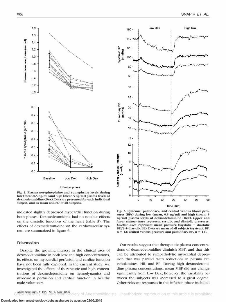
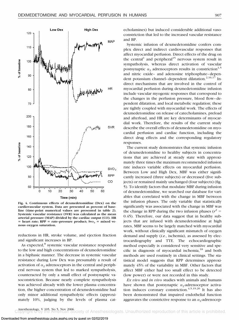
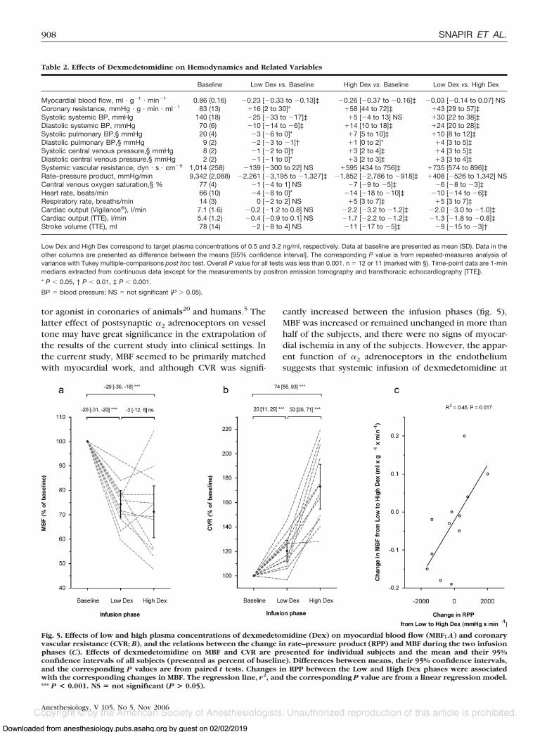
Plasma levels of epinephrine and norepinephrine de-creased on the average by approximately 70% duringLow Dex, and only slight further decreases were notedduring High Dex (fig. 2). Dexmedetomidine had a bipha-sic effect on hemodynamics expressed by reduced bloodpressures and HR during Low Dex, and substantial in-creases in blood pressures (systemic, pulmonary, andvenous) and peripheral vascular resistance with corre-sponding decreases in HR, mixed venous oxygen satura-tion, and CO during High Dex (figs. 3 and 4 and table 2).
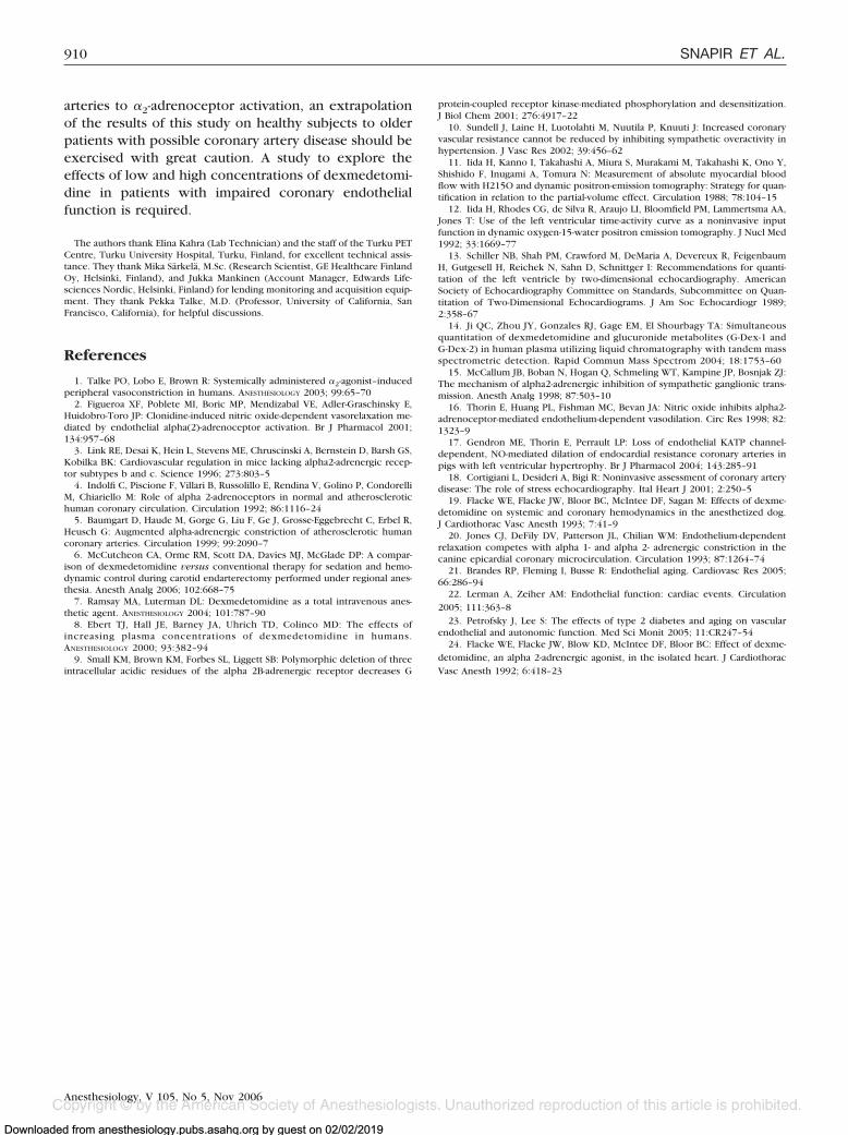
As assessed with 15O-labeled water and PET, dexme-detomidine had significant effects on MBF and CVR. Thelow dexmedetomidine concentration produced a 22%increase in CVR and a 27% reduction in MBF. The highdexmedetomidine concentration did not affect MBF interms of population mean; however, great variability wasobserved in the subjects’ responses in this phase (fig. 5).CVR was further increased during the High Dex infusionphase (fig. 5). The only variable that was significantlycorrelated with the change in MBF after the increase inthe dexmedetomidine infusion rate was the difference inRPP between Low and High Dex phases; increased RPPwas associated with increased MBF and vice versa (r2 �0.45, P � 0.017) (fig. 5). No signs of myocardial ischemia(as assessed by electrocardiography and TTE) were ob-served throughout the study.
Dexmedetomidine had only small effects on systolicmyocardial function. Compared with baseline, changesin ejection fraction, contractility index (preejection pe-riod divided by the left ventricle ejection time), anddisplacements of the mitral and tricuspid valve annuli
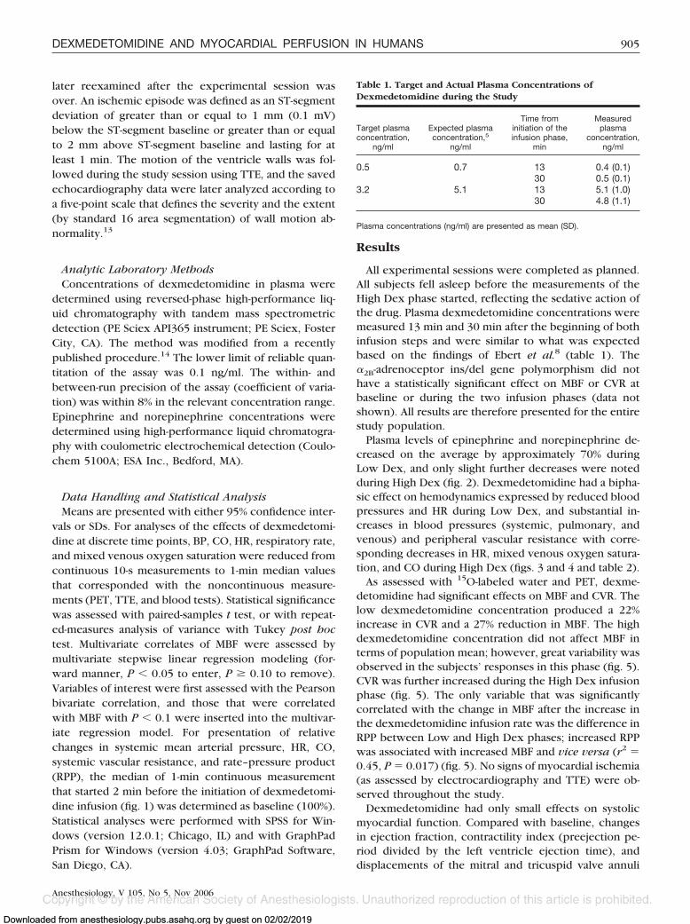
Table 1. Target and Actual Plasma Concentrations ofDexmedetomidine during the Study
Target plasmaconcentration,
ng/ml
Expected plasmaconcentration,5
ng/ml
Time frominitiation of theinfusion phase,
min
Measuredplasma
concentration,ng/ml
0.5 0.7 13 0.4 (0.1)30 0.5 (0.1)
3.2 5.1 13 5.1 (1.0)30 4.8 (1.1)
Plasma concentrations (ng/ml) are presented as mean (SD).
905DEXMEDETOMIDINE AND MYOCARDIAL PERFUSION IN HUMANS
Anesthesiology, V 105, No 5, Nov 2006
Downloaded from anesthesiology.pubs.asahq.org by guest on 02/02/2019
indicated slightly depressed myocardial function duringboth phases. Dexmedetomidine had no notable effectson the diastolic functions of the heart (table 3). Theeffects of dexmedetomidine on the cardiovascular sys-tem are summarized in figure 6.
Discussion
Despite the growing interest in the clinical uses ofdexmedetomidine in both low and high concentrations,its effects on myocardial perfusion and cardiac functionhave not been fully explored. In the current study, weinvestigated the effects of therapeutic and high concen-trations of dexmedetomidine on hemodynamics andmyocardial perfusion and cardiac function in healthymale volunteers.
Our results suggest that therapeutic plasma concentra-tions of dexmedetomidine diminish MBF, and that thiscan be attributed to sympatholytic myocardial depres-sion that was parallel with reductions in plasma cat-echolamines, HR, and BP. During high dexmedetomi-dine plasma concentrations, mean MBF did not changesignificantly from Low Dex; however, the variability be-tween the subjects was increased to a great degree.Other relevant responses in this infusion phase included
Fig. 2. Plasma norepinephrine and epinephrine levels duringlow (mean 0.5 ng/ml) and high (mean 5 ng/ml) plasma levels ofdexmedetomidine (Dex). Data are presented for each individualsubject, and as mean and SD of all subjects.
Fig. 3. Systemic, pulmonary, and central venous blood pres-sures (BPs) during low (mean, 0.5 ng/ml) and high (mean, 5ng/ml) plasma levels of dexmedetomidine (Dex). Upper andlower thinner lines represent systolic and diastolic pressures.Thicker lines represent mean pressure ([systolic � diastolicBP]/3 � diastolic BP). Data are mean of all subjects (systemic BP,n � 12; central venous pressure and pulmonary BP, n � 11).
906 SNAPIR ET AL.
Anesthesiology, V 105, No 5, Nov 2006
Downloaded from anesthesiology.pubs.asahq.org by guest on 02/02/2019
reductions in HR, stroke volume, and ejection fractionand significant increases in BP.
As expected,8 systemic vascular resistance respondedto the low and high concentrations of dexmedetomidinein a biphasic manner. The decrease in systemic vascularresistance during Low Dex was presumably a result ofactivation of �2 adrenoceptors in the central and periph-eral nervous system that led to marked sympatholysis,counteracted by only a small effect of postsynaptic va-soconstriction. Because nearly complete sympatholysiswas achieved already with the lower plasma concentra-tion, the higher concentration of dexmedetomidine hadonly minor additional sympatholytic effects (approxi-mately 10%, judging by the levels of plasma cat-
echolamines) but induced considerable additional vaso-constriction that led to the increased vascular resistanceand BP.
Systemic infusion of dexmedetomidine confers com-plex direct and indirect cardiovascular responses thataffect myocardial perfusion. Direct effects of the drug onthe central1 and peripheral15 nervous system result insympatholysis, whereas direct activation of vascularpostsynaptic �2 adrenoceptors results in constriction1,5
and nitric oxide– and adenosine triphosphate–depen-dent potassium channel–dependent dilatation.2,16,17 In-direct mechanisms that are involved in the control ofmyocardial perfusion during dexmedetomidine infusioninclude vascular myogenic responses that correspond tothe changes in the perfusion pressure, blood flow–de-pendent dilatation, and local metabolic regulation; theseare tightly coupled with myocardial work. The effects ofdexmedetomidine on release of catecholamines, preloadand afterload, and HR are key determinants of myocar-dial work. Therefore, the results of the current studydescribe the overall effects of dexmedetomidine on myo-cardial perfusion and cardiac function, including thedirect drug effects and the corresponding regulatoryresponses.
The current study demonstrates that systemic infusionof dexmedetomidine to healthy subjects in concentra-tions that are achieved at steady state with approxi-mately three times the maximum recommended infusionrate induces variable effects on myocardial perfusion.Between Low and High Dex, MBF was either signifi-cantly increased (three subjects) or decreased (five sub-jects) or remained mainly unchanged (four subjects) (fig.5). To identify factors that modulate MBF during infusionof dexmedetomidine, we searched our database for vari-ables that correlated with the change in MBF betweenthe infusion phases. The only variable that statisticallysignificantly was associated with the change in MBF wasthe change in RPP during the two infusion phases (r2 �0.45). Therefore, our data suggest that in healthy sub-jects that are infused with dexmedetomidine at highrates, MBF seems to be largely matched with myocardialwork, without clinically significant mismatch of oxygendemand and supply (i.e., ischemia), as assessed by elec-trocardiography and TTE. The echocardiographicmethod especially is considered very sensitive and spe-cific in diagnosis of myocardial ischemia,18 and bothmethods are used routinely in clinical settings. The sta-tistical model suggests that RPP determines approxi-mately 45% of the variability in MBF. Other factors thataffect MBF either had too small effect to be detected(low power) or were not recorded in this study.
Ex vivo and in vivo studies with animals and humanshave shown that postsynaptic �2-adrenoceptor activa-tion induces coronary constriction.4,5,19,20 It has alsobeen demonstrated that impaired endothelial functionaggravates the constrictive response to an �2-adrenocep-
Fig. 4. Continuous effects of dexmedetomidine (Dex) on thecardiovascular system. Data are presented as percent of base-line (time-point numerical values are presented in table 2).Systemic vascular resistance (SVR) was calculated as the meanarterial pressure (MAP) divided by the cardiac output (CO). HR� heart rate; RPP � rate–pressure product; SvO2 � mixed ve-nous oxygen saturation.
907DEXMEDETOMIDINE AND MYOCARDIAL PERFUSION IN HUMANS
Anesthesiology, V 105, No 5, Nov 2006
Downloaded from anesthesiology.pubs.asahq.org by guest on 02/02/2019
tor agonist in coronaries of animals20 and humans.5 Thelatter effect of postsynaptic �2 adrenoceptors on vesseltone may have great significance in the extrapolation ofthe results of the current study into clinical settings. Inthe current study, MBF seemed to be primarily matchedwith myocardial work, and although CVR was signifi-
cantly increased between the infusion phases (fig. 5),MBF was increased or remained unchanged in more thanhalf of the subjects, and there were no signs of myocar-dial ischemia in any of the subjects. However, the appar-ent function of �2 adrenoceptors in the endotheliumsuggests that systemic infusion of dexmedetomidine at
Table 2. Effects of Dexmedetomidine on Hemodynamics and Related Variables
Baseline Low Dex vs. Baseline High Dex vs. Baseline Low Dex vs. High Dex
Myocardial blood flow, ml · g�1 · min�1 0.86 (0.16) �0.23 [�0.33 to �0.13]‡ �0.26 [�0.37 to �0.16]‡ �0.03 [�0.14 to 0.07] NSCoronary resistance, mmHg · g · min · ml�1 83 (13) �16 [2 to 30]* �58 [44 to 72]‡ �43 [29 to 57]‡Systolic systemic BP, mmHg 140 (18) �25 [�33 to �17]‡ �5 [�4 to 13] NS �30 [22 to 38]‡Diastolic systemic BP, mmHg 70 (6) �10 [�14 to �6]‡ �14 [10 to 18]‡ �24 [20 to 28]‡Systolic pulmonary BP,§ mmHg 20 (4) �3 [�6 to 0]* �7 [5 to 10]‡ �10 [8 to 12]‡Diastolic pulmonary BP,§ mmHg 9 (2) �2 [�3 to �1]† �1 [0 to 2]* �4 [3 to 5]‡Systolic central venous pressure,§ mmHg 8 (2) �1 [�2 to 0]† �3 [2 to 4]‡ �4 [3 to 5]‡Diastolic central venous pressure,§ mmHg 2 (2) �1 [�1 to 0]* �3 [2 to 3]‡ �3 [3 to 4]‡Systemic vascular resistance, dyn · s · cm�5 1,014 (258) �139 [�300 to 22] NS �595 [434 to 756]‡ �735 [574 to 896]‡Rate–pressure product, mmHg/min 9,342 (2,088) �2,261 [�3,195 to �1,327]‡ �1,852 [�2,786 to �918]‡ �408 [�526 to 1,342] NSCentral venous oxygen saturation,§ % 77 (4) �1 [�4 to 1] NS �7 [�9 to �5]‡ �6 [�8 to �3]‡Heart rate, beats/min 66 (10) �4 [�8 to 0]* �14 [�18 to �10]‡ �10 [�14 to �6]‡Respiratory rate, breaths/min 14 (3) 0 [�2 to 2] NS �5 [3 to 7]‡ �5 [3 to 7]‡Cardiac output (Vigilance®), l/min 7.1 (1.6) �0.2 [�1.2 to 0.8] NS �2.2 [�3.2 to �1.2]‡ �2.0 [�3.0 to �1.0]‡Cardiac output (TTE), l/min 5.4 (1.2) �0.4 [�0.9 to 0.1] NS �1.7 [�2.2 to �1.2]‡ �1.3 [�1.8 to �0.8]‡Stroke volume (TTE), ml 78 (14) �2 [�8 to 4] NS �11 [�17 to �5]‡ �9 [�15 to �3]†
Low Dex and High Dex correspond to target plasma concentrations of 0.5 and 3.2 ng/ml, respectively. Data at baseline are presented as mean (SD). Data in theother columns are presented as difference between the means [95% confidence interval]. The corresponding P value is from repeated-measures analysis ofvariance with Tukey multiple-comparisons post hoc test. Overall P value for all tests was less than 0.001. n � 12 or 11 (marked with §). Time-point data are 1-minmedians extracted from continuous data (except for the measurements by positron emission tomography and transthoracic echocardiography [TTE]).
* P � 0.05, † P � 0.01, ‡ P � 0.001.
BP � blood pressure; NS � not significant (P � 0.05).
Fig. 5. Effects of low and high plasma concentrations of dexmedetomidine (Dex) on myocardial blood flow (MBF; A) and coronaryvascular resistance (CVR; B), and the relations between the change in rate–pressure product (RPP) and MBF during the two infusionphases (C). Effects of dexmedetomidine on MBF and CVR are presented for individual subjects and the mean and their 95%confidence intervals of all subjects (presented as percent of baseline). Differences between means, their 95% confidence intervals,and the corresponding P values are from paired t tests. Changes in RPP between the Low and High Dex phases were associatedwith the corresponding changes in MBF. The regression line, r2, and the corresponding P value are from a linear regression model.*** P < 0.001. NS � not significant (P > 0.05).
908 SNAPIR ET AL.
Anesthesiology, V 105, No 5, Nov 2006
Downloaded from anesthesiology.pubs.asahq.org by guest on 02/02/2019

high rates to patients with impaired endothelial func-tion, such as older patients21 and patients with vasculardiseases (e.g., coronary artery disease,22 diabetes23), mayhave deleterious effects on myocardial (and possiblyother organ) perfusion.
The results of our echocardiography measurementspropose that dexmedetomidine induces myocardial de-pression that is similar to the effect of treatment with �blockers, and that this effect is achieved already at ther-apeutic concentrations. A previous study on isolated dogheart has shown that dexmedetomidine does not havedirect effects on cardiac function.24 Therefore, a proba-ble explanation for this observation is the reduction inrelease of catecholamines and therefore reduction inmyocardial inotropy. Contractility (denoted by preejec-tion period divided by the left ventricle ejection time;positive changes mean less contractility), contraction
(denoted by the movement of the lateral annuli), andejection fraction were reduced during High Dex com-pared with Baseline; however, the differences betweenthe Low and the High Dex phases were relatively smalland may be largely explained by the increased afterloadduring this phase.
We conclude that in healthy subjects, dexmedetomi-dine plasma concentrations that correspond to the rec-ommended infusion rates reduce MBF by sympatholysisand reduction in cardiac work, and that high concentra-tions of dexmedetomidine that correspond with concen-trations that are achieved at steady state with approxi-mately three times the maximum recommended infusionrate do not further reduce MBF and do not induce clin-ically evident mismatch between cardiac oxygen de-mand and supply. Because of the apparent significanceof the vascular endothelium for the overall response of
Table 3. Effects of Dexmedetomidine on Myocardial Function
Baseline Low Dex vs. Baseline High Dex vs. Baseline High Dex vs. Low Dex Overall P
Diastolic functionE=/A=§ 2.0 (0.5) �0.2 [�0.3 to 0.7] NS �0.1 [�0.4 to 0.6] NS �0.1 [�0.6 to 0.4] NS 0.66E/E=§ 3.1 (0.6) �0.4 [�0.9 to 0.1] NS �0.5 [�1.0, 0] NS �0.0 [�0.5 to 0.5] NS 0.054
Systolic functionTDI lateral wall, m/s 0.19 (0.04) �0.02 [�0.02 to 0.06] NS �0.00 [�0.04 to 0.04] NS �0.03 [�0.07 to 0.01] NS 0.14M-mode MV lateral annulus, cm 1.5 (0.3) �0.1 [�0.2 to 0] NS �0.2 [�0.3 to �0.1]‡ �0.1 [�0.2 to 0] NS �0.001M-mode TCV lateral annulus, cm 2.4 (0.3) �0.3 [�0.5 to �0.1]† �0.3 [�0.5 to 0.1]† �0.0 [�0.2 to 0.2] NS 0.001PEP/LVET 0.33 (0.06) �0.07 [0.01 to 0.13]† �0.10 [0.04 to 0.16]‡ �0.02 [�0.04 to 0.08] NS �0.001Ejection fraction, % 64 (9) �2 [�7 to 3] NS �8 [�10 to �6]† �6 [�8 to �4]* 0.004Left ventricle diameter,§ cm 5.4 (0.4) 0.0 [�0.2 to 0.2] NS �0.2 [0 to 0.4] NS �0.2 [0 to 0.4]* 0.019
Low Dex and High Dex correspond to dexmedetomidine target plasma concentrations of 0.5 and 3.2 ng/ml, respectively. Data at baseline are presented as mean(SD). Data in the other columns are presented as difference between the means [95% confidence interval]. The corresponding P values are from repeated-measures analysis of variance with Tukey multiple-comparisons post hoc test. n � 12 or 11 (marked with §).
* P � 0.05, † P � 0.01, ‡ P � 0.001 (figure 1).
A= � late diastolic blood flow velocity through the mitral valve annulus; E � peak velocity of early filling; E= � early diastolic blood flow velocity through the mitralvalve annulus; MV � mitral valve; NS � not significant (P � 0.05); PEP � time from the Q wave in the electrocardiogram to the second heart sound minus leftventricular ejection time; LVET � left ventricular ejection time; TCV � tricuspid valve; TDI � tissue Doppler imaging.
Fig. 6. Summary of the effects of dexmedetomidine (Dex) on the cardiovascular system. The data are differences between means and their95% confidence intervals between Low Dex (mean plasma dexmedetomidine, 0.5 ng/ml) and Baseline (plasma dexmedetomidine 0ng/ml) and between High Dex (mean plasma dexmedetomidine, 5 ng/ml) and Low Dex. P values are from paired t tests. * P < 0.05, ** P< 0.01, *** P < 0.001. BP � blood pressure; NS � not significant (P > 0.05).
909DEXMEDETOMIDINE AND MYOCARDIAL PERFUSION IN HUMANS
Anesthesiology, V 105, No 5, Nov 2006
Downloaded from anesthesiology.pubs.asahq.org by guest on 02/02/2019
arteries to �2-adrenoceptor activation, an extrapolationof the results of this study on healthy subjects to olderpatients with possible coronary artery disease should beexercised with great caution. A study to explore theeffects of low and high concentrations of dexmedetomi-dine in patients with impaired coronary endothelialfunction is required.
The authors thank Elina Kahra (Lab Technician) and the staff of the Turku PETCentre, Turku University Hospital, Turku, Finland, for excellent technical assis-tance. They thank Mika Sarkela, M.Sc. (Research Scientist, GE Healthcare FinlandOy, Helsinki, Finland), and Jukka Mankinen (Account Manager, Edwards Life-sciences Nordic, Helsinki, Finland) for lending monitoring and acquisition equip-ment. They thank Pekka Talke, M.D. (Professor, University of California, SanFrancisco, California), for helpful discussions.
References
1. Talke PO, Lobo E, Brown R: Systemically administered �2-agonist–inducedperipheral vasoconstriction in humans. ANESTHESIOLOGY 2003; 99:65–70
2. Figueroa XF, Poblete MI, Boric MP, Mendizabal VE, Adler-Graschinsky E,Huidobro-Toro JP: Clonidine-induced nitric oxide-dependent vasorelaxation me-diated by endothelial alpha(2)-adrenoceptor activation. Br J Pharmacol 2001;134:957–68
3. Link RE, Desai K, Hein L, Stevens ME, Chruscinski A, Bernstein D, Barsh GS,Kobilka BK: Cardiovascular regulation in mice lacking alpha2-adrenergic recep-tor subtypes b and c. Science 1996; 273:803–5
4. Indolfi C, Piscione F, Villari B, Russolillo E, Rendina V, Golino P, CondorelliM, Chiariello M: Role of alpha 2-adrenoceptors in normal and atherosclerotichuman coronary circulation. Circulation 1992; 86:1116–24
5. Baumgart D, Haude M, Gorge G, Liu F, Ge J, Grosse-Eggebrecht C, Erbel R,Heusch G: Augmented alpha-adrenergic constriction of atherosclerotic humancoronary arteries. Circulation 1999; 99:2090–7
6. McCutcheon CA, Orme RM, Scott DA, Davies MJ, McGlade DP: A compar-ison of dexmedetomidine versus conventional therapy for sedation and hemo-dynamic control during carotid endarterectomy performed under regional anes-thesia. Anesth Analg 2006; 102:668–75
7. Ramsay MA, Luterman DL: Dexmedetomidine as a total intravenous anes-thetic agent. ANESTHESIOLOGY 2004; 101:787–90
8. Ebert TJ, Hall JE, Barney JA, Uhrich TD, Colinco MD: The effects ofincreasing plasma concentrations of dexmedetomidine in humans.ANESTHESIOLOGY 2000; 93:382–94
9. Small KM, Brown KM, Forbes SL, Liggett SB: Polymorphic deletion of threeintracellular acidic residues of the alpha 2B-adrenergic receptor decreases G
protein-coupled receptor kinase-mediated phosphorylation and desensitization.J Biol Chem 2001; 276:4917–22
10. Sundell J, Laine H, Luotolahti M, Nuutila P, Knuuti J: Increased coronaryvascular resistance cannot be reduced by inhibiting sympathetic overactivity inhypertension. J Vasc Res 2002; 39:456–62
11. Iida H, Kanno I, Takahashi A, Miura S, Murakami M, Takahashi K, Ono Y,Shishido F, Inugami A, Tomura N: Measurement of absolute myocardial bloodflow with H215O and dynamic positron-emission tomography: Strategy for quan-tification in relation to the partial-volume effect. Circulation 1988; 78:104–15
12. Iida H, Rhodes CG, de Silva R, Araujo LI, Bloomfield PM, Lammertsma AA,Jones T: Use of the left ventricular time-activity curve as a noninvasive inputfunction in dynamic oxygen-15-water positron emission tomography. J Nucl Med1992; 33:1669–77
13. Schiller NB, Shah PM, Crawford M, DeMaria A, Devereux R, FeigenbaumH, Gutgesell H, Reichek N, Sahn D, Schnittger I: Recommendations for quanti-tation of the left ventricle by two-dimensional echocardiography. AmericanSociety of Echocardiography Committee on Standards, Subcommittee on Quan-titation of Two-Dimensional Echocardiograms. J Am Soc Echocardiogr 1989;2:358–67
14. Ji QC, Zhou JY, Gonzales RJ, Gage EM, El Shourbagy TA: Simultaneousquantitation of dexmedetomidine and glucuronide metabolites (G-Dex-1 andG-Dex-2) in human plasma utilizing liquid chromatography with tandem massspectrometric detection. Rapid Commun Mass Spectrom 2004; 18:1753–60
15. McCallum JB, Boban N, Hogan Q, Schmeling WT, Kampine JP, Bosnjak ZJ:The mechanism of alpha2-adrenergic inhibition of sympathetic ganglionic trans-mission. Anesth Analg 1998; 87:503–10
16. Thorin E, Huang PL, Fishman MC, Bevan JA: Nitric oxide inhibits alpha2-adrenoceptor-mediated endothelium-dependent vasodilation. Circ Res 1998; 82:1323–9
17. Gendron ME, Thorin E, Perrault LP: Loss of endothelial KATP channel-dependent, NO-mediated dilation of endocardial resistance coronary arteries inpigs with left ventricular hypertrophy. Br J Pharmacol 2004; 143:285–91
18. Cortigiani L, Desideri A, Bigi R: Noninvasive assessment of coronary arterydisease: The role of stress echocardiography. Ital Heart J 2001; 2:250–5
19. Flacke WE, Flacke JW, Bloor BC, McIntee DF, Sagan M: Effects of dexme-detomidine on systemic and coronary hemodynamics in the anesthetized dog.J Cardiothorac Vasc Anesth 1993; 7:41–9
20. Jones CJ, DeFily DV, Patterson JL, Chilian WM: Endothelium-dependentrelaxation competes with alpha 1- and alpha 2- adrenergic constriction in thecanine epicardial coronary microcirculation. Circulation 1993; 87:1264–74
21. Brandes RP, Fleming I, Busse R: Endothelial aging. Cardiovasc Res 2005;66:286–94
22. Lerman A, Zeiher AM: Endothelial function: cardiac events. Circulation2005; 111:363–8
23. Petrofsky J, Lee S: The effects of type 2 diabetes and aging on vascularendothelial and autonomic function. Med Sci Monit 2005; 11:CR247–54
24. Flacke WE, Flacke JW, Blow KD, McIntee DF, Bloor BC: Effect of dexme-detomidine, an alpha 2-adrenergic agonist, in the isolated heart. J CardiothoracVasc Anesth 1992; 6:418–23
910 SNAPIR ET AL.
Anesthesiology, V 105, No 5, Nov 2006
Downloaded from anesthesiology.pubs.asahq.org by guest on 02/02/2019


![INDEX [ebooks.asmedigitalcollection.asme.org]ebooks.asmedigitalcollection.asme.org/pdfaccess.ashx?url=/data/...INDEX Actual Mating ... ASME Y14.43-2003 Dimensioning and Tolerancing](https://img.pdfslide.us/doc/110x75/5a9ff5fb7f8b9a89178d5fd2/pdfindex-actual-mating-asme-y1443-2003-dimensioning-and-tolerancing-principles.jpg)