Embed Size (px)
Citation preview
Effects of Isotretinoin on Meibomian Glands
Allison Moy*, Nancy A. McNamara†, and Meng C. Lin‡
ABSTRACTThe authors have reviewed the potential etiology and long-standing consequences of isotretinoin use in the development ofdry eye symptoms in the absence of significant clinical findings. Despite the normal appearance of meibomian glandstructure on meibography and minimal signs of eyelid margin inflammation, the secretory function of these glands is re-duced and symptoms of dryness can greatly impact a patient’s quality of life. The available literature indicates thatisotretinoin’s effect on the meibomian glands likely mimics its effects on the sebaceous glands of the skin in the treatment ofacne. Several representative cases seen at the University of California Berkeley School of Optometry Dry Eye Clinic providea clinical paradigm with the goal of raising awareness of the potential prevalence of this disease in patients who experiencesymptoms of dry eye. These cases highlight the importance of meibomian gland expression in determining whether there ispoor quality and/or quantity of meibum secondary to reduced gland function. Currently, there is no definitive method torestore the structure and function of damaged meibomian glands; thus, treatment options for isotretinoin-associatedmeibomian gland dysfunction are primarily palliative to manage patient symptoms.(Optom Vis Sci 2015;92:925Y930)
Key Words: isotretinoin, meibomian glands, keratitis sicca, nonobvious meibomian gland dysfunction, acne, Accutane,13 cis-retinoic acid, review
Keratoconjunctivitis sicca, more commonly known as dryeye, is a common condition with a wide array of etiologies.Patients with symptoms of ocular dryness are often clas-
sified as having either evaporative dry eye or aqueous-deficient dryeye, although, in reality, dryness is a common symptom thatoccurs in many disorders affecting the ocular surface. Aqueous-deficient dry eye refers to a deficiency in the watery componentof the tear film, whereas evaporative dry eye is most often associatedwith altered quality or quantity of oils secreted from meibomianglands located along the upper and lower eyelid margins. Evapo-rative dry eye can be further classified based on meibomian glandsecretion: a low-delivery versus high-delivery state.1 A low-deliverystate can be caused by either hyposecretion of meibum or ob-struction of the gland, whereas a high-delivery state results in a largevolume of lipid secretion that is visible with gland expression.1
Physical obstruction of the meibomian gland duct is the mostcommon form of meibomian gland dysfunction (MGD) and isreferred to clinically as obstructive MGD.2 The typical signs of
obstructive MGD are orifice pouting, capped or ulcerated glands,irregular or thickened eyelid margins, telangiectasia, and serrationof the eyelid margin.2
Recently, Blackie et al.2 proposed a subcategory of MGD, re-ferred to as nonobvious MGD, which they speculated to be aprecursor to obstructive MGD. Patients with nonobvious MGDdemonstrated characteristic changes in the quality and quantity ofsebum secretion but in the absence of clinically apparent in-flammation. Blackie et al.2 suggested that the prevalence ofnonobvious MGD might be higher than expected because itsdiagnosis requires physical expression of the glands, which is notroutinely performed.
Obstruction of meibomian glands in the absence of inflammationwas first described in 1980 by Korb and Henriquez.3 In that study, agroup of contact lensYintolerant patients (mean age, 34 years; range,19 to 82 years) demonstrated significant meibomian gland obstructionin the absence of any obvious clinical signs of ocular inflammation.Further analysis of histopathologic smears obtained from scrapings ofthe eyelid margin in the affected and control groups exhibited dif-ferences in cell population.3 In the affected group, the smears revealedan increased turnover rate of epidermal cells along with the presence ofdesquamated epithelial cells aggregating into keratotic clusters, whichwere absent in the control group.3 Korb and Henriquez3 suggest thatthese clusters of desquamated epithelial cells are significant in themechanism of obstruction to the meibomian glands.
1040-5488/15/9209-0925/0 VOL. 92, NO. 9, PP. 925Y930
OPTOMETRY AND VISION SCIENCE
Copyright * 2015 American Academy of Optometry
REVIEW
Optometry and Vision Science, Vol. 92, No. 9, September 2015
*OD†OD, PhD‡OD, PhD, FAAO
School of Optometry (all authors), Vision Science Program (NAMc, MCL),
University of California, Berkeley, Berkeley, California; Proctor Foundation, Uni-
versity of California, San Francisco, San Francisco, California (NAMc); and Clinical
Research Center, University of California, Berkeley, Berkeley, California (MCL).
Copyright © American Academy of Optometry. Unauthorized reproduction of this article is prohibited.
The etiology of nonobvious MGD is not entirely understood,but one cause may be the use of systemic medications, suchas isotretinoin. Isotretinoin was first marketed as Accutane byHoffman-La Roche as a treatment of severe acne. Between 30 and50% of patients report dry eye symptoms while undergoing iso-tretinoin treatment of acne.4Y6 Isotretinoin appears to cause anonobvious, low-delivery state MGD through hyposecretion ofmeibum owing to poorly functioning glands.
Here, we review the potential etiology and long-standing con-sequences of isotretinoin use in the development of nonobviousMGD. We report a series of representative cases to demonstrate theclinical paradigm with the goal of raising awareness of the potentialprevalence of this disease in patients who experience symptoms ofdry eye in the absence of significant clinical findings.
Isotretinoin’s Mechanism of Action forAcne Treatment
Isotretinoin (also known as 13 cis-retinoic acid) has been themain drug of choice for severe acne particularly when othertreatment modalities fail.7 The standard oral dosage for acnetreatment ranges from 0.5 to 2 mg/kg per day over a span of 4 to6 months,7 and previous studies have shown that with a dose of 0.5to 1.0 mg/kg per day, isotretinoin can reduce sebum excretion by90% within 6 weeks through a mechanism that includes decreasedsebocyte proliferation and suppression of sebum production.8,9
Isotretinoin also alters the microenvironment in the gland by de-creasing the amount of Propionibacterium acnes colonization, whichin turn decreases inflammation.8 The general consensus is thatisotretinoin affects the function of meibomian glands in a similarfashion to its effect on sebaceous glands in the skin.9,10 Evidencefrom translational research suggests that isotretinoin’s upregulationof FoxO transcription factors is responsible for inhibiting sebocytelipid synthesis, decreasing sebocyte proliferation, inducing sebocyteapoptosis, and arresting the sebocyte cell cycle.9 Isotretinoin-mediated stimulation of FoxO-mediated gene expression couldexplain the development of dry eye in these patients.9
Similarly, Ding et al.10 used proliferation assays and cell deathanalyses to show that 13 cis-retinoic acid alters meibomian glandepithelial cell gene expression, reduces the activity of cell survivalmediators, inhibits proliferation, and induces meibocyte cell death.
Animal Studies Demonstrating Isotretinoin’s Effectson Tear Gland Morphology
Using animal models to explore the effects of isotretinoin at acellular level, studies have shown that low-dose isotretinoin (2 mg/kgper day) administered orally to rabbits for 60 days was found todecrease goblet cells, increase thickening and keratinization of themeibomian gland ducts, and decrease acini size and lipid content ofthe meibomian glands.11 Similar to findings in human patients, therewas also no evidence of an inflammatory reaction in the meibomianglands themselves or the surrounding eyelid tissue.11 Meibomiangland morphology has also been analyzed in hamster models where areduction of up to 75% in the mean volume of meibomian acinartissue resulted from the oral intubation of powdered 13 cis-retinoicacid over a 30-day period.12 The selected dosage was based on theclinical dose used for cystic acne therapy. Although ocular surface and
tear composition were not assessed, the hamsters developed eyelidcrusting and conjunctival erythema.12
Interestingly, isotretinoin’s effects appear to be specific to themeibomian gland without impacting the overall level of aqueoustear production. When administered to rabbits over 5 monthsusing a dosage 5 to 10 times higher than that used clinically, therewas no significant change in tear secretion measured using theSchirmer test compared with the untreated control group.13
Additionally, histologic analysis showed no significant alterationsin morphology of the lacrimal gland in treated and untreatedrabbits. This further indicates that ocular dryness associated withisotretinoin use is dependent on pathologic changes of themeibomian glands, rather than reduced lacrimal gland function.13
Clinical Studies Demonstrating Isotretinoin’s Effectson the Ocular Surface
Using meibography, Mathers et al.14 demonstrated atrophy andreduced density of meibomian glands in patients after a 4-monthtreatment of acne. A unique finding with meibography indicatedthat all the glands were equally affected by isotretinoin use, which,in contrast to MGD secondary to blepharitis or rosacea, causessome glands to be severely affected whereas others remain fullyintact.14 The degree of atrophy was determined using a predefinedscale from 0 to 4 based on the appearance of atrophy. Beforetreatment, the amount of atrophy was zero, but increased to 2.5 T1.2 (p G 0.005). These morphological changes were accompaniedby a decrease in meibum volume and an increase in meibumthickness when performing gland expression. The meibum vol-ume and thickness were also graded on a predefined scale basedon appearance from 1 to 4. The meibum volume decreased from1.52 T 0.68 before treatment to 1.0 T 0.30 during treatment (p G0.05), whereas the meibum thickness increased from 1.7 T 0.9 to3.10 T 1.26 (p G 0.005). Many patients with normal secreta beforetreatment developed thick, toothpaste-like secreta by the end ofthe study.14 Increased viscosity of meibum may be attributed tomultiple factors. Isotretinoin is known to alter the composition ofmeibomian gland secretions by increasing cholesterol content anddecreasing waxy esters.14 It also causes stasis of secretion secondaryto increased keratinization of the meibomian gland duct, whichcould subsequently interfere with the degradation of meibum byresident bacterial or epithelial lipase enzymes.14 Additionally,isotretinoin increases tear osmolarity during treatment, mostlikely because of a compromised lipid function and destabilizationof the tear film, which facilitates an increased rate of tear evap-oration.14 Tear samples were taken before and during treatmentand increased from 304.9 T 11 to 316.3 T 10 mOsm/L (p G0.005). Similar to animal studies, aqueous tear production by thelacrimal gland appears to remain stable during treatment as in-dicated by a normal Schirmer test.14
In addition to its lipid and aqueous components, the tear filmcontains a layer of mucus that serves important roles both in innatedefense and in maintaining a moist, smooth ocular surface. A studyby de Queiroga et al.15 used conjunctival impression cytology toanalyze the effect of isotretinoin on goblet cells, which are re-sponsible for the production of mucin. Subjects were on isotreti-noin for 3 months of varying dosage from 0.35 to 0.88 mg/kgper day. Alterations to the conjunctival epithelial tissue includes
926 Effects of Isotretinoin on Meibomian GlandsVMoy et al.
Optometry and Vision Science, Vol. 92, No. 9, September 2015
Copyright © American Academy of Optometry. Unauthorized reproduction of this article is prohibited.
squamous metaplasiaVa process whereby nonkeratinized mucosalepithelial cells lining the ocular surface are transformed to patho-logically keratinized, nonsecretory epithelial cells with a reducednumber of mucin-secreting goblet cells.15 de Queiroga et al.demonstrated an isotretinoin-induced toxicity that was associatedwith morphologic changes in the conjunctiva. These findingssuggested that in addition to altering the composition and secretionof meibum, isotretinoin further compromises tear quality by in-terfering with the production of gel-forming mucins.15
Altered tear film stability was noted by Rismondo and Ubels,who measured isotretinoin levels in the tears of both rabbits andhuman subjects. The presence of isotretinoin in the tear film wasbelieved to exacerbate ocular surface disease by further increasingtear osmolarity.16 Additionally, Rismondo and Ubels16 measuredisotretinoin levels in the tear film of human subjects after 1, 2, 3,and 4 months of treatment and found that isotretinoin levels wereconstant, indicating that concentration was not related to durationof treatment. The maximum levels of isotretinoin in the tear filmwere achieved after 3 days of treatment, suggesting that the amountof isotretinoin secreted by the lacrimal gland is limited.16 Karalezliet al.17 found that pretreatment TBUT was 12.84 seconds anddecreased to 7.84 seconds after 30 days on isotretinoin. Similarly,Egger et al.18 found that although none of their subjects had TBUTless than 10 seconds before therapy, after 12 weeks on isotretinoin,69.1% (38 of 55) developed pathologic TBUT (G10 seconds).Interestingly, tear stability recovered to pretreatment levels 1 monthafter discontinuing the medication in both studies.17,18
Although isotretinoin’s detrimental effects on the meibomianglands and tear film are well established, the extent to whichthedosage of isotretinoin influences disease severity is unclear.One study of meibomian gland morphology or excreta compo-sition noted no difference in disease severity,14 whereas anotherfound clinically significant differences in TBUT values betweenlow-dose (G0.5 mg/kg per day) and high-dose (90.5 mg/kg perday) treatment groups, which subsequently returned to similarlynormal values within 1 month after treatment.19 Additionalstudies are required to address the extent to which varying dosageaffects the severity of MGD.
Along with isotretinoin’s effect on the tear film, the medicationalso increases the conjunctival bacterial flora. Egger et al.18 notedan increase in colonization of Staphylococcus aureus in the con-junctival sac present in 7.3% of subjects before treatment and thatit increased to 61.8% of subjects during therapy, which remainedelevated throughout the 16 weeks of treatment. After 1 month ofdiscontinuing isotretinoin, conjunctival flora went back to normallevels.18 Oner et al.20 also found an increase in Staphylococcus insubjects on isotretinoin. Staphylococcus was cultured in 22% ofsubjects before isotretinoin therapy and this increased to 36%during therapy.
The extent to which isotretinoin’s effects on meibomian glandstructure and function persist after discontinuation of therapy isunclear. Most studies have found that clinical signs related toocular surface integrity return to normal within 1 to 2 months ofdiscontinuing isotretinoin.15,17,19 However, there are several re-ports of keratoconjunctivitis sicca that persists for several yearsafter isotretinoin therapy.20,21 For example, in a retrospective study,Fraunfelder et al.22 found 16 cases among 1741 subjects (1%) withirreversible keratoconjunctivitis sicca. Prospective studies that
delineate the prevalence of long-term damage to meibomian glandsafter isotretinoin therapy and potential predictors or risk factors areunavailable. Information regarding isotretinoin’s long-term effectsis incomplete, and until additional studies are performed on humansubjects, it is unknown if damage to the meibomian glands persistsand in some cases may be permanent.
Treatment for Isotretinoin-Induced MGD
Treatment options for isotretinoin-associated, nonobvious MGDare severely limited, as there are few methods to restore the structureand function of damaged meibomian glands. Most treatment op-tions are primarily palliative in nature but can effectively reducesymptoms and improve quality of life in patients with chronic andoften debilitating symptoms of dryness. Patients may benefit fromstandard MGD treatment to promote sebum secretion. These in-clude warm compresses, eyelid massage, and eyelid hygiene. In ad-dition, supplementing ocular hydration through the complementaryuse of artificial tears and lubricants may be beneficial. Lastly,LipiFlow by TearScience is an in-office instrument used to heat themeibomian glands from the inner eyelid using a specialized contactlens that protects the cornea while providing therapeutic heat andpulsating pressure to express obstructed meibomian glands. A singleLipiFlow treatment is at least as effective as a 3-month, twice-dailyeyelid margin hygiene regimen for MGD.23 This procedure mayhave a potential benefit, but further research is necessary to look intoits effect on isotretinoin-associated MGD.
Oral antibiotics may improve patient symptoms and improvemeibum quality; however, there is no evidence that antibiotics canrelieve meibomian gland obstruction.2 Oral antibiotics such asdoxycycline (100 mg bid) or tetracycline (250 mg qid) taken6 weeks to several months24 alter the fatty acid composition ofthe meibomian glands, which provides a more stable tear filmwith less tear evaporation. Studies have also shown that oralazithromycin (i.e., 500 mg/d for 3 days in 7-day intervals for4 weeks) improves ocular surface signs.25,26 Topical azithromycin(1%) rubbed on the eyelid margins twice a day has shown off-label-use success in improving meibomian gland secretion qual-ity, eyelid redness, and overall symptomatic relief.27
In recent in vitro studies, azithromycin has been shown tostimulate the function of meibomian glands and may be a prom-ising medication for isotretinoin-associated MGD. Azithromycinhas demonstrated its ability to act directly on human meibomianglands to enhance the quality and quantity of meibum and promotetheir holocrine secretion.28Y30 Liu et al.28 exposed immortalizedhuman meibomian gland epithelial cells to azithromycin, andwithin 3 days, the number and size of lipid-containing vesiclesmarkedly increased compared with the control cells. The samegroup also compared azithromycin to doxycycline, minocycline,and tetracycline and found that only azithromycin increased lipidcontent in the meibomian glands.29 This makes azithromycin’sactions unique, and it is suggested that its cationic amphiphilicstructure plays an important role.29 The increase in lipid primarilyoccurs within lysosomes that subsequently release lipid content byholocrine secretion.29 Cationic amphiphilic compounds are knownto promote lysosome propagation and phospholipid accumulation.30
Doxycycline, minocycline, and tetracycline do not contain thisstructure30; thus, the therapeutic benefits of tetracycline derivatives
Effects of Isotretinoin on Meibomian GlandsVMoy et al. 927
Optometry and Vision Science, Vol. 92, No. 9, September 2015
Copyright © American Academy of Optometry. Unauthorized reproduction of this article is prohibited.
are more likely attributed to their anti-inflammatory and anti-bacterial actions with MGD.29 Although azithromycin has similaranti-inflammatory and antibacterial effects, the additional lipid-promoting quality noted in these studies may prove more beneficialfor isotretinoin-associated MGD, as it appears to promote lipid se-cretion. Further in vivo studies are necessary to test the therapeuticbenefits of azithromycin in patients with isotretinoin-associated MGD.
CLINICAL CASES
To illustrate the potential long-term consequences of isotretinointherapy on ocular surface integrity, we present three cases repre-sentative of patients with moderate to severe symptoms of ocular
dryness (Ocular Surface Disease Index scores were 27, 72, and 62,respectively), but without clinical signs to support their symptoms.These patients all took isotretinoin in their teens and graduallydeveloped dry eye symptoms that persisted for several years afterdiscontinuing the medication. Before presenting to University ofCalifornia Berkeley’s Dry Eye Clinic, these patients had pursuedmultiple treatment modalities, such as warm compresses, fish oilsupplements, and artificial tears, but with limited success.
Examination Findings
Several common findings and pertinent negatives were noted inthese patients. For example, there was either trace or no corneal
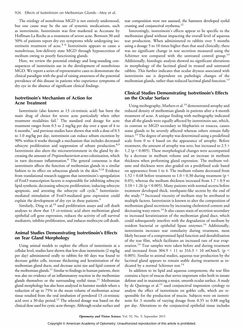
FIGURE 2.Meibography for case 2 (left, OD; right, OS).
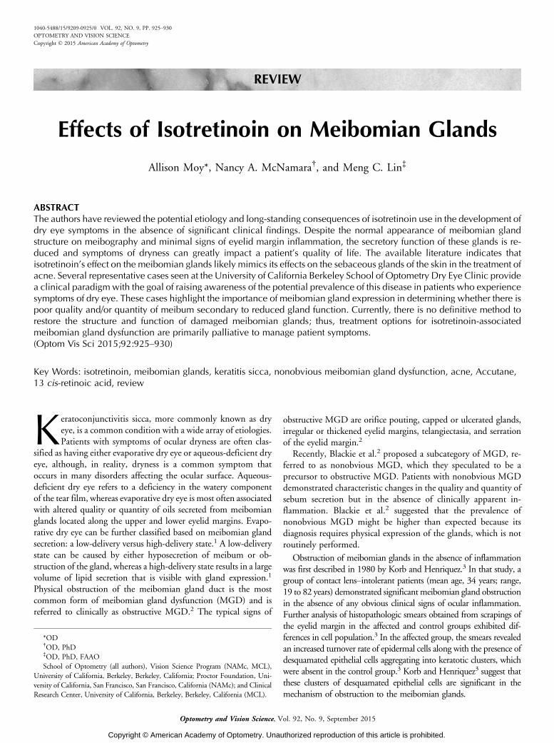
FIGURE 1.Meibography for case 1 (left, OD; right, OS).
928 Effects of Isotretinoin on Meibomian GlandsVMoy et al.
Optometry and Vision Science, Vol. 92, No. 9, September 2015
Copyright © American Academy of Optometry. Unauthorized reproduction of this article is prohibited.
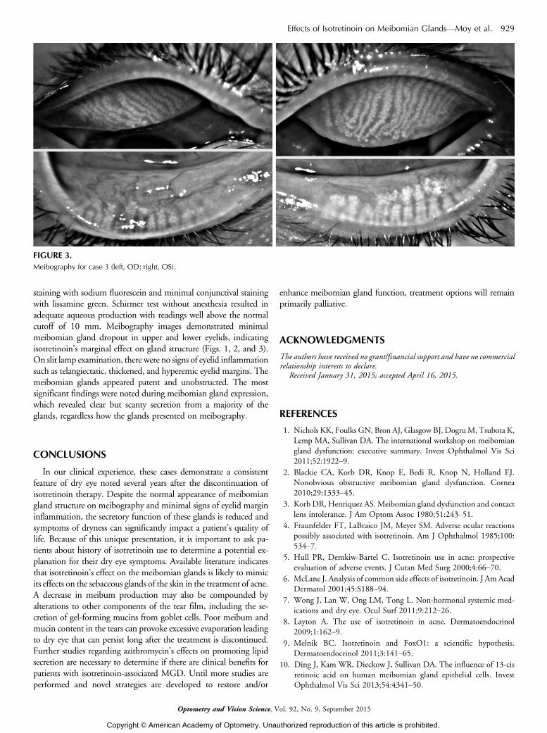
staining with sodium fluorescein and minimal conjunctival stainingwith lissamine green. Schirmer test without anesthesia resulted inadequate aqueous production with readings well above the normalcutoff of 10 mm. Meibography images demonstrated minimalmeibomian gland dropout in upper and lower eyelids, indicatingisotretinoin’s marginal effect on gland structure (Figs. 1, 2, and 3).On slit lamp examination, there were no signs of eyelid inflammationsuch as telangiectatic, thickened, and hyperemic eyelid margins. Themeibomian glands appeared patent and unobstructed. The mostsignificant findings were noted during meibomian gland expression,which revealed clear but scanty secretion from a majority of theglands, regardless how the glands presented on meibography.
CONCLUSIONS
In our clinical experience, these cases demonstrate a consistentfeature of dry eye noted several years after the discontinuation ofisotretinoin therapy. Despite the normal appearance of meibomiangland structure on meibography and minimal signs of eyelid margininflammation, the secretory function of these glands is reduced andsymptoms of dryness can significantly impact a patient’s quality oflife. Because of this unique presentation, it is important to ask pa-tients about history of isotretinoin use to determine a potential ex-planation for their dry eye symptoms. Available literature indicatesthat isotretinoin’s effect on the meibomian glands is likely to mimicits effects on the sebaceous glands of the skin in the treatment of acne.A decrease in meibum production may also be compounded byalterations to other components of the tear film, including the se-cretion of gel-forming mucins from goblet cells. Poor meibum andmucin content in the tears can provoke excessive evaporation leadingto dry eye that can persist long after the treatment is discontinued.Further studies regarding azithromycin’s effects on promoting lipidsecretion are necessary to determine if there are clinical benefits forpatients with isotretinoin-associated MGD. Until more studies areperformed and novel strategies are developed to restore and/or
enhance meibomian gland function, treatment options will remainprimarily palliative.
ACKNOWLEDGMENTS
The authors have received no grant/financial support and have no commercialrelationship interests to declare.
Received January 31, 2015; accepted April 16, 2015.
REFERENCES
1. Nichols KK, Foulks GN, Bron AJ, Glasgow BJ, Dogru M, Tsubota K,Lemp MA, Sullivan DA. The international workshop on meibomian
gland dysfunction: executive summary. Invest Ophthalmol Vis Sci2011;52:1922Y9.
2. Blackie CA, Korb DR, Knop E, Bedi R, Knop N, Holland EJ.Nonobvious obstructive meibomian gland dysfunction. Cornea2010;29:1333Y45.
3. Korb DR, Henriquez AS. Meibomian gland dysfunction and contact
lens intolerance. J Am Optom Assoc 1980;51:243Y51.
4. Fraunfelder FT, LaBraico JM, Meyer SM. Adverse ocular reactions
possibly associated with isotretinoin. Am J Ophthalmol 1985;100:534Y7.
5. Hull PR, Demkiw-Bartel C. Isotretinoin use in acne: prospectiveevaluation of adverse events. J Cutan Med Surg 2000;4:66Y70.
6. McLane J. Analysis of common side effects of isotretinoin. J Am AcadDermatol 2001;45:S188Y94.
7. Wong J, Lan W, Ong LM, Tong L. Non-hormonal systemic med-
ications and dry eye. Ocul Surf 2011;9:212Y26.
8. Layton A. The use of isotretinoin in acne. Dermatoendocrinol
2009;1:162Y9.
9. Melnik BC. Isotretinoin and FoxO1: a scientific hypothesis.Dermatoendocrinol 2011;3:141Y65.
10. Ding J, Kam WR, Dieckow J, Sullivan DA. The influence of 13-cisretinoic acid on human meibomian gland epithelial cells. InvestOphthalmol Vis Sci 2013;54:4341Y50.
FIGURE 3.Meibography for case 3 (left, OD; right, OS).
Effects of Isotretinoin on Meibomian GlandsVMoy et al. 929
Optometry and Vision Science, Vol. 92, No. 9, September 2015
Copyright © American Academy of Optometry. Unauthorized reproduction of this article is prohibited.
11. Kremer I, Gaton DD, David M, Gaton E, Shapiro A. Toxic effectsof systemic retinoids on meibomian glands. Ophthalmic Res 1994;26:124Y8.
12. Lambert RW, Smith RE. Effects of 13-cis-retinoic acid on thehamster meibomian gland. J Invest Dermatol 1989;92:321Y5.
13. Rismondo V, Ubels JL, Osgood TB. Tear secretion and lacrimal
gland function of rabbits treated with isotretinoin. J Am AcadDermatol 1988;19:280Y5.
14. Mathers WD, Shields WJ, Sachdev MS, Petroll WM, Jester JV.Meibomian gland morphology and tear osmolarity: changes withAccutane therapy. Cornea 1991;10:286Y90.
15. de Queiroga IB, Antonio Vieira L, Barros Jde N, Melo Diniz Mde F,
de Morais LC. Conjunctival impression cytology changes induced byoral isotretinoin. Cornea 2009;28:1009Y13.
16. Rismondo V, Ubels JL. Isotretinoin in lacrimal gland fluid and tears.Arch Ophthalmol 1987;105:416Y20.
17. Karalezli A, Borazan M, Altinors DD, Dursun R, Kiyici H, AkovaYA. Conjunctival impression cytology, ocular surface, and tear-film
changes in patients treated with systemic isotretinoin. Cornea 2009;28:46Y50.
18. Egger SF, Huber-Spitzy V, Bohler K, Raff M, Scholda C, Barisani T,Vecsei VP. Ocular side effects associated with 13-cis-retinoic acidtherapy for acne vulgaris: clinical features, alterations of tearfilm and
conjunctival flora. Acta Ophthalmol Scand 1995;73:355Y7.
19. Cumurcu T, Sezer E, Kilic R, Bulut Y. Comparison of dose-relatedocular side effects during systemic isotretinoin administration. Eur JOphthalmol 2009;19:196Y200.
20. Oner A, Ferahbas A, Karakucuk S, Utas S, Karaman B, Kutlugun C,Somdas M, Mirza E. Ocular side effects associated with systemic
isotretinoin. J Toxicol-Cutan Ocul 2004;23:189Y95.
21. Lerman S. Ocular side effects of accutane therapy. Lens Eye ToxicRes 1992;9:429Y38.
22. Fraunfelder FT, Fraunfelder FW, Edwards R. Ocular side effectspossibly associated with isotretinoin usage. Am J Ophthalmol 2001;132:299Y305.
23. Finis D, Hayajneh J, Konig C, Borrelli M, Schrader S, Geerling G.
Evaluation of an automated thermodynamic treatment (LipiFlow(R))
system for meibomian gland dysfunction: a prospective, randomized,
observer-masked trial. Ocul Surf 2014;12:146Y54.
24. Geerling G, Tauber J, Baudouin C, Goto E, Matsumoto Y, O’Brien
T, Rolando M, Tsubota K, Nichols KK. The international workshop
on meibomian gland dysfunction: report of the subcommittee on
management and treatment of meibomian gland dysfunction. Invest
Ophthalmol Vis Sci 2011;52:2050Y64.
25. Bakar O, Demircay Z, Toker E, Cakir S. Ocular signs, symptoms
and tear function tests of papulopustular rosacea patients receiving
azithromycin. J Eur Acad Dermatol Venereol 2009;23:544Y9.
26. Igami TZ, Holzchuh R, Osaki TH, Santo RM, Kara-Jose N, Hida
RY. Oral azithromycin for treatment of posterior blepharitis. Cornea
2011;30:1145Y9.
27. Luchs J. Efficacy of topical azithromycin ophthalmic solution 1% in
the treatment of posterior blepharitis. Adv Ther 2008;25:858Y70.
28. Liu Y, Kam WR, Ding J, Sullivan DA. Effect of azithromycin on
lipid accumulation in immortalized human meibomian gland epi-
thelial cells. JAMA Ophthalmol 2014;132:226Y8.
29. Liu Y, Kam WR, Ding J, Sullivan DA. Can tetracycline antibiotics
duplicate the ability of azithromycin to stimulate human meibomian
gland epithelial cell differentiation? Cornea 2015;34:342Y6.
30. Liu Y, Kam WR, Ding J, Sullivan DA. One man’s poison is another
man’s meat: using azithromycin-induced phospholipidosis to pro-
mote ocular surface health. Toxicology 2014;320:1Y5.
Allison MoySchool of Optometry
University of CaliforniaBerkeley, CA 94720
e-mail: [email protected]
930 Effects of Isotretinoin on Meibomian GlandsVMoy et al.
Optometry and Vision Science, Vol. 92, No. 9, September 2015
Copyright © American Academy of Optometry. Unauthorized reproduction of this article is prohibited.