Embed Size (px)
Citation preview
EFFECTS OF IONIZING RADIATIONUnited Nations Scientific Committee on the
Effects of Atomic Radiation
UNSCEAR 2006 Report to the General Assembly
with Scientific Annexes
VOLUME IIScientific Annexes C, D and E
UNITED NATIONSNew York, 2009
NOTE
The report of the Committee without its annexes appears as Official Records of the General Assembly, Sixty-first Session, Supplement No. 46 and corrigendum (A/61/46 and Corr. 1). The report reproduced here includes the corrections of the corrigendum.
The designation employed and the presentation of material in this publication do not imply the expression of any opinion whatsoever on the part of the Secretariat of the United Nations con-cerning the legal status of any country, territory, city or area, or of its authorities, or concerning the delimitation of its frontiers or boundaries.
The country names used in this document are, in most cases, those that were in use at the time the data were collected or the text prepared. In other cases, however, the names have been updated, where this was possible and appropriate, to reflect political changes.
UNITED NATIONS PUBLICATION
Sales No. E.09.IX.5
ISBN 978-92-1-142270-2
1
ANNEX C
Non-targeted and delayed effects of exposure to ionizing radiation
ContentsPage
INTRODUCTION . . . . . . . . . . . . . . . . . . . . . . . . . . . . . . . . . . . . . . . . . . . . . . . . . . . . . . . . . . . . . . . . . . . . . . . . . . . . . 3
I. RADIATION-INDUCED GENOMIC INSTABILITY . . . . . . . . . . . . . . . . . . . . . . . . . . . . . . . . . . . . . . . . . . . . . . . . . . . 5 A. Radiation-induced genomic instability in vitro . . . . . . . . . . . . . . . . . . . . . . . . . . . . . . . . . . . . . . . . . . . . . . . 5 B. Induced genomic instability after in vivo irradiation followed by in vitro analysis . . . . . . . . . . . . . . . . . . . . . 12 C. Induced genomic instability after in vitro irradiation followed by in vivo analysis . . . . . . . . . . . . . . . . . . . . . 12 D. Radiation-induced genomic instability in vivo . . . . . . . . . . . . . . . . . . . . . . . . . . . . . . . . . . . . . . . . . . . . . . . . 13 1. Mouse models for radiation-induced genomic instability in vivo . . . . . . . . . . . . . . . . . . . . . . . . . . . . . . 17 2. Human studies . . . . . . . . . . . . . . . . . . . . . . . . . . . . . . . . . . . . . . . . . . . . . . . . . . . . . . . . . . . . . . . . . . . 18 E. Genomic instability and radiation-induced leukaemia . . . . . . . . . . . . . . . . . . . . . . . . . . . . . . . . . . . . . . . . . . 21 1. Mouse models . . . . . . . . . . . . . . . . . . . . . . . . . . . . . . . . . . . . . . . . . . . . . . . . . . . . . . . . . . . . . . . . . . . 21 2. Human studies . . . . . . . . . . . . . . . . . . . . . . . . . . . . . . . . . . . . . . . . . . . . . . . . . . . . . . . . . . . . . . . . . . . 21 F. Role of telomeres and telomerase in radiation-induced genomic instability . . . . . . . . . . . . . . . . . . . . . . . . . 21 G. Conclusions . . . . . . . . . . . . . . . . . . . . . . . . . . . . . . . . . . . . . . . . . . . . . . . . . . . . . . . . . . . . . . . . . . . . . . . . . 22
II. BYSTANDER EFFECTS AND RADIATION EXPOSURE . . . . . . . . . . . . . . . . . . . . . . . . . . . . . . . . . . . . . . . . . . . . . . 23 A. Bystander effects in vitro . . . . . . . . . . . . . . . . . . . . . . . . . . . . . . . . . . . . . . . . . . . . . . . . . . . . . . . . . . . . . . . 23 1. Bystander effects after cytoplasmic irradiation . . . . . . . . . . . . . . . . . . . . . . . . . . . . . . . . . . . . . . . . . . . 25 2. Bystander effects after low fluences of alpha particle irradiation . . . . . . . . . . . . . . . . . . . . . . . . . . . . . 25 3. Bystander effects after irradiation with a charged-particle microbeam . . . . . . . . . . . . . . . . . . . . . . . . . 27 4. Bystander effects after transfer of medium from irradiated cells . . . . . . . . . . . . . . . . . . . . . . . . . . . . . 31 B. Bystander effects in vivo . . . . . . . . . . . . . . . . . . . . . . . . . . . . . . . . . . . . . . . . . . . . . . . . . . . . . . . . . . . . . . . 33
III. RELATIONSHIP BETwEEN RADIATION-INDUCED GENOMIC INSTABILITY AND BYSTANDER EFFECTS . . . . . . . . . 37 A. Relationship between radiation hypersensitivity at low doses and bystander effects . . . . . . . . . . . . . . . . . 37 B. Relationship between radiation adaptive response and bystander effects . . . . . . . . . . . . . . . . . . . . . . . . . . 38 C. Conclusions . . . . . . . . . . . . . . . . . . . . . . . . . . . . . . . . . . . . . . . . . . . . . . . . . . . . . . . . . . . . . . . . . . . . . . . . . 38
IV. ABSCOPAL EFFECTS OF RADIATION . . . . . . . . . . . . . . . . . . . . . . . . . . . . . . . . . . . . . . . . . . . . . . . . . . . . . . . . . . 39 A. Review . . . . . . . . . . . . . . . . . . . . . . . . . . . . . . . . . . . . . . . . . . . . . . . . . . . . . . . . . . . . . . . . . . . . . . . . . . . . 39 B. Conclusions . . . . . . . . . . . . . . . . . . . . . . . . . . . . . . . . . . . . . . . . . . . . . . . . . . . . . . . . . . . . . . . . . . . . . . . . . 40
V. CLASTOGENIC FACTORS INDUCED BY IONIZING RADIATION . . . . . . . . . . . . . . . . . . . . . . . . . . . . . . . . . . . . . . . 41 A. Review . . . . . . . . . . . . . . . . . . . . . . . . . . . . . . . . . . . . . . . . . . . . . . . . . . . . . . . . . . . . . . . . . . . . . . . . . . . . 41 B. Conclusions . . . . . . . . . . . . . . . . . . . . . . . . . . . . . . . . . . . . . . . . . . . . . . . . . . . . . . . . . . . . . . . . . . . . . . . . . 43
VI. IMPACT OF NON-TARGETED AND DELAYED EFFECTS OF RADIATION ON FUTURE GENERATIONS . . . . . . . . . . . 45 A. Studies in non-mammalian species . . . . . . . . . . . . . . . . . . . . . . . . . . . . . . . . . . . . . . . . . . . . . . . . . . . . . . . 45 B. Mouse studies . . . . . . . . . . . . . . . . . . . . . . . . . . . . . . . . . . . . . . . . . . . . . . . . . . . . . . . . . . . . . . . . . . . . . . . 46 1. Irradiation of the mouse zygote . . . . . . . . . . . . . . . . . . . . . . . . . . . . . . . . . . . . . . . . . . . . . . . . . . . . . . 46
Page
2. Pre-implantation embryo chimera assay . . . . . . . . . . . . . . . . . . . . . . . . . . . . . . . . . . . . . . . . . . . . . . . . 46 3. Mouse mutation assays . . . . . . . . . . . . . . . . . . . . . . . . . . . . . . . . . . . . . . . . . . . . . . . . . . . . . . . . . . . . 47 4. Alterations in tandem repeat DNA sequences . . . . . . . . . . . . . . . . . . . . . . . . . . . . . . . . . . . . . . . . . . . 47 5. Tumour induction in the offspring of irradiated parents . . . . . . . . . . . . . . . . . . . . . . . . . . . . . . . . . . . . . 49 C. Malformation induction in the offspring of irradiated parents . . . . . . . . . . . . . . . . . . . . . . . . . . . . . . . . . . . . 50 D. Human studies . . . . . . . . . . . . . . . . . . . . . . . . . . . . . . . . . . . . . . . . . . . . . . . . . . . . . . . . . . . . . . . . . . . . . . 50 E. Cancer incidence in the offspring of irradiated humans . . . . . . . . . . . . . . . . . . . . . . . . . . . . . . . . . . . . . . . . 51 F. Pregnancy outcomes in the offspring of irradiated humans . . . . . . . . . . . . . . . . . . . . . . . . . . . . . . . . . . . . . 52 G. Genetic damage and malformation induction in the offspring of irradiated parents . . . . . . . . . . . . . . . . . . . 52 H. Impact of non-targeted and delayed effects of radiation on future generations . . . . . . . . . . . . . . . . . . . . . . 52
VII. IMPLICATIONS OF NON-TARGETED AND DELAYED EFFECTS . . . . . . . . . . . . . . . . . . . . . . . . . . . . . . . . . . . . . . . . 55
CONCLUDING REMARKS . . . . . . . . . . . . . . . . . . . . . . . . . . . . . . . . . . . . . . . . . . . . . . . . . . . . . . . . . . . . . . . . . . . . . . 57
References . . . . . . . . . . . . . . . . . . . . . . . . . . . . . . . . . . . . . . . . . . . . . . . . . . . . . . . . . . . . . . . . . . . . . . . . . . . . . . 59
2
3
INTRODUCTION
1. The goal of this annex is to summarize the evidence for non-targeted and delayed effects of exposure to ionizing radiation in vitro and in vivo. Currently, human health risk estimates for effects associated with radiation exposures are based primarily on the view that the detrimental effects of irradiation occur only in irradiated cells. Over the years, a number of non-targeted effects of radiation exposure have been described that challenge this concept. These non- targeted effects include genomic instability occurring in the progeny of an irradiated cell, bystander effects, clastogenic factors produced in plasma from irradiated individuals that can cause chromosome damage when cultured with non-irradiated cells, and heritable effects of parental irradiation
that can manifest across generations. This annex considers whether these effects pose new challenges to evaluating risks associated with radiation exposure, understanding radiation-induced carcinogenesis and interpreting epidemiological data on radiation exposure.
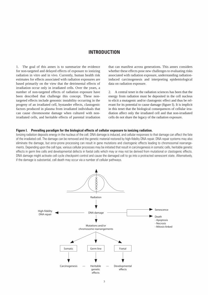
2. A central tenet in the radiation sciences has been that the energy from radiation must be deposited in the cell nucleus to elicit a mutagenic and/or clastogenic effect and thus be rel-evant for its potential to cause damage (figure I). It is implicit in this tenet that the biological consequences of cellular irra-diation affect only the irradiated cell and that non-irradiated cells do not share the legacy of the radiation exposure.
Figure I. Prevailing paradigm for the biological effects of cellular exposure to ionizing radiation. Ionizing radiation deposits energy in the nucleus of the cell. DNA damage is induced, and cellular responses to that damage can affect the fate of the irradiated cell. The damage can be removed and the genetic material restored by high-fidelity DNA repair. DNA repair systems may also eliminate the damage, but error-prone processing can result in gene mutations and clastogenic effects leading to chromosomal rearrange-ments. Depending upon the cell type, various cellular processes may be initiated that result in carcinogenesis in somatic cells, heritable genetic effects in germ line cells and developmental defects in foetal cells which may or may not be derived from mutational or clastogenic effects. DNA damage might activate cell cycle checkpoint control and cause the damaged cell to go into a protracted senescent state. Alternatively, if the damage is substantial, cell death may occur via a number of cellular pathways.
Radiation
DNA damageSenescence
Death- Apoptosis- Necrosis- Mitosis-linkedMutations and/or
chromosome rearrangements
High-fidelityDNA repair
Heritablegeneticeffects
Developmentaleffects
Carcinogenesis — —
Germ lineSomatic Foetal
4 UNSCEAR 2006 REPORT: VOLUME II
3. When ionizing radiation is absorbed in biological mate-rial, excitations and ionizations occur that are non-randomly distributed along localized tracks. The spatial distribution of these ionization/excitation events produced by different particles varies considerably depending on the quality of radiation. The term “linear energy transfer” (LET) is used to classify radiation quality according to the average energy transferred per unit length of the track. For the purposes of this annex, X- and gamma rays are considered to be low-LET radiation, protons and neutrons are considered to be intermediate LET radiation, and alpha particles and heavy ions are considered to be high-LET radiation.
4. In contrast to the risks associated with exposures to low doses of ionizing radiation (less than about 200 mSv, UNSCEAR 2000 Report [U2]), the risks of cancer after high and moderate doses of radiation are relatively well under-stood. This understanding is based on data from detailed epidemiological studies of the survivors of the atomic bombings in Japan and other exposed groups, e.g. clinically irradiated populations and those exposed as a result of the Chernobyl accident (UNSCEAR 2000 Report, annex I). However, risks at low doses are generally extrapolated from the high-dose data, applying dose and dose-rate effective-ness factors. Estimating risk is further complicated because environmental exposures are predominantly protracted, low-dose, low-dose-rate exposures, or high-dose-rate exposures delivered in small fractions (see annex A, “Epidemiological studies of radiation and cancer”). This contrasts with the majority of laboratory studies and clinical exposure situa-tions, where exposures are usually acute, high-dose, high-
dose-rate exposures. In addition, inherent in many models of radiation risk is that only those cells or tissues actually irradiated are burdened by the legacy of the radiation expo-sure. A number of non-targeted delayed effects of radiation exposure have been described; the purpose of this annex is to summarize the evidence for these effects and indicate present hypotheses on how they may affect the assessment of health hazards associated with radiation exposure and radiation-induced carcinogenesis.
5. For the purposes of this annex, “non-targeted effects” refers to radiation-induced effects manifesting in cells whose nucleus was not subject to a direct hit by the radia-tion, i.e. no ionization events due to cellular irradiation were deposited within the volume of that nucleus. In such instances the radiation may have hit the cytoplasm, or neigh-bouring cells, tissues or organs, or even cells in another culture vessel, and a response is communicated from these irradiated cells to non-irradiated cells to elicit an effect. It must be stressed at this stage that the non- targeted effects of ionizing radiation described in this annex do not imply that the well-documented targeted effects of radiation are irrelevant or unimportant, or that the concept of “dose” needs to be revised. That is not the case. Rather, the goal of this annex is to summarize the literature on non-targeted effects associated with exposure to ionizing radiation and, where possible, to evaluate how such effects may affect risks associated with radiation exposure, the understanding of radiation-induced carcinogenesis, and the mechanistic basis for interpreting epidemiological data on radiation effects.
5
I. RADIATION-INDUCED GENOMIC INSTABILITY
A. Radiation-induced genomic instability in vitro
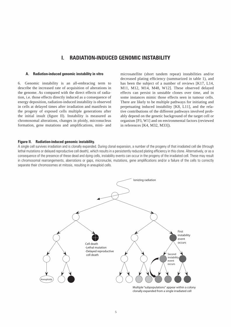
6. Genomic instability is an all-embracing term to describe the increased rate of acquisition of alterations in the genome. As compared with the direct effects of radia-tion, i.e. those effects directly induced as a consequence of energy deposition, radiation-induced instability is observed in cells at delayed times after irradiation and manifests in the progeny of exposed cells multiple generations after the initial insult (figure II). Instability is measured as chromo somal alterations, changes in ploidy, micronucleus formation, gene mutations and amplifications, mini- and
microsatellite (short tandem repeat) instabilities and/or decreased plating efficiency (summarized in table 1), and has been the subject of a number of reviews [K17, L14, M11, M12, M14, M48, W12]. These observed delayed effects can persist in unstable clones over time, and in some instances mimic those effects seen in tumour cells. There are likely to be multiple pathways for initiating and perpetuating induced instability [K8, L11], and the rela-tive contributions of the different pathways involved prob-ably depend on the genetic background of the target cell or organism [P3, W1] and on environmental factors (reviewed in references [K4, M32, M33]).
Figure II. Radiation-induced genomic instability. A single cell survives irradiation and is clonally expanded. During clonal expansion, a number of the progeny of that irradiated cell die (through lethal mutations or delayed reproductive cell death), which results in a persistently reduced plating efficiency in this clone. Alternatively, or as a consequence of the presence of these dead and dying cells, instability events can occur in the progeny of the irradiated cell. These may result in chromosomal rearrangements, aberrations or gaps, micronuclei, mutations, gene amplifications and/or a failure of the cells to correctly separate their chromosomes at mitosis, resulting in aneuploid cells.
Cell death-Lethal mutation-Delayed reproductivecell death
Ionizing radiation
Firstinstabilityeventoccurs
Multiple “subpopulations“ appear within a colonyclonally expanded from a single irradiated cell
Secondinstabilityeventoccurs
Aneuploidy
6 UNSCEAR 2006 REPORT: VOLUME II
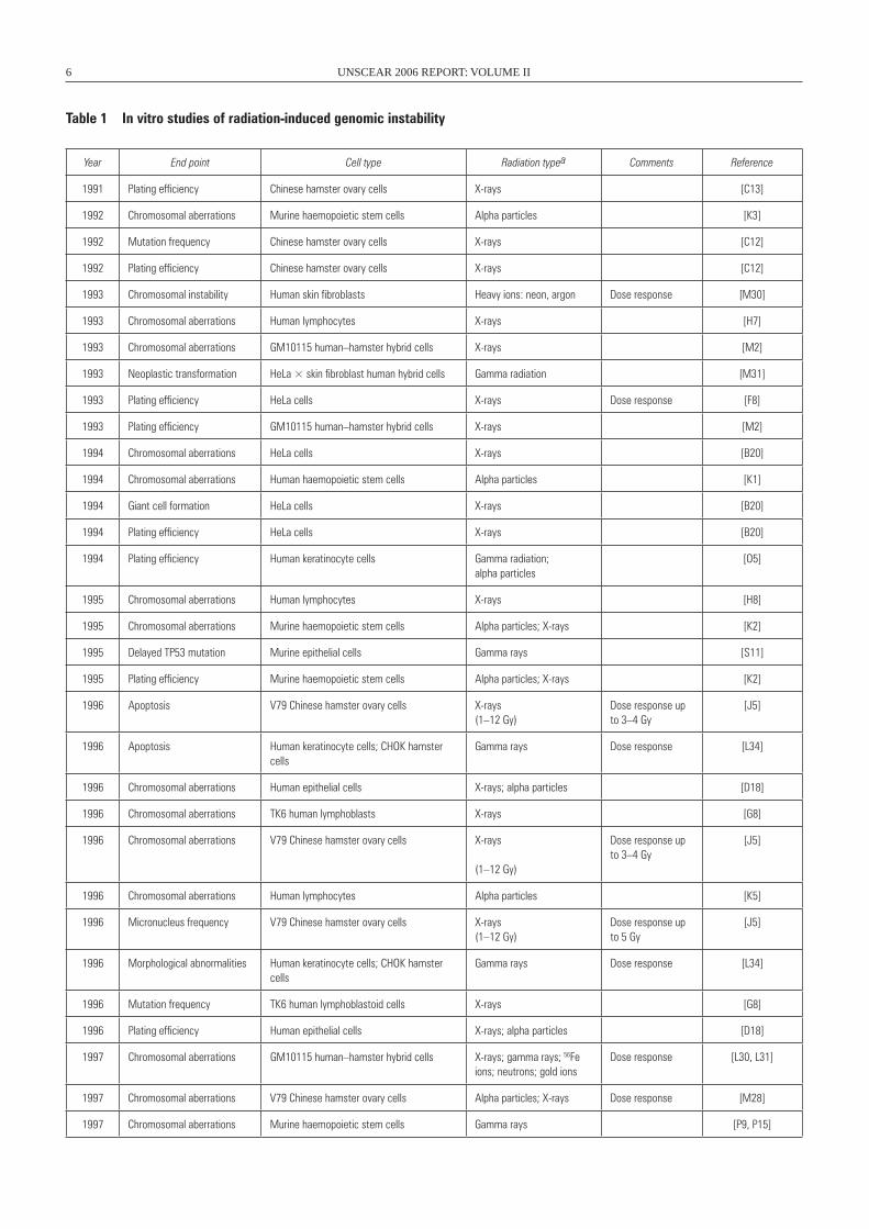
Year End point Cell type Radiation typea Comments Reference
1991 Plating efficiency Chinese hamster ovary cells X-rays [C13]
1992 Chromosomal aberrations Murine haemopoietic stem cells Alpha particles [K3]
1992 Mutation frequency Chinese hamster ovary cells X-rays [C12]
1992 Plating efficiency Chinese hamster ovary cells X-rays [C12]
1993 Chromosomal instability Human skin fibroblasts Heavy ions: neon, argon Dose response [M30]
1993 Chromosomal aberrations Human lymphocytes X-rays [H7]
1993 Chromosomal aberrations GM10115 human–hamster hybrid cells X-rays [M2]
1993 Neoplastic transformation HeLa × skin fibroblast human hybrid cells Gamma radiation [M31]
1993 Plating efficiency HeLa cells X-rays Dose response [F8]
1993 Plating efficiency GM10115 human–hamster hybrid cells X-rays [M2]
1994 Chromosomal aberrations HeLa cells X-rays [B20]
1994 Chromosomal aberrations Human haemopoietic stem cells Alpha particles [K1]
1994 Giant cell formation HeLa cells X-rays [B20]
1994 Plating efficiency HeLa cells X-rays [B20]
1994 Plating efficiency Human keratinocyte cells Gamma radiation; alpha particles
[O5]
1995 Chromosomal aberrations Human lymphocytes X-rays [H8]
1995 Chromosomal aberrations Murine haemopoietic stem cells Alpha particles; X-rays [K2]
1995 Delayed TP53 mutation Murine epithelial cells Gamma rays [S11]
1995 Plating efficiency Murine haemopoietic stem cells Alpha particles; X-rays [K2]
1996 Apoptosis V79 Chinese hamster ovary cells X-rays(1–12 Gy)
Dose response up to 3–4 Gy
[J5]
1996 Apoptosis Human keratinocyte cells; CHOK hamster cells
Gamma rays Dose response [L34]
1996 Chromosomal aberrations Human epithelial cells X-rays; alpha particles [D18]
1996 Chromosomal aberrations TK6 human lymphoblasts X-rays [G8]
1996 Chromosomal aberrations V79 Chinese hamster ovary cells X-rays
(1–12 Gy)
Dose response up to 3–4 Gy
[J5]
1996 Chromosomal aberrations Human lymphocytes Alpha particles [K5]
1996 Micronucleus frequency V79 Chinese hamster ovary cells X-rays(1–12 Gy)
Dose response up to 5 Gy
[J5]
1996 Morphological abnormalities Human keratinocyte cells; CHOK hamster cells
Gamma rays Dose response [L34]
1996 Mutation frequency TK6 human lymphoblastoid cells X-rays [G8]
1996 Plating efficiency Human epithelial cells X-rays; alpha particles [D18]
1997 Chromosomal aberrations GM10115 human–hamster hybrid cells X-rays; gamma rays; 56Fe ions; neutrons; gold ions
Dose response [L30, L31]
1997 Chromosomal aberrations V79 Chinese hamster ovary cells Alpha particles; X-rays Dose response [M28]
1997 Chromosomal aberrations Murine haemopoietic stem cells Gamma rays [P9, P15]
Table 1 In vitro studies of radiation-induced genomic instability
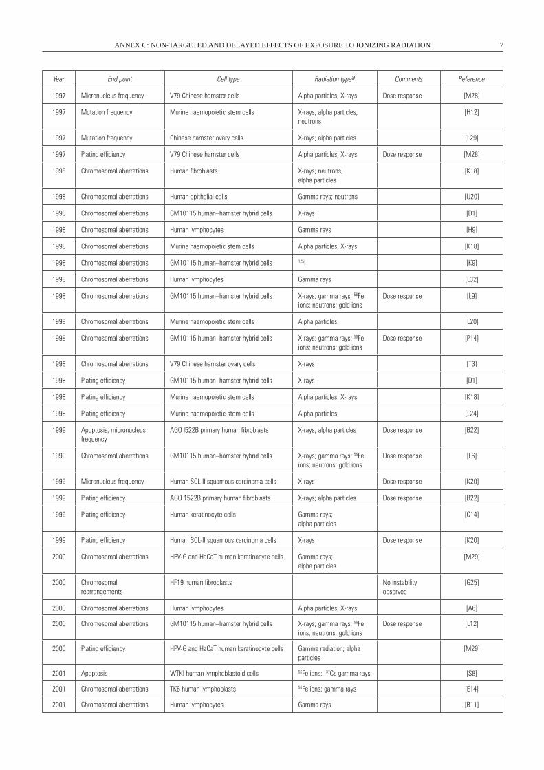
ANNEX C: NON-TARGETED AND DELAYED EFFECTS OF EXPOSURE TO IONIZING RADIATION 7
Year End point Cell type Radiation typea Comments Reference
1997 Micronucleus frequency V79 Chinese hamster cells Alpha particles; X-rays Dose response [M28]
1997 Mutation frequency Murine haemopoietic stem cells X-rays; alpha particles; neutrons
[H12]
1997 Mutation frequency Chinese hamster ovary cells X-rays; alpha particles [L29]
1997 Plating efficiency V79 Chinese hamster cells Alpha particles; X-rays Dose response [M28]
1998 Chromosomal aberrations Human fibroblasts X-rays; neutrons; alpha particles
[K18]
1998 Chromosomal aberrations Human epithelial cells Gamma rays; neutrons [U20]
1998 Chromosomal aberrations GM10115 human–hamster hybrid cells X-rays [D1]
1998 Chromosomal aberrations Human lymphocytes Gamma rays [H9]
1998 Chromosomal aberrations Murine haemopoietic stem cells Alpha particles; X-rays [K18]
1998 Chromosomal aberrations GM10115 human–hamster hybrid cells 125I [K9]
1998 Chromosomal aberrations Human lymphocytes Gamma rays [L32]
1998 Chromosomal aberrations GM10115 human–hamster hybrid cells X-rays; gamma rays; 56Fe ions; neutrons; gold ions
Dose response [L9]
1998 Chromosomal aberrations Murine haemopoietic stem cells Alpha particles [L20]
1998 Chromosomal aberrations GM10115 human–hamster hybrid cells X-rays; gamma rays; 56Fe ions; neutrons; gold ions
Dose response [P14]
1998 Chromosomal aberrations V79 Chinese hamster ovary cells X-rays [T3]
1998 Plating efficiency GM10115 human–hamster hybrid cells X-rays [D1]
1998 Plating efficiency Murine haemopoietic stem cells Alpha particles; X-rays [K18]
1998 Plating efficiency Murine haemopoietic stem cells Alpha particles [L24]
1999 Apoptosis; micronucleus frequency
AGO l522B primary human fibroblasts X-rays; alpha particles Dose response [B22]
1999 Chromosomal aberrations GM10115 human–hamster hybrid cells X-rays; gamma rays; 56Fe ions; neutrons; gold ions
Dose response [L6]
1999 Micronucleus frequency Human SCL-II squamous carcinoma cells X-rays Dose response [K20]
1999 Plating efficiency AGO 1522B primary human fibroblasts X-rays; alpha particles Dose response [B22]
1999 Plating efficiency Human keratinocyte cells Gamma rays; alpha particles
[C14]
1999 Plating efficiency Human SCL-II squamous carcinoma cells X-rays Dose response [K20]
2000 Chromosomal aberrations HPV-G and HaCaT human keratinocyte cells Gamma rays; alpha particles
[M29]
2000 Chromosomal rearrangements
HF19 human fibroblasts No instability observed
[G25]
2000 Chromosomal aberrations Human lymphocytes Alpha particles; X-rays [A6]
2000 Chromosomal aberrations GM10115 human–hamster hybrid cells X-rays; gamma rays; 56Fe ions; neutrons; gold ions
Dose response [L12]
2000 Plating efficiency HPV-G and HaCaT human keratinocyte cells Gamma radiation; alpha particles
[M29]
2001 Apoptosis wTKI human lymphoblastoid cells 56Fe ions; 137Cs gamma rays [S8]
2001 Chromosomal aberrations TK6 human lymphoblasts 56Fe ions; gamma rays [E14]
2001 Chromosomal aberrations Human lymphocytes Gamma rays [B11]
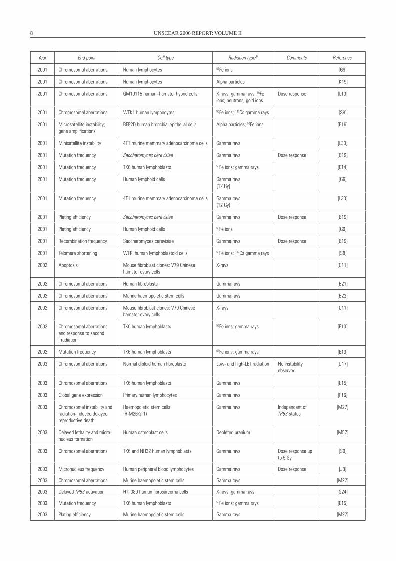
8 UNSCEAR 2006 REPORT: VOLUME II
Year End point Cell type Radiation typea Comments Reference
2001 Chromosomal aberrations Human lymphocytes 56Fe ions [G9]
2001 Chromosomal aberrations Human lymphocytes Alpha particles [K19]
2001 Chromosomal aberrations GM10115 human–hamster hybrid cells X-rays; gamma rays; 56Fe ions; neutrons; gold ions
Dose response [L10]
2001 Chromosomal aberrations wTK1 human lymphocytes 56Fe ions; 137Cs gamma rays [S8]
2001 Microsatellite instability; gene amplifications
BEP2D human bronchial epithelial cells Alpha particles; 56Fe ions [P16]
2001 Minisatellite instability 4T1 murine mammary adenocarcinoma cells Gamma rays [L33]
2001 Mutation frequency Saccharomyces cerevisiae Gamma rays Dose response [B19]
2001 Mutation frequency TK6 human lymphoblasts 56Fe ions; gamma rays [E14]
2001 Mutation frequency Human lymphoid cells Gamma rays(12 Gy)
[G9]
2001 Mutation frequency 4T1 murine mammary adenocarcinoma cells Gamma rays(12 Gy)
[L33]
2001 Plating efficiency Saccharomyces cerevisiae Gamma rays Dose response [B19]
2001 Plating efficiency Human lymphoid cells 56Fe ions [G9]
2001 Recombination frequency Saccharomyces cerevisiae Gamma rays Dose response [B19]
2001 Telomere shortening wTKI human lymphoblastoid cells 56Fe ions; 137Cs gamma rays [S8]
2002 Apoptosis Mouse fibroblast clones; V79 Chinese hamster ovary cells
X-rays [C11]
2002 Chromosomal aberrations Human fibroblasts Gamma rays [B21]
2002 Chromosomal aberrations Murine haemopoietic stem cells Gamma rays [B23]
2002 Chromosomal aberrations Mouse fibroblast clones; V79 Chinese hamster ovary cells
X-rays [C11]
2002 Chromosomal aberrations and response to second irradiation
TK6 human lymphoblasts 56Fe ions; gamma rays [E13]
2002 Mutation frequency TK6 human lymphoblasts 56Fe ions; gamma rays [E13]
2003 Chromosomal aberrations Normal diploid human fibroblasts Low- and high-LET radiation No instability observed
[D17]
2003 Chromosomal aberrations TK6 human lymphoblasts Gamma rays [E15]
2003 Global gene expression Primary human lymphocytes Gamma rays [F16]
2003 Chromosomal instability and radiation-induced delayed reproductive death
Haemopoietic stem cells(R-M26/2-1)
Gamma rays Independent of TP53 status
[M27]
2003 Delayed lethality and micro-nucleus formation
Human osteoblast cells Depleted uranium [M57]
2003 Chromosomal aberrations TK6 and NH32 human lymphoblasts Gamma rays Dose response up to 5 Gy
[S9]
2003 Micronucleus frequency Human peripheral blood lymphocytes Gamma rays Dose response [J8]
2003 Chromosomal aberrations Murine haemopoietic stem cells Gamma rays [M27]
2003 Delayed TP53 activation HTI 080 human fibrosarcoma cells X-rays; gamma rays [S24]
2003 Mutation frequency TK6 human lymphoblasts 56Fe ions; gamma rays [E15]
2003 Plating efficiency Murine haemopoietic stem cells Gamma rays [M27]
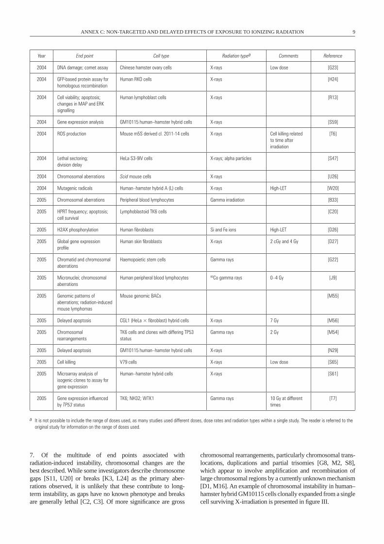
ANNEX C: NON-TARGETED AND DELAYED EFFECTS OF EXPOSURE TO IONIZING RADIATION 9
Year End point Cell type Radiation typea Comments Reference
2004 DNA damage; comet assay Chinese hamster ovary cells X-rays Low dose [G23]
2004 GFP-based protein assay for homologous recombination
Human RKO cells X-rays [H24]
2004 Cell viability; apoptosis; changes in MAP and ERK signalling
Human lymphoblast cells X-rays [R13]
2004 Gene expression analysis GM10115 human–hamster hybrid cells X-rays [S59]
2004 ROS production Mouse m5S derived cl. 2011-14 cells X-rays Cell killing related to time after irradiation
[T6]
2004 Lethal sectoring; division delay
HeLa S3-9IV cells X-rays; alpha particles [S47]
2004 Chromosomal aberrations Scid mouse cells X-rays [U26]
2004 Mutagenic radicals Human–hamster hybrid A (L) cells X-rays High-LET [w20]
2005 Chromosomal aberrations Peripheral blood lymphocytes Gamma irradiation [B33]
2005 HPRT frequency; apoptosis; cell survival
Lymphoblastoid TK6 cells [C20]
2005 H2AX phosphorylation Human fibroblasts Si and Fe ions High-LET [D26]
2005 Global gene expression profile
Human skin fibroblasts X-rays 2 cGy and 4 Gy [D27]
2005 Chromatid and chromosomal aberrations
Haemopoietic stem cells Gamma rays [G22]
2005 Micronuclei; chromosomal aberrations
Human peripheral blood lymphocytes 60Co gamma rays 0–4 Gy [J9]
2005 Genomic patterns of aberrations; radiation-induced mouse lymphomas
Mouse genomic BACs [M55]
2005 Delayed apoptosis CGL1 (HeLa × fibroblast) hybrid cells X-rays 7 Gy [M56]
2005 Chromosomal rearrangements
TK6 cells and clones with differing TP53 status
Gamma rays 2 Gy [M54]
2005 Delayed apoptosis GM10115 human–hamster hybrid cells X-rays [N29]
2005 Cell killing V79 cells X-rays Low dose [S65]
2005 Microarray analysis of isogenic clones to assay for gene expression
Human–hamster hybrid cells X-rays [S61]
2005 Gene expression influenced by TP53 status
TK6; NH32; wTK1 Gamma rays 10 Gy at different times
[T7]
a It is not possible to include the range of doses used, as many studies used different doses, dose rates and radiation types within a single study. The reader is referred to the original study for information on the range of doses used.
7. Of the multitude of end points associated with radiation-induced instability, chromosomal changes are the best described. While some investigators describe chromosome gaps [S11, U20] or breaks [K3, L24] as the primary aber-rations observed, it is unlikely that these contribute to long-term instability, as gaps have no known phenotype and breaks are generally lethal [C2, C3]. Of more significance are gross
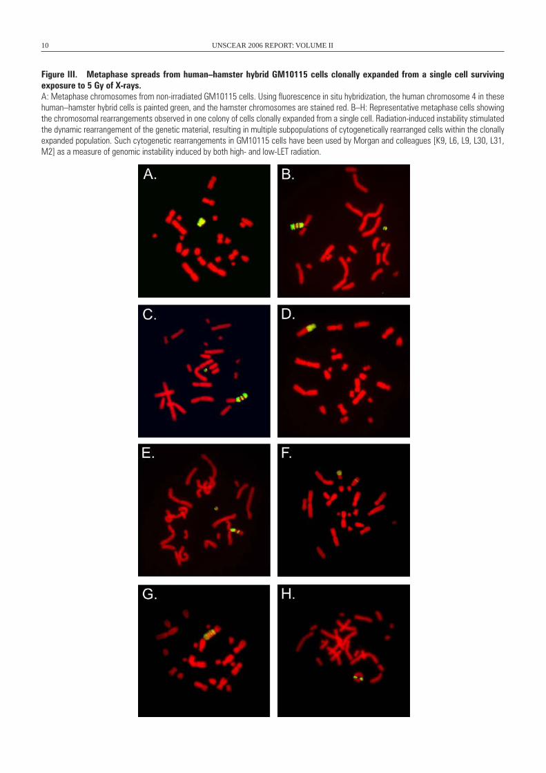
chromosomal rearrangements, particularly chromosomal trans-locations, duplications and partial trisomies [G8, M2, S8], which appear to involve amplification and recombination of large chromosomal regions by a currently unknown mechanism [D1, M16]. An example of chromosomal instability in human–hamster hybrid GM10115 cells clonally expanded from a single cell surviving X-irradiation is presented in figure III.
10 UNSCEAR 2006 REPORT: VOLUME II
Figure III. Metaphase spreads from human–hamster hybrid GM10115 cells clonally expanded from a single cell surviving exposure to 5 Gy of X-rays. A: Metaphase chromosomes from non-irradiated GM10115 cells. Using fluorescence in situ hybridization, the human chromosome 4 in these human–hamster hybrid cells is painted green, and the hamster chromosomes are stained red. B–H: Representative metaphase cells showing the chromosomal rearrangements observed in one colony of cells clonally expanded from a single cell. Radiation-induced instability stimulated the dynamic rearrangement of the genetic material, resulting in multiple subpopulations of cytogenetically rearranged cells within the clonally expanded population. Such cytogenetic rearrangements in GM10115 cells have been used by Morgan and colleagues [K9, L6, L9, L30, L31, M2] as a measure of genomic instability induced by both high- and low-LET radiation.
ANNEX C: NON-TARGETED AND DELAYED EFFECTS OF EXPOSURE TO IONIZING RADIATION 11
8. Instability is a frequent event in colonies of surviving cells. Kadhim et al. [K3] reported karyotypic abnormalities in 40–60% of murine stem cells exposed to doses of alpha particles that would produce about one hit per cell. Sabatier and colleagues [S44] observed late passage non-random chro-mosomal instability in >50% of metaphase cells from human dermal fibroblasts irradiated with a wide range of high-LET radiations (386 to 13,600 keV/µm). Likewise, Limoli et al. [L6] observed that X-rays induced chromosomal instabil-ity in ~3% of surviving human–hamster hybrid GM10115 clones per gray of radiation. This increased to ~4% Gy–1 after high-LET iron ion exposure [L12, L13]. This observed frequency of instability is grossly in excess of the reported frequency for gene mutations at similar doses. Therefore it is unlikely that mutation in a single gene or gene family is responsible for the unstable phenotype in unstable clones. Instead it is reasonable to suppose that factors contributing to maintaining genomic instability over time include critical pathways in DNA damage and repair [C10, H11, M48, Y3], chromosomal replication [B32], cellular homeostasis [B3, B6, M24, M48] and alterations in gene expression [S59, S60, S61, S62].
9. The high frequency of induced instability observed in different systems raises the intriguing question as to what is really being measured and of the significance of these obser-vations. While the numerous in vitro studies summarized in table 1 have established the occurrence of radiation-induced genomic instability, many of the cell lines used were not “normal” initially, and in many cases they involved tumour-derived cell lines. Dugan and Bedford [D17] have pointed out that instability is sometimes not observed in apparently nor-mal cells after irradiation. However, there are many reports of instability in the progeny of irradiated normal human and murine bone marrow cells [K1, K2] and in cultured human lymphocytes [B11, H7, H8, H9]. Some of this confusion may relate to the role of a functional TP53 tumour suppres-sor gene. Both TP53-dependent [S9] and TP53-independent pathways have been proposed [K5, L8], and Moore et al. [M54] showed that genomic instability might differ both quantitatively and qualitatively as a consequence of altered TP53 expression. Furthermore, differences in cellular prolif-eration patterns and susceptibility to mutation between cells and cell lines might also influence the reported results [C19]. Because the majority of reports indicate that irradiated normal primary cells readily demonstrate the instability phenotype (table 1), this implicates ionizing radiation as the causative agent. It also establishes induced instability as a phenotype associated with radiation exposure. As always, however, caution should be exercised when extrapolating from in vitro cell culture systems to the human situation in vivo.
10. When cells or tissues are directly exposed to ionizing radiation, biological effects are generally induced in a dose-dependent manner. One perplexing feature of radiation-induced genomic instability, and of non-targeted effects in general, is the lack of a well-defined dose response profile. Most investigators report that non-targeted effects are inde-pendent of dose. However, some investigators have observed
a dose response at lower doses that tends to saturate at higher doses, and a few investigators have observed consistent dose-related effects. These data are summarized in table 1 and are reviewed in references [L6, M32]. Furthermore, radiation-induced instability appears independent of dose rate, although this has not been extensively investigated to date [L6]. In addi-tion, there does not appear to be a significant LET effect for radiation-induced genomic instability, with both high- and low-LET radiations being effective [H2, K34, L12].
11. Many of the genomic changes described under the title of induced instability are changes of the same type as observed in human tumours. Radiation-induced cancers have no known molecular signature, and continued investigation aimed at understanding the processes and pathways by which radiation induces genomic alterations in the progeny of irra-diated cells should provide insights into the mechanisms of radiation-induced transformation and carcinogenesis [M62]. Kennedy et al. [K10] demonstrated replication dependence of radiation-induced transformation of C3H 10T½ cells. A similar replication dependence was also reported for radiation-induced mammary cancers in rats, in which expres-sion of epigenetic initiation required replication of irradiated mammary stem cells in the tissue microenvironment [K7].
12. While the multiple phenotypes associated with radiation-induced genomic instability are relatively well characterized, the molecular, biochemical and cellular events that initiate and perpetuate instability remain unknown. Directly induced DNA damage, e.g. induced DNA double-strand breaks, is probably not causative [M13]. Instead, deficiencies in cellular responses to DNA damage [C10, Y3], changes in gene expression [B6] or perturbations in cellular homeo stasis [B3] are more likely to be involved, and provide a rational explanation as to why the unstable phenotype can persist. In the GM10115 cell system, clones of unstable cells continue to show the dynamic production of novel chromosomal rearrangements for over four years post-irradiation [N1]. Attempts to define the target for induced instability indicate that, while the nucleus may be the ultimate target [B6, K9], there is evidence for a persist-ent increase in reactive oxygen species (ROS) in cultures of cells showing radiation-induced genomic instability. A role for enhanced oxidative stress in perpetuating the unstable phenotype was first described by Clutton et al. [C5], and was later confirmed in studies by Limoli et al. [L7, L9, L10] and Redpath and Gutierrez [R3]. A persistent induction of ROS has also been shown to cause delayed reinduction of TP53 in normal human fibro blasts [R6]. A study by Roy et al. [R5] revealed that hypoxia (2% oxygen) significantly reduced X-ray-induced delayed effects, specifically cell death, giant cell formation and chromosomal aberrations, compared with cells cultured under their “normal” 20% oxygen conditions. The role of ROS in radiation-induced genomic instability has been reviewed in detail by Mikhailov and colleagues [B45, M48]. It should be noted that oxygen tension in normal tissue shows a typical Gaussian distribution of values with a median between 40 and 60 mm Hg, and no values below 10 mm Hg [A13]. Tumours, on the other hand, invariably
12 UNSCEAR 2006 REPORT: VOLUME II
show a distribution with much lower oxygen tension [B44]. As will be described later, a role for ROS in non-targeted radiation-induced bystander effects has also been described, suggesting a potential commonality in processes involved in these delayed effects of exposure to ionizing radiation.
13. It is well known that most mammalian cells do not divide indefinitely in vitro or in vivo, owing to a process termed replicative senescence. In human cells, replica-tive senescence can be caused by telomere shortening, but murine cells senesce despite having long, stable telomeres. Parrinello et al. [P19] showed that the phenotypes of senes-cent human fibroblasts and mouse embryonic fibroblasts (MEFs) differ under standard culture conditions that include 20% oxygen. The MEFs did not senesce in physiological (3%) oxygen levels, but underwent a spontaneous event that allowed indefinite proliferation in 20% oxygen. The prolifer-ation and cytogenetic profiles of DNA repair-deficient MEFs suggested that DNA damage limits MEF proliferation in 20% oxygen. Indeed, MEFs accumulated more DNA damage in 20% oxygen than in 3% oxygen, and more damage than human fibroblasts in 20% oxygen. These results identify oxygen sensitivity as a critical difference between mouse and human cells, explaining their proliferative differences in culture, and possibly their different rates of cancer induction and ageing. Furthermore, they may contribute to explaining some of the differences between mouse and human studies described later in this annex.
14. There is emerging evidence implicating a role for extranuclear and even extracellular events in initiating and perpetuating radiation-induced chromosomal instability. Kadhim et al. [K3] analysed chromosomal instability in murine haemopoietic stem cells following alpha particle irra-diation. Many of the surviving cells were those that were not traversed by an alpha particle during irradiation. Expanding these studies, Wright and colleagues used a protective metal grid to shield regions of the cell culture flask and lethally irradiated the non-shielded regions of the flask. They then cultured the non-irradiated, shielded cells and examined the clonal progeny for induced chromosomal instability. A high frequency of instability was observed in the progeny of cells that were not directly hit by radiation [L24]. Clearly, induced instability has an extracellular component, and signals from irradiated cells can stimulate chromosomal rearrangements in non-targeted cells within the radiation environment (reviewed in reference [M10]). These observations have implications for the fate of cells surviving radiation exposure in that some of these surviving cells may develop genomic instability. These observations also indicate that even cells outside the radiation field can manifest phenotypes similar to those of irradiated cells.
B. Induced genomic instability after in vivo irradiation followed by in vitro analysis
15. Weissenborn and Streffer were the first to describe induction of genomic instability after irradiation in vivo
followed by analysis in vitro. They reported structural and numerical chromosomal anomalies as well as micronuclei at the first, second and third mitosis after in vivo irradiation of one- or two-cell mouse embryos with X-rays or neutrons [W6, W7]. These observations were extended by Ullrich and Davis [U18], who irradiated inbred BALB/c mice and at varying intervals after irradiation removed and cultured the mammary glands in vitro. Cytogenetic analysis indicated that instability could develop and persist in situ in a mature, fully differentiated tissue after in vivo irradiation. Further-more, there was a dose-dependent increase in the frequency of delayed aberrations at low doses (0.1–1 Gy) that reached a plateau at higher doses [U18].
16. Cellular studies on radiation-induced murine mam-mary cancer demonstrated strain-dependent differences in susceptibility, presumably resulting from differences in sensitivity to neoplastic initiation [U17]. Similar strain sus-ceptibility is apparent for in vivo irradiation followed by in vitro analysis of induced instability. Mammary cells from BALB/c mice are more susceptible to radiation-induced genomic instability than those from C57BL/6 or F
1 hybrid
crosses of C57BL/6 and BALB/c mice [P9, U20]. Studies of DNA repair in the radiosensitive BALB/c mouse revealed inefficient end-joining of gamma-ray-induced double-strand breaks in DNA. This is apparently due to reduced expression of the DNA-PKcs protein and lowered DNA-PK activity in these mice. This may impair the animals’ ability to appro-priately respond to induced damage and may thus account for the increased instability [O4, Y3]. Most DNA repair pro-cesses have evolved to prevent genomic instability induced by endogenous lesions [L49] and induced DNA damage (for a comprehensive discussion, see the BEIR VII [C23] and French Academies [T8] reports).
C. Induced genomic instability after in vitro irradiation followed by in vivo analysis
17. Conversely, instability induced in vitro can be trans-mitted in vivo following transplantation of irradiated cells into recipient animals. Paquette and Little [P3] irradi-ated C3H 10T½ cells and cultured half in cell culture in vitro; the other half was transplanted into syngeneic and non- immunosuppressed C3H mice. Interestingly, a higher frequency of minisatellite instability was observed in those irradiated cells injected into mice than those cultured in vitro. Watson et al. [W3] reported the induction and long-term persistence of chromosomal instability after murine bone marrow cells were irradiated in vitro and then trans-planted into female CBA/H mice that had received 10 Gy of X-irradiation less than two hours before to eradicate the host bone marrow. These studies were later extended to demon-strate that instability induced by X-ray or neutron irradiation in vitro can be transmitted in vivo [W4]. A recent analysis of a series of radiation-induced sarcomas [G10] showed a prevalence (53%) of somatic TP53 mutations, which was significantly higher than that for sporadic sarcomas (16.8%).
ANNEX C: NON-TARGETED AND DELAYED EFFECTS OF EXPOSURE TO IONIZING RADIATION 13
The mutations were inactivating and associated with the loss of the other TP53 allele. This loss of heterozygosity was due to the loss of a large fragment of the chromosome or of the whole chromosome, probably indicating a more general chromosomal instability similar to that previously described [L1].
18. Watson et al. [W2] have provided convincing evidence that the induction of genomic instability following in vitro irradiation and in vivo expression can result from a non-targeted bystander-like effect. That is, rather than resulting from the direct effect of radiation exposure being passed on to the progeny of that irradiated cell, instability might also result from soluble signals being passed from irradi-ated cells to non-irradiated cells. When non-irradiated cells were mixed with cells irradiated with 0.5 Gy of neutrons at 0.04 Gy/min and then transplanted into recipient CBA/H mice, instability was observed in the non-irradiated cell population [W2]. An elegantly conceived chromosomal marker system allowed the investigators to distinguish between the irradiated and non-irradiated transplanted cells and cells derived from the host mouse. Irradiated and non-irradiated cells were distinguished by using marrow from CBA/H mice (40XY cells) and the congenic CBA/H strain (40XY6T6 cells) homozygous for the stable T6 recipro-cal translocation between chromosomes 14 and 15. Using this system, unambiguous evidence for non-clonal chro-mosomal aberrations was observed in clonal populations derived in vitro from neutron-irradiated bone marrow cells. Furthermore, after transplantation with neutron- irradiated cells, translocations and deletions were observed for a period of 3–13 months. Significantly, there was also a higher frequency of unstable aberrations in the bone marrow of the recipient mouse. These results implicate an in vivo bystander-like mechanism in the induction of chromosomal instability, and suggest that the instability observed in the non-irradiated cells is not an artefact of clonal selection. This result was confirmed by Xue et al. [X2], who injected nude mice with a mixture of human colon LS174T adeno-carcinoma cells and LS174T-cells prelabelled with lethal doses of DNA-incorporated 5-[125I]iodo-2’-deoxyuridine (125IUdR). A distinct inhibitory effect on the growth of the unlabelled LS174T tumour cells was observed. Because 125IUdR is incorporated into DNA, almost all the electrons emitted during radioactive decay have a subcellular range of <0.5 µm. This led the authors to conclude that the inhibi-tory result was due to a bystander effect generated in vivo by factors present within and/or released by the 125IUdR-labelled cells. However, it is also possible that debris and breakdown products from the heavily irradiated cells might affect bystander cells, and these non-labelled cells might even incorporate 125I released from dying cells.
19. Currently the mechanisms underlying the induction and persistence of instability are not understood. The induc-tion of chromosomal aberrations in vivo by a bystander-like mechanism might provide insights into the mechanisms as well as link instability to bystander effects. Bystander effects can be mediated by cell-to-cell gap junction communication
and secretion of soluble factors. These secreted factors [S39] might include extracellular cytokine-like factors [L2, N8] that are able to increase intracellular levels of ROS in non-irradiated cells [L23, M10, M16]. Lorimore et al. recently proposed a potential mechanism for these in vivo radiation-induced bystander effects [L22]. They found per-sistent macro phage activation combined with neutrophil infiltration following 4 Gy whole-body irradiation of mice. The inflammatory nature of the observed responses may provide a mechanism for the long-term production of genetic damage by a bystander effect, ultimately con-tributing to radiation-induced instability and potentially leukaemo genesis. This will be discussed in more detail in the section on radiation-induced genomic instability and bystander effects.
D. Radiation-induced genomic instability in vivo
20. In reviewing the literature on in vivo non-targeted effects of ionizing radiation, it becomes obvious when considering the mouse studies that many of the observed effects are highly dependent upon the mouse strain used and the sex of the animal studied [M58]. Consequently, there has been an effort throughout this annex to identify the mouse strain used when comparing conflicting data. It is also apparent that even the same mouse strain can vary significantly when bred in different colonies in differ-ent laboratories. Differences due to sex might also exist, but not all of the studies provide adequate details on the sex of the animals used. While much has been learned from animal models [F7], caution should be exercised when extrapolating from the animal studies to the human situation.
21. The reports of radiation-induced genomic instability in vivo are summarized in table 2, which also lists the end point used to assay instability, the model system, the type of radiation used and whether or not genomic instability was observed. In this section the methods of analysing instabil-ity in vivo will be highlighted along with potential areas of conflict and associated caveats.
22. Nowell [N19] first proposed that genomic instabil-ity might be a driving force in tumorigenesis and a hall-mark of many cancers [C7, L3]. There is accumulating evidence suggesting that instability may represent a criti-cal step in the genesis of certain radiation-induced cancers [L14, S3, U20]. Implicit in this annex is the hypothesis that radiation-induced genomic instability provides relevant underlying mechanistic contributions to some radiation-induced cancers. While the precise relationship between radiation-induced genomic instability and radiation car-cinogenesis remains to be determined, understanding the mechanisms of induced instability might provide valuable insights into health risks associated with radiation exposure and the carcino genesis process in general.
14 UNSCEAR 2006 REPORT: VOLUME II
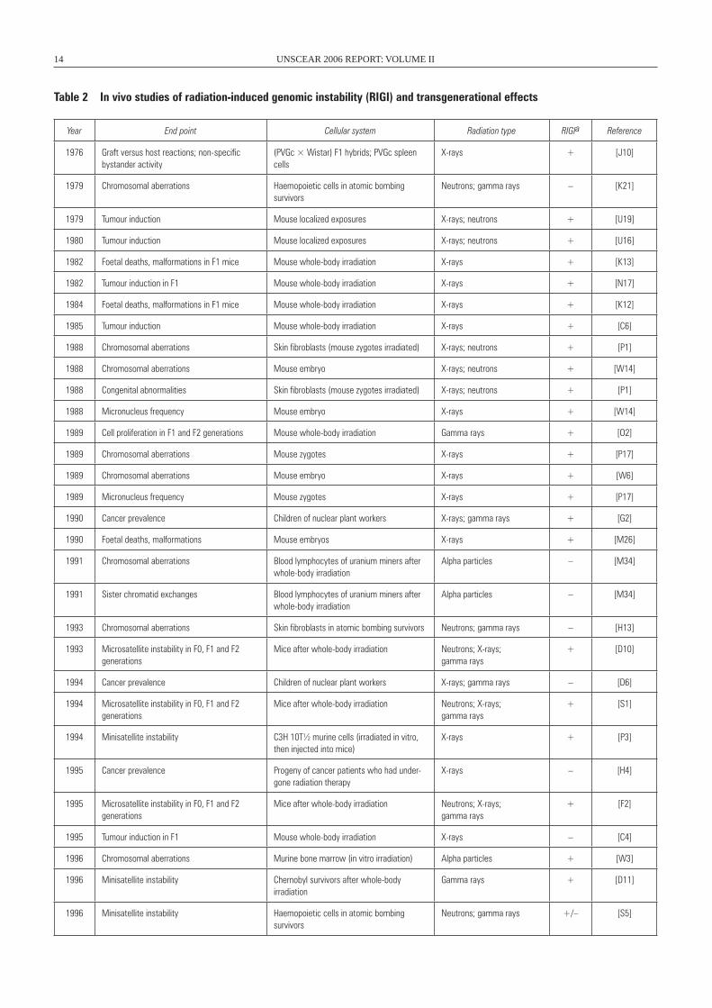
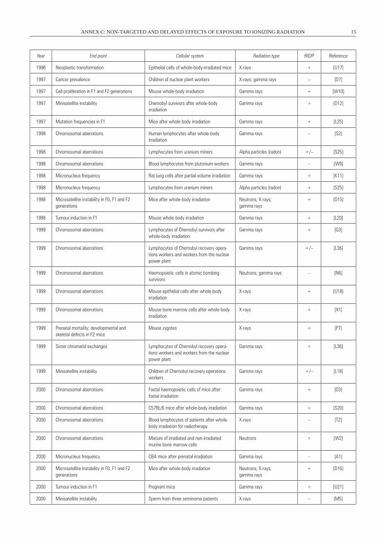
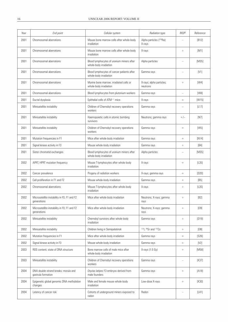
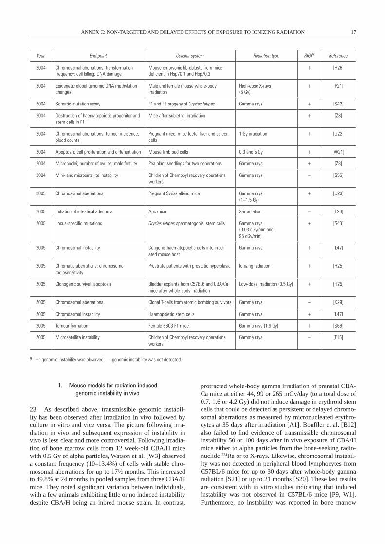
Table 2 In vivo studies of radiation-induced genomic instability (RIGI) and transgenerational effects
Year End point Cellular system Radiation type RIGIa Reference
1976 Graft versus host reactions; non-specific bystander activity
(PVGc × wistar) F1 hybrids; PVGc spleen cells
X-rays + [J10]
1979 Chromosomal aberrations Haemopoietic cells in atomic bombing survivors
Neutrons; gamma rays – [K21]
1979 Tumour induction Mouse localized exposures X-rays; neutrons + [U19]
1980 Tumour induction Mouse localized exposures X-rays; neutrons + [U16]
1982 Foetal deaths, malformations in F1 mice Mouse whole-body irradiation X-rays + [K13]
1982 Tumour induction in F1 Mouse whole-body irradiation X-rays + [N17]
1984 Foetal deaths, malformations in F1 mice Mouse whole-body irradiation X-rays + [K12]
1985 Tumour induction Mouse whole-body irradiation X-rays + [C6]
1988 Chromosomal aberrations Skin fibroblasts (mouse zygotes irradiated) X-rays; neutrons + [P1]
1988 Chromosomal aberrations Mouse embryo X-rays; neutrons + [w14]
1988 Congenital abnormalities Skin fibroblasts (mouse zygotes irradiated) X-rays; neutrons + [P1]
1988 Micronucleus frequency Mouse embryo X-rays + [w14]
1989 Cell proliferation in F1 and F2 generations Mouse whole-body irradiation Gamma rays + [O2]
1989 Chromosomal aberrations Mouse zygotes X-rays + [P17]
1989 Chromosomal aberrations Mouse embryo X-rays + [w6]
1989 Micronucleus frequency Mouse zygotes X-rays + [P17]
1990 Cancer prevalence Children of nuclear plant workers X-rays; gamma rays + [G2]
1990 Foetal deaths, malformations Mouse embryos X-rays + [M26]
1991 Chromosomal aberrations Blood lymphocytes of uranium miners after whole-body irradiation
Alpha particles – [M34]
1991 Sister chromatid exchanges Blood lymphocytes of uranium miners after whole-body irradiation
Alpha particles – [M34]
1993 Chromosomal aberrations Skin fibroblasts in atomic bombing survivors Neutrons; gamma rays – [H13]
1993 Microsatellite instability in F0, F1 and F2 generations
Mice after whole-body irradiation Neutrons; X-rays; gamma rays
+ [D10]
1994 Cancer prevalence Children of nuclear plant workers X-rays; gamma rays – [D6]
1994 Microsatellite instability in F0, F1 and F2 generations
Mice after whole-body irradiation Neutrons; X-rays; gamma rays
+ [S1]
1994 Minisatellite instability C3H 10T½ murine cells (irradiated in vitro, then injected into mice)
X-rays + [P3]
1995 Cancer prevalence Progeny of cancer patients who had under-gone radiation therapy
X-rays – [H4]
1995 Microsatellite instability in F0, F1 and F2 generations
Mice after whole-body irradiation Neutrons; X-rays; gamma rays
+ [F2]
1995 Tumour induction in F1 Mouse whole-body irradiation X-rays – [C4]
1996 Chromosomal aberrations Murine bone marrow (in vitro irradiation) Alpha particles + [w3]
1996 Minisatellite instability Chernobyl survivors after whole-body irradiation
Gamma rays + [D11]
1996 Minisatellite instability Haemopoietic cells in atomic bombing survivors
Neutrons; gamma rays +/– [S5]
ANNEX C: NON-TARGETED AND DELAYED EFFECTS OF EXPOSURE TO IONIZING RADIATION 15
Year End point Cellular system Radiation type RIGIa Reference
1996 Neoplastic transformation Epithelial cells of whole-body-irradiated mice X-rays + [U17]
1997 Cancer prevalence Children of nuclear plant workers X-rays; gamma rays – [D7]
1997 Cell proliferation in F1 and F2 generations Mouse whole-body irradiation Gamma rays + [w10]
1997 Minisatellite instability Chernobyl survivors after whole-body irradiation
Gamma rays + [D12]
1997 Mutation frequencies in F1 Mice after whole-body irradiation Gamma rays + [L25]
1998 Chromosomal aberrations Human lymphocytes after whole-body irradiation
Gamma rays – [S2]
1998 Chromosomal aberrations Lymphocytes from uranium miners Alpha particles (radon) +/– [S25]
1998 Chromosomal aberrations Blood lymphocytes from plutonium workers Gamma rays – [w9]
1998 Micronucleus frequency Rat lung cells after partial-volume irradiation Gamma rays + [K11]
1998 Micronucleus frequency Lymphocytes from uranium miners Alpha particles (radon) + [S25]
1998 Microsatellite instability in F0, F1 and F2 generations
Mice after whole-body irradiation Neutrons; X-rays; gamma rays
+ [D15]
1998 Tumour induction in F1 Mouse whole-body irradiation Gamma rays + [L20]
1999 Chromosomal aberrations Lymphocytes of Chernobyl survivors after whole-body irradiation
Gamma rays + [G3]
1999 Chromosomal aberrations Lymphocytes of Chernobyl recovery opera-tions workers and workers from the nuclear power plant
Gamma rays +/– [L36]
1999 Chromosomal aberrations Haemopoietic cells in atomic bombing survivors
Neutrons; gamma rays – [N6]
1999 Chromosomal aberrations Mouse epithelial cells after whole-body irradiation
X-rays + [U18]
1999 Chromosomal aberrations Mouse bone marrow cells after whole-body irradiation
X-rays + [X1]
1999 Prenatal mortality; developmental and skeletal defects in F2 mice
Mouse zygotes X-rays + [P7]
1999 Sister chromatid exchanges Lymphocytes of Chernobyl recovery opera-tions workers and workers from the nuclear power plant
Gamma rays + [L36]
1999 Minisatellite instability Children of Chernobyl recovery operations workers
Gamma rays +/– [L18]
2000 Chromosomal aberrations Foetal haemopoietic cells of mice after foetal irradiation
Gamma rays + [D3]
2000 Chromosomal aberrations C57BL/6 mice after whole-body irradiation Gamma rays + [S20]
2000 Chromosomal aberrations Blood lymphocytes of patients after whole-body irradiation for radiotherapy
X-rays – [T2]
2000 Chromosomal aberrations Mixture of irradiated and non-irradiated murine bone marrow cells
Neutrons + [w2]
2000 Micronucleus frequency CBA mice after prenatal irradiation Gamma rays – [A1]
2000 Microsatellite instability in F0, F1 and F2 generations
Mice after whole-body irradiation Neutrons; X-rays; gamma rays
+ [D16]
2000 Tumour induction in F1 Pregnant mice Gamma rays + [U21]
2000 Minisatellite instability Sperm from three seminoma patients X-rays – [M5]
16 UNSCEAR 2006 REPORT: VOLUME II
Year End point Cellular system Radiation type RIGIa Reference
2001 Chromosomal aberrations Mouse bone marrow cells after whole-body irradiation
Alpha particles (224Ra); X-rays
– [B12]
2001 Chromosomal aberrations Mouse bone marrow cells after whole-body irradiation
X-rays + [M1]
2001 Chromosomal aberrations Blood lymphocytes of uranium miners after whole-body irradiation
Alpha particles – [M35]
2001 Chromosomal aberrations Blood lymphocytes of cancer patients after whole-body irradiation
Gamma rays – [V1]
2001 Chromosomal aberrations Murine bone marrow; irradiated cells or whole-body irradiation
X-rays; alpha particles; neutrons
+ [w4]
2001 Chromosomal aberrations Blood lymphocytes from plutonium workers Gamma rays – [w8]
2001 Ductal dysplasia Epithelial cells of ATM+/- mice X-rays + [w15]
2001 Minisatellite instability Children of Chernobyl recovery operations workers
Gamma rays – [L17]
2001 Minisatellite instability Haemopoietic cells in atomic bombing survivors
Neutrons; gamma rays +/– [N7]
2001 Minisatellite instability Children of Chernobyl recovery operations workers
Gamma rays + [w5]
2001 Mutation frequencies in F1 Mice after whole-body irradiation Gamma rays + [N14]
2001 Signal kinase activity in F3 Mouse whole-body irradiation Gamma rays + [B4]
2001 Sister chromatid exchanges Blood lymphocytes of uranium miners after whole-body irradiation
Alpha particles – [M35]
2002 APRT, HPRT mutation frequency Mouse T-lymphocytes after whole-body irradiation
X-rays + [L35]
2002 Cancer prevalence Progeny of radiation workers X-rays; gamma rays + [D20]
2002 Cell proliferation in F1 and F2 Mouse whole-body irradiation Gamma rays + [B5]
2002 Chromosomal aberrations Mouse T-lymphocytes after whole-body irradiation
X-rays + [L35]
2002 Microsatellite instability in F0, F1 and F2 generations
Mice after whole-body irradiation Neutrons; X-rays; gamma rays
+ [B2]
2002 Microsatellite instability in F0, F1 and F2 generations
Mice after whole-body irradiation Neutrons; X-rays; gamma rays
+ [D9]
2002 Minisatellite instability Chernobyl survivors after whole-body irradiation
Gamma rays + [D19]
2002 Minisatellite instability Children living in Semipalatinsk 131I, 90Sr and 137Cs + [D8]
2002 Mutation frequencies in F1 Mice after whole-body irradiation Gamma rays + [S26]
2002 Signal kinase activity in F3 Mouse whole-body irradiation Gamma rays + [V2]
2003 ROS content; state of DNA structure Bone marrow cells of male mice after whole-body irradiation
X-rays (1.5 Gy) + [M58]
2003 Minisatellite instability Children of Chernobyl recovery operations workers
Gamma rays – [K37]
2004 DNA double-strand breaks; morula and gastrula formation
Oryzias latipes F3 embryos derived from male founders
Gamma rays + [A18]
2004 Epigenetic global genomic DNA methylation changes
Male and female mouse whole-body irradiation
Low-dose X-rays + [K30]
2004 Latency of cancer risk Cohorts of underground miners exposed to radon
Radon – [L41]
ANNEX C: NON-TARGETED AND DELAYED EFFECTS OF EXPOSURE TO IONIZING RADIATION 17
Year End point Cellular system Radiation type RIGIa Reference
2004 Chromosomal aberrations; transformation frequency; cell killing; DNA damage
Mouse embryonic fibroblasts from mice deficient in Hsp70.1 and Hsp70.3
+ [H26]
2004 Epigenetic global genomic DNA methylation changes
Male and female mouse whole-body irradiation
High-dose X-rays(5 Gy)
+ [P21]
2004 Somatic mutation assay F1 and F2 progeny of Oryzias latipes Gamma rays + [S42]
2004 Destruction of haematopoietic progenitor and stem cells in F1
Mice after sublethal irradiation + [Z8]
2004 Chromosomal aberrations; tumour incidence; blood counts
Pregnant mice; mice foetal liver and spleen cells
1 Gy irradiation + [U22]
2004 Apoptosis; cell proliferation and differentiation Mouse limb bud cells 0.3 and 5 Gy + [w21]
2004 Micronuclei; number of ovules; male fertility Pea plant seedlings for two generations Gamma rays + [Z8]
2004 Mini- and microsatellite instability Children of Chernobyl recovery operations workers
Gamma rays – [S55]
2005 Chromosomal aberrations Pregnant Swiss albino mice Gamma rays(1–1.5 Gy)
+ [U23]
2005 Initiation of intestinal adenoma Apc mice X-irradiation – [E20]
2005 Locus-specific mutations Oryzias latipes spermatogonial stem cells Gamma rays(0.03 cGy/min and 95 cGy/min)
+ [S43]
2005 Chromosomal instability Congenic haematopoietic cells into irradi-ated mouse host
Gamma rays + [L47]
2005 Chromatid aberrations; chromosomal radiosensitivity
Prostrate patients with prostatic hyperplasia Ionizing radiation + [H25]
2005 Clonogenic survival; apoptosis Bladder explants from C57BL6 and CBA/Ca mice after whole-body irradiation
Low-dose irradiation (0.5 Gy) + [H25]
2005 Chromosomal aberrations Clonal T-cells from atomic bombing survivors Gamma rays – [K29]
2005 Chromosomal instability Haemopoietic stem cells Gamma rays + [L47]
2005 Tumour formation Female B6C3 F1 mice Gamma rays (1.9 Gy) + [S66]
2005 Microsatellite instability Children of Chernobyl recovery operations workers
Gamma rays – [F15]
a +: genomic instability was observed; –: genomic instability was not detected.
1. Mouse models for radiation-induced genomic instability in vivo
23. As described above, transmissible genomic instabil-ity has been observed after irradiation in vivo followed by culture in vitro and vice versa. The picture following irra-diation in vivo and subsequent expression of instability in vivo is less clear and more controversial. Following irradia-tion of bone marrow cells from 12 week-old CBA/H mice with 0.5 Gy of alpha particles, Watson et al. [W3] observed a constant frequency (10–13.4%) of cells with stable chro-mosomal aberrations for up to 17½ months. This increased to 49.8% at 24 months in pooled samples from three CBA/H mice. They noted significant variation between individuals, with a few animals exhibiting little or no induced instability despite CBA/H being an inbred mouse strain. In contrast,
protracted whole-body gamma irradiation of prenatal CBA-Ca mice at either 44, 99 or 265 mGy/day (to a total dose of 0.7, 1.6 or 4.2 Gy) did not induce damage in erythroid stem cells that could be detected as persistent or delayed chromo-somal aberrations as measured by micronucleated erythro-cytes at 35 days after irradiation [A1]. Bouffler et al. [B12] also failed to find evidence of transmissible chromosomal instability 50 or 100 days after in vivo exposure of CBA/H mice either to alpha particles from the bone-seeking radio-nuclide 224Ra or to X-rays. Likewise, chromosomal instabil-ity was not detected in peripheral blood lymphocytes from C57BL/6 mice for up to 30 days after whole-body gamma radiation [S21] or up to 21 months [S20]. These last results are consistent with in vitro studies indicating that induced instability was not observed in C57BL/6 mice [P9, W1]. Furthermore, no instability was reported in bone marrow
18 UNSCEAR 2006 REPORT: VOLUME II
cells from Swiss mice up to 100 days after exposure to 3 Gy of X-rays [X1]. These results were initially presumed to indicate that the Swiss mouse strain, like the C57BL/6 mouse strain, is refractory to radiation-induced instability. However, this does not appear to be the case. When Swiss albino mice were exposed to 0.25–1.5 Gy of gamma radia-tion on day 14 or 17 of gestation, significant dose-dependent increases in chromosomal aberrations, micronuclei and/or changes in ploidy were observed in the bone marrow at 12 months of age [D3]. The investigators concluded that radiation-induced genomic instability in the foetal haemo-poietic cells of the mouse persisted post-natally [D3]. These data are summarized in table 2.
24. Although not designed to specifically investigate radiation-induced genomic instability, a number of studies have examined the persistence of cytogenetic rearrangements in animals at delayed times after irradiation. Hande et al. [H18, H19, H20, H21] used female Swiss mice to study the induction and persistence of dicentrics and translocations in splenocytes up to 112 days after exposure to 2 Gy of whole-body X irradiation. The frequencies of dicentrics decreased exponentially with time, while the frequencies of transloca-tions were constant in the period 0–7 days and then decreased linearly or exponentially. No new chromosomal rearrange-ments were observed, suggesting that there was no delayed cytogenetic instability in these animals. Similar studies using other mouse models have reported similar results [T5].
25. In attempting to reconcile the apparently conflicting results described above, Bouffler et al. [B12] have noted the sensitivity of mouse bone marrow cells to perturbations through transplantation and culture, and emphasized the need for sound control experiments to be performed con-currently. For instance, some of the radiation-induced trans-missible chromosomal instability reported by Watson et al. [W3] could be attributed to the low background frequency of aberrations observed in the control repopulating cells. This is in contrast to the higher background described by Bouffler et al. [B12]. It is also possible that the disparate literature on radiation-induced genomic instability in vivo reflects the inherent variability between the inbred mouse strains used and differences due to sex within the animal strains used.
26. To investigate the in vivo non-targeted effects of low-LET radiation, Lorimore et al. [L47] used the same congenic sex-mismatch bone marrow transplantation protocol as used by Watson et al. [W2] to repopulate the haemopoietic system from a mixture of gamma-irradiated and non-irradiated hae-mopoietic stem cells such that host-, irradiated donor- and non-irradiated donor-derived cells could be distinguished. Chromosomal instability in the progeny of irradiated haemo-poietic stem cells accompanied by a reduction in their contri-bution to the repopulated haemopoietic system was observed and is consistent with a delayed genomic instability pheno-type being expressed in vivo. However, chromosomal insta-bility was also shown in the progeny of the non- irradiated haemopoietic stem cells, implicating a bystander-like mechanism. Studies of the influence of irradiated recipient
stromal microenvironment and experiments replacing irradi-ated cells with irradiated cell-conditioned medium revealed the source of the in vivo bystander effect to be the descend-ants of irradiated cells rather than the irradiated cells them-selves. Lorimore et al. [L47] speculated that it is possible that a radiation-induced genomic instability phenotype in vivo need not necessarily be a reflection of intrinsically unstable cells but the response to ongoing production of inflammatory-type damaging signals [L22] as a long-term unexpected consequence of the initial radiation.
27. While the literature is replete with apparently contradic-tory reports of radiation-induced instability in mouse model systems, these results clearly indicate that genetic factors can play a major role in the instability phenotype and that analy-sis of radiation-induced genomic instability in vivo is signifi-cantly more complicated than in vitro. Critical analysis of radiation-induced genomic instability in vivo is not a trivial undertaking. In any animal model there is likely to be some inherent genomic instability that complicates the selection of appropriate control populations. Such experiments generally involve inbred strains of mice, and even in radiation- sensitive populations only a small percentage, generally <50%, will exhibit an instability phenotype. Further more, extrapolating such results to other mouse strains or outbred populations is difficult at best. Until a careful study involving sound and relevant controls as well as statistically relevant numbers of animals exposed to a homogeneous quality of radiation is carried out, the induction of radiation-induced genomic instability in vivo will remain controversial.
2. Human studies
28. Radiation therapy has improved over recent years, and many of the cancer patients treated with radiation are surviv-ing longer than did those in the past. Second cancers occur-ring in the irradiated field have been reported in some of these patients, suggesting a direct role of the radiation exposure [B37, B38]. Data on second cancers occurring in children irradiated for cancer indicate that some genetic predisposi-tion to cancer may also predispose them to radiotherapy- related second cancers [D22, E19, F14]. Nevertheless, it is still difficult to identify the radiation-induced lesions initiating the second malignancy. At the time of diagnosis, multiple genomic alterations are present in the tumours, and the majority are likely to represent secondary events occur-ring during tumour evolution and subsequent selection. This underscores that caution must be applied to analysis of radiation-induced genomic instability and its role in human carcinogenesis. The subsequent discussion in this section highlights the controversies and contradictions inherent in the human studies. To this end it is reasonable to expect that analysis of normal, healthy populations of individuals would not provide evidence of instability regardless of the individu-als’ radiation history. Indeed, the majority of studies inves-tigating instability in radiation-exposed populations have analysed samples from normal, healthy individuals and did not find evidence of instability [T2, T4]. It is also reasonable
ANNEX C: NON-TARGETED AND DELAYED EFFECTS OF EXPOSURE TO IONIZING RADIATION 19
to expect that analysis of instability in individuals manifest-ing phenotypic effects of radiation exposure, e.g. cancer or leukaemia, might well show evidence of induced instabil-ity. Once again, limited studies indicate that this is the case [N6, N7]. Whether or not the observed instability is a direct or non-targeted effect of radiation exposure, or a secondary selective effect of disease evolution, cannot be definitively determined at present. Furthermore, this question is unlikely to be resolved in the foreseeable future. This caveat should be kept in mind in the following discussion.
29. As has been described utilizing the mouse as a model system, both induction and lack of induction of transmissi-ble radiation-induced genomic instability have been reported in humans, and once again genetic factors appear to play a role in the observed instability [K4]. Induced chromo-somal instability has been described in long-term cultures of human lymphocytes following irradiation and culture in vitro [H9]. Using the same lymphocyte culture protocol, chromosomal instability was reported in blood samples from individuals exposed during the radiation accident in Estonia in 1994 [S2]. Radiation exposure was variable, pro-tracted and not precisely determined. Furthermore, blood samples were taken well after radiation exposure. No dose response was apparent, and contrary to previous studies from the Lambert laboratory, chromosomal instability was also observed in long-term cultures from non-exposed con-trols [S2]. In contrast, cytogenetic analysis of 18 individu-als who had received between 35 and 80 Gy of fractionated radiation therapy for different cancers showed no increase in aberrant cell types as a function of time after complet-ing therapy. Thus no cytogenetic evidence that fractionated radiotherapy induced a persistent or late-manifesting state of genomic instability was found [T2]. It should be stressed that the majority of patients treated for different malignan-cies received localized, partial-body irradiation with empha-sis on minimizing damage to normal tissue. Consequently, different proportions of bone marrow stem cell populations and peripheral blood lymphocytes would have been exposed to the radiation. It is likely that more cells than the number actually analysed (<200 per patient), would have to be inter-rogated before evidence of persistent transmissible chromo-somal instability would be observed in these individuals, if it indeed existed [T2].
30. The availability of cultured lymphocyte preparations from radiation workers with internal deposits of plutonium has provided the opportunity to examine whether protracted irradiation of bone marrow cells had induced a transmissi-ble genomic instability in descendant cells in the peripheral blood [W8]. Bone marrow dose calculations provided indi-vidual cumulative estimates at the time of sampling ranging up to 1.8 Sv. Chromosome analysis revealed no significant differences, either in comparisons between the total group of plutonium workers and controls for comparable periods or when the comparisons were restricted to a group of plu-tonium workers with initial bone marrow plutonium doses of greater than 0.25 Sv. There was therefore no evidence from this study for the induction of persistent transmissible
genomic instability in the bone marrow of radiation workers with internal deposits of plutonium [W8]. Likewise, clonally expanded T-cell lymphocyte populations did not demonstrate increased chromosomal instability using either G-band anal-ysis or multicolour fluorescence in situ hybridization [K29].
31. The long-term effect of radiation exposure on uranium miners employed by the Wismut uranium mining company in the former German Democratic Republic was investigated by scoring the frequency and percentage of micronuclei with and without a centromere. Kryscio et al. [K38] reported that genomic instability had occurred in the lymphocytes of miners, especially those with cancer.
32. A number of investigators have studied the alpha radia-tion risks in patients who received injections of Thorotrast, an X-ray contrast medium used in Europe, Japan and the United States from the late 1920s to 1955. Thorotrast was composed of thorium dioxide and contained 232Th, a natu-rally occurring radionuclide. Because the physical half-life of 232Th is 14 billion years and Thorotrast is not appreci-ably eliminated from the body, the tissues in which it was deposited are irradiated by alpha rays for the entire lifetime of the subject. The major causes of death among the Thoro-trast patients are liver cancer, liver cirrhosis, leukaemia and other cancers. Mutation analyses of the TP53 gene and loss of heterozygosity (LOH) studies at the 17p locus were per-formed by Ishikawa et al. [I2] to characterize the genetic changes in Thorotrast-induced liver tumours. LOH was not frequent; most mutations were transitions, suggesting that genetic changes in Thorotrast-induced cancers were mainly delayed mutations and not the result of the direct effects of radiation.
33. Likewise Iwamoto et al. [I3] analysed mutations in TP53 from 20 Thorotrast recipients who developed can-cer, mostly of hepatic bile duct and blood vessel origin. Of the 20 cases, 19 had TP53 point mutations. Moreover, the accompanying non-tumour tissues from these patients also had TP53 mutations, albeit at lower frequency. The distri-bution pattern of the point mutations was significantly dif-ferent between the non-tumour and tumour tissues, with most mutations in malignant tissues located in the highly conserved domains of the TP53 gene. These results support the idea that TP53 mutations are important in the genesis of Thorotrast-induced tumours but that these point mutations are a secondary outcome of genomic instability induced by the irradiation. A similar result was reported by Kamikawa et al. [K24], who investigated mutations of the RAS and the TP53 genes in archival sections of liver cancers induced by Thorotrast. These investigators were unable to rule out the possibility that genetic insults occurred indirectly in the pro-liferating cells adjacent to the necrosis rather than being a direct effect of alpha particles.
34. Wada et al. [W17] also investigated genetic changes in the TP53 gene in 19 autopsy cases of liver malignan-cies. LOH at the 17p13 locus and mutations in TP53 were analysed. A number of cases were informative: four cases
20 UNSCEAR 2006 REPORT: VOLUME II
showed LOH and eight contained mutations. The direct action of alpha particles was thought to result in relatively large deletions, such as those detected by LOH. Therefore the low frequency of such changes (27%) compared with point mutations (47%) suggests that the genetic changes in the TP53 gene in the liver tumours related to Thorotrast were not caused mainly by direct actions of alpha particles but rather by indirect effects that may have been due to cycles of necrosis and regeneration. This study was recently expanded to compare Thorotrast-induced liver cancers to those not associated with Thorotrast exposure. LOH at 37 loci was investigated. Liu et al. [L46] found frequent LOH at micro-satellite markers D4S1538, D16S2624 and D17S1303 to be common to all the subtypes of liver cancer, independent of the specific carcinogenic agent. In contrast, LOH at marker D4S1652 was generally not observed in Thorotrast-induced cancers. LOH analysis revealed that Thorotrast-induced can-cers share some LOH features with cancers not induced by Thorotrast, and Liu and colleagues concluded that induced LOH is not simply due to direct insult to DNA by alpha par-ticles, but can occur through complex mechanisms, includ-ing bystander effects [L46]. Such a conclusion is reasonable given the analysis of Goto et al. [G18], who used imaging plate autoradiography to examine the microdistribution of alpha particles in pathological sections of tissues from Thorotrast patients. They found that the amount of thorium deposited in tumour tissue was correlated with that in non-tumour tissue, and that Thorotrast deposition was not asso-ciated with DNA damage determined by histochemistry. Goto et al. [G18] concluded that radioactive thorium always migrates in macrophages within the deposited organs, and that the organs are evenly exposed to alpha particles.
35. In an evaluation of Thorotrast-induced genomic insta-bility, Liu et al. [L45] analysed microsatellite instability in Thorotrast-induced liver cancers. The frequency of micro-satellite instability cases was 62.5% in Thorotrast-induced cancers, whereas it was 22.7% in non-Thorotrast induced cancers. Liu and colleagues suggested that microsatellite instability induced by exposure to Thorotrast mainly reflects clonal expansion of cancer cells and is partly due to inactiva-tion of the DNA mismatch repair gene hMLH1 by hyper-methylation. A recent finding also suggests that methylation changes in DNA can be associated with radiation-induced genomic instability [K35].
36. Littlefield et al. [L16] examined the cumulative genetic damage in a 72-year-old man who was treated with a 32 mL bolus of Thorotrast during cerebral angiography performed more than 40 years earlier. Peripheral T-lymphocytes were cultured to quantify the frequencies and cellular distribu-tions of asymmetrical and symmetrical types of chromo-somal aberrations. Assays of glycophorin A (GPA) muta-tions in red blood cells were also performed. Their results revealed that approximately 30% of the lymphocytes in this patient contained one or more chromosomal aberrations, the majority of which were of the “stable” type. About one third of the lymphocytes with chromosome damage carried multiple aberrations, suggesting that significant numbers of
stem cells survived exposures to alpha particle radiation that induced complex genomic alterations. Increased frequen-cies of GPA mutations were observed, demonstrating that genomic damage was also induced in erythroid progenitors. Despite the relatively severe burden of somatic cell damage induced by 40 years of internal alpha particle irradiation, the patient remained free of any serious illness. Furthermore, these results provided no in vivo evidence for the contin-ued expression of genomic instability. A similar observation was reported by Hande et al. [H3], using a fluorescence in situ hybridization technique that made possible the detec-tion of intrachromosomal rearrangements and deletions. They described the quantification of stable intrachromo-somal aberrations in lymphocytes of healthy former nuclear weapons workers who were exposed to plutonium. Even many years after occupational exposure, more than half the blood cells of the healthy plutonium workers contained large (>6 Mb) intrachromosomal rearrangements. The yield of these aberrations was highly correlated with plutonium dose to the bone marrow. It is significant that, despite the relatively high frequency of intrachromosomal aberrations, there was no evidence of transmissible chromosomal insta-bility and no obvious detrimental health consequences in the populations sampled.
37. Nevertheless, a role for radiation-induced genomic instability has been described for solid tumours develop-ing after radiotherapy for bilateral retinoblastoma [L1]. Genome alterations of second tumours (five osteosarcomas, one malignant peripheral sheath nerve tumour, one leio-myosarcoma) occurring in the field of irradiation of seven patients treated for bilateral retinoblastoma were studied. Because of a germ line mutation in the retinoblastoma gene (RB1), these patients were predisposed to develop radiation-induced tumours. In all radiation-induced tumours ana-lysed, the normal RB1 allele was lost, whereas the germ line mutated allele was retained and the two TP53 alleles were inactivated. A comparison of these tumours with the non-radiation-induced tumours led Lefevre et al. [L1] to conclude that this loss was due to the radiation-induced chromosomal instability rather than a direct effect of ionizing radiation. A similar observation was reported by Ryabchenko and col-leagues [R12] after analysis of chromosomal aberrations in peripheral lymphocytes taken from Hodgkin’s disease patients after prolonged (up to 31 years) remission periods. The mean frequency and patterns of aberrations in remission patients were significantly different from comparison groups (healthy donors and primary Hodgkin’s disease patients). New cancer cases were diagnosed in a number of the remis-sion patients, leading the investigators to suggest that the tumorigenic potential of radiochemotherapy is mediated via induction of genomic instability in exposed cells. Long after the therapy, the instability may become an initiating event in the development of new malignancies in affected tissues, whereas the instability induced in haemopoietic stem cells may reveal itself in peripheral lymphocytes derived from previously exposed precursor cells. The caveat, of course, is that these individuals were cancer patients and may have been inherently predisposed to second cancers.
ANNEX C: NON-TARGETED AND DELAYED EFFECTS OF EXPOSURE TO IONIZING RADIATION 21
E. Genomic instability and radiation-induced leukaemia
1. Mouse models
38. Plumb et al. [P8] have reviewed the relationship between radiation-induced genomic instability and radiation-induced leukaemia. They presented evidence that genomic instabil-ity plays a role during radiation leukaemogenesis. How-ever, with the exception of a high incidence of non-clonal chromatid-type cytogenetic aberrations in neutron-induced acute myeloid leukaemia in mice [B26], the genetic lesions described (including non-clonal chromosomal aberrations, LOH and minisatellite/microsatellite mutations) were sim-ilar to those detected in de novo leukaemias and cancers. This damage was not transmissible and the authors inter-preted these observations as evidence of apoptosis or other cell death. Nevertheless, this radiation-induced damage in vivo was indistinguishable from de novo multistage leu-kaemogenesis. Thus it is not yet possible to define a type of genomic instability in radiation-induced leukaemias in mice that demonstrates a specific characteristic of immediate or delayed effects of the initiating exposure to ionizing radiation. Evidence for radiation-induced genomic instability in mouse leukaemia and haemopoietic stem cells led MacDonald et al. [M1] to conclude that the induced instability contributed sig-nificantly to the induced leukaemia. A similar conclusion was reached by Ban et al. [B1], who suggested that loss of TP53 function triggers the tumorigenic process leading to stem cell leukaemia through the induction of chromosomal instability. These authors also pointed out that the aetiology of stem cell leukaemia is likely to differ from that of myeloid leukaemia, because different results were observed in acute myeloid leukaemia (AML) [B1].
39. Interestingly, however, susceptibility to radiation-induced leukaemia is genetically separable from sensitivity to radiation-induced genomic instability [B13]. A series of matings, backcrosses and intercrosses between CBA/H mice susceptible to radiation-induced acute myeloid leukaemia and radiation-resistant C57BL/6 mice was carried out, and acute myeloid leukaemia and thymic lymphoma susceptibil-ity was analysed. No simple genetic relationship between susceptibility to radiation-induced leukaemia and the sensi-tivity of the haemopoietic stem cells to induced instability was found.
2. Human studies
40. Cytogenetic analysis of leukaemia patients among the survivors of the atomic bombings in Japan revealed that patients exposed to >2 Gy exhibited a higher incidence of chromosomal aberrations and more complex chromosomal rearrangements than did patients exposed to lower radiation doses or unexposed patients [T1]. A more recent cytogenetic analysis of the heavily exposed patients with acute myelo-cytic leukaemia or myelodysplastic syndrome in the cohort of bombing survivors indicated persistent chromosomal insta-bility [N6]. These cytogenetic observations are supported
by studies demonstrating high frequencies of micro satellite instability in those bombing survivors with acute myelo-cytic leukaemia and a history of high exposure [N7]. These investigators concluded that this persistent instability might strongly influence the development of leukaemia in humans exposed to ionizing radiation. This study stimulated two letters to the editor of the journal in which it was published. The first, by Little [L40], claimed that although there was evidence that the microsatellite instability rate was higher in the AML cases among the bombing survivors than in the control group, the evidence that this higher rate was related to the radiation dose these cases received was weak. Little went on to show that the number of loci for which microsatellite mutation data were not detectable was higher in the bombing survivor cases than in the control group. The second letter, by Cox and Edwards [C15], raised the issue of the statistical strength of the dose-related association between expression of genomic instability in AML and the probability of causa-tion by radiation. In response to these letters, Plumb [P18] pointed out that, from a biological perspective, the striking similarities in the leukaemias that arose in the bombing sur-vivors and in therapy patients indicated that a significant proportion of the AML cases among the bombing survivors described by Nakanishi et al. [N6, N7] could indeed have been induced by the radiation. Reconciling mathematical models [L40] and statistical analysis [C15] with the biologi-cal observations will always be difficult, however, particu-larly with the small number of AMLs observed in this unique population.
41. Mazurik and colleagues [M63] investigated molec-ular, biochemical and cytogenetic parameters in blood samples from 17 radiation accident victims who between 1.7 and 43.8 years previously had suffered acute radiation sickness ranging in severity from grade I to grade IV. All patients showed ~25–30% reduction in oxidative status and increased levels of both stable and unstable chromosomal aberrations that correlated with the severity of the acute radiation sickness. These data were interpreted as evidence for delayed genomic instability in these radiation exposed individuals.
F. Role of telomeres and telomerase in radiation-induced genomic instability
42. Telomeres are specialized DNA–protein complexes at the ends of linear chromosomes. They are composed of a repetitive DNA sequence and associated proteins. Telomere alterations, caused by replication-mediated shortening, direct damage or defective telomere-associated proteins, can generate chromosomal instability, which can be observed in senescence and during the immortalization process (reviewed in reference [M47]). Telomeres are essential for proper maintenance of chromosomes and may play a role in ageing and cancer [G13, G14]. Telomere length can be main-tained by telomerase [B25], or by the alternative mechanism of telomere lengthening which is telomerase-independent [M7, S50]. Telomere length abnormalities observed in
22 UNSCEAR 2006 REPORT: VOLUME II
radiosensitive cells suggest that, in some human cells, short telomeres might correlate with radiation sensitivity [M49]. This may or may not be the case in mouse cells, as both hypersensitivity [G19] and the absence of an effect have been described [M49]. To complicate the matter further, long but dysfunctional telomeres have been found to corre-late with chromosomal radiosensitivity in a mouse AML cell line 7926 [F13].
43. Another role for telomerase appears to be the de novo formation of telomeres, or chromosome healing to stabilize broken chromosomes [M41, S35]. Telomere loss results in sister chromatid fusion and prolonged breakage–fusion–bridge cycles leading to extensive DNA amplification and large deletions (reviewed in reference [M37]). Significantly, the loss of a single telomere can result in the instability of multiple chromosomes [S46] and generate many of the types of cytogenetic rearrangements commonly associated with human cancer. Telomere dysfunction can also trigger exten-sive DNA fragmentation and the evolution of complex chro-mosomal abnormalities in tumours in mice [A10] and humans [G11, M37]. This also appears to involve repeated cycles of dicentric chromosome formation, anaphase bridging, subse-quent breakage and refusion events [G12]. In addition, telo-mere dysfunction can result in increased mutation rates and genomic instability (reviewed in references [F9, M37]).
44. It is noteworthy that exposure to ionizing radiation can induce telomerase activity both in vitro [H23, N27] and in vivo [H22]. This appears to be TP53-dependent [N26] and has been suggested as a measure for monitoring the radio-curability of tumour cells [S49]. Radiation-induced telom-erase activation depends on dose rate, is not related to cell cycle redistribution or to the induction of cell death, and is likely to be the consequence of specific regulatory responses to ionizing radiation [P20].
45. Telomere repeat-like sequences are also seen as dis-crete bands at distinct intrachromosomal sites in a number of vertebrate species [M42]. These interstitial telomere sites probably represent ancestral telomere fusion events or ampli-fication of the repeat sequences in ancestral karyotypes as latent telomeres [L39, M42]. There is compelling evidence that some of these interstitial telomere sites may be hot spots for both spontaneous [A11, H15] and radiation-induced chromosome damage [A9, B27, S36, S57, S58]. While it is clear that interstitial telomere sequences can influence the radiation sensitivity of chromosomes, many of these studies were performed in hamster cells where the interstitial telo-meres are located at or near pericentromeric heterochroma-tin. Analyses of radiation sensitivity in a naturally occurring short interstitial telomere in a human chromosome (2q31, [A16]) and of a transfected 800bp telomeric repeat in human chromosome 4q indicate that human interstitial telomere sequences might not be prone to spontaneous [D24] or radiation-induced [D25] breakage.
46. The involvement of telomeric repeats in radiation-induced chromosomal instability was first described by
Sabatier et al. [S44] in high-LET-irradiated human fibrob-lasts. They demonstrated that instability acquired by human chromosomes recurrently involved telomeric associations. This was not due to drastic telomere shortening [S45]. A role for recombination involving interstitial telomere-like repeat sequences in inducing chromosomal instability was later demonstrated by Day et al. [D1] in an in vitro Chinese human–hamster hybrid model system. In this instance it was apparent that rearrangements involving unstable chro-mosomes occurred preferentially at the sites of interstitial telomere sequences rather than at the true terminal telo-meres as observed by Sabatier [S45]. This difference is likely to be due to the telomeres on transformed hamster chromosomes being very small relative to human telo meres [S56]. Ojima et al. [O6] have reported that telomeres are destabilized several generations after X-irradiation in nor-mal human fibroblasts. Their data suggest X-irradiation might not affect telomeres directly, but rather by inducing a delayed instability.
47. Initially it was thought that an interstitial telomere-like repeat sequence on chromosome 2 played an important role in the deletions in somatic haemopoietic cells that charac-terized the earliest phases of radiation-induced AML in the CBA/H mouse [B28]. Subsequent detailed molecular analy-sis of break points suggested that, while telomere sequences were located close to regions frequently involved, break points were not exactly coincident with telomere sequences [F10]. The regions of frequent breakage appeared to have properties expected of matrix/scaffold attachment regions [F10]. At this stage one can only speculate on whether telo-meric changes are a cause, an effect or a combination of both in contributing to genomic instability independent of radia-tion exposure.
G. Conclusions
48. Radiation-induced genomic instability is now well established in a number of normal and transformed cell lines in vitro. Instability can manifest as multiple dif-ferent end points and can result from both targeted and non-targeted events. The results of studies of induced instability in vivo are more complex and contradictory. In mice, observation of the instability phenotype appears to be dependent upon the mouse strain used and the power of the investigation in terms of the numbers of animals investigated. The evidence for induced instability in exposed human populations is controversial, with both positive and negative effects being reported. It is possi-ble that radiation-induced genomic instability provides the impetus for those genomic alterations associated with radiation carcinogenesis. This view should be tempered by the high frequency with which instability is observed both in vitro and in vivo, and the general lack of a dose–response curve. Overall, a specific role for induced insta-bility in the genesis of radiation-induced cancers has yet to be definitively demonstrated.
23
II. BYSTANDER EFFECTS AND RADIATION EXPOSURE
49. There has been a resurgence of interest in radiation-induced bystander effects, largely because of the develop-ment of single-cell charged-particle irradiators. The term “bystander effect” was adopted from the gene therapy lit-erature, where it usually refers to the killing of several sub-populations of tumour cells by targeting only one “type” of cell within a heterogeneous population [F6]. For the pur-poses of this annex, the definition proposed by Djordjevic will be used [D5]. That is, “bystander effect” describes the ability of cells affected by an agent to convey manifestations of damage to other cells not directly targeted by the agent or not necessarily susceptible to it per se. Thus radiation-induced bystander effects are effects manifesting in cells that were non-irradiated neighbours of irradiated cells or that received factors secreted or shed by irradiated cells. It is implicit in this review that in vitro bystander effects are the result of a signal generated by an irradiated cell interacting with a non-irradiated cell [C22] and are not the result of radi-ation-induced changes in the culture medium [Z2], or due to experimental variables such as the cell culture environment
[M50]. This underscores the critical role of adequate and appropriate controls. A historical perspective describing key events in the study of radiation-induced bystander effects has been presented by Mothersill and Seymour [M45].
A. Bystander effects in vitro
50. Radiation-induced bystander effects in vitro embrace a number of different non-targeted experimental effects, some of which are likely to be detrimental to the cell, whereas others are not. Different effects are observed in differ-ent cell types, and depend on the cell type producing the bystander signal after irradiation and the cell type receiving the bystander signal (summarized in table 3). Consequently, no rigid rules can be applied to the multitude of responses occurring in cells not targeted by radiation. For convenience, bystander effects have been divided into four, not necessarily mutually exclusive, subcategories.
Table 3 In vitro studies of the bystander effect
Year Origin of bystander effect End point Cell type Radiation type Reference
1992 Low-fluence alpha particle irradiation
Sister chromatid exchanges Human fibroblasts and epithelial cells
Alpha particles [N4]
1996 Low-fluence alpha particle irradiation
Sister chromatid exchanges Human lung fibroblasts Alpha particles [D2]
1997 Medium transfer Sister chromatid exchanges Human lung fibroblasts Alpha particles [L2]
1997 Medium transfer Clonogenic survival Human epithelial cells; human fibroblasts
Gamma rays [M18]
1997 Medium transfer Clonogenic survival Human keratinocytes Gamma rays [S12]
1997 Microbeam irradiation Si- mutants induced AL human–hamster hybrid cells Alpha particles [H14]
1998 Low-fluence alpha particle irradiation
p53, p21, MDM2, CDC2, RAD5 1 protein levels
Human fibroblasts Alpha particles [A3]
1998 Low-fluence alpha particle irradiation
Plating efficiency; chromosomal aberrations
Murine bone marrow cells Alpha particles [L24]
1998 Medium transfer Clonogenic survival Human keratinocytes Gamma rays [M22]
1998 Microbeam irradiation Micronucleus frequency; apoptosis Human fibroblasts Alpha particles [P10]
1999 Cytoplasmic irradiation by microbeam
Clonogenic survival; CD59 mutation frequency
AL human–hamster hybrid cells Alpha particles [w13]
1999 Low-fluence alpha particle irradiation
HPRT mutations Chinese hamster ovary cells Alpha particles [N5]
24 UNSCEAR 2006 REPORT: VOLUME II
Year Origin of bystander effect End point Cell type Radiation type Reference
2000 Cytoplasmic irradiation by microbeam
Clonogenic survival; CD59 mutation frequency
AL human–hamster hybrid cells Alpha particles [Z1]
2000 Low-fluence alpha particle irradiation
Gl checkpoint Human fibroblasts Alpha particles [A8]
2000 Medium transfer Clonogenic survival; AP-endonuclease; TP53; ROS
Human lung fibroblasts (HFL-l) Alpha particles [I10]
2000 Medium transfer Clonogenic survival; intracellular cal-cium levels; mitochondrial membrane potential; ROS levels
Human keratinocytes Gamma rays [L28]
2000 Medium transfer Clonogenic survival Human keratinocytes Gamma rays [S13]
2001 Co-culture Clonogenic survival V79 Chinese hamster cells (3-D tissue culture model)
3H beta particles [B24]
2001 Co-culture p53, HSP72 protein levels A-172 human glioblastoma cells X-rays [M3]
2001 Co-culture Enhanced plating efficiency; micronucleus frequency
Human salivary gland cells X-rays or carbon beam
[S30]
2001 Low-fluence alpha particle irradiation
Changes in gene expression; induction of DNA damage
Human fibroblasts and epithelial cells
Alpha particles [A4]
2001 Medium transfer Clonogenic survival; apoptosis; trans-formation frequency
CGLI human HeLa × skin fibroblast hybrid cells
X-rays [L5]
2001 Medium transfer Clonogenic survival; intracellular cal-cium levels; mitochondrial membrane potential; ROS levels
Human keratinocytes Gamma rays [L38]
2001 Medium transfer Clonogenic survival Human urothelium cells Gamma rays [M39]
2001 Microbeam irradiation Micronucleus frequency Primary human fibroblasts Alpha particles [B9]
2001 Microbeam irradiation Transformation frequency C3H 10T½ murine fibroblasts Alpha particles [S6]
2001 Microbeam irradiation Clonogenic survival C3H l0T½ murine fibroblasts Alpha particles [A7]
2002 Co-culture Enhanced plating efficiency and proliferation
Human salivary gland cells Carbon beam [S15]
2002 Co-culture on double Mylar dishes Clonogenic survival; CD59 mutation frequency
AL human–hamster hybrid cells Alpha particles [Z4]
2002 Low-fluence alpha particle irradiation
Micronucleus frequency Human fibroblasts Alpha particles [L43]
2002 Low-fluence alpha particle irradiation
HPRT mutations; sister chromatid exchanges
Human fibroblasts and epithelial cells
Alpha particles [L37]
2002 Low-fluence alpha particle irradiation
Chromosomal aberrations Chinese hamster ovary cells Alpha particles [N3]
2002 Low-fluence alpha particle irradiation
HPRT mutations; sister chromatid exchanges
Chinese hamster ovary cells Alpha particles [N21]
2002 Medium transfer Clonogenic survival; AP-endonuclease; TP53; ROS
Human lung fibroblasts (HFL-l) Alpha particles [I1, I5]
2002 Medium transfer Clonogenic survival; intracellular cal-cium levels; mitochondrial membrane potential; ROS levels
Human keratinocytes Gamma rays [L26, L27]
2002 Medium transfer Clonogenic survival Thirteen cell lines: human epithelial carcinoma cells, Sw48 human colon carcinoma cells
Gamma rays [M21, M38]
2002 Microbeam irradiation Micronucleus frequency; apoptosis Sections of human and porcine ureter
Alpha particles [B7]
ANNEX C: NON-TARGETED AND DELAYED EFFECTS OF EXPOSURE TO IONIZING RADIATION 25
Year Origin of bystander effect End point Cell type Radiation type Reference
2002 Microbeam irradiation Clonogenic survival V79 Chinese hamster cells Alpha particles [S27]
2003 Co-culture Micronucleus frequency Human fibroblasts Carbon-ion beam: beta particles
[S28]
2003 Co-culture Enhanced plating efficiency and proliferation
Human salivary gland cells Carbon beams [S29]
2003 Low-fluence alpha particle irradiation
HPRT mutations Chinese hamster ovary cells Alpha particles [N20]
2003 Medium transfer Clonogenic survival Human keratinocytes Gamma rays [M27]
2003 Microbeam irradiation Micronucleus frequency; apoptosis Sections of human and porcine ureter
Alpha particles [B8]
2003 Medium transfer Apoptosis; micronuclei Human–hamster hybrid cells (GM10115; Fe10-3; LS12)
X-rays [N1, N2]
2003 Carbon ion beam Micronuclei; gap junctions; ROS Human fibroblasts Carbon ion beam [S28]
2003 Helium ion microbeam Micronuclei; nitric oxide T98G cell nuclei from human glioblastoma
Helium ion beam [S16]
2003 Microbeam irradiation AL cell mutagenic assay AL cells Charged particle microbeam; alpha particles; X-rays
[Z6]
2004 Medium transfer RPA expression Primary human fibroblasts Gamma irradiation [B42]
2004 Medium transfer Micronucleus frequency Chinese hamster ovary cells Ultrasoft X-ray microprobe
[K31]
2004 Medium transfer Transformation frequency HeLa × skin fibroblast hybrid cells Low-dose X-rays [K32]
2004 Co-culture Cell proliferation using 3H-TdR incorporation
Antigen presenting cells (J774A.1) and T-lymphocytes(EL-4)
Low-dose irradiation [L44]
2004 Chronic and acute irradiation iNOS accumulation wTP53 cells Chronic gamma rays; acute X-rays
[M59]
2004 Co-culture and medium transfer Oncogenic transformation frequency C3H 10T½ cells High- and low-dose X-rays
[M51]
2004 Cell–cell contact during irradiation Oncogenic transformation frequency C3H 10T½ cells X-rays [M43]
2004 Co-culture and medium transfer Cloning efficiency; cell numbers Repair-deficient human cell lines Gamma rays [M44]
2004 Co-culture Chromosomal instability Human fibroblast BJ1-htert Alpha irradiation [P22]
2004 Co-culture Apoptosis; necrosis LY (L5178Y) suspension cells; human salivary gland cells
Carbon ions; X-rays [S31]
2004 Co-culture Micronuclei; nitric oxide Glioma cells; primary human fibroblasts
He ion particles [S32]
2004 Co-culture AL cell mutagenic assay AL cells Low-dose X-rays [Z10]
2004 Microbeam irradiation Micronuclei and cell cycle delays Human fibroblasts 90 keV/µm alpha particles
[P6]
2005 Co-culture Transposition of chromosomal loci Human lymphocytes X-rays (10 cGy) [T4]
2005 Medium transfer Micronucleus frequency; apoptosis X-rays [K33]
2005 Medium transfer Mutation and deletion in mitochondrial DNA
Human keratinocytes (HPV-G) Gamma irradiation [M60]
2005 Medium transfer Micronucleus frequency; HPRT mutation frequency
GM10115 human–hamster hybrid cells
X-rays [N30]
2005 Co-culture Sister chromatid exchanges and chromosomal aberrations
Hamster cell lines (V3 and irs3) Low-fluence alpha particles
[N25]
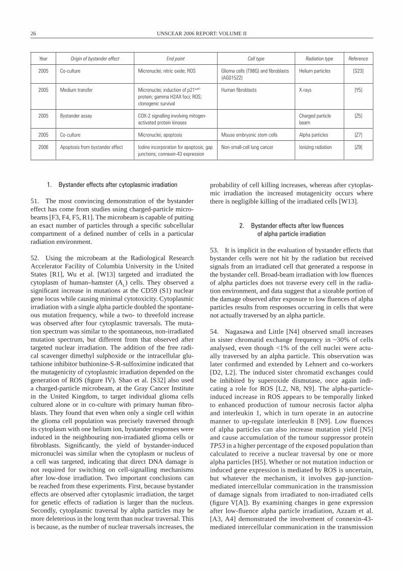
26 UNSCEAR 2006 REPORT: VOLUME II
Year Origin of bystander effect End point Cell type Radiation type Reference
2005 Co-culture Micronuclei; nitric oxide; ROS Glioma cells (T98G) and fibroblasts (AG01522)
Helium particles [S23]
2005 Medium transfer Micronuclei; induction of p21waf1 protein; gamma H2AX foci; ROS; clonogenic survival
Human fibroblasts X-rays [Y5]
2005 Bystander assay COX-2 signalling involving mitogen-activated protein kinases
Charged particle beam
[Z5]
2005 Co-culture Micronuclei; apoptosis Mouse embryonic stem cells Alpha particles [Z7]
2006 Apoptosis from bystander effect Iodine incorporation for apoptosis; gap junctions; connexin-43 expression
Non-small-cell lung cancer Ionizing radiation [Z9]
1. Bystander effects after cytoplasmic irradiation
51. The most convincing demonstration of the bystander effect has come from studies using charged-particle micro-beams [F3, F4, F5, R1]. The microbeam is capable of putting an exact number of particles through a specific subcellular compartment of a defined number of cells in a particular radiation environment.
52. Using the microbeam at the Radiological Research Accelerator Facility of Columbia University in the United States [R1], Wu et al. [W13] targeted and irradiated the cytoplasm of human–hamster (A
L) cells. They observed a
significant increase in mutations at the CD59 (S1) nuclear gene locus while causing minimal cytotoxicity. Cytoplasmic irradiation with a single alpha particle doubled the spontane-ous mutation frequency, while a two- to threefold increase was observed after four cytoplasmic traversals. The muta-tion spectrum was similar to the spontaneous, non-irradiated mutation spectrum, but different from that observed after targeted nuclear irradiation. The addition of the free radi-cal scavenger dimethyl sulphoxide or the intracellular glu-tathione inhibitor buthionine-S-R-sulfoximine indicated that the mutagenicity of cytoplasmic irradiation depended on the generation of ROS (figure IV). Shao et al. [S32] also used a charged-particle microbeam, at the Gray Cancer Institute in the United Kingdom, to target individual glioma cells cultured alone or in co-culture with primary human fibro-blasts. They found that even when only a single cell within the glioma cell population was precisely traversed through its cytoplasm with one helium ion, bystander responses were induced in the neighbouring non-irradiated glioma cells or fibroblasts. Significantly, the yield of bystander-induced micronuclei was similar when the cytoplasm or nucleus of a cell was targeted, indicating that direct DNA damage is not required for switching on cell-signalling mechanisms after low-dose irradiation. Two important conclusions can be reached from these experiments. First, because bystander effects are observed after cytoplasmic irradiation, the target for genetic effects of radiation is larger than the nucleus. Secondly, cytoplasmic traversal by alpha particles may be more deleterious in the long term than nuclear traversal. This is because, as the number of nuclear traversals increases, the
probability of cell killing increases, whereas after cytoplas-mic irradiation the increased mutagenicity occurs where there is negligible killing of the irradiated cells [W13].
2. Bystander effects after low fluences of alpha particle irradiation
53. It is implicit in the evaluation of bystander effects that bystander cells were not hit by the radiation but received signals from an irradiated cell that generated a response in the bystander cell. Broad-beam irradiation with low fluences of alpha particles does not traverse every cell in the radia-tion environment, and data suggest that a sizeable portion of the damage observed after exposure to low fluences of alpha particles results from responses occurring in cells that were not actually traversed by an alpha particle.
54. Nagasawa and Little [N4] observed small increases in sister chromatid exchange frequency in ~30% of cells analysed, even though <1% of the cell nuclei were actu-ally traversed by an alpha particle. This observation was later confirmed and extended by Lehnert and co-workers [D2, L2]. The induced sister chromatid exchanges could be inhibited by superoxide dismutase, once again indi-cating a role for ROS [L2, N8, N9]. The alpha-particle-induced increase in ROS appears to be temporally linked to enhanced production of tumour necrosis factor alpha and interleukin 1, which in turn operate in an autocrine manner to up-regulate interleukin 8 [N9]. Low fluences of alpha particles can also increase mutation yield [N5] and cause accumulation of the tumour suppressor protein TP53 in a higher percentage of the exposed population than calculated to receive a nuclear traversal by one or more alpha particles [H5]. Whether or not mutation induction or induced gene expression is mediated by ROS is uncertain, but whatever the mechanism, it involves gap-junction-mediated inter cellular communication in the transmission of damage signals from irradiated to non-irradiated cells (figure V[A]). By examining changes in gene expression after low-fluence alpha particle irradiation, Azzam et al. [A3, A4] demonstrated the involvement of connexin-43-mediated intercellular communication in the transmission
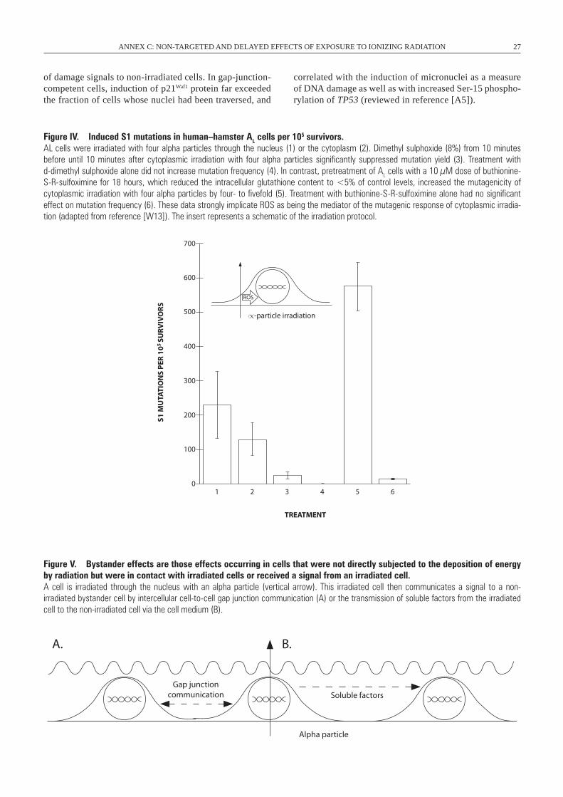
ANNEX C: NON-TARGETED AND DELAYED EFFECTS OF EXPOSURE TO IONIZING RADIATION 27
Figure IV. Induced S1 mutations in human–hamster AL cells per 105 survivors.AL cells were irradiated with four alpha particles through the nucleus (1) or the cytoplasm (2). Dimethyl sulphoxide (8%) from 10 minutes before until 10 minutes after cytoplasmic irradiation with four alpha particles significantly suppressed mutation yield (3). Treatment with d-dimethyl sulphoxide alone did not increase mutation frequency (4). In contrast, pretreatment of AL cells with a 10 µM dose of buthionine-S-R-sulfoximine for 18 hours, which reduced the intracellular glutathione content to <5% of control levels, increased the mutagenicity of cytoplasmic irradiation with four alpha particles by four- to fivefold (5). Treatment with buthionine-S-R-sulfoximine alone had no significant effect on mutation frequency (6). These data strongly implicate ROS as being the mediator of the mutagenic response of cytoplasmic irradia-tion (adapted from reference [w13]). The insert represents a schematic of the irradiation protocol.
of damage signals to non-irradiated cells. In gap-junction-competent cells, induction of p21Waf1 protein far exceeded the fraction of cells whose nuclei had been traversed, and
correlated with the induction of micronuclei as a measure of DNA damage as well as with increased Ser-15 phospho-rylation of TP53 (reviewed in reference [A5]).
Figure V. Bystander effects are those effects occurring in cells that were not directly subjected to the deposition of energy by radiation but were in contact with irradiated cells or received a signal from an irradiated cell. A cell is irradiated through the nucleus with an alpha particle (vertical arrow). This irradiated cell then communicates a signal to a non- irradiated bystander cell by intercellular cell-to-cell gap junction communication (A) or the transmission of soluble factors from the irradiated cell to the non-irradiated cell via the cell medium (B).
700
600
500
400
300
200
100
01 2 3 4 5 6
TREATMENT
S1 M
UTA
TIO
NS
PER
10
5 SU
RV
IVO
RS
∝-particle irradiation
ROS
A. B.
Gap junctioncommunication Soluble factors
Alpha particle
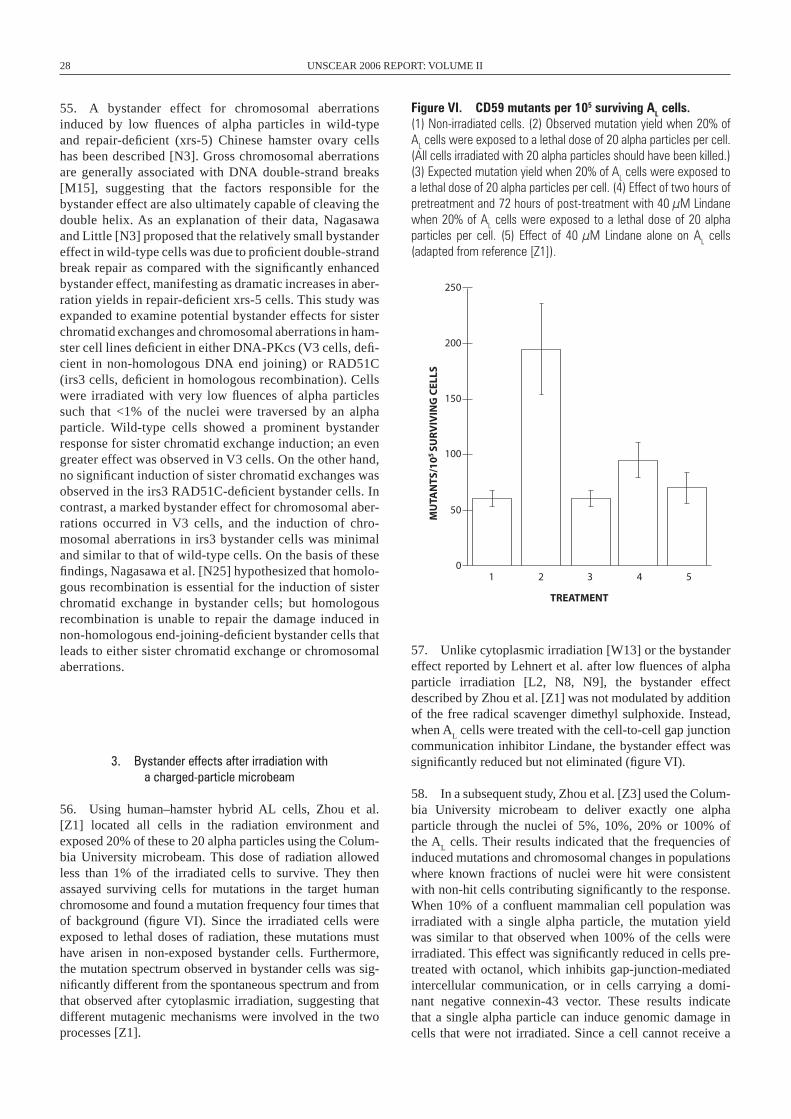
28 UNSCEAR 2006 REPORT: VOLUME II
55. A bystander effect for chromosomal aberrations induced by low fluences of alpha particles in wild-type and repair-deficient (xrs-5) Chinese hamster ovary cells has been described [N3]. Gross chromosomal aberrations are generally associated with DNA double-strand breaks [M15], suggesting that the factors responsible for the bystander effect are also ultimately capable of cleaving the double helix. As an explanation of their data, Nagasawa and Little [N3] proposed that the relatively small bystander effect in wild-type cells was due to proficient double-strand break repair as compared with the significantly enhanced bystander effect, manifesting as dramatic increases in aber-ration yields in repair-deficient xrs-5 cells. This study was expanded to examine potential bystander effects for sister chromatid exchanges and chromosomal aberrations in ham-ster cell lines deficient in either DNA-PKcs (V3 cells, defi-cient in non-homologous DNA end joining) or RAD51C (irs3 cells, deficient in homologous recombination). Cells were irradiated with very low fluences of alpha particles such that <1% of the nuclei were traversed by an alpha particle. Wild-type cells showed a prominent bystander response for sister chromatid exchange induction; an even greater effect was observed in V3 cells. On the other hand, no significant induction of sister chromatid exchanges was observed in the irs3 RAD51C-deficient bystander cells. In contrast, a marked bystander effect for chromosomal aber-rations occurred in V3 cells, and the induction of chro-mosomal aberrations in irs3 bystander cells was minimal and similar to that of wild-type cells. On the basis of these findings, Nagasawa et al. [N25] hypothesized that homolo-gous recombination is essential for the induction of sister chromatid exchange in bystander cells; but homologous recombination is unable to repair the damage induced in non-homologous end-joining-deficient bystander cells that leads to either sister chromatid exchange or chromosomal aberrations.
3. Bystander effects after irradiation with a charged-particle microbeam
56. Using human–hamster hybrid AL cells, Zhou et al. [Z1] located all cells in the radiation environment and exposed 20% of these to 20 alpha particles using the Colum-bia University microbeam. This dose of radiation allowed less than 1% of the irradiated cells to survive. They then assayed surviving cells for mutations in the target human chromosome and found a mutation frequency four times that of background (figure VI). Since the irradiated cells were exposed to lethal doses of radiation, these mutations must have arisen in non-exposed bystander cells. Furthermore, the mutation spectrum observed in bystander cells was sig-nificantly different from the spontaneous spectrum and from that observed after cytoplasmic irradiation, suggesting that different mutagenic mechanisms were involved in the two processes [Z1].
Figure VI. CD59 mutants per 105 surviving AL cells. (1) Non-irradiated cells. (2) Observed mutation yield when 20% of AL cells were exposed to a lethal dose of 20 alpha particles per cell. (All cells irradiated with 20 alpha particles should have been killed.) (3) Expected mutation yield when 20% of AL cells were exposed to a lethal dose of 20 alpha particles per cell. (4) Effect of two hours of pretreatment and 72 hours of post-treatment with 40 µM Lindane when 20% of AL cells were exposed to a lethal dose of 20 alpha particles per cell. (5) Effect of 40 µM Lindane alone on AL cells (adapted from reference [Z1]).
57. Unlike cytoplasmic irradiation [W13] or the bystander effect reported by Lehnert et al. after low fluences of alpha particle irradiation [L2, N8, N9], the bystander effect described by Zhou et al. [Z1] was not modulated by addition of the free radical scavenger dimethyl sulphoxide. Instead, when A
L cells were treated with the cell-to-cell gap junction
communication inhibitor Lindane, the bystander effect was significantly reduced but not eliminated (figure VI).
58. In a subsequent study, Zhou et al. [Z3] used the Colum-bia University microbeam to deliver exactly one alpha particle through the nuclei of 5%, 10%, 20% or 100% of the A
L cells. Their results indicated that the frequencies of
induced mutations and chromosomal changes in populations where known fractions of nuclei were hit were consistent with non-hit cells contributing significantly to the response. When 10% of a confluent mammalian cell population was irradiated with a single alpha particle, the mutation yield was similar to that observed when 100% of the cells were irradiated. This effect was significantly reduced in cells pre-treated with octanol, which inhibits gap-junction-mediated inter cellular communication, or in cells carrying a domi-nant negative connexin-43 vector. These results indicate that a single alpha particle can induce genomic damage in cells that were not irradiated. Since a cell cannot receive a
250
200
150
100
50
01 2 3 4 5
TREATMENT
MU
TAN
TS/1
05 SU
RV
IVIN
G C
ELLS
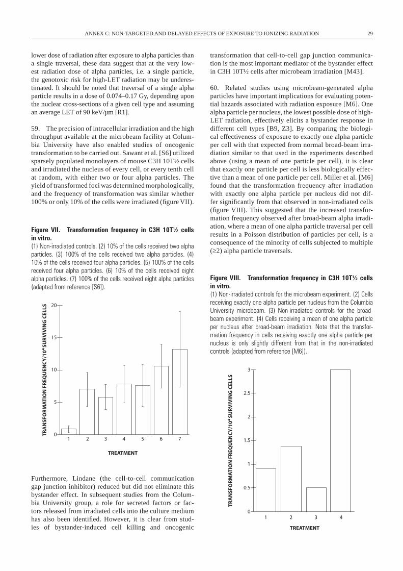
ANNEX C: NON-TARGETED AND DELAYED EFFECTS OF EXPOSURE TO IONIZING RADIATION 29
lower dose of radiation after exposure to alpha particles than a single traversal, these data suggest that at the very low-est radiation dose of alpha particles, i.e. a single particle, the genotoxic risk for high-LET radiation may be underes-timated. It should be noted that traversal of a single alpha particle results in a dose of 0.074–0.17 Gy, depending upon the nuclear cross-sections of a given cell type and assuming an average LET of 90 keV/µm [R1].
59. The precision of intracellular irradiation and the high throughput available at the microbeam facility at Colum-bia University have also enabled studies of oncogenic transformation to be carried out. Sawant et al. [S6] utilized sparsely populated monolayers of mouse C3H 10T½ cells and irradiated the nucleus of every cell, or every tenth cell at random, with either two or four alpha particles. The yield of transformed foci was determined morphologically, and the frequency of transformation was similar whether 100% or only 10% of the cells were irradiated (figure VII).
transformation that cell-to-cell gap junction communica-tion is the most important mediator of the bystander effect in C3H 10T½ cells after microbeam irradiation [M43].
60. Related studies using microbeam-generated alpha particles have important implications for evaluating poten-tial hazards associated with radiation exposure [M6]. One alpha particle per nucleus, the lowest possible dose of high-LET radiation, effectively elicits a bystander response in different cell types [B9, Z3]. By comparing the biologi-cal effectiveness of exposure to exactly one alpha particle per cell with that expected from normal broad-beam irra-diation similar to that used in the experiments described above (using a mean of one particle per cell), it is clear that exactly one particle per cell is less biologically effec-tive than a mean of one particle per cell. Miller et al. [M6] found that the transformation frequency after irradiation with exactly one alpha particle per nucleus did not dif-fer significantly from that observed in non-irradiated cells (figure VIII). This suggested that the increased transfor-mation frequency observed after broad-beam alpha irradi-ation, where a mean of one alpha particle traversal per cell results in a Poisson distribution of particles per cell, is a consequence of the minority of cells subjected to multiple (≥2) alpha particle traversals.
20
15
10
5
01 2 3 4 5 6 7
TREATMENT
TRA
NSF
OR
MA
TIO
N F
REQ
UEN
CY
/10
4 SU
RV
IVIN
G C
ELLS
3
2
2.5
1.5
1
0.5
01 2 3 4
TREATMENT
TRA
NSF
OR
MA
TIO
N F
REQ
UEN
CY
/10
4 SU
RV
IVIN
G C
ELLS
Figure VII. Transformation frequency in C3H 10T½ cells in vitro. (1) Non-irradiated controls. (2) 10% of the cells received two alpha particles. (3) 100% of the cells received two alpha particles. (4) 10% of the cells received four alpha particles. (5) 100% of the cells received four alpha particles. (6) 10% of the cells received eight alpha particles. (7) 100% of the cells received eight alpha particles (adapted from reference [S6]).
Figure VIII. Transformation frequency in C3H 10T½ cells in vitro. (1) Non-irradiated controls for the microbeam experiment. (2) Cells receiving exactly one alpha particle per nucleus from the Columbia University microbeam. (3) Non-irradiated controls for the broad-beam experiment. (4) Cells receiving a mean of one alpha particle per nucleus after broad-beam irradiation. Note that the transfor-mation frequency in cells receiving exactly one alpha particle per nucleus is only slightly different from that in the non-irradiated controls (adapted from reference [M6]).
Furthermore, Lindane (the cell-to-cell communication gap junction inhibitor) reduced but did not eliminate this bystander effect. In subsequent studies from the Colum-bia University group, a role for secreted factors or fac-tors released from irradiated cells into the culture medium has also been identified. However, it is clear from stud-ies of bystander-induced cell killing and oncogenic
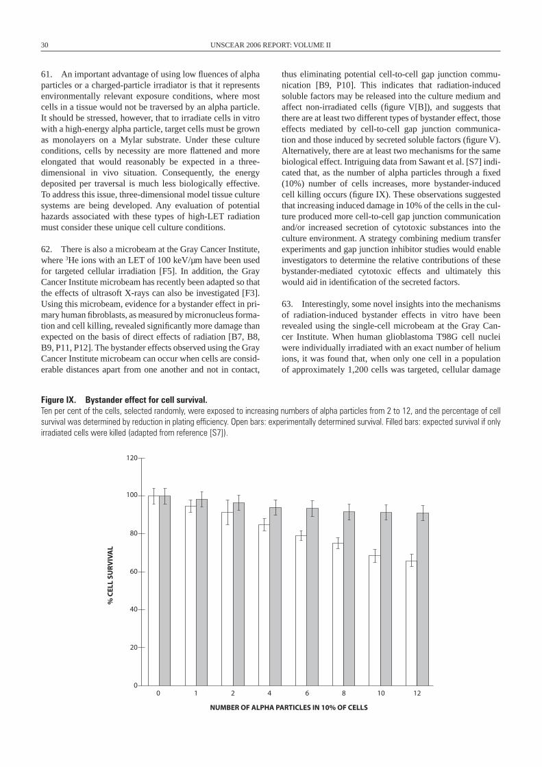
30 UNSCEAR 2006 REPORT: VOLUME II
61. An important advantage of using low fluences of alpha particles or a charged-particle irradiator is that it represents environmentally relevant exposure conditions, where most cells in a tissue would not be traversed by an alpha particle. It should be stressed, however, that to irradiate cells in vitro with a high-energy alpha particle, target cells must be grown as monolayers on a Mylar substrate. Under these culture conditions, cells by necessity are more flattened and more elongated that would reasonably be expected in a three-dimensional in vivo situation. Consequently, the energy deposited per traversal is much less biologically effective. To address this issue, three-dimensional model tissue culture systems are being developed. Any evaluation of potential hazards associated with these types of high-LET radiation must consider these unique cell culture conditions.
62. There is also a microbeam at the Gray Cancer Institute, where 3He ions with an LET of 100 keV/µm have been used for targeted cellular irradiation [F5]. In addition, the Gray Cancer Institute microbeam has recently been adapted so that the effects of ultrasoft X-rays can also be investigated [F3]. Using this microbeam, evidence for a bystander effect in pri-mary human fibroblasts, as measured by micronucleus forma-tion and cell killing, revealed significantly more damage than expected on the basis of direct effects of radiation [B7, B8, B9, P11, P12]. The bystander effects observed using the Gray Cancer Institute microbeam can occur when cells are consid-erable distances apart from one another and not in contact,
thus eliminating potential cell-to-cell gap junction commu-nication [B9, P10]. This indicates that radiation-induced soluble factors may be released into the culture medium and affect non-irradiated cells (figure V[B]), and suggests that there are at least two different types of bystander effect, those effects mediated by cell-to-cell gap junction communica-tion and those induced by secreted soluble factors (figure V). Alternatively, there are at least two mechanisms for the same biological effect. Intriguing data from Sawant et al. [S7] indi-cated that, as the number of alpha particles through a fixed (10%) number of cells increases, more bystander-induced cell killing occurs (figure IX). These observations suggested that increasing induced damage in 10% of the cells in the cul-ture produced more cell-to-cell gap junction communication and/or increased secretion of cytotoxic substances into the culture environment. A strategy combining medium transfer experiments and gap junction inhibitor studies would enable investigators to determine the relative contributions of these bystander-mediated cytotoxic effects and ultimately this would aid in identification of the secreted factors.
63. Interestingly, some novel insights into the mechanisms of radiation-induced bystander effects in vitro have been revealed using the single-cell microbeam at the Gray Can-cer Institute. When human glioblastoma T98G cell nuclei were individually irradiated with an exact number of helium ions, it was found that, when only one cell in a population of approximately 1,200 cells was targeted, cellular damage
100
120
80
60
40
20
00 1 2 4 6 8 10 12
NUMBER OF ALPHA PARTICLES IN 10% OF CELLS
% C
ELL
SUR
VIV
AL
Figure IX. Bystander effect for cell survival. Ten per cent of the cells, selected randomly, were exposed to increasing numbers of alpha particles from 2 to 12, and the percentage of cell survival was determined by reduction in plating efficiency. Open bars: experimentally determined survival. Filled bars: expected survival if only irradiated cells were killed (adapted from reference [S7]).
ANNEX C: NON-TARGETED AND DELAYED EFFECTS OF EXPOSURE TO IONIZING RADIATION 31
measured as induced micronuclei was increased by 20% [S16]. When the percentage of cells individually targeted was increased from 1% to 20%, the yield of micronuclei in the population greatly exceeded that predicted on the basis of the yield when all of the cells were targeted assuming no bystander effect. However, when 2-(4-carboxyphenyl)-4, 4, 5, 5-tetramethylimidazoline-1-oxyl-3-oxide (c-PTIO), a nitric-oxide-specific scavenger, was present in the culture medium, the micronucleus yields decreased to the predicted values. This indicates that nitric oxide contributed to the bystander effect. Moreover, the medium harvested from cells targeted with helium ions showed a cytotoxic effect by inducing micronuclei in non-irradiated T98G cells, and this bystander response was also inhibited by c-PTIO treatment. The induc-tion of micronuclei in the population could also be decreased by c-PTIO treatment when 100% of cells were individually targeted by one or two helium ions, indicating a complex interaction of direct irradiation and bystander signals. A role for nitric oxide in the medium-mediated bystander effect has now been described in a number of studies [S15, S16, S31, S32]. Furthermore, the secretion of nitric oxide as a bystander effect has been linked to the induction of radioresistance in recipient cells [M3, M4]. However, it is not clear whether nitric-oxide-mediated bystander effects occur in conjunction with, or independent of, increased levels of ROS, and how this might affect the characterization of subsequent radiation sensitivity.
64. Microbeam experiments using high-Z elements (460 MeV 40Ar, 1,260 keV/µm and 260 MeV 20Ne, 380 keV/µm) have also been performed at the Japan Atomic Energy Research Institute [S33]. Confluent normal human fibrob-lasts were targeted and the induced micronuclei evaluated after replating the cells. Even when only a single cell was hit, a 1.4-fold increase in the frequency of micronuclei was observed, indicative of a bystander effect. The observed increase in micronucleus frequency saturated when four cells were targeted and could be suppressed when dimethyl sulphoxide (a scavenger of ROS) or PMA (an inhibitor of gap junction communication) was present at the time of irra-diation. Thus a role for nitric oxide, ROS and cell-to-cell gap junctions has been invoked in bystander responses.
65. It is likely, however, that there are multiple bystander pathways. By using the Columbia University charged parti-cle beam in conjunction with a strip dish design, Zhou et al. [Z5] show that the cyclooxygenase-2 (COX-2, also known as prostaglandin endoperoxide synthase-2) signalling cascade plays an essential role in the bystander process. Treatment of bystander cells with NS-398, which suppresses COX-2 activity, significantly reduced the bystander effect. This pro-vided evidence that the COX-2-related pathway, which is essential in mediating cellular inflammatory response, is a critical signalling link for the bystander phenomenon in this assay system. Furthermore, any signalling pathway likely to be involved is complicated by the genotype of the exposed organism. For example, Zhu et al. [Z7] examined the ability of mouse embryonic stem cells differing in the status of the DNA repair gene, Rad9, to express a bystander effect after
exposure to alpha particles. All populations, when confluent, demonstrated a dose-independent bystander effect with respect to cell killing and apoptosis. Minimal alpha particle induction of micronuclei in bystander cells was observed, except for the Rad9-/- mutant, where a significant increase above background was detected. Therefore the Rad9 null mutation selectively sensitizes mouse embryonic stem cells to spontaneous and high-LET-radiation-induced bystander apoptosis and micro-nucleus formation, but it has much less impact on cell killing by direct or bystander alpha particle exposure.
66. At present there is no detailed information on potential bystander effects occurring after cellular exposure to low-LET radiation in non-irradiated cells subsequently cultured in the same physical proximity. This is because the develop-ment of focused low-LET radiation sources has lagged well behind that of high-LET charged particle microbeams, owing to practical and technical complications. Gamma rays inter-act with matter in a number of ways, including photoelectric absorption, Compton scattering and pair production. Since the electrons will undergo scattering as they exit the vacuum system, it is not practical at present to irradiate single cells or subcellular structures, as is possible with the charged parti-cle microbeams described above. However, by using pulsed electron beams, a low-LET microbeam can be constructed that will mimic many important aspects of the interaction of gamma rays with cells. Such devices are currently avail-able at the University of Maryland [S37] and Texas A&M University [W16] in the United States. Consequently, it is anticipated that in the foreseeable future many of the studies that have been performed with high-LET alpha particles or intermediate-LET ultrasoft X-rays can be replicated using these new low-LET microbeams. In addition, the ability to experimentally vary the LET distribution by changing the incident beam energy will allow investigation of the rela-tive biological importance of various parts of the energetic electrons’ track and hence evaluate how this might modulate bystander responses [S38].
4. Bystander effects after transfer of medium from irradiated cells
67. Mothersill and Seymour [M18] demonstrated a sig-nificant reduction in plating efficiency in non-irradiated cultures of human epithelial cells (but not fibroblasts) that received culture medium from irradiated cultures. These observations indicated that irradiated cells secreted a toxic substance, a “bystander factor”, into the culture medium that can kill non-irradiated cells (figure X). Medium irra-diated in the absence of cells had no effect on survival when transferred to non-irradiated cells. Not all cells are capable of producing the toxic bystander factor, nor are all cells capable of receiving and responding to the secreted signal [M17, M18, M22, M25]. The effect was depend-ent on the cell number at the time of irradiation, could be observed as early as 30 minutes post-irradiation, and was still effective when medium transfer occurred 60 hours after irradiation.
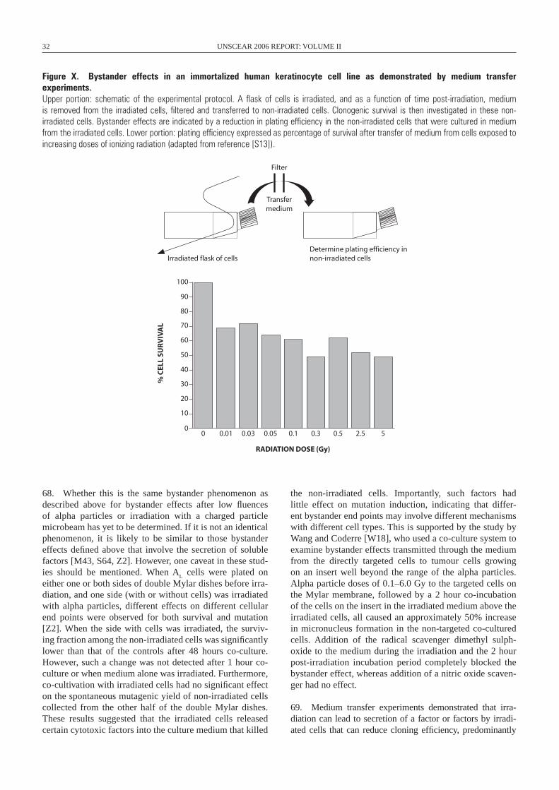
32 UNSCEAR 2006 REPORT: VOLUME II
Figure X. Bystander effects in an immortalized human keratinocyte cell line as demonstrated by medium transfer experiments. Upper portion: schematic of the experimental protocol. A flask of cells is irradiated, and as a function of time post-irradiation, medium is removed from the irradiated cells, filtered and transferred to non-irradiated cells. Clonogenic survival is then investigated in these non- irradiated cells. Bystander effects are indicated by a reduction in plating efficiency in the non-irradiated cells that were cultured in medium from the irradiated cells. Lower portion: plating efficiency expressed as percentage of survival after transfer of medium from cells exposed to increasing doses of ionizing radiation (adapted from reference [S13]).
68. Whether this is the same bystander phenomenon as described above for bystander effects after low fluences of alpha particles or irradiation with a charged particle microbeam has yet to be determined. If it is not an identical phenomenon, it is likely to be similar to those bystander effects defined above that involve the secretion of soluble factors [M43, S64, Z2]. However, one caveat in these stud-ies should be mentioned. When A
L cells were plated on
either one or both sides of double Mylar dishes before irra-diation, and one side (with or without cells) was irradiated with alpha particles, different effects on different cellular end points were observed for both survival and mutation [Z2]. When the side with cells was irradiated, the surviv-ing fraction among the non-irradiated cells was significantly lower than that of the controls after 48 hours co-culture. However, such a change was not detected after 1 hour co-culture or when medium alone was irradiated. Furthermore, co-cultivation with irradiated cells had no significant effect on the spontaneous mutagenic yield of non-irradiated cells collected from the other half of the double Mylar dishes. These results suggested that the irradiated cells released certain cytotoxic factors into the culture medium that killed
the non-irradiated cells. Importantly, such factors had little effect on mutation induction, indicating that differ-ent bystander end points may involve different mechanisms with different cell types. This is supported by the study by Wang and Coderre [W18], who used a co-culture system to examine bystander effects transmitted through the medium from the directly targeted cells to tumour cells growing on an insert well beyond the range of the alpha particles. Alpha particle doses of 0.1–6.0 Gy to the targeted cells on the Mylar membrane, followed by a 2 hour co-incubation of the cells on the insert in the irradiated medium above the irradiated cells, all caused an approximately 50% increase in micronucleus formation in the non-targeted co-cultured cells. Addition of the radical scavenger dimethyl sulph-oxide to the medium during the irradiation and the 2 hour post-irradiation incubation period completely blocked the bystander effect, whereas addition of a nitric oxide scaven-ger had no effect.
69. Medium transfer experiments demonstrated that irra-diation can lead to secretion of a factor or factors by irradi-ated cells that can reduce cloning efficiency, predominantly
100
90
70
80
60
50
40
20
30
10
00 0.01 0.03 0.05 0.1 0.3 0.5 2.5 5
RADIATION DOSE (Gy)
% C
ELL
SUR
VIV
AL
Irradiated flask of cellsDetermine plating efficiency innon-irradiated cells
Filter
Transfermedium
ANNEX C: NON-TARGETED AND DELAYED EFFECTS OF EXPOSURE TO IONIZING RADIATION 33
by stimulating apoptosis [L26, L27, L28, S12], increasing neoplastic transformation [L5] or inducing genomic instabil-ity [S12] in non-irradiated cells. The first detectable effect on recipient cells after transfer of medium containing the bystander factor from irradiated cells was a rapid calcium pulse (1–2 minutes) followed 30–120 minutes later by changes in mitochondrial membrane permeability and the induction of ROS [L28, L38, M24]. Cell-to-cell contact during irradia-tion was not required to induce killing of bystander cells, but medium from cell cultures irradiated at high densities resulted in the greatest amount of bystander-induced cell death [M22]. Furthermore, the use of apoptosis inhibitors or medium from lactate dehydrogenase or glucose-6-phosphate dehydrogenase mutant cells reduced or prevented the bystander effect [M24]. Treatment with the antioxidants L-lactate and L-deprenyl prevented bystander-factor-associated cell killing [M24], sug-gesting that energy/redox metabolism may be involved in the medium-mediated bystander response.
70. Mothersill and co-workers [M44] showed that repair-deficient human cell lines produced a moderate to severe amount of bystander-induced cell death after medium transfer to autologous cells or to a reporter cell line. Nor-mal “repair-proficient” lines have much less severe, or no, bystander-induced effects on cloning efficiency. These results are in agreement with the observations of Little and colleagues [L42, N3]. Mothersill et al. [M44] interpreted these data as supporting the hypothesis that bystander effects play a protective role in biological systems by terminating division in cells containing DNA damage. Thus the repair-deficient cells, irrespective of the actual repair defect, may respond to the occurrence of DNA damage in the population by removing large numbers of cells from the proliferating pool. It should be noted that repair-deficient cell lines tend to show an increased frequency of induced genomic insta-bility, again suggesting a commonality in the mechanism of radiation-induced genomic instability and bystander effects.
71. The majority of medium transfer experiments reported have utilized low-LET radiation to induce the bystander effect and have come primarily from a single laboratory (reviewed in references [M19, M20]). A fascinating observation from these studies, but one that is difficult to reconcile with those of other studies, concerns the radiation doses required to elicit a bystander response. In human keratino cytes, low-LET 60Co gamma ray doses of 0.01–0.5 Gy reduced clono genic sur-vival after transfer of irradiated medium. This was entirely due to bystander effects. The magnitude of cell killing was relatively constant and appeared to saturate at doses in the range 0.03–0.05 Gy [S13]. At doses of greater than 0.5 Gy, cell killing was the result of the direct effects of radiation as well as the dose-independent bystander effect [S13]. These observations are difficult to explain in terms of the classical cell survival curve, where there is very little if any cell kill-ing observed at radiation doses of a few milligrays in directly irradiated cells.
72. In contrast to the cell-killing effects reported by Mothersill and Seymour after medium transfer from gamma-
irradiated cultures, Iyer and Lehnert [I4] have observed quite different cellular responses in human fibroblast cells cul-tured in supernatants from alpha-irradiated fibroblasts. They observed decreases in basal levels of TP53 and CDKN1AQ in non-irradiated cells, rather than increases as described by others (e.g. [A4, H5]). These decreases were accompanied by increases in proliferating cell nuclear antigen and CDC2, apparently mediated by TGF-beta 1 and the induction of intracellular ROS [N8]. In contrast to the detrimental effects on cell well-being characteristic of bystander effects so far described, Iyer and Lehnert [I4] showed that their decreased TP53/CDKN1A bystander effect correlated with enhanced cell proliferation.
73. Attempting to reconcile these conflicting results raises a number of questions. While the quality of radiation and the cell types under investigation are different, these stud-ies highlight the family of responses characterizing the bystander effect. Mechanistically, factors transferred via cell-to-cell gap junction communication or secreted into the culture medium may interact with those non-irradiated cells in an antagonistic manner, ultimately killing the non-irradiated cells (reviewed in reference [S39]), but the reasons why cultured cells should secrete cytotoxic factors and why no such dramatic reduction in plating efficiency is observed when populations of cells are irradiated with low doses of low-LET radiation are not immediately obvious. Likewise, the pro-mitogenic response reported by Lehnert et al. [L2, L48] after medium transfer is contradictory to those direct effects observed in irradiated cells where irradiation can inhibit cell growth. Cell proliferation following irradiation appears contrary to the long-term well-being of the cell, tissue, organ or organism, whereas cell death would be likely to protect against the possibility of detrimental mutations, chromosomal rearrangements, and so on. Clearly bystander effects can modify cellular responses to radiation, and it remains to be determined whether these effects character-ized in non-irradiated cells in vitro have a major role in the response of irradiated cells in vitro or in irradiated and non-irradiated cells in vivo.
B. Bystander effects in vivo
74. Bystander effects have been observed predominantly by using single-cell in vitro systems that do not have real-istic multicellular morphology. Given that the bystander pheno menon must involve cell-to-cell interactions, the rele-vance of such single-cell in vitro studies is questionable. However, Belyakov et al. [B36] have described bystander responses in a three-dimensional, normal human tissue sys-tem. While not a true in vivo assay, this model skin system does provide some semblance of multicellular interactions. End points were induction of micronucleated and apoptotic cells. Non- irradiated cells up to 1 mm distant from irradi-ated cells showed a significant enhancement in the effect over background levels, with an average increase in effect of 1.7-fold for micronuclei and 2.8-fold for apoptosis. The surprisingly long range of bystander signals in a human
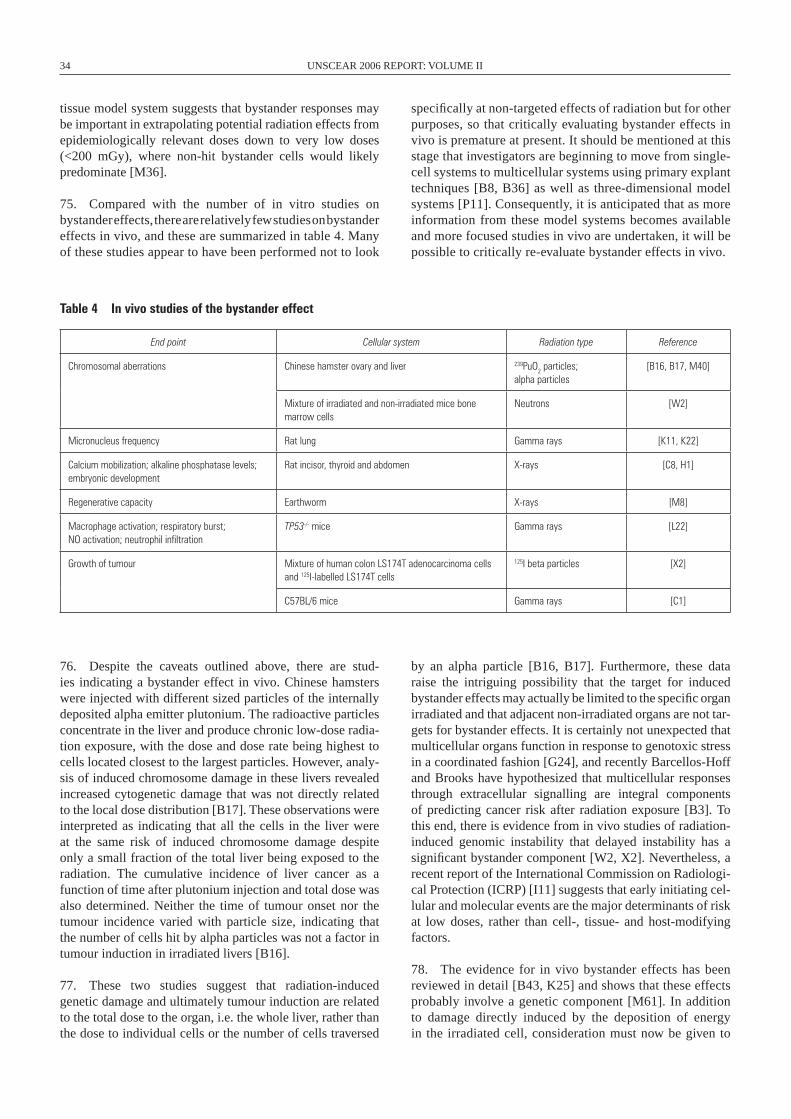
34 UNSCEAR 2006 REPORT: VOLUME II
tissue model system suggests that bystander responses may be important in extrapolating potential radiation effects from epidemiologically relevant doses down to very low doses (<200 mGy), where non-hit bystander cells would likely predominate [M36].
75. Compared with the number of in vitro studies on bystander effects, there are relatively few studies on bystander effects in vivo, and these are summarized in table 4. Many of these studies appear to have been performed not to look
specifically at non-targeted effects of radiation but for other purposes, so that critically evaluating bystander effects in vivo is premature at present. It should be mentioned at this stage that investigators are beginning to move from single-cell systems to multicellular systems using primary explant techniques [B8, B36] as well as three-dimensional model systems [P11]. Consequently, it is anticipated that as more information from these model systems becomes available and more focused studies in vivo are undertaken, it will be possible to critically re-evaluate bystander effects in vivo.
Table 4 In vivo studies of the bystander effect
End point Cellular system Radiation type Reference
Chromosomal aberrations Chinese hamster ovary and liver 239PuO2 particles; alpha particles
[B16, B17, M40]
Mixture of irradiated and non-irradiated mice bone marrow cells
Neutrons [w2]
Micronucleus frequency Rat lung Gamma rays [K11, K22]
Calcium mobilization; alkaline phosphatase levels; embryonic development
Rat incisor, thyroid and abdomen X-rays [C8, H1]
Regenerative capacity Earthworm X-rays [M8]
Macrophage activation; respiratory burst; NO activation; neutrophil infiltration
TP53-/- mice Gamma rays [L22]
Growth of tumour Mixture of human colon LS174T adenocarcinoma cells and 125I-labelled LS174T cells
125I beta particles [X2]
C57BL/6 mice Gamma rays [C1]
76. Despite the caveats outlined above, there are stud-ies indicating a bystander effect in vivo. Chinese hamsters were injected with different sized particles of the internally deposited alpha emitter plutonium. The radioactive particles concentrate in the liver and produce chronic low-dose radia-tion exposure, with the dose and dose rate being highest to cells located closest to the largest particles. However, analy-sis of induced chromosome damage in these livers revealed increased cytogenetic damage that was not directly related to the local dose distribution [B17]. These observations were interpreted as indicating that all the cells in the liver were at the same risk of induced chromosome damage despite only a small fraction of the total liver being exposed to the radiation. The cumulative incidence of liver cancer as a function of time after plutonium injection and total dose was also determined. Neither the time of tumour onset nor the tumour incidence varied with particle size, indicating that the number of cells hit by alpha particles was not a factor in tumour induction in irradiated livers [B16].
77. These two studies suggest that radiation-induced genetic damage and ultimately tumour induction are related to the total dose to the organ, i.e. the whole liver, rather than the dose to individual cells or the number of cells traversed
by an alpha particle [B16, B17]. Furthermore, these data raise the intriguing possibility that the target for induced bystander effects may actually be limited to the specific organ irradiated and that adjacent non-irradiated organs are not tar-gets for bystander effects. It is certainly not unexpected that multicellular organs function in response to genotoxic stress in a coordinated fashion [G24], and recently Barcellos-Hoff and Brooks have hypothesized that multicellular responses through extracellular signalling are integral components of predicting cancer risk after radiation exposure [B3]. To this end, there is evidence from in vivo studies of radiation-induced genomic instability that delayed instability has a significant bystander component [W2, X2]. Nevertheless, a recent report of the International Commission on Radiologi-cal Protection (ICRP) [I11] suggests that early initiating cel-lular and molecular events are the major determinants of risk at low doses, rather than cell-, tissue- and host-modifying factors.
78. The evidence for in vivo bystander effects has been reviewed in detail [B43, K25] and shows that these effects probably involve a genetic component [M61]. In addition to damage directly induced by the deposition of energy in the irradiated cell, consideration must now be given to
ANNEX C: NON-TARGETED AND DELAYED EFFECTS OF EXPOSURE TO IONIZING RADIATION 35
these indirect effects of radiation, and a model quantifying these considerations has been proposed [B14]. An irradi-ated cell can send out a signal and induce a response in a cell whose nucleus was not hit by radiation. Thus a detri-mental bystander effect, e.g. chromosomal aberrations, in essence “modifies” the biological effectiveness of a given radiation dose by increasing the number of cells that expe-rience adverse effects over that directly exposed to the radiation. Significantly, these bystander effects appear to be limited to the organ irradiated, i.e. are organ-specific
[B43]. Thus, at the present state of our knowledge, it is reasonable to assume that any bystander effect induced in vivo is accounted for in models of organ risk evaluation. As a result, it is unlikely that the resurgence of interest in these non-targeted radiation effects will substantially alter risk estimates as discussed in detail in the BEIR VII report [C23]. Nevertheless, it cannot be excluded that increasing the knowledge basis for in vivo bystander effects at low doses and low dose rates in specific organs may affect current organ risk estimates.

37
III. RELATIONSHIP BETwEEN RADIATION-INDUCED GENOMIC INSTABILITY AND BYSTANDER EFFECTS
79. The evidence for effects occurring in cells that them-selves were not irradiated but are the progeny of irradiated cells (radiation-induced genomic instability), and non- targeted cellular effects usually associated with direct expo-sure to ionizing radiation occurring in non-irradiated cells (bystander effects), has been reviewed in the previous para-graphs. Many of the end points associated with these two phenomena are the same: induced chromosomal rearrange-ments, micronuclei, increased mutation, increased transfor-mation and cell killing. So what is the relationship, if any, between induced instability and bystander effects? Chromo-somal instability in haemopoietic cells can be induced by an indirect, non-targeted bystander type of mechanism [K3, L24]. Persistently increased levels of intracellular ROS have been reported in chromosomally unstable cells [C5, L7, L8, L9, L10, R3, R6], and provide a plausible mechanism for perpetuating instability over time (reviewed in references [M10, M16]). Considerable experimental support for this hypothesis comes from Wright and co-workers [L22, W2, W4], and this has recently been reviewed in detail [L23]. The resultant intercellular signalling cascades, cytokine produc-tion, nitric oxide production and persistent free radicals all have the potential to mediate both instability and bystander effects (reviewed in references [L23, M10]).
80. To critically evaluate the hypothesis that chromosomally unstable GM10115 clones perpetuate instability by secreting a bystander-like factor into the culture medium, thus driv-ing the delayed production of chromosomal rearrangements, Nagar et al. took medium from a chromosomally unstable clone of GM10115 human–hamster hybrid cells, filtered it and cultured non-irradiated GM10115 cells in this medium. None of the non-irradiated GM10115 cells were able to sur-vive and form colonies in medium from the unstable clone. Nagar et al. called this novel effect, by which cells cultured in medium from chromosomally unstable GM10115 cells die, the death-inducing effect [N1]. The unstable clones showing the death-inducing effect also showed increased numbers of apoptotic cells and elevated levels of intracellular ROS [N2], either or both of which might contribute factors to the cul-ture medium responsible for the death-inducing effect. Fur-thermore, Nagar et al. [N1] have interpreted this observation as indicating that unstable clones of cells do secrete factors that, while generally not toxic to the unstable clone, most likely contribute to the perpetuation of the unstable pheno-type. It should be stressed that the death-inducing effect is separate from the bystander effect observed after trans-ferring medium from irradiated cells. Nagar et al. did not observe a reduction in plating efficiency when medium from irradiated GM10115 cells was transferred to non-irradiated
GM10115 cells. This indicates that GM10115 cells either do not secrete a cytotoxic bystander factor or are not susceptible to a bystander factor, and that the death-inducing effect is not the same as the bystander effect described by Mothersill and co-workers. Likewise, chromosomal instability was not detected in GM10115 cells after transferring medium from irradiated cells as described by Mothersill and Seymour [M18, M21] for induced bystander effects.
81. It should be noted that Mothersill et al. [M44] have also reported that they do not find a bystander response in some Chinese hamster cell lines after medium transfer. This implies that, like the human–hamster hybrid GM10115 cell line, CHOK1 hamster cells may be deficient in produc-ing a bystander signal or in responding to that signal. It is interesting that when medium from irradiated CHOK1 cells was added to either non-irradiated CHOK1 cells or repair-deficient XR1 hamster cells, the plating efficiency actually increased rather than decreased [M44]. However, signifi-cantly increased bystander effects after cellular exposure to low fluences of alpha particles have been described in repair-deficient Chinese hamster cells compared with wild-type hamster cells [N3, N20, N25].
82. Evidence increasingly suggests that induced instabil-ity and bystander effects are linked (reviewed in references [L23, M10, M16]), and it is likely, given the commonality of the end points observed, that both phenomena could be mani-festations of the same non-targeted processes [M10, M16]. Furthermore, a significant contribution from bystander-like factors could help explain the high frequency of radiation-induced instability reported, for example, in references [K3, L6, M2].
83. The Committee continues to hold the view that mecha-nistic information is important for its recommendations on radiation-induced health effects at doses of below ~200 mSv and for risk assessment for individuals. The latter aspect is important because epidemiology always refers to popula-tions, and genomic instability, for example, varies among individuals.
A. Relationship between radiation hypersensitivity at low doses and bystander effects
84. To date, investigation of the relationship between radiation hypersensitivity at low doses and bystander effects has been limited to a single study. Joiner et al. [J4] described radiation hypersensitivity at radiation doses where
38 UNSCEAR 2006 REPORT: VOLUME II
the in vitro bystander effect would be expected to pre-dominate. Interestingly, an investigation into the relation-ship between radiation-induced low-dose hypersensitivity and the bystander effect indicated a weak inverse correla-tion between these two low-dose phenomena. Specifically, those cells exhibiting a large bystander effect did not show radiation hypersensitivity [M23]. Should these results be confirmed, they would suggest that, at very low radiation doses, bystander effects might dominate the overall cellu-lar response. Furthermore, such bystander effects at doses of a few milligrays could have some bearing on the apparent elimination of damaged cells and the absence of repair of radiation-induced DNA double-strand breaks in the very low dose range (1.2 mGy) in X-irradiated normal human fibro-blasts as reported by Rothkamm and Lobrich [R14].
B. Relationship between radiation adaptive response and bystander effects
85. The radiation adaptive response refers to the phe-nomenon by which cells irradiated with a sublethal dose of ionizing radiation (an “adaptive” dose of a few centigrays) become less susceptible to subsequent exposure to high doses of radiation (a “challenge” dose of several grays). There is a vast literature on “adaptive responses”, and this section is not intended to be an exhaustive literature review. Instead, the goal is to provide examples of the main evidence for low doses of radiation protecting against a subsequent high-dose radiation challenge. The adaptive response to radiation was first described as a reduction in chromosomal aberration fre-quency in stimulated human lymphocytes [O7]. Subsequent reported adaptive responses include reduction of cell killing [I6], micronucleus formation and sister chromatid exchange [I7, I8], mutation [K26, R11, U25] and transformation [A17, R8]. An adaptive response has also been described after clinical [M53], environmental [G16] or occupational [B34] exposures to radiation. The mechanism for this radioadapta-tion is thought to be that low radiation doses enhance DNA repair ability and antioxidant activity, resulting in more pro-ficient cellular responses to the subsequent challenge [G21, I9, S48].
86. Reports of the adaptive response to radiation are con-flicting, however, because radioadaptation is not consistently a robust effect in all cell systems [A14, B35, B40, H16]. The variation among different studies could be related to a number of factors, including cell type [R7], cell culture conditions, cell
cycle effects, types of radiation used, doses and dose rates, as well as time interval between irradiations [S54]. Current uncertainties in interpreting experimental data from both adaptive response and bystander investigations do not per-mit any firm conclusions regarding the relationships between these two low-dose phenomena to be reached at present.
87. Nevertheless, radiation-induced bystander effects can be considered a competing phenomenon with respect to an adaptive response [S7]. Using the Columbia University charged particle microbeam and the A
L cell mutagenic assay,
Zhou et al. [Z6] showed that pretreatment of cells with a low dose of X-rays four hours before alpha particle irradiation significantly decreased this bystander mutagenic response. Furthermore, bystander cells showed an increase in sensitiv-ity after a subsequent challenging dose of X-rays. Using the same irradiation system, Mitchell et al. [M51] found that an adaptive dose of 2 cGy of X-rays cancelled out the major-ity of the bystander effect produced by alpha particles. For oncogenic transformation, but not cell survival, radioadapta-tion could occur in non-irradiated cells via a transmissible signal.
C. Conclusions
88. Ionizing-radiation-induced bystander effects are those effects occurring in cells that were not traversed by radiation but were induced by signals from irradiated cells. Mecha-nistically, the signal is passed from cell to cell by gap junc-tion communication or is secreted into the culture medium where it can be transferred to non-irradiated cells. Both positive effects for the cell (e.g. increased cell proliferation or an induced radioprotective adaptive response) and nega-tive effects (e.g. cytogenetic damage or cytotoxic bystander responses) have been described. Bystander effects induced by high-LET radiation have been described in a number of different cell types in studies using either low radiation flu-ences of alpha particles or charged particle microbeams. A bystander effect induced by low-LET radiation is less well established and to date has only been demonstrated after medium transfer experiments. Experimental verification in different laboratories and the use of newly developed low-LET microbeams will extend these observations. To date, low-LET bystander effects appear to be a low-dose phenom-enon, and reconciling low-dose bystander cytotoxicity with the lack of directly induced cell killing at these same doses is perplexing.
39
IV. ABSCOPAL EFFECTS OF RADIATION
A. Review
89. An abscopal effect may be defined as a significant tissue response to irradiation in tissues definitively separate from the region exposed to radiation. The response must be measurable, and the distance separating the responding tissues and the portal(s) of irradiation must be great enough to rule out any possible effect of scattered radiation [N15]. Originally described by Mole [M52] in 1953, the word abscopal comes from the Latin ab (position away from) and scopus (mark or target). The mechanism of the abscopal effect is unknown, although a variety of underlying biologi-cal events can be hypothesized, including a possible role for the immune system [M46, U24].
90. An example illustrating radiation-induced abscopal effects is the study of early DNA damage induced in rat lung cells following single-dose, partial-volume (lung base and lung apex) irradiation [K11]. When the lungs were removed at 16–18 hours after whole-lung irradiation, an average of ~0.85 micronuclei per binucleate cell were observed in the irradiated animals, compared with 0.02 micronuclei per binucleate cell in the lungs from control animals. When only the lung base was irradiated, the frequency of micronuclei in cells from the irradiated field was 0.85. However, non-irradiated cells from the out-of-field lung apex also showed a significant increase in the frequency of micronuclei, 0.43 per binucleate cell, significantly higher than the non- irradiated control value. Cells from the lungs of rats injected with superoxide dismutase within one hour prior to irradiation of the lung base and processed 16–18 hours after irradia-tion showed a reduction in the number of micronuclei in the shielded lung apex, indicating the potential involvement of oxygen radicals [K11].
91. Ohba et al. [O3] described the case of a 76-year-old Japanese man with hepatocellular carcinoma that regressed after radiotherapy for thoracic vertebral bone metastasis. Serum levels of tumour necrosis factor alpha increased after radiotherapy, and the investigators suggested that such abscopal-related regression might be associated with host immune response, involving cytokines such as tumour necrosis factor alpha. To understand the potential mecha-nisms, Camphausen et al. [C1] examined whether the absco-pal effect was mediated through TP53. Non-tumour-bearing legs of C57BL/6 (wild-type TP53) and TP53 null B6.129S2-Trp53(tm1Tyj) mice were irradiated to determine whether an abscopal effect could be observed against Lewis lung car-cinoma and T241 (fibrosarcoma) implanted at a distant site. In the TP53 wild-type mice, both the Lewis lung carcinoma
and T241 tumour cells implanted into the midline dorsum grew at a significantly slower rate when the leg of the animal was exposed to five 10 Gy fractions of radiation compared with sham-irradiated animals. This suggests that the absco-pal effect is not tumour-specific. When the radiation dose to the leg was reduced (12 × 2 Gy), the inhibition of Lewis lung carcinoma tumour growth was decreased, indicating a radia-tion dose dependency for the abscopal effect. In contrast, when the legs of TP53 null animals or wild-type TP53 mice treated with pifithrin-alpha (a TP53 blocker) were irradiated (5 ×10 Gy), tumour growth was not delayed. These data implicate TP53 as a key mediator of the radiation-induced abscopal effect and suggest that pathways downstream of TP53 are important in eliciting this response.
92. Ionizing radiation can reduce tumour growth outside the field of radiation [A15, E16, K27, R9, R10], but this abscopal effect remains a rare and poorly understood event. Ionizing radiation generates inflammatory signals and in principle could provide both tumour-specific antigens from dying cells and maturation stimuli that are necessary for den-dritic cells to activate tumour-specific T-cells. Demaria et al. [D23] tested the hypothesis that the abscopal effect elicited by radiation is immune-mediated. Mice bearing a syngeneic mammary carcinoma, 67NR, in both flanks were treated with growth factor Flt3-Ligand daily for 10 days after local radia-tion therapy to only one of the two tumours at a single dose of 2 or 6 Gy. The second, non-irradiated tumour was used as indicator of the abscopal effect. Radiation alone led to growth delay exclusively of the irradiated 67NR tumour. However, growth of the non-irradiated tumour was also impaired by the combination of radiation and ligand. Importantly, the abscopal effect was shown to be tumour-specific, because growth of a non-irradiated A20 lymphoma in the same mice containing a treated 67NR tumour was not affected. More-over, no growth delay of non-irradiated 67NR tumours was observed when T-cell-deficient (nude) mice were treated with the combination of radiation and ligand. These results demonstrate that in this cell system the abscopal effect is in part immune-mediated and that T-cells are required to mediate distant tumour inhibition induced by radiation.
93. Abscopal effects after partial-body irradiation have also been described in earthworms [M8], White Leghorn cock-erels [M9] and rats [C8, H1]. Abscopal reactions have also been described in patients with chronic leukaemias, wherein irradiation of an enlarged spleen or liver will induce a gen-eralized remission with return of the bone marrow, white blood cell count and peripheral blood cells to normal ranges [N15]. In fact, there are a number of well-recognized effects
40 UNSCEAR 2006 REPORT: VOLUME II
described by radiation therapists that occur beyond the radia-tion field. Goldberg and Lehnert [G7] have recently reviewed these bystander-like effects but concluded that the clinical literature does not provide strong evidence for or against the existence of radiation bystander effects, and by extension abscopal effects, in vivo. They argue that many studies can be interpreted as suggesting non-targeted effects in vivo, and recommend that prospective clinical trials be carried out that involve detailed field and dose information combined with well-documented patient risk factors in order to investigate potential bystander effects after radiation therapy [G7].
B. Conclusions
94. The few studies discussed in this section that describe the potential abscopal effects of ionizing radiation are gener-ally descriptive in nature and provide little or no interpre-tation in terms of the mechanism underlying the response. This together with the lack of confirmatory studies means that definitive conclusions on the impact of any poten-tial abscopal effects are not possible. Additional focused research involving well-designed prospective clinical trials could clarify this issue.
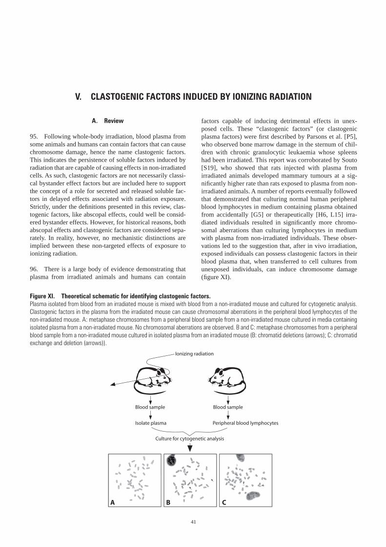
41
V. CLASTOGENIC FACTORS INDUCED BY IONIZING RADIATION
A. Review
95. Following whole-body irradiation, blood plasma from some animals and humans can contain factors that can cause chromosome damage, hence the name clastogenic factors. This indicates the persistence of soluble factors induced by radiation that are capable of causing effects in non-irradiated cells. As such, clastogenic factors are not necessarily classi-cal bystander effect factors but are included here to support the concept of a role for secreted and released soluble fac-tors in delayed effects associated with radiation exposure. Strictly, under the definitions presented in this review, clas-togenic factors, like abscopal effects, could well be consid-ered bystander effects. However, for historical reasons, both abscopal effects and clastogenic factors are considered sepa-rately. In reality, however, no mechanistic distinctions are implied between these non-targeted effects of exposure to ionizing radiation.
96. There is a large body of evidence demonstrating that plasma from irradiated animals and humans can contain
factors capable of inducing detrimental effects in unex-posed cells. These “clastogenic factors” (or clastogenic plasma factors) were first described by Parsons et al. [P5], who observed bone marrow damage in the sternum of chil-dren with chronic granulocytic leukaemia whose spleens had been irradiated. This report was corroborated by Souto [S19], who showed that rats injected with plasma from irradiated animals developed mammary tumours at a sig-nificantly higher rate than rats exposed to plasma from non-irradiated animals. A number of reports eventually followed that demonstrated that culturing normal human peripheral blood lymphocytes in medium containing plasma obtained from accidentally [G5] or therapeutically [H6, L15] irra-diated individuals resulted in significantly more chromo-somal aberrations than culturing lymphocytes in medium with plasma from non-irradiated individuals. These obser-vations led to the suggestion that, after in vivo irradiation, exposed individuals can possess clastogenic factors in their blood plasma that, when transferred to cell cultures from unexposed individuals, can induce chromosome damage (figure XI).
Figure XI. Theoretical schematic for identifying clastogenic factors. Plasma isolated from blood from an irradiated mouse is mixed with blood from a non-irradiated mouse and cultured for cytogenetic analysis. Clastogenic factors in the plasma from the irradiated mouse can cause chromosomal aberrations in the peripheral blood lymphocytes of the non-irradiated mouse. A: metaphase chromosomes from a peripheral blood sample from a non-irradiated mouse cultured in media containing isolated plasma from a non-irradiated mouse. No chromosomal aberrations are observed. B and C: metaphase chromosomes from a peripheral blood sample from a non-irradiated mouse cultured in isolated plasma from an irradiated mouse (B: chromatid deletions (arrows); C: chromatid exchange and deletion (arrows)).
Ionizing radiation
Blood sample
Isolate plasma Peripheral blood lymphocytes
Culture for cytogenetic analysis
Blood sample
A B C
42 UNSCEAR 2006 REPORT: VOLUME II
97. Clastogenic factors have been described in plasma from survivors of the atomic bombings in Japan [P2], per-sonnel involved in salvage operations after the Chernobyl accident [E8, E10] and children exposed as a consequence of the Chernobyl accident [E11, G3]. In addition, clastogenic factors have been reported in human [G1, L19, S10] and rat blood plasma [F1] after in vitro irradiation. Clastogenic fac-tors can be induced within 15 minutes of irradiation [F1] and appear to be very persistent or continuously regener-ated: times of 10 weeks post-irradiation have been reported for rats [F1], 7–10½ years for irradiated humans [E8, G5, G6] and >30 years for the atomic bombing survivors [P2]. Clastogenic factors reflect neither radiation-induced deple-tion of protective factors nor radiation-induced changes in normal plasma components, but rather represent products secreted or excreted by cellular elements as a result of irradi-ation [F1]. In vitro induction of clastogenic factors does not appear to be related to the dose [S10] or quality of radiation [G1]; however, this might not be the case in vivo [E8].
98. Emerit et al. [E8] investigated clastogenic factors in the plasma of 32 civil workers from Armenia who had been engaged as emergency workers around the Chernobyl atomic power station in 1986. They also included 15 emergency workers who had emigrated from the former Soviet Union to Israel. Reference plasma samples were obtained from 41 blood donors from the Armenian Blood Center in Yerevan. The samples were tested for their clastogenic activity in blood cultures from healthy donors. The samples from the first Armenian group, with the higher average radiation dose (0.6 ± 0.6 Gy), were more clastogenic than those from the second group, which had been exposed to 0.2 ± 0.2 Gy. The samples from the Israeli emergency workers also induced significantly increased aberration rates (14.0 ± 3.9% aber-rant cells). The clastogenic activity described above could be inhibited by superoxide dismutase [E8], indicating that the chromosome-damaging effects of radiation-induced clasto-genic factors are exerted via the intermediation of super-oxide radicals, as is known for clastogenic factors of different origin [E18, F12].
99. It should be stressed that there is variability between individuals in their ability to produce clastogenic factors [E11, G3], and not all irradiated individuals exhibit this effect [L4]. Indeed it is difficult to evaluate how common is the induction of clastogenic factors in the human population. On the one hand, blood samples from irradiated individuals are a valuable commodity, usually studied for more conven-tional biomarkers of radiation exposure. On the other hand, it is perhaps not surprising that there are very few reports failing to detect clastogenic factors, as such negative results are generally less likely to be published.
100. The precise nature of clastogenic factors is unknown, but endogenous viruses and compounds that interfere with DNA repair and/or increase the production of free radi-cals have all been implicated [E1, E2, E3]. On the basis of a number of inhibitor studies, the bulk of evidence sug-gests that the mechanism of action of clastogenic factors
is probably mediated by free radicals. Free radical scaven-gers such as superoxide dismutase, penicillamine, cysteine and various antioxidant plant extracts all reduce or elimi-nate clastogenic factor activity [E1, E2, E3]. The molecu-lar mechanisms for this effect and the specific nature of the factors capable of persisting, or of being regenerated over protracted time intervals (>30 years in the case of some of the atomic bombing survivors [P2]), remain unknown. Never theless, it is tempting to speculate on the poten-tial relationship between clasto genic factors and factors involved in the bystander effect. Both can be induced by ionizing radiation and the factors produced can cause genetic damage in non-irradiated cells. Given the current interest in bystander effects resulting from secreted fac-tors produced after cellular irradiation [S39], it may be an appropriate time to revisit clastogenic factors and evaluate the biological significance and nature of these radiation-induced secreted factors.
101. At present, the biological significance of clastogenic factors remains unclear [H11]. Furthermore, it would be misleading to imply that clastogenic factors are unique to radiation exposure. Transferable clastogenic effects have been described in blood plasma after whole-body stresses as diverse as asbestos exposure [E7] and ischaemia reper-fusion injury [E6], and occur spontaneously in patients with HIV-1 [E1], hepatitis C [E12], Crohn’s disease [E5] and scleroderma [A2]. The reports of diffusible clastogenic factors induced by irradiation also resemble the reports of clastogenic activity in the plasma of patients with certain inherited disorders, including Bloom’s syndrome [E4], ataxia-telangiectasia [S14] and Fanconi’s anaemia [E9]. Individuals with these chromosome breakage syndromes show an increased incidence of cancer, which begs the question as to the role of clastogenic factors in creating a cellular environment predisposed to increased genomic instability and ultimately neoplastic transformation [H11, W11].
102. Emerit and colleagues have carried out many of the studies investigating clastogenic factors. These investiga-tors have described these factors in plasma from individu-als exposed to different types of radiation under a variety of exposure conditions. In addition, Emerit’s group has described clastogenic factors after exposure to other DNA-damaging agents, as well as in individuals with a number of medical conditions and various genetic diseases. It would be misleading to imply that Emerit and colleagues are the only ones to describe clastogenic factors in various disease states. Reports from other laboratories lend support to these obser-vations (e.g. [B10, G1, S14]), but the biological significance and potential health hazards associated with clastogenic f actors remain to be determined.
103. Nevertheless, the presence of clastogenic factors in peripheral blood samples from some irradiated individuals raises intriguing questions concerning the role of chromo-somal rearrangements as dosimeters of radiation exposure [S10]. Cytogenetic analysis of first division metaphase
ANNEX C: NON-TARGETED AND DELAYED EFFECTS OF EXPOSURE TO IONIZING RADIATION 43
cells from irradiated individuals reveals both asymmetri-cal (dicentric and polycentric chromosomes as well as ring chromosomes) and symmetrical (reciprocal translocations) exchange-type aberrations, insertions, inversions and chro-mosomal breaks (reviewed in reference [C9]). The asymmet-rical exchange-type aberrations generally lead to prolifera-tive cell death in subsequent mitoses and decline over time. Symmetrical translocations, on the other hand, are generally stable over time and can persist, although they also appear to decline, albeit at a much slower rate than asymmetrical aber-rations [S20]. Since clastogenic factors from some irradiated individuals can induce chromosome damage, a role for these factors in the well-described persistence of chromosomal
rearrangements in blood samples from irradiated individuals is possible but unlikely.
B. Conclusions
104. It remains difficult to establish a clear description of the relevance of clastogenic factors to overall cellular responses to ionizing radiation, particularly at low doses. In part, this is due to the paucity of data on the nature of the factors and on their mechanism of action. Furthermore, how such clastogenic factors might influence the dose–response curve at low doses is not possible to discern at this time.

45
VI. IMPACT OF NON-TARGETED AND DELAYED EFFECTS OF RADIATION ON FUTURE GENERATIONS
105. It is important to consider whether non-targeted effects of radiation influence our consideration of the consequences of irradiation of a parent on end points in offspring. The fol-lowing sections summarize the data from non-human and human studies and consider them in the context of the Com-mittee’s current position on heritable effects of irradiation. Several diverse studies have provided a somewhat confusing picture of the potential non-targeted or delayed effects of ion-izing radiation in humans, mice and other organisms. Many of the studies present technical difficulties and ambigui-ties in interpretation, for example, with respect to uncertain radiation doses in human studies, potential strain dependency of responses in mouse studies, and poorly defined criteria used to define effects in some molecular studies. These com-plicating factors will be highlighted throughout this section.
106. Many of the studies examine effects in the F1 off-spring of irradiated parents, while other studies consider F2 and later generations. It is only these studies of F2 and later generations that can be unambiguously considered transgen-erational. This is because the F1 studies may be revealing mutations that occur during parental germ cell development. Thus F1 studies address “heritable effects”, while F2 and later generation studies address “transgenerational effects”.
A. Studies in non-mammalian species
107. The first demonstrated transmission of genomic insta-bility to subsequent generations was in Drosophila treated with ionizing radiation and mustard gas. However, the end point used was lethality, which revealed little about the nature or the mechanisms of the processes involved [A12].
108. Shima and colleagues have developed a “specific locus” test system using the Japanese medaka fish, Oryzias latipes [S40]. The genetic end points available are dominant lethal mutations, total “specific locus” mutations and viable “specific locus” mutations. The medaka has a transparent egg membrane and embryo body, and both visible mosaics and whole-body mutations can be detected during development at an early-expressed pigmentation locus [S40]. When wild-type +/+ males were gamma-irradiated and then mated with wl/wl females, the frequency of F
1 embryos with both wild-
type orange leucophores (wl/+) and mutant-type white leuco-phores (wl/wl*) (mosaic mutants) was ~5.7 × 10⁻³ Gy–1. The frequency of embryos with only white leucophores (whole-body mutants) was ~1.3 × 10⁻³ Gy–1. These results suggest that delayed mutations arise frequently in medaka fish embryos that have been fertilized with irradiated sperm [S41].
109. There was also a significant dose-rate effect for this type of mutation. Shimada et al. [S43] determined the fre-quency of “specific locus” mutations at five pigmentation loci in medaka spermatogonial stem cells after gamma irradiation at 0.03 cGy/min and 95 cGy/min. At each total dose, the mutation frequency was significantly lower in the 0.03 cGy/min group than in the 95 cGy/min group. The ratio of the induced mutation frequency at 0.03 cGy/min to that at 95 cGy/min was approximately 0.42 for doses of less than 1.9 cGy and approximately 0.33 for doses of 1.9–4.75 Gy [S43]. There was some specificity as to when such mutation events can be induced during spermatogenesis. When sperm and late spermatids were irradiated, the mutation frequency within non-irradiated maternally derived alleles was approx-imately three times higher than in the control group. In the F
2
generation, however, no increase in mutation frequency was observed. Similarly, there was no significant increase in the F
1 mutation frequency when stem cell spermatogonia were
irradiated. These data suggest that irradiation of sperm and late spermatids can induce indirect mutations in F
1 somatic
cells, supporting the idea that genomic instability arises dur-ing F
1 embryonic development. Moreover, such instability
apparently arises most frequently when eggs are fertilized just after the sperm are irradiated [S42]. It should be noted that dose-rate effects and germ-cell-stage specificities for mutational response were previously demonstrated for “spe-cific locus” mutations in mice, and such findings have been factored into past decisions of the Committee regarding the estimation of the risk of hereditary effects. Although interest-ing, these new findings in fish provide no reason to modify those estimates.
110. Microsatellite mutations have also been studied in plants grown in heavily contaminated areas near Chernobyl [K15, K16]. Kovalchuk et al. [K15] investigated the muta-tion rates of 13 microsatellite loci in wheat plants grown in a contaminated (900 Ci/km2) versus a control (<1 Ci/km2) area, and found a 3.6-fold increase in germinal muta-tion rate in the contaminated versus the control plot. Elle-gren et al. [E17] reported an increased frequency of partial albinism, a morphological aberration associated with loss of fitness, among barn swallows, Hirundo rustica, breed-ing close to Chernobyl. Heritability estimates indicate that mutations causing albinism were at least partly of germ line origin. Furthermore, evidence for an increased germ line mutation rate was obtained from segregation analysis at two hypervariable microsatellite loci, indicating that muta-tion events in barn swallows from Chernobyl were two- to tenfold higher than in birds from control areas in Ukraine and Italy.
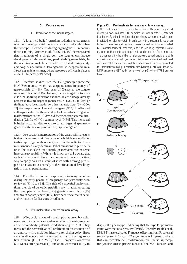
46 UNSCEAR 2006 REPORT: VOLUME II
B. Mouse studies
1. Irradiation of the mouse zygote
111. A long-held belief regarding radiation teratogenesis was that developmental defects are only inducible when the conceptus is irradiated during organogenesis. In contra-diction to this, Streffer et al. [M26, P1, P7] demonstrated that irradiation of a single cell, the zygote, can induce developmental abnormalities, particularly gastroschisis, in the resulting animal. Indeed, when irradiated during early embryogenesis, induced teratogenesis is suppressed in a TP53-dependent manner, where apoptotic cell death plays a critical role [K23, N23, N24].
112. Streffer’s studies used the Heiligenberger (now the HLG/Zte) mouse, which has a spontaneous frequency of gastroschisis of ~3%. One gray of X-rays to the zygote increased this to ~11%, leading the investigators to con-clude that ionizing radiation enhances latent damage already present in this predisposed mouse strain [H27, S34]. Similar findings have been made by other investigators [G4, G20, J7] after exposure to chemical mutagens [G15]. Streffer and colleagues extended these studies to demonstrate congenital malformations in the 19-day-old foetuses after paternal irra-diation (2.8 Gy of 137Cs gamma rays) [M64]. This increased lethality occurred after exposure of all stages of spermato-genesis with the exception of early spermatogonia.
113. One possible interpretation of the gastroschisis results is that this mouse strain has a peculiarly high susceptibility to this type of gross abnormality and that the radiation treat-ments induced many dominant lethal mutations in germ cells or in the pronucleus that greatly exacerbated this extreme inherent susceptibility. While it is important to be aware that such situations exist, there does not seem to be any practical way to apply data on a strain of mice with a strong predis-position to a serious anomaly to the estimation of hereditary risk in human populations.
114. The effect of in utero exposure to ionizing radiation during the early phases of pregnancy has previously been reviewed [J7, P1, S34]. The risk of congenital malforma-tions, the role of genomic instability after irradiation during the pre-implantation phase [S63], genetic susceptibility [J6] and health consequences [H17] have been reviewed in detail and will not be further considered here.
2. Pre-implantation embryo chimera assay
115. Wiley et al. have used a pre-implantation embryo chi-mera assay to demonstrate adverse effects in embryos after acute whole-body paternal irradiation (figure XII). They measured the competitive cell proliferation disadvantage of an embryo with a radiation history after challenge by direct cell-to-cell contact with a normal embryo in an aggrega-tion chimera [O1, O2, W10]. The F1 embryos conceived 6–7 weeks after paternal F0 irradiation were most likely to
display the phenotype, indicating that the type B spermato-gonia were the most sensitive [W10]. Recently, Baulch et al. [B4, B5] have evaluated F3 mouse offspring from F0 paternal mice exposed to 1 Gy of 137Cs gamma rays for gene products that can modulate cell proliferation rate, including recep-tor tyrosine kinase, protein kinase C and MAP kinases, and
1 Gy 137Cs gamma rays
Type BSpermatogonia
Radiation history
Litter mates
ANALYSIS
F0
F1
F2
F3
Figure XII. Pre-implantation embryo chimera assay. F0 CD1 male mice were exposed to 1 Gy of 137Cs gamma rays and mated to non-irradiated CD1 females six weeks after F0 paternal irradiation. F1 animals with a radiation history were mated with non-irradiated females to obtain F2 embryos with a paternal F0 radiation history. These four-cell embryos were paired with non-irradiated CD1 control four-cell embryos, and the resulting chimeras were cultured to the blastocyst stage and transferred to a foster mother. The pups resulting from the transfer were screened, and those with and without a paternal F0 radiation history were identified and bred with normal females. Sex-matched pairs could then be evaluated for competitive cell proliferation disadvantage, protein kinase C, MAP kinase and GST activities, as well as p21waf1 and TP53 protein levels.
ANNEX C: NON-TARGETED AND DELAYED EFFECTS OF EXPOSURE TO IONIZING RADIATION 47
protein levels of nuclear TP53 and p21waf1. All three-protein kinase activities were altered, and nuclear levels of TP53 and p21waf1 protein were higher in F3 offspring with a pater-nal F0 radiation history than in non-irradiated litter-mates. While there is clear evidence of effects, it is unclear how this rather novel and artificial phenotype can be related to clini-cally important hereditary effects, and thus no attempt will be made to apply these results to hereditary risk estimation in humans.
3. Mouse mutation assays
116. To investigate whether preconception paternal irra-diation can lead to the heritable transmission of genomic instability in mice, Luke et al. [L25] measured mutation fre-quency in a transgenic mouse model tagged with a lambda shuttle vector. This assay system allowed mutations in the lacI gene from the shuttle vector to be analysed in vitro after the animal had been irradiated in vivo. The results indicated that, as parental dose increased, there was a trend towards higher mutation frequency in vectors recovered from DNA from the bone marrow of F1 progeny. These data demonstrate heritable transmission of factors leading to genomic instabil-ity in F
1 progeny following paternal pre-conception irradia-
tion, although the results with a lambda shuttle vector would be complicated to apply in quantitative risk estimation for humans because they do not involve mammalian genes.
117. An increase in micronucleus frequency in bone marrow erythrocytes from the F1 progeny of male BALB/c mice exposed to chronic low-dose gamma irradiation was observed by Fomenko et al. [F11]. Mice were irradiated with 10, 25 or 50 cGy at dose rates of 1, 5 and 15 cGy/day, and were mated with non-irradiated females on day 15 after irradiation. The obtained offspring had an elevated micro-nucleus frequency in bone marrow erythrocytes at the age of two months. This suggests the transmission of genomic instability from damaged germ line cells of irradiated male parents to somatic cells of the progeny. It is unclear, how-ever, if the effects on micronuclei represent chromosome damage of consequence to clinical diseases beyond those already covered by the Committee’s current methods to estimate hereditary risk.
118. The pun mouse background allows visual detection of ~70 kb DNA deletions in the pink-eyed unstable (pun) locus in developing mouse embryos. These are scored as black spots on the light gray fur or black cells on the transparent retinal epithelium of the offspring [R2]. In the fur spot assay, 10-day-old pups are observed for black spots on the light gray fur, and the number of animals with fur spots is counted. In the eye-spot assay, mice are sacrificed at ~20 days, the eyes are removed and the number of black cells in whole mounts of the unpigmented retinal pigment epithelium are determined [R2]. The C57BL/6Jpun/pun mouse strain contains a 70 kb tandem duplication of the pink-eyed dilution (p) gene [B29], the pun mutation. The pun mutation is autosomal recessive and results in a dilute, light grey coat colour and pink eyes.
Intrachromosomal homologous recombination between the 70 kb repeats that delete one copy of a duplicated 70 kb DNA fragment at the pun locus restores the p gene and produces black pigment in the hair and retinal epithelium in wild-type mice. On the C57BL/6Jpun/pun inbred background, 5–10% of the mice spontaneously display fur spots and from four to six eye-spots per unpigmented retinal pigment epithelium.
119. The pun fur spot assay was used to demonstrate that exposure of the parental germ line to ionizing radiation results in induction of delayed DNA deletions in mouse off-spring [C16, S17]. Male pun assay/pun assay mice were irra-diated with 1 Gy of X-rays and mated 28 days later with non-irradiated pun assay/pun assay females. The offspring showed a higher frequency of large fur spots. Since deletions occurring early in embryogenesis should yield larger spots than events occurring later, the large spots indicated deletion events occurring early in embryo development and many cell divisions after irradiation [C16]. Shiraishi et al. [S17] irradiated male mice with 6 Gy and observed an increase in pun assay reversions resulting in eye-spots, in the irradi-ated paternal pun assay allele as well as in the non-irradiated maternal pun assay allele, indicating untargeted recombina-tion in the offspring. The number of spots per retinal epithe-lium increased twofold when the male was irradiated at the spermatozoa stage [S17], but pun assay instability was not observed when radiation was delivered either to spermato-gonial stem cells or to late spermatids. It is noteworthy that radiation-induced instability of the pun assay allele has been observed in F1 mice but not in the F2 generation (reported in reference [N22]).
120. Estimates by the Committee of hereditary risk for exposures of males have been based on damage to sperma-togonial stem cells because they are the only germ cells in males that can accumulate appreciable doses under low-dose-rate exposure conditions. The finding that there is no induced pun instability in spermatogonial stem cells suggests that there is no need to revise current risk estimates upward.
4. Alterations in tandem repeat DNA sequences
121. Alterations in tandem repeat DNA sequences, such as minisatellite DNA and expanded simple tandem repeats (ESTRs), in the genome have been used as markers of genetic change. Such loci have high spontaneous rates of mutation, which facilitates the measurement of induced mutation in relatively small numbers of samples. Alterations (mutations) are manifested as gains or losses in repeat units and are detected either by pedigree screening or by amplifi-cations [N22, Y4]. Mutations in both minisatellite sequences and ESTRs appear to arise via indirect mechanisms rather than by direct damage to the repeat locus itself [Y4]. The significance of these DNA sequence changes is unknown. If they are genetically neutral they will not affect risk; never-theless, the fact that they happen indicates that exposure to ionizing radiation can lead to genomic destabilization that may occur by a non-targeted mechanism.
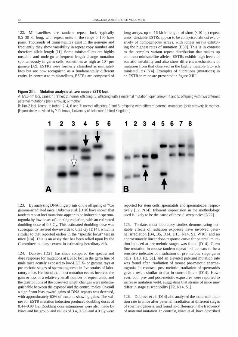
48 UNSCEAR 2006 REPORT: VOLUME II
122. Minisatellites are tandem repeat loci, typically 0.5–30 kb long, with repeat units in the range 6–100 base pairs. Thousands of minisatellites exist in the genome and frequently they show variability in repeat copy number and therefore allele length [J1]. Some minisatellites are highly unstable and undergo a frequent length change mutation spontaneously in germ cells, sometimes as high as 10-1 per gamete [J2]. ESTRs were formerly classified as minisatel-lites but are now recognized as a fundamentally different entity. In contrast to minisatellites, ESTRs are composed of
long arrays, up to 16 kb in length, of short (<10 bp) repeat units. Unstable ESTRs appear to be comprised almost exclu-sively of homogeneous arrays, with longer arrays exhibit-ing the highest rates of mutation [B30]. This is in contrast to the complex variant repeat distribution that makes up common minisatellite alleles. ESTRs exhibit high levels of somatic instability and also show different mechanisms of mutation from that observed in the highly mutable GC-rich mini satellites [Y4]. Examples of alterations (mutations) in an ESTR in mice are presented in figure XIII.
Figure XIII. Mutation analysis at two mouse ESTR loci. A: Ms6-hm loci. Lanes: 1: father; 2: normal offspring; 3: offspring with a maternal mutation (open arrow); 4 and 5: offspring with two different paternal mutations (dark arrows); 6: mother. B: Hm-2 loci. Lanes: 1: father; 2, 4, 6 and 7: normal offspring; 3 and 5: offspring with different paternal mutations (dark arrows); 8: mother. [Figure kindly provided by Y. Dubrova, University of Leicester, United Kingdom.]
123. By analysing DNA fingerprints of the offspring of 60Co gamma-irradiated mice, Dubrova et al. [D10] have shown that tandem repeat loci mutations appear to be induced in sperma-togonia by low doses of ionizing radiation, with an estimated doubling dose of 0.5 Gy. This estimated doubling dose was subsequently revised downwards to 0.33 Gy [D14], which is similar to that reported earlier in the “specific locus” test in mice [R4]. This is an assay that has been relied upon by the Committee to a large extent in estimating hereditary risk.
124. Dubrova [D21] has since compared the spectra and dose response for mutations at ESTR loci in the germ line of male mice acutely exposed to low-LET X- or gamma rays at pre-meiotic stages of spermatogenesis in five strains of labo-ratory mice. He found that most mutation events involved the gain or loss of a relatively small number of repeat units, and the distributions of the observed length changes were indistin-guishable between the exposed and the control males. Overall a significant bias toward gains of DNA repeats was detected, with approximately 60% of mutants showing gains. The val-ues for ESTR mutation induction produced doubling doses of 0.44–0.98 Gy. Doubling dose estimations were also made by Niwa and his group, and values of 3.4, 0.893 and 4.0 Gy were
reported for stem cells, spermatids and spermatozoa, respec-tively [F2, N14]. Inherent imprecision in the methodology used is likely to be the cause of these discrepancies [N22].
125. To date, most laboratory studies demonstrating her-itable effects of radiation exposure have involved pater-nal irradiation [B4, B5, D14, D15, N14, S1, W10], and an approximately linear dose-response curve for paternal muta-tion induced at pre-meiotic stages was found [D14]. Germ line mutation in mouse tandem repeat loci appears to be a sensitive indicator of irradiation of pre-meiotic stage germ cells [D10, F2, S1], and an elevated paternal mutation rate was found after irradiation of mouse pre-meiotic sperma-togonia. In contrast, post-meiotic irradiation of spermatids gave a result similar to that in control litters [D14]. How-ever, both pre- and post-meiotic exposures were reported to increase mutation yield, suggesting that strains of mice may differ in stage susceptibility [F2, N14, S1].
126. Dubrova et al. [D14] also analysed the maternal muta-tion rate in mice after paternal irradiation at different stages of spermatogenesis, and found no difference in the frequency of maternal mutation. In contrast, Niwa et al. have described
ANNEX C: NON-TARGETED AND DELAYED EFFECTS OF EXPOSURE TO IONIZING RADIATION 49
a small but statistically significant increase in the maternally derived Ms6hm allele, in addition to the paternally derived allele, when only male parents were irradiated at the sper-matozoa stage [F2, N14, S1]. Niwa and Kominami [N14] demonstrated a statistically significant increase in mater-nal allelic mutation rate in F1 mice born to irradiated male parents. The authors concluded that, as a consequence of male (sperm) irradiation, genomic instability is triggered in the zygote, which then mutates the paternally derived allele in cis and the maternally derived allele in trans.
127. Mutation rates at two ESTR loci have been studied in the germ line of first- and second-generation offspring of inbred male CBA/H, C57BL/6 and BALB/c mice exposed to either neutrons or X-rays. Paternal CBA/H exposure resulted in increased mutation rates in the germ line of two subse-quent generations. Comparable transgenerational effects were observed also in neutron-irradiated C57BL/6 and X-irradiated BALB/c mice. The spontaneous mutation rates and radiation-induced transgenerational instability varied between strains (BALB/c>CBA/H>C57BL/6). Pre- and post-meiotic paternal exposure resulted in similar increases in mutation rate in the germ line of both generations of CBA/H mice, which suggests that radiation-induced expanded simple tandem repeat insta-bility is manifested in diploid cells after fertilization [B2]. Although there are some difficulties in interpreting these data [B41], they do suggest that excess transgenerational muta-tion at the unstable loci may be detected after irradiation of developing male germ cells.
128. Analysis of ESTR mutation induction in the mouse germ line suggests that mutations are likely to be due to pausing of the replication fork and subsequent polymerase slippage events [B31, D21]. The frequency of induced muta-tion appears to be significantly greater than the predicted fre-quency of radiation-induced DNA damage at a given tandem repeat locus. This has led many investigators to conclude that the increased mutation rates observed at these repeats are not necessarily the result of directly induced DNA damage at the “specific locus”, but result from radiation-induced damage elsewhere in the genome or cell [D13, D14, D15, F2]. While alterations in tandem repeat DNA sequences may provide useful biomarkers of induced germ line effects, the biological significance of such mutations remains unknown. The similarity of the calculated doubling doses to those pub-lished for “specific-locus” mutations in mice supports the validity of the tandem repeat DNA sequence data, although the uncertainty in doubling doses in “specific-locus” experi-ments needs to be borne in mind [U1]. The comparative data between germ cell stages for mutations that affect tandem repeat DNA sequences remain of interest and potential importance.
5. Tumour induction in the offspring of irradiated parents
129. Nomura first reported a significant increase in lung tumours, mostly benign adenomas, in the F1 offspring of X-irradiated ICR parental mice [N17, N18, N28]. This has
been confirmed in some studies [H10, L20, L21, V3] but not all. For example, Cattanach et al. [C4], using the optimal experimental conditions defined by Nomura [N17], found that tumour incidence was no higher in the offspring of irra-diated BALB/cJ mice than in the non-irradiated controls. They did find that the proportion of fertile females and mean litter size were affected by the radiation, showing a dose-dependent, dominant lethal response. In attempting to rec-oncile these differences, Cattanach et al. [C4] proposed that inconsistencies in the animal experiments may in part be due to lack of an appropriate concurrent control, whose periodic or cyclic variation in tumour incidence may have been out of phase with that in the treated animals. Alternatively, the reported differences could reflect strain differences in the mice used.
130. Selby and Priest [S52] reported no induced leukae-mias when male CBA/Ca mice were injected with 239Pu citrate solutions at nominal activities of 6 and 60 Bq/g, to give absorbed doses of approximately 0.3 and 4.0 cGy, and were mated to females of the same strain 54–68 days later. Nomura [N16] found no increase in leukaemia in the off-spring of ICR mice derived from spermatogonia after acute irradiation. In contrast, when spermatogonia from the N5 mouse strain were irradiated, Nomura found a 10-fold-greater incidence of acute lymphocytic leukaemia in the off-spring than in the non-irradiated controls. Once again, this may be explained by differences in genetic predisposition to leukaemia induction by radiation in these mouse strains.
131. Extending his original findings, Nomura hypothe-sized that, if radiation-induced mutations in the germ line led to heritable lung tumours in offspring, then all the cells in the lung should be at increased tumorigenic risk. Following a subsequent challenge with the carcinogen urethane, Nomura described significantly increased clusters of tumour nodules in the lung [N17, N18]. Vorobtsova and Kitaev [V3] reported similar findings, but Cattanach et al. [C21] were unable to replicate these results in C3H/HeH mice. Selby et al. [S53] suggested that the explanation for the surprisingly high rates of induction of dominant mutations that cause tumours sug-gested by the experiments of Nomura and others might result from the confounding effect of the radiation-induced domi-nant lethality that often occurs in such experiments. Other possible non-mutational explanations for the high mutation rates reported for dominant tumour mutations by Nomura have been suggested by Selby [S51] and Cattanach et al. [C4, C21].
132. Lord et al. have also investigated the heritable effects of pre-conception paternal irradiation from injected pluto-nium (239Pu alpha particle irradiation), 137Cs gamma rays or the Auger-electron-emitting radionuclide 55Fe. They demon-strated perturbed haemopoiesis in offspring, as well as enhanced sensitivity to methylnitrosourea (MNU, 50 mg/kg) as a secondary carcinogenic insult [H10, L20, L21]. A major difference from the tumour experiments discussed above was that Lord et al. used much lower doses of radiation. As a result, there would have been little or no induced dominant
50 UNSCEAR 2006 REPORT: VOLUME II
lethality, and the possibility that this was a confounding effect is therefore removed. The mutation rates calculated from these results suggest that Nomura’s results actually underestimated the extent of this phenomenon. Since these studies rely on a secondary treatment with a carcinogen, there seems to be no way to apply their results to revise the Committee’s estimates of hereditary risk, which are made for radiation alone.
C. Malformation induction in the offspring of irradiated parents
133. There are several studies addressing the question of malformation induction after irradiation of either female [K13, L50, M65, N17, N31, N32, N34, W22, W23] or male [K12, K36, M64, N17, N31, N32, N34, R15] mice and looking for malformations in the next or subsequent [L50, L51, N33, P7] generations. In all of these studies, condi-tions were found that resulted in transmission of radiation-induced germ cell effects in the form of malformations to the F
1 or subsequent generations. The mechanism for the
development of these malformations is apparently differ-ent under these conditions from that which is responsible for the development of malformations induced by radiation exposures during major organogenesis [S34]. The following conclusions can be drawn from the experiments: compara-tively high doses (>1 Gy) are required for transgenerational malformation effects to be detected. The effects seen after female exposure are not due to indirect effects because of radiation sickness of the mother, but must have, at least partly, a genetic background [L50, W22, W23]. This genetic background is quite obvious after radiation exposure of male mice. These transgenerational effects are not restricted to low-LET radiation [K36]. It seems unlikely that there is a direct relationship between chromosomal translocations and congenital anomalies [L50, N34], although some suspicion in that direction has been expressed [R15]. The basis of the malformations observed seems to be heterogeneous: some are due to genetic changes of high penetrance that are rap-idly eliminated; some are due to modification of genes of low penetrance; and some are probably of non-genetic origin [L50]. There are strain-specific differences as to the extent of the transgenerational effect [R15]. There are indications that a major proportion of mutations are eliminated in the first generation and that only a minor proportion are transmitted to later generations [L50].
134. At present there are too many uncertainties about those data suggesting that dominant mutations and/or genomic instability cause tumours in progeny of irradiated mice to be able to apply such data in hereditary risk estimation.
D. Human studies
135. To date, no radiation-induced genetic, i.e. heredi-tary, diseases have been demonstrated in human popula-tions exposed to ionizing radiation. Neither the offspring of
individuals treated with chemotherapy and/or radiotherapy for cancer [B18] nor the offspring of women treated with radiation during infancy for haemangiomas [K6] demon-strated any significant effects attributable to parental expo-sure to chemicals or radiation [U1]. Furthermore, a number of studies involving the children of survivors of the Hiro-shima and Nagasaki atomic bombings have failed to detect any transmitted genetic effects of radiation exposure [K14, N12, N13, S4, S5]. A cohort of 31,150 children born to par-ents who were within 2 kilometres of the hypocentre at the time of the bombing was compared with a control cohort of 41,066 children. During the children’s early years, congeni-tal defects, sexual development, physical development and survival were all investigated. Later, cytogenetic studies and the electrophoretic properties of a series of serum proteins or erythrocytic enzymes were analysed, in addition to a com-plete medical evaluation. None of these indicators was sig-nificantly modified by parental radiation exposure (reviewed in reference [N10]).
136. In addition, in a study that examined 50 families exposed after the Hiroshima and Nagasaki atomic bombings with 64 children, and 50 control families with 60 children [K14], minisatellite analyses revealed no genetic effects at six human tandem repeat. This study was later expanded to include analysis of mutations at eight hypervariable mini-satellite loci in the offspring (61 from exposed families, in 60 of which only one parent was exposed, and 58 from unexposed parents) of atomic bombing survivors with mean doses of >1 Sv. They found 44 mutations in paternal alleles and 8 mutations in maternal alleles, with no indication that the high doses of acutely applied radiation had caused significant genetic effects [K28].
137. In contrast to these observations, Dubrova et al. [D11] described elevated mutation rates in DNA tandem repeat sequences in humans living in rural areas of the Mogilev district of Belarus, which was heavily contaminated with radionuclides from the Chernobyl reactor accident. The fre-quency of mutation was assayed both by DNA fingerprint-ing using one multilocus probe and by single-locus analysis using four probes, and this revealed mutation rates approxi-mately twofold higher in the offspring of exposed parents when compared with an unexposed population from the United Kingdom. These initial observations were expanded to include analysis of families from rural areas in the Kiev and Zhytomir regions of Ukraine, which were heavily con-taminated by radionuclides after the Chernobyl accident. A statistically significant 1.6-fold increase in mutation rate was found in the germ line of exposed fathers, whereas the maternal germ line mutation rate in the exposed families was not elevated [D9].
138. The initial report by Dubrova et al. [D11] generated commentary centred largely on the selection of a non-exposed population from the United Kingdom for comparison with exposed parents from Belarus [S4], the failure to exclude other contaminants such as pollutants and viral infections [N11], and questions regarding the biological significance of
ANNEX C: NON-TARGETED AND DELAYED EFFECTS OF EXPOSURE TO IONIZING RADIATION 51
increased mutations in hypervariable tandem repeat alleles [N10]. These criticisms initially appeared to detract from the significance of the findings of Dubrova et al. However, in a subsequent report, Dubrova et al. [D12] recruited more fami-lies from the affected region and used five additional mini-satellite probes, and once again their data indicated a twofold higher mutation rate in exposed families than in non- irradiated families from the United Kingdom. They also used individual radiation doses for external and internal chronic exposure to 137Cs as an indicator of long-term population exposure and found a significant positive correlation between radiation dose and mutation rate over multiple loci with the exposed families. Significantly, there were no obvious differences in the muta-tion spectrum observed between the exposed and the con-trol families. In subsequent studies, children born to parents who participated in recovery operations after the Chernobyl accident also showed an elevated mutation rate at some loci [L18, W5], as did children born to parents living around the S emipalatinsk nuclear test site in Kazakhstan [D8].
139. It should be stressed that the analysis of tandem repeat loci mutation rate demands sophisticated molecular biology, and it is important that observed mutants be vali-dated. Jeffreys and Dubrova [J3] suggested that technical artefacts might explain the sevenfold increase in mutation rate in child ren of Chernobyl recovery operations work-ers described by Weinberg et al. [W5]. Questions of valid paternity, sample mix-up, variation between DNA samples and the demands of the required technology are all potential sources of variability and could explain differences in results between different investigators.
140. Nevertheless, other studies have failed to reproduce these positive findings [F15, L17, S55]. A way of reconcil-ing these apparently contradictory results was offered by Livshits et al. [L17]. They measured the frequency of inher-ited mutant alleles at seven hypermutable minisatellite loci in 183 children born to Chernobyl recovery operations work-ers and in 163 children born to control families living in non-irradiated areas of Ukraine. No significant difference in the frequency of inherited mutant alleles was found between the exposed and the control groups. The exposed group was then divided into two subgroups according to the time at which the children were conceived in relation to the fathers’ work at the power plant. Eighty-eight children were conceived either while their fathers were employed at the facility or up to 2 months later (subgroup 1). The other 95 children were conceived at least 4 months after their fathers had stopped working at the Chernobyl site (subgroup 2). The frequencies of mutant alleles were higher for the majority of loci in sub-group 1 than in subgroup 2, suggesting that the timing of irra-diation during spermatogenesis had affected its mutagenic potential.
141. Given the frequency of mutations in these hyper variable alleles and the lack of evidence for significant differences in the mutation spectrum between control and exposed families [D12], it is unlikely that the minisatellite loci themselves are the direct targets of the radiation. If the increased mutation
rate is not caused by DNA damage directly, it might well result from non-targeted events associated with radiation-induced genomic instability [D13, D16, F2, N14, S1].
142. The reasons for the discrepancy between the data collected from the children of survivors of the Hiroshima and Nagasaki bombings and the induction of human germ line mutation in the majority of studies involving the off-spring of parents living in radiation-contaminated environ-ments are not easily reconciled. The types of radiation to which individuals were exposed differed between the two populations, and there are uncertainties associated with the doses. The bombings resulted in a single acute exposure to predominantly gamma radiation and a small amount of neutron radiation, whereas contamination from Chernobyl resulted in chronic exposures to internalized 131I. However, this is unlikely to be the only reason for the discrepancy, because there are also negative results for the children of recovery operations workers exposed to low-dose-rate external/internal exposures [F15, S55].
143. The controversy surrounding the induction of muta-tions in tandem repeat sequences is far from resolved. May et al. [M5] examined the mutation frequency at hyper variable tandem repeats in sperm from three seminoma patients following hemipelvic radiotherapy. Scattered radiation doses to the testicles were monitored, and the mutation rates in pre-treatment sperm DNA were compared with sperm derived from irradiated meiotic and post-meiotic cells. No evidence for radiation-induced germ line mutation at these hyper variable loci was observed even though the patients were monitored for a period of 1 to 11 months.
144. Of all the reported studies, it is only those of the popu-lations living in the Semipalatinsk region of Kazakhstan that permit comment on the potential transgenerational effects (i.e. F2+ generations). In one such study, the F2 offspring of those parents that received the highest external and internal doses did not show any elevation of minisatellite mutation frequency [D8].
145. These and related studies have been summarized in table 2 and have recently been subject to critical review [B41]. To summarize the conclusions of Bouffler and the expert review panel [B41]: only limited data are available indicating that germ line mutation of minisatellites can be detected in irradiated human populations. The data are incon-sistent and show only limited evidence of dose dependence, and the panel found that additional work would be necessary to establish the radiation responsiveness of these loci. Fur-thermore, the data on mutation of human tandemly repeated DNA loci do not warrant a dramatic revision of germ line or cancer risk estimates for radiation at present [B41].
E. Cancer incidence in the offspring of irradiated humans
146. The risk of cancer in the offspring of humans irra-diated prior to conception is also controversial. No excess
52 UNSCEAR 2006 REPORT: VOLUME II
cancer incidence has been reported in children born to par-ents exposed to ionizing radiation by the atomic bombings in Japan [Y1, Y2] or in the offspring of cancer patients treated with radiotherapy [H4].
147. A major event early in this controversy was the con-clusion by Gardner et al. [G2] from a case-control study that the increased incidence of leukaemia and non-Hodgkin’s lymphoma among children living near the nuclear reprocess-ing plant in Sellafield in the United Kingdom was associated with paternal employment and the recorded external dose of whole-body radiation during work at the plant before con-ception. This conclusion was controversial [D6] and was not supported by the excess of childhood leukaemia observed at nearby Seascale [P4], or by an extensive study of radiation workers and childhood cancers [D7]. Nevertheless, Dickin-son and Parker [D4] published results of a cohort study that supports the initial association with paternal radiation dose, which suggests that it still remains a possible explanation [B15], although population mixing [G17] is regarded as a potentially important factor in this particular cluster.
148. The United Kingdom Committee on Medical Aspects of Radiation in the Environment (COMARE), in their seventh report, reviewed the evidence concerning the incidence of childhood leukaemia and other cancers in the offspring of parents occupationally exposed to radia-tion prior to conception [C17]. The COMARE agreed that, while a link between parental exposure and such effects in the offspring was possible in principle, the epidemio-logical evidence from the offspring of radiation workers in the United Kingdom, the United States and Germany failed to support an increased rate of solid tumours in children. Furthermore, COMARE concluded that the balance of evi-dence indicated that the likelihood of developing childhood leukaemia or non-Hodgkin’s lymphoma was not related to radiation dose.
F. Pregnancy outcomes in the offspring of irradiated humans
149. In their eighth report, COMARE reviewed pregnancy outcomes following pre-conception exposure to ionizing radiation in humans [C18]. They found that the available epidemiological data were inadequate to allow definitive statements about the effect of pre-conception radiation expo-sure on pregnancy outcomes. This was due to difficulties in obtaining reliable figures for the end points of concern and to the possibility that the radiation exposures in most studies may have been too low to produce a detectable effect. The conclusion reached by COMARE was that, from all the epi-demiological data examined, there was little evidence that adverse reproductive outcomes in general are related to parental radiation exposure. Similarly, the limited data avail-able did not link miscarriage or neonatal death with parental irradiation. COMARE did point out, however, that almost all of the published studies on pregnancy outcome follow-ing parental exposure to radiation in human populations
lack statistical power; this is probably due to the low doses to which the populations were exposed and to the small population sample size.
150. The Scientific Committee takes note of these conclu-sions by COMARE, which support its own view that the types of genetic data discussed in this annex imply no modification of its own estimates of hereditary risk. It has been the Scien-tific Committee’s position that the mutation rates in experi-mental organisms upon which it has based its estimates of hereditary risk are sufficiently low as to make it unlikely that analyses of the radiation-exposed human populations avail-able for study would show statistically significant increases in hereditary diseases.
G. Genetic damage and malformation induction in the offspring of irradiated parents
151. The densely populated costal regions of Kerala state in southwest India have deposits of radioactive monazite-bearing sand and provide a unique opportunity to investigate the effects of high levels of natural radiation on human popu-lations [N35]. The background radiation levels range from ≤1.0 mGy to >35.0 mGy per year owing to naturally occur-ring thorium and its decay products. There is a comprehen-sive programme to assess the biological and health effects of this radiation exposure in humans, focusing mainly on constitutional chromosome abnormalities and the incidence of congenital malformations in newborns. To date, the data do not reveal any effect on cytogenetic aberrations in lym-phocytes [C24] or the incidence of congenital malformations in newborns [J11] that can be associated with exposure to ionizing radiation.
H. Impact of non-targeted and delayed effects of radiation on future generations
152. Heritable effects are observed in first-generation off-spring and/or in later generations after one or both parents have been irradiated prior to conception. Since it was estab-lished, the Committee has made estimates of the genetic effects of radiation in humans in offspring of irradiated parents based upon clear demonstrations that mutations can be induced in experimental organisms, including experimen-tal mammals. The UNSCEAR 2001 Report on the hereditary effects of radiation emphasized that no radiation-induced genetic (i.e. hereditary) diseases have so far been demon-strated in human populations exposed to ionizing radia-tion [U1]. No demonstrable adverse reproductive outcomes were described for the survivors of the atomic bombings in Japan, or for women irradiated during infancy for skin hae-mangiomas. No demonstrable hereditary effects of radiation exposure resulting from the Chernobyl accident have been described [U2]. Likewise, no increase in cytogenetic abnor-malities [W19] or genetic effects [B39] has been reported in survivors of childhood cancer exposed to ionizing radiation before reproduction.
ANNEX C: NON-TARGETED AND DELAYED EFFECTS OF EXPOSURE TO IONIZING RADIATION 53
153. Ionizing radiation is considered a universal mutagen. Experimental studies in plants and animals have demon-strated that radiation can induce hereditary effects, and humans are unlikely to be an exception in this regard. It was for this reason that the Committee estimated hereditary risk in humans in the absence of direct evidence in humans. This annex presents a re-evaluation of some of the controversial data, in view of newer findings, and also a review of some types of damage not considered in earlier UNSCEAR reports. Two assumptions are commonly made in the estimation of genetic risk: (1) that the seven “specific loci” in the mouse constitute a suitable basis for extrapolation to genetic dis-ease in humans; and (2) that heritable mutations are induced by radiation damage (energy-loss events leading to double-strand damage) occurring within the genome and are induced linearly with dose, at least at low doses. The issues of main importance in this section are whether the information on
the types of mutation considered below might (1) be used to improve the Committee’s estimates of hereditary risk, and (2) indicate some type of genetic instability that could lead to modification of risk through subsequent generations.
154. Overall it is clear that irradiation of the parent can lead to some changes in the offspring, but it is likely that most of these are due to the manifestation of direct damage caused by radiation in the original germ cell. The high incidence of off-spring with “mutations” in ESTRs and the detection of muta-tions in maternal alleles after paternal irradiation suggest that non-targeted instability may be induced in specific circum-stances. There is only very limited evidence that instability is transmitted across into the F2 generation, and human data are negative. Therefore the Committee considers that these data are insufficient to justify modification of current risk estimates for hereditary effects or cancer in humans.

55
VII. IMPLICATIONS OF NON-TARGETED AND DELAYED EFFECTS
155. A wealth of information has been reviewed that deals with possible radiation-induced non-targeted and delayed genetic effects, as well as genetic end points that can occur spontaneously. Most of the estimates of hereditary risk made by the Committee in the past have been based on classical mutation experiments. Such studies often require exceedingly large samples of offspring. For the reasons described above, it does not appear that the new findings necessitate changes in the Committee’s estimates of hereditary risk. It is possible that certain types of genetic damage detected by some of the assays have no relationship to clinically important phenotypes. It is also possible that the methods of hereditary risk estimation used by the Committee in the past adequately incorporate any genetic risks of clinical rele vance that might be associated with the damage detected by the various assays considered. While it is clear that some of the assays permit demonstration of effects of radiation using much smaller sample sizes than more classical methods, uncertainty remains as to whether these effects correlate with the rare types of mutation that cause clinically serious conditions. There is no convincing evidence of transgenerational instability in humans caused by radiation that would lead to the propagation of clinically important effects over succeeding generations. Because the Committee’s current risk estimates already assume transmis-sion of many of the effects found in the first generation to later generations, rare instances of such propagation would have little impact on total risk estimates.
156. The relevance of non-targeted and delayed effects to the development of cancer and hereditary effects is not yet clear. Carcinogenesis involves a progression of genetic events that are associated with specific stages of the malig-nant process. It is tempting to speculate that induced genomic instability can drive the progression of genetic changes and thus provide the impetus for acquiring those genomic alterations associated with carcino genesis. Yet this must be tempered by the high frequency with which instability is observed both in vitro and in vivo, and the observation that instability generally tends to saturate at low doses of radiation. Nevertheless, if radiation exposure induces a transgenerational instability that could be passed through the germ line and increase a child’s susceptibility to cancer or genetic effects, this would have health rami-fications. Bystander effects could also have significant implications for human exposures, particularly to very low fluences of high-LET radiation, e.g. to radon, where only a small fraction of the cell population would be hit, i.e. subject to energy deposition events. However, bystander effects appear to be limited to the irradiated organ, and since risk estimates are to an organ and not a cell, bystander
effects are essentially encompassed in current radiation risk estimates for carcinogenesis.
157. Radiation-induced instability and the existence of bystander effects are well established and incontrovertible. A common observation of these responses is that they dominate at low doses and saturate with increasing dose (reviewed in references [P13, S18]). In addition to damage directly induced by the deposition of energy in the nucleus of the irradiated cell, consideration must now be given to these indirect effects of radiation. An irradiated cell can send out a signal and induce a response in a cell whose nucleus was not hit by radiation. This might result in genetic damage, genomic instability or lethality in non-irradiated cells. These non-targeted effects in essence “amplify” the biological effectiveness of a given radi-ation dose by increasing the number of cells that experience effects over those directly exposed to the radiation.
158. Understanding of these non-targeted effects is still in its infancy, and much of the data to date have been obtained from in vitro studies. While the significance of these indirect effects for human health remains to be elucidated, it would seem prudent to consider the implications of non-targeted delayed effects of radiation exposure when considering models of radiation carcinogenesis, particularly at low doses.
159. In vivo non-targeted effects are not new and have pre-viously been implicated in radiation-induced carcinogenesis [S22]. The recent revival of interest in these non- targeted effects and the subsequent influx of new data suggest that it is time to re-examine the concepts of radiation dose and target size. Many of the indirect effects described indicate that the tissue volume in which detrimental effects of radiation may be observed is larger than the precise volume irradiated. This issue may have important implications for human health. Life exists in a radiation environment, the use of radiation has become an integral part of modern life, and the applications of radiation in medicine and industry bring tremendous bene fits to society. Over time, biological systems have demonstrated a remarkable ability to adapt to environments to which they are gradually exposed, and low doses of radiation are no excep-tion. Higher doses of radiation can cause neoplasia. This pre-sumably occurs through a combination of direct damage and non-targeted effects, and models of radiation-induced car-cinogenesis should incorporate both direct and indirect effects when evaluating radiation risks. Ultimately, understanding the multitude of multicellular responses to radiation may pro-vide a framework for evaluating health risks associated with radiation exposure and a logical means of intervening in the development of suspected radiation-induced cancers.

57
CONCLUDING REMARKS
160. This annex has reviewed a multitude of studies on non-targeted and delayed effects of exposure to ionizing radia-tion. In addition to damage directly induced by the deposition of energy in the nucleus of an irradiated cell, consideration should now be given to these indirect effects of radiation. An irradiated cell can send out signals and induce a response in a cell whose nucleus was not subject to energy deposition events following irradiation. These non-targeted effects in essence “amplify” the biological effectiveness of a given radiation dose by increasing the number of cells that experience effects over those directly exposed to the radiation.
161. In spite of the large body of new information avail-able, considerable disagreement remains concerning any definitive relationship between these non-targeted effects and the observed health effects attributable to radiation.
162. The Committee stresses that direct epidemiologi-cal observations and associated quantification of the health effects of radiation incorporate all mechanistic elements, including the targeted (direct) effects of irradiation as well as the non-targeted and delayed effects described in this report.
163. A specific role for non-targeted effects in the observed health effects associated with radiation exposure cannot be
determined directly. Such effects can provide mechanistic information at doses of below ~200 mGy that could be per-tinent to evaluating health effects at these low doses. How-ever, in ascribing a mechanism to a particular biological effect, the data in question should be independently repli-cated and show a strong coherence with the particular end point considered. The UNSCEAR 2000 Report considered the conventional view that the deposition of energy in the nucleus and the subsequent cellular processing of induced DNA damage were consistent with the observed cancer/ heritable effects induced by ionizing radiation.
164. In light of these considerations, the overall view of the Committee is that the data currently available do not require changes in radiation risk coefficients for cancer and hereditary effects of radiation in humans. The Com-mittee will maintain surveillance of developments in the area of non-targeted and delayed effects, and recommends that future research pay particular attention to study design emphasizing replication, low-dose responses and associa-tions with health effects particularly in the human popula-tion. Ultimately, understanding the range and multitude of multicellular responses to radiation will provide mechanis-tic insights into how radiation induces its observed health effects.

ANNEX C: NON-TARGETED AND DELAYED EFFECTS OF EXPOSURE TO IONIZING RADIATION 59
References
A1 Abramsson-Zetterberg, L., G. Zetterberg, S. Sundell-Bergman et al. Absence of genomic instability in mice following prenatal low dose-rate gamma-irradiation. Int. J. Radiat. Biol. 76(7): 971-977 (2000).
A2 Auclair, C., A. Gouyette, A. Levy et al. Clastogenic inosine nucleotide as components of the chromo-some breakage factor in scleroderma patients. Arch. Biochem. Biophys. 278(1): 238-244 (1990).
A3 Azzam, E.I., S.M. de Toledo, T. Gooding et al. Intercellular communication is involved in the bystander regulation of gene expression in human cells exposed to very low fluences of alpha parti-cles. Radiat. Res. 150(5): 497-504 (1998).
A4 Azzam, E.I., S.M. de Toledo and J.B. Little. Direct evi-dence for the participation of gap junction- mediated intercellular communication in the transmission of damage signals from alpha-particle irradiated to non-irradiated cells. Proc. Natl. Acad. Sci. U.S.A. 98(2): 473-478 (2001).
A5 Azzam, E.I., S.M. de Toledo and J.B. Little. Oxida-tive metabolism, gap junctions and the ionizing radi-ation-induced bystander effect. Oncogene 22(45): 7050-7057 (2003).
A6 Anderson, R.M., S.J. Marsden, E.G. Wright et al. Complex chromosome aberrations in peripheral blood lymphocytes as a potential biomarker of expo-sure to high-LET alpha-particles. Int. J. Radiat. Biol. 76(1): 31-42 (2000).
A7 Azzam, E.I., S.M. de Toledo, D.R. Spitz et al. Oxi-dative metabolism modulates signal transduction and micronucleus formation in bystander cells from alpha-particle-irradiated normal human fibroblast cultures. Cancer Res. 62(19): 5436-5442 (2002).
A8 Azzam, E.I., S.M. de Toledo, A.J. Waker et al. High and low fluences of alpha-particles induce a G1 checkpoint in human diploid fibroblasts. Cancer Res. 60(10): 2623-2631 (2000).
A9 Alvarez, L., J.W. Evans, R. Wilks et al. Chromosomal radiosensitivity at intrachromosomal telomeric sites. Genes Chrom. Cancer 8(1): 8-14 (1993).
A10 Artandi, S.E., S. Chang, S.L. Lee et al. Telomere dys-function promotes non-reciprocal translocations and epithelial cancers in mice. Nature 406(6796): 641-645 (2000).
A11 Ashley, T. and D.C. Ward. A “hot spot” of recombina-tion coincides with an interstitial telomeric sequence in the Armenian hamster. Cytogenet. Cell Genet. 62(2-3): 169-171 (1993).
A12 Auerbach, C. and B.J. Kilbey. Mutation in eukaryotes. Annu. Rev. Genet. 5: 163-218 (1971).
A13 Adam, M.F., E.C. Gabalski, D.A. Bloch et al. Tissue oxygen distribution in head and neck cancer patients. Head Neck 21(2): 146-153 (1999).
A14 Andersson, H.C. and S. Na Chiangmai. No adaptive response of Chinese hamster ovary cells to low doses of ionizing radiation. Hereditas 117(3): 215-222 (1992).
A15 Antoniades, J., L.W. Brady and D.A. Lightfoot. Lym-phangiographic demonstration of the abscopal effect in patients with malignant lymphomas. Int. J. Radiat. Oncol. Biol. Phys. 2(1-2): 141-147 (1977).
A16 Azzalin, C.M., S.G. Nergadze and E. Giulotto. Human intrachromosomal telomeric-like repeats: sequence organization and mechanisms of origin. Chromosoma 110(2): 75-82 (2001).
A17 Azzam, E.I., G.P. Raaphorst and R.E. Mitchel. Radia-tion-induced adaptive response for protection against micronucleus formation and neoplastic transforma-tion in C3H 10T½ mouse embryo cells. Radiat. Res. 138 (Suppl. 1): S28-S31 (1994).
A18 Aizawa, K., H. Mitani, N. Kogure et al. Identification of radiation-sensitive mutants in the Medaka, Oryzias latipes. Mech. Dev. 121(7-8): 895-902 (2004).
B1 Ban, N., K. Yoshida, S. Aizawa et al. Cytogenetic analy-sis of radiation-induced leukemia in Trp53-deficient C3H/He mice. Radiat. Res. 158(1): 69-77 (2002).
B2 Barber, R., M.A. Plumb, E. Boulton et al. Elevated mutation rates in the germ line of first- and second-generation offspring of irradiated male mice. Proc. Natl. Acad. Sci. U.S.A. 99(10): 6877-6882 (2002).
B3 Barcellos-Hoff, M.H. and A.L. Brooks. Extracellular signalling through the microenvironment: a hypo-thesis relating carcinogenesis, bystander effects, and genomic instability. Radiat. Res. 156(5): 618-627 (2001).
B4 Baulch, J.E., O.G. Raabe and L.M. Wiley. Heritable effects of paternal irradiation in mice on signalling protein kinase activities in F3 offspring. Mutagenesis 16(1): 17-23 (2001).
B5 Baulch, J.E., O.G. Raabe, L.M. Wiley et al. Germline drift in chimeric male mice possessing an F
2 compo-
nent with a paternal F0 radiation history. Mutagenesis 17(1): 9-13 (2002).
B6 Baverstock, K. Radiation-induced genomic instabil-ity: a paradigm-breaking phenomenon and its rel-evance to environmentally induced cancer. Mutat. Res. 454(1-2): 89-109 (2000).
B7 Belyakov, O.V., M. Folkard, C. Mothersill et al. Bystander-induced apoptosis and premature differ-entiation in primary urothelial explants after charged particle microbeam irradiation. Radiat. Prot. Dosim. 99(1-4): 249-251 (2002).
B8 Belyakov, O.V., M. Folkard, C. Mothersill et al. A proliferation-dependent bystander effect in primary porcine and human urothelial explants in response to targeted irradiation. Br. J. Cancer 88(5): 767-774 (2003).
B9 Belyakov, O.V., A.M. Malcolmson, M. Folkard et al. Direct evidence for a bystander effect of ionizing radiation in primary human fibroblasts. Br. J. Cancer 84(5): 674-679 (2001).
B10 Bertho, J.M. and P. Gourmelon. Human thymic stromal cell irradiation reduces intra-thymic T cell
60 UNSCEAR 2006 REPORT: VOLUME II
precursor proliferation: evidence for a soluble media-tor. Int. J. Radiat. Biol. 74(3): 387-396 (1998).
B11 Bortoletto, E., M. Mognato, P. Ferraro et al. Chromo-some instability induced in the cell progeny of human T lymphocytes irradiated in G(0) with gamma-rays. Mutagenesis 16(6): 529-537 (2001).
B12 Bouffler, S.D., J.W. Haines, A.A. Edwards et al. Lack of detectable transmissible chromosomal instabil-ity after in vivo or in vitro exposure of mouse bone marrow cells to 224Ra alpha particles. Radiat. Res. 155(2): 345-352 (2001).
B13 Boulton, E., H. Cleary, D. Papworth et al. Suscep-tibility to radiation-induced leukaemia/lymphoma is genetically separable from sensitivity to radiation-induced genomic instability. Int. J. Radiat. Biol. 77(1): 21-29 (2001).
B14 Brenner, D.J., J.B. Little and R.K. Sachs. The bystander effect in radiation oncogenesis: II. A quan-titative model. Radiat. Res. 155(3): 402-408 (2001).
B15 Bridges, B.A. Radiation and germline mutation at repeat sequences: are we in the middle of a paradigm shift? Radiat. Res. 156(5): 631-641 (2001).
B16 Brooks, A.L., S.A. Benjamin, F.F. Hahn et al. The induction of liver tumors by 239Pu citrate or 239PuO2 particles in the Chinese hamster. Radiat. Res. 96(1): 135-151 (1983).
B17 Brooks, A.L., J.C. Retherford and R.O. McClellan. Effect of 239PuO2 particle number and size on the frequency and distribution of chromosome aberra-tions in the liver of the Chinese hamster. Radiat. Res. 59(3): 693-709 (1974).
B18 Byrne, J., S.A. Rasmussen, S.C. Steinhorn et al. Genetic disease in offspring of long-term survivors of childhood and adolescent cancer. Am. J. Hum. Genet. 62(1): 45-52 (1998).
B19 Brennan, R.J. and R.H. Schiestl. Persistent genomic instability in the yeast Saccharomyces cerevisiae induced by ionizing radiation and DNA-damaging agents. Radiat. Res. 155(6): 768-777 (2001).
B20 Brown, D.C. and K.R. Trott. Clonal heterogeneity in the progeny of HeLa cells which survive X- irradiation. Int. J. Radiat. Biol. 66(2): 151-155 (1994).
B21 Boyle, J.M., A.R. Spreadborough, M.J. Greaves et al. Delayed chromosome changes in gamma-irradiated normal and Li-Fraumeni fibroblasts. Radiat. Res. 157(2): 158-165 (2002).
B22 Belyakov, O.V., K.M. Prise, K.R. Trott et al. Delayed lethality, apoptosis and micronucleus formation in human fibroblasts irradiated with X-rays or alpha-particles. Int. J. Radiat. Biol. 75(8): 985-993 (1999).
B23 Bassing, C.H., K.F. Chua, J. Sekiguchi et al. Increased ionizing radiation sensitivity and genomic instability in the absence of histone H2AX. Proc. Natl. Acad. Sci. U.S.A. 99(12): 8173-8178 (2002).
B24 Bishayee, A., H.Z. Hill, D. Stein et al. Free radical-initiated and gap junction-mediated bystander effect due to nonuniform distribution of incorporated radio-activity in a three-dimensional tissue culture model. Radiat. Res. 155(2): 335-344 (2001).
B25 Blackburn, E.H., C.W. Greider, E. Henderson et al. Recognition and elongation of telomeres by telomer-ase. Genome 31(2): 553-560 (1989).
B26 Bouffler, S.D., E.I. Meijne, R. Huiskamp et al. Chro-mosomal abnormalities in neutron-induced acute myeloid leukemias in CBA/H mice. Radiat. Res. 146(3): 349-352 (1996).
B27 Bouffler, S.D., W.F. Morgan, T.K. Pandita et al. The involvement of telomeric sequences in chromosomal aberrations. Mutat. Res. 366(2): 129-135 (1996).
B28 Bouffler, S., A. Silver, D. Papworth et al. Murine radiation myeloid leukaemogenesis: relationship between interstitial telomere-like sequences and chromosome 2 fragile sites. Genes Chromosomes Cancer 6(2): 98-106 (1993).
B29 Brilliant, M.H., Y. Gondo and E.M. Eicher. Direct molecular identification of the mouse pink-eyed unstable mutation by genome scanning. Science 252(5005): 566-569 (1991).
B30 Bois, P.R., L. Southgate and A.J. Jeffreys. Length of uninterrupted repeats determines instability at the unstable mouse expanded simple tandem repeat family MMS10 derived from independent SINE B1 elements. Mamm. Genome 12(2): 104-111 (2001).
B31 Barber, R.C., L. Miccoli, P.P. van Buul et al. Germline mutation rates at tandem repeat loci in DNA-repair deficient mice. Mutat. Res. 554(1-2): 287-295 (2004).
B32 Breger, K.S., L. Smith, M.S. Turker et al. Ioniz-ing radiation induces frequent translocations with delayed replication and condensation. Cancer Res. 64(22): 8231-8238 (2004).
B33 Bozsakyova, E., L. Wsolova and I. Chalupa. Spon-taneous and gamma-ray-induced sister chromatid exchanges in patients with carcinoma of cervix uteri. Int. J. Radiat. Biol. 81(2): 177-185 (2005).
B34 Barquinero, J.F., L. Barrios, M.R. Caballin et al. Occupational exposure to radiation induces an adap-tive response in human lymphocytes. Int. J. Radiat. Biol. 67(2): 187-191 (1995).
B35 Bauchinger, M., E. Schmid, H. Braselmann et al. Absence of adaptive response to low-level irradiation from tritiated thymidine and X-rays in lymphocytes of two individuals examined in serial experiments. Mutat. Res. 227(2): 103-107 (1989).
B36 Belyakov, O.V., S.A. Mitchell, D. Parikh et al. Bio-logical effects in unirradiated human tissue induced by radiation damage up to 1 mm away. Proc. Natl. Acad. Sci. U.S.A. 102(40): 14203-14208 (2005).
B37 Boice, J.D. Jr., M. Blettner, R.A. Kleinerman et al. Radiation dose and leukemia risk in patients treated for cancer of the cervix. J. Natl. Cancer Inst. 79(6): 1295-1311 (1987).
B38 Boice, J.D. Jr., G. Engholm, R.A. Kleinerman et al. Radiation dose and second cancer risk in patients treated for cancer of the cervix. Radiat. Res. 116(1): 3-55 (1988).
B39 Boice, J.D. Jr., E.J. Tawn, J.F. Winther et al. Genetic effects of radiotherapy for childhood cancer. Health Phys. 85(1): 65-80 (2003).
ANNEX C: NON-TARGETED AND DELAYED EFFECTS OF EXPOSURE TO IONIZING RADIATION 61
B40 Bosi, A. and G. Olivieri. Variability of the adaptive response to ionizing radiations in humans. Mutat. Res. 211(1): 13-17 (1989).
B41 Bouffler, S.D., B.A. Bridges, D.N. Cooper et al. Assessing radiation-associated mutational risk to the germline: repetitive DNA sequences as mutational targets and biomarkers. Radiat. Res. 165(3): 249-268 (2006).
B42 Balajee, A.S., B. Ponnaiya, R. Baskar et al. Induction of replication protein A in bystander cells. Radiat. Res. 162(6): 677-686 (2004).
B43 Brooks, A.L. Evidence for “bystander effects” in vivo. Hum. Exp. Toxicol. 23(2): 67-70 (2004).
B44 Brown, J.M. The hypoxic cell: A target for selective cancer therapy—Eighteenth Bruce F. Cain Memo-rial Award lecture. Cancer Res. 59(23): 5863-5870 (1999).
B45 Burlakova, E.B., V.F. Mikhailov and V.K. Mazurik. The redox homeostasis system in radiation-induced genome instability. Radiat. Biol. Radioecol. 41(5): 489-499 (2001).
C1 Camphausen, K., M.A. Moses, C. Menard et al. Radi-ation abscopal antitumor effect is mediated through p53. Cancer Res. 63(8): 1990-1993 (2003).
C2 Carrano, A.V. and J.A. Heddle. The fate of chro-mosome aberrations. J. Theor. Biol. 38(2): 289-304 (1973).
C3 Carrano, A.V., J. Minkler and D. Piluso. On the fate of stable chromosomal aberrations. Mutat. Res. 30(1): 153-156 (1975).
C4 Cattanach, B.M., G. Patrick, D. Papworth et al. Inves-tigation of lung tumour induction in BALB/cJ mice following paternal X-irradiation. Int. J. Radiat. Biol. 67(5): 607-615 (1995).
C5 Clutton, S.M., K.M. Townsend, C. Walker et al. Radiation-induced genomic instability and persist-ing oxidative stress in primary bone marrow cultures. Carcinogenesis 17(8): 1633-1639 (1996).
C6 Coggle, J.E., D.M. Peel and J.D. Tarling. Lung tumour induction in mice after uniform and non- uniform external thoracic X-irradiation. Int. J. Radiat. Biol. Relat. Stud. Phys. Chem. Med. 48(1): 95-106 (1985).
C7 Coleman, W.B. and G.J. Tsongalis. The role of genomic instability in human carcinogenesis. Anti-cancer Res. 19(6A): 4645-4664 (1999).
C8 Collett, W.K., J.A. Watson and N. Wald. Abscopal and direct effects on calcium mobilization, alkaline phosphatase levels, and dentin formation following x-irradiation of either the rat incisor or the thyroid-parathyroid region. J. Dent. Res. 45(5): 1529-1538 (1966).
C9 Cornforth, M.N. Radiation-induced damage and the formation of chromosomal aberrations. p. 559-585 in: DNA Damage and Repair, Vol. 2 (M.F. Hoekstra and J.A. Nickoloff, eds.). Humana Press Inc., Totowa, New Jersey, 1999.
C10 Cui, X., M. Brenneman, J. Meyne et al. The XRCC2 and XRCC3 repair genes are required for chromosome
stability in mammalian cells. Mutat. Res. 434(2): 75-88 (1999).
C11 Crompton, N.E., Y.Q. Shi, F. Wuergler et al. A single low dose of x-rays induces high frequencies of genetic instability (aneuploidy) and heritable damage (apop-tosis), dependent on cell type and p53 status. Mutat. Res. 517(1-2): 173-186 (2002).
C12 Chang, W.P. and J.B. Little. Persistently elevated fre-quency of spontaneous mutations in progeny of CHO clones surviving X-irradiation: association with delayed reproductive death phenotype. Mutat. Res. 270(2): 191-199 (1992).
C13 Chang, W.P. and J.B. Little. Delayed reproductive death in X-irradiated Chinese hamster ovary cells. Int. J. Radiat. Biol. 60(3): 483-496 (1991).
C14 Creane, M., C.B. Seymour and C. Mothersill. Effect of docetaxel (Taxotere) on expression of radiation-induced lethal mutations in human cell lines. Int. J. Radiat. Biol. 75(6): 725-730 (1999).
C15 Cox, R. and A.A. Edwards. Comments on the paper: Microsatellite instability in acute myelocytic leukae-mia developed from A-bomb survivors—and related cytogenic data. Int. J. Radiat. Biol. 78(5): 443-445 (2002).
C16 Carls, N. and R.H. Schiestl. Effect of ionizing radia-tion on transgenerational appearance of pun rever-sions in mice. Carcinogenesis 20(12): 2351-2354 (1999).
C17 Committee on Medical Aspects of Radiation in the Environment (COMARE). Seventh Report. Parents occupationally exposed to radiation prior to the con-ception of their children. A review of the evidence concerning the incidence of cancer in their children. National Radiological Protection Board, Chilton, 2002.
C18 Committee on Medical Aspects of Radiation in the Environment (COMARE). Eighth Report. Review of pregnancy outcomes following preconceptional exposure to radiation. National Radiological Protec-tion Board, Chilton, 2004.
C19 Cairns, J. Mutation selection and the natural history of cancer. Nature 255(5505): 197-200 (1975).
C20 Canova, S., F. Fiorasi, M. Mognato et al. Modeled microgravity affects cell response to ionizing radia-tion and increases genomic damage. Radiat. Res. 163(2): 191-199 (2005).
C21 Cattanach, B.M., D. Papworth, G. Patrick et al. Inves-tigation of lung tumour induction in C3H/HeH mice, with and without tumour promotion with urethane, following paternal X-irradiation. Mutat. Res. 403(1-2): 1-12 (1998).
C22 Coates, P.J., S.A. Lorimore and E.G. Wright. Dam-aging and protective cell signalling in the untargeted effects of ionizing radiation. Mutat. Res. 568(1): 5-20 (2004).
C23 Committee on the Biological Effects of Ionizing Radiation (BEIR VII). Health Risks from Exposure to Low Levels of Ionizing Radiation (BEIR VII). National Academy of Sciences, National Research
62 UNSCEAR 2006 REPORT: VOLUME II
Council. National Academy Press, Washington, 2006.
C24 Cheriyan, V.D., C.J. Kurien, B. Das et al. Genetic monitoring of the human population from high-level natural radiation areas of Kerala on the southwest coast of India. II. Incidence of numerical and struc-tural chromosomal aberrations in the lymphocytes of newborns. Radiat. Res. 152 (Suppl. 6): S154-S158 (1999).
D1 Day, J.P., C.L. Limoli and W.F. Morgan. Recom-bination involving interstitial telomere repeat-like sequences promotes chromosomal instability in Chi-nese hamster cells. Carcinogenesis 19(2): 259-265 (1998).
D2 Deshpande, A., E.H. Goodwin, S.M. Bailey et al. Alpha-particle-induced sister chromatid exchange in normal human lung fibroblasts: evidence for an extra-nuclear target. Radiat. Res. 145(3): 260-267 (1996).
D3 Devi, P.U. and M. Hossain. Induction of chromo-somal instability in mouse hemopoietic cells by fetal irradiation. Mutat. Res. 456(1-2): 33-37 (2000).
D4 Dickinson, H.O. and L. Parker. Leukaemia and non-Hodgkin’s lymphoma in children of male Sellafield radiation workers. Int. J. Cancer 99(3): 437-444 (2002).
D5 Djordjevic, B. Bystander effects: a concept in need of clarification. Bioessays 22(3): 286-290 (2000).
D6 Doll, R., H.J. Evans and S.C. Darby. Paternal exposure not to blame. Nature 367(6465): 678-680 (1994).
D7 Draper, G.J., M.P. Little, T. Sorahan et al. Cancer in the offspring of radiation workers: a record linkage study. Br. Med. J. 315(7117): 1181-1188 (1997).
D8 Dubrova, Y.E., R.I. Bersimbaev, L.B. Djansugurova et al. Nuclear weapons tests and human germline mutation rate. Science 295(5557): 1037 (2002).
D9 Dubrova, Y.E., G. Grant, A.A. Chumak et al. Elevated minisatellite mutation rate in the post-Chernobyl families from Ukraine. Am. J. Hum. Genet. 71(4): 801-809 (2002).
D10 Dubrova, Y.E., A.J. Jeffreys and A.M. Malashenko. Mouse minisatellite mutations induced by ionizing radiation. Nat. Genet. 5(1): 92-94 (1993).
D11 Dubrova, Y.E., V.N. Nesterov, N.G. Krouchinsky et al. Human minisatellite mutation rate after the Cherno-byl accident. Nature 380(6576): 683-686 (1996).
D12 Dubrova, Y.E., V.N. Nesterov, N.G. Krouchinsky. Further evidence for elevated human mini satellite mutation rate in Belarus eight years after the Chernobyl accident. Mutat. Res. 381(2): 267-278 (1997).
D13 Dubrova, Y.E., M. Plumb, J. Brown et al. Induction of minisatellite mutations in the mouse germline by low-dose chronic exposure to gamma-radiation and fission neutrons. Mutat. Res. 453(1): 17-24 (2000).
D14 Dubrova, Y.E., M. Plumb, J. Brown et al. Stage spe-cificity, dose response, and doubling dose for mouse minisatellite germ-line mutation induced by acute radiation. Proc. Natl. Acad. Sci. U.S.A. 95(11): 6251-6255 (1998).
D15 Dubrova, Y.E., M. Plumb, J. Brown et al. Radiation-induced germline instability at minisatellite loci. Int. J. Radiat. Biol. 74(6): 689-696 (1998).
D16 Dubrova, Y.E., M. Plumb, B. Gutierrez et al. Trans-generational mutation by radiation. Nature 405(6782): 37 (2000).
D17 Dugan, L.C. and J.S. Bedford. Are chromosomal instabilities induced by exposure of cultured normal human cells to low- or high-LET radiation? Radiat. Res. 159(3): 301-311 (2003).
D18 Durante, M., G.F. Grossi and T.C. Yang. Radiation-induced chromosomal instability in human mammary epithelial cells. Adv. Space Res. 18(1-2): 99-108 (1996).
D19 Dubrova, Y.E. and M.A. Plumb. Ionising radiation and mutation induction at mouse minisatellite loci. The story of the two generations. Mutat. Res. 499(2): 143-150 (2002).
D20 Dickinson, H.O. and L. Parker. Leukaemia and non-Hodgkin’s lymphoma in children of Sellafield male radiation workers. Letter to the Editor. Int. J. Cancer 101(1): 100 (2002).
D21 Dubrova, Y.E. Radiation-induced mutation at tandem repeat DNA Loci in the mouse germline: spectra and doubling doses. Radiat. Res. 163(2): 200-207 (2005).
D22 de Vathaire, F., P. Francois, M. Schlumberger et al. Epidemiological evidence for a common mechanism for neuroblastoma and differentiated thyroid tumour. Br. J. Cancer 65(3): 425-428 (1992).
D23 Demaria, S., B. Ng, M.L. Devittt et al. Ionizing radia-tion inhibition of distant untreated tumors (abscopal effect) is immune mediated. Int. J. Radiat. Oncol. Biol. Phys. 58(3): 862-870 (2004).
D24 Desmaze, C., C. Alberti, L. Martins et al. The influ-ence of interstitial telomeric sequences on chromo-some instability in human cells. Cytogenet. Cell Genet. 86(3-4): 288-295 (1999).
D25 Desmaze, C., L.M. Pirzio, R. Blaise et al. Interstitial telomeric repeats are not preferentially involved in radiation-induced chromosome aberrations in human cells. Cytogenet. Genome Res. 104(1-4): 123-30 (2004).
D26 Desai, N., M. Durante, Z.W. Lin et al. High LET-induced H2AX phosphorylation around the Bragg curve. Adv. Space Res. 35(2): 236-242 (2005).
D27 Ding, L.H., M. Shingyoji, F. Chen et al. Gene expres-sion profiles of normal human fibroblasts after expo-sure to ionizing radiation: a comparative study of low and high doses. Radiat. Res. 164(1): 17-26 (2005).
E1 Edeas, M.A., I. Emerit, Y. Khalfoun et al. Clastogenic factors in plasma of HIV-1 infected patients activate HIV-1 replication in vitro: inhibition by superoxide dismutase. Free Radic. Biol. Med. 23(4): 571-578 (1997).
E2 Emerit, I. Clastogenic factors: detection and assay. Methods Enzymol. 186: 555-564 (1990).
E3 Emerit, I. Reactive oxygen species, chromosome mutation, and cancer: possible role of clastogenic
ANNEX C: NON-TARGETED AND DELAYED EFFECTS OF EXPOSURE TO IONIZING RADIATION 63
factors in carcinogenesis. Free Radic. Biol. Med. 16(1): 99-109 (1994).
E4 Emerit, I., P.A. Cerutti, A. Levy et al. Chromosome breakage factor in the plasma of two Bloom’s syn-drome patients. Hum. Genet. 61(1): 65-67 (1982).
E5 Emerit, I., J. Emerit, A. Levy et al. Chromosomal breakage in Crohn’s disease: anticlastogenic effect of D-penicillamine and L-cysteine. Hum. Genet. 50(1): 51-57 (1979).
E6 Emerit, I., J.N. Fabiani, A. Levy et al. Plasma from patients exposed to ischemia reperfusion contains clastogenic factors and stimulates the chemilumi-nescence response of normal leukocytes. Free Radic. Biol. Med. 19(4): 405-415 (1995).
E7 Emerit, I., M.C. Jaurand, L. Saint-Etienne et al. For-mation of a clastogenic factor by asbestos-treated rat pleural mesothelial cells. Agents Actions 34(3-4): 410-415 (1991).
E8 Emerit, I., A. Levy, L. Cernjavski et al. Transferable clastogenic activity in plasma from persons exposed as salvage personnel of the Chernobyl reactor. J. Cancer Res. Clin. Oncol. 120(9): 558-561 (1994).
E9 Emerit, I., A. Levy, G. Pagano et al. Transferable clas-togenic activity in plasma from patients with Fanconi anemia. Hum. Genet. 96(1): 14-20 (1995).
E10 Emerit, I., N. Oganesian, T. Sarkisian et al. Clasto-genic factors in the plasma of Chernobyl accident recovery workers: anticlastogenic effect of Ginkgo biloba extract. Radiat. Res. 144(2): 198-205 (1995).
E11 Emerit, I., M. Quastel, J. Goldsmith et al. Clastogenic factors in the plasma of children exposed at Cherno-byl. Mutat. Res. 373(1): 47-54 (1997).
E12 Emerit, I., F. Serejo, P. Filipe et al. Clastogenic fac-tors as biomarkers of oxidative stress in chronic hepatitis C. Digestion 62(2-3): 200-207 (2000).
E13 Evans, H.H., M.F. Horng, M. Ricanati et al. Char-acteristics of genomic instability in clones of TK6 human lymphoblasts surviving exposure to 56Fe ions. Radiat. Res. 158(6): 687-698 (2002).
E14 Evans, H.H., M.F. Horng, M. Ricanati et al. Diverse delayed effects in human lymphoblastoid cells sur-viving exposure to high-LET 56Fe particles or low-LET 137Cs gamma radiation. Radiat. Res. 156(3): 259-271 (2001).
E15 Evans, H.H., M.F. Horng, M. Ricanati et al. Induc-tion of genomic instability in TK6 human lymphob-lasts exposed to 137Cs γ radiation: comparison to the induction by exposure to accelerated 56Fe particles. Radiat. Res. 159(6): 737-747 (2003).
E16 Ehlers, G. and M. Fridman. Abscopal effect of radia-tion in papillary adenocarcinoma. Br. J. Radiol. 46(543): 220-222 (1973).
E17 Ellegren, H., G. Lindgren, C.R. Primmer et al. Fit-ness loss and germline mutations in barn swallows breeding in Chernobyl. Nature 389(6651): 593-596 (1997).
E18 Emerit, I., N. Oganesian, R. Arutyunian et al. Oxi-dative stress-related clastogenic factors in plasma from Chernobyl liquidators: protective effects of
antioxidant plant phenols, vitamins and oligo-elements. Mutat. Res. 377(2): 239-246 (1997).
E19 Eng, C., F.P. Li, D.H. Abramson et al. Mortality from second tumors among long-term survivors of retino-blastoma. J. Natl. Cancer Inst. 85(14): 1121-1128 (1993).
E20 Ellender, M., J.D. Harrison, A.A. Edwards et al. Direct single gene mutational events account for radiation-induced intestinal adenoma yields in Apc(Min/+) mice. Radiat. Res. 163(5): 552-556 (2005).
F1 Faguet, G.B., S.M. Reichard and D.A. Welter. Radiation-induced clastogenic plasma factors. Cancer Genet. Cytogenet. 12(1): 73-83 (1984).
F2 Fan, Y.J., Z. Wang, S. Sadamoto et al. Dose-response of a radiation induction of a germline mutation at a hypervariable mouse minisatellite locus. Int. J. Radiat. Biol. 68(2): 177-183 (1995).
F3 Folkard, M., G. Schettino, B. Vojnovic et al. A focused ultrasoft x-ray microbeam for targeting cells individually with submicrometer accuracy. Radiat. Res. 156(6): 796-804 (2001).
F4 Folkard, M., B. Vojnovic, K.J. Hollis et al. A charged-particle microbeam: II. A single-particle micro- collimation and detection system. Int. J. Radiat. Biol. 72(4): 387-395 (1997).
F5 Folkard, M., B. Vojnovic, K.M. Prise et al. A charged-particle microbeam: I. Development of an experimen-tal system for targeting cells individually with counted particles. Int. J. Radiat. Biol. 72(4): 375-385 (1997).
F6 Freeman, S.M., C.N. Abboud, K.A. Whartenby et al. The “bystander effect”: tumor regression when a fraction of the tumor mass is genetically modified. Cancer Res. 53(21): 5274-5283 (1993).
F7 Fry, R.J.M. Mice, myths, and men. NCRP 18: 9-45 (1995).
F8 Fitzek, M. and K.R. Trott. Clonal heterogeneity in delayed decrease of plating efficiency of irradiated HeLa cells. Radiat. Environ. Biophys. 32(1): 33-39 (1993).
F9 Feldser, D.M., J.A. Hackett and C.W. Greider. Tel-omere dysfunction and the initiation of genome instability. Nat. Rev. Cancer 3(8): 623-627 (2003).
F10 Finnon, R., J. Moody, E. Meijne et al. A major break-point cluster domain in murine radiation-induced acute myeloid leukemia. Mol. Carcinog. 34(2): 64-71 (2002).
F11 Fomenko, L.A., G.V. Vasil’eva and V.G. Bezlepkin. Micronucleus frequency is increased in bone marrow erythrocytes from offspring of male mice exposed to chronic low-dose gamma irradiation. Izv. Akad. Nauk Ser. Biol. 4: 419-423 (2001). (In Russian).
F12 Filipe, P., I. Emerit, A. Alaoui Youssefi et al. Oxyrad-ical-mediated clastogenic plasma factors in psoriasis: increase in clastogenic activity after PUVA. Photo-chem. Photobiol. 66(4): 497-501 (1997).
F13 Finnon, P., H.P. Wong, A.R. Silver et al. Long but dysfunctional telomeres correlate with chromosomal radiosensitivity in a mouse AML cell line. Int. J. Radiat. Biol. 77(12): 1151-1162 (2001).
64 UNSCEAR 2006 REPORT: VOLUME II
F14 Fletcher, O., D. Easton, K. Anderson et al. Lifetime risks of common cancers among retinoblastoma sur-vivors. J. Natl. Cancer Inst. 96(5): 357-363 (2004).
F15 Furitsu, K., H. Ryo, K.G. Yeliseeva et al. Microsatel-lite mutations show no increases in the children of the Chernobyl liquidators. Mutat. Res. 581(1-2): 69-82 (2005).
F16 Falt, S., K. Holmberg, B. Lambert et al. Long-term global gene expression patterns in irradiated human lymphocytes. Carcinogenesis 24(11): 1837-1845 (2003).
G1 Gajendiran, N., K. Tanaka and N. Kamada. Comet assay to assess the non-target effect of neutron-radiation in human peripheral blood. J. Radiat. Res. (Tokyo) 42: 157-163 (2001).
G2 Gardner, M.J., M.P. Snee, A.J. Hall et al. Results of case-control study of leukaemia and lymphoma among young people near Sellafield nuclear plant in West Cumbria. Br. Med. J. 300(6722): 423-429 (1990).
G3 Gemignani, F., M. Ballardin, F. Maggiani et al. Chro-mosome aberrations in lymphocytes and clastogenic factors in plasma detected in Belarus children 10 years after Chernobyl accident. Mutat. Res. 446(2): 245-253 (1999).
G4 Generoso, W.M., A.G. Shourbaji, W.W. Piegorsch et al. Developmental response of zygotes exposed to similar mutagens. Mutat. Res. 250(1-2): 439-446 (1991).
G5 Goh, K. and H. Sumner. Breaks in normal human chromosomes: are they induced by a transferable sub-stance in the plasma of persons exposed to total-body irradiation? Radiat. Res. 35(1): 171-181 (1968).
G6 Goh, K.O. Total-body irradiation and human chro-mosomes: cytogenetic studies of the peripheral blood and bone marrow leukocytes seven years after total-body irradiation. Radiat. Res. 35(1): 155-170 (1968).
G7 Goldberg, Z. and B.E. Lehnert. Radiation-induced effects in unirradiated cells: a review and implica-tions in cancer. Int. J. Oncol. 21(2): 337-349 (2002).
G8 Grosovsky, A.J., K.K. Parks, C.R. Giver et al. Clonal analysis of delayed karyotypic abnormalities and gene mutations in radiation-induced genetic instabil-ity. Mol. Cell. Biol. 16(11): 6252-6262 (1996).
G9 Grosovsky, A., H. Bethel, K. Parks et al. Genomic instability in human lymphoid cells exposed to 1 GeV/amu Fe ions. Phys. Med. 17 (Suppl. 1): 238-240 (2001).
G10 Gonin-Laurent, N., A. Gibaud, M. Huygue et al. Spe-cific TP53 mutation pattern in radiation-induced sar-comas. Carcinogenesis 27(6): 1266-1272 (2006).
G11 Gisselsson, D., T. Jonson, A. Petersen et al. Telomere dysfunction triggers extensive DNA fragmentation and evolution of complex chromosome abnormalities in human malignant tumors. Proc. Natl. Acad. Sci. U.S.A. 98(22): 12683-12688 (2001).
G12 Gisselsson, D., L. Pettersson, M. Hoglund et al. Chromosomal breakage-fusion-bridge events cause
genetic intratumor heterogeneity. Proc. Natl. Acad. Sci. U.S.A. 97(10): 5357-5362 (2000).
G13 Greider, C.W. Telomere length regulation. Annu. Rev. Biochem. 65: 337-365 (1996).
G14 Greider, C.W. and E.H. Blackburn. Telomeres, tel-omerase and cancer. Sci. Am. 274(2): 92-97 (1996).
G15 Generoso, W.M., J.C. Rutledge, K.T. Cain et al. Exposure of female mice to ethylene oxide within hours after mating leads to fetal malformation and death. Mutat. Res. 176(2): 269-274 (1987).
G16 Ghiassi-nejad, M., S.M. Mortazavi, J.R. Cameron et al. Very high background radiation areas of Ramsar, Iran: preliminary biological studies. Health Phys. 82(1): 87-93 (2002).
G17 Gilham, C., J. Peto, J. Simpson et al. Day care in infancy and risk of childhood acute lymphoblastic leukaemia: findings from UK case-control study. Br. Med. J. 330(7503): 1294 (2005).
G18 Goto, A., Y. Takebayashi, D. Liu et al. Microdistri-bution of alpha particles in pathological sections of tissues from thorotrast patients detected by imaging plate autoradiography. Radiat. Res. 158(1): 54-60 (2002).
G19 Goytisolo, F.A., E. Samper, J. Martín-Caballero et al. Short telomeres result in organismal hypersensi-tivity to ionizing radiation in mammals. J. Exp. Med. 192(11): 1625-1636 (2000).
G20 Gu, Y., M. Kai and T. Kusama. The embryonic and fetal effects in ICR mice irradiated in the various stages of the preimplantation period. Radiat. Res. 147(6): 735-740 (1997).
G21 Guo, G., Y. Yan-Sanders, B.D. Lyn-Cook et al. Man-ganese superoxide dismutase-mediated gene expres-sion in radiation-induced adaptive responses. Mol. Cell. Biol. 23(7): 2362-2378 (2003).
G22 Gowans, I.D., S.A. Lorimore, J.M. McIlrath et al. Genotype-dependent induction of transmissible chromosomal instability by gamma-radiation and the benzene metabolite hydroquinone. Cancer Res. 65(9): 3527-3530 (2005).
G23 Guerci, A.M., F.N. Dulout and A.I. Seoane. DNA damage in Chinese hamster cells repeatedly exposed to low doses of X-rays. Cytogenet. Genome Res. 104(1-4): 173-177 (2004).
G24 Gasser, S., S. Orsulic, E.J. Brown et al. The DNA damage pathway regulates innate immune system ligands of the NKG2D receptor. Nature 436(7054): 1186-1190 (2005).
G25 Griffin, C.S., A. Neshasateh-Riz, R.J. Mairs et al. Absence of delayed chromosomal instability in a normal human fibroblast cell line after 125I iodo-deoxyuridine. Int. J. Radiat. Biol. 76(7): 963-969 (2000).
H1 Hahn, E.W. and S.M. Feingold. Abscopal delay of embryonic development after prefertilization x-irradiation. Radiat. Res. 53(2): 267-272 (1973).
H2 Hall, E.J. and T.K. Hei. Genomic instability and bystander effects induced by high-LET radiation. Oncogene 22(45): 7034-7042 (2003).
ANNEX C: NON-TARGETED AND DELAYED EFFECTS OF EXPOSURE TO IONIZING RADIATION 65
H3 Hande, M.P., T.V. Azizova, C.R. Geard et al. Past exposure to densely ionizing radiation leaves a unique permanent signature in the genome. Am. J. Hum. Genet. 72(5): 1162-1170 (2003).
H4 Hawkins, M.M., G.J. Draper and D.L. Winter. Cancer in the offspring of survivors of childhood leukaemia and non-Hodgkin lymphoma. Br. J. Cancer 71(6): 1335-1339 (1995).
H5 Hickman, A.W., R.J. Jaramillo, J.F. Lechner et al. Alpha-particle-induced p53 protein expression in a rat lung epithelial cell strain. Cancer Res. 54(22): 5797-5800 (1994).
H6 Hollowell, J.G. Jr. and L.G. Littlefield. Chromosome damage induced by plasma of x-rayed patients: an indirect effect of x-ray. Proc. Soc. Exp. Biol. Med. 129(1): 240-244 (1968).
H7 Holmberg, K., S. Falt, A. Johansson et al. Clonal chromosome aberrations and genomic instability in X-irradiated human T-lymphocyte cultures. Mutat. Res. 286(2): 321-330 (1993).
H8 Holmberg, K., A.E. Meijer, G. Auer et al. Delayed chromosomal instability in human T-lymphocyte clones exposed to ionizing radiation. Int. J. Radiat. Biol. 68(3): 245-255 (1995).
H9 Holmberg, K., A.E. Meijer, M. Harms-Ringdahl et al. Chromosomal instability in human lymphocytes after low dose rate gamma-irradiation and delayed mitogen stimulation. Int. J. Radiat. Biol. 73(1): 21-34 (1998).
H10 Hoyes, K.P., B.I. Lord, C. McCann et al. Trans-generational effects of preconception paternal con-tamination with (55)Fe. Radiat. Res. 156(5): 488-494 (2001).
H11 Huang, L., A.R. Snyder and W.F. Morgan. Radiation-induced genomic instability and its implications for radiation carcinogenesis. Oncogene 22(37): 5848-5854 (2003).
H12 Harper, K., S.A. Lorimore and E.G. Wright. Delayed appearance of radiation-induced mutations at the Hprt locus in murine hemopoietic cells. Exp. Hema-tol. 25(3): 263-269 (1997).
H13 Honda, T., N. Sadamori, M. Itoh et al. Clonal fibrob-lastic cell lines established from a heavily exposed atomic bomb survivor. Mutat. Res. 291(2): 125-133 (1993).
H14 Hei, T.K., L.J. Wu, S.X. Liu et al. Mutagenic effects of a single and an exact number of alpha particles in mammalian cells. Proc. Natl. Acad. Sci. U.S.A. 94(8): 3765-3770 (1997).
H15 Hastie, N.D. and R.C. Allshire. Human telomeres: fusion and interstitial sites. Trends Genet. 5(10): 326-331 (1989).
H16 Hain, J., R. Jaussi and W. Burkart. Lack of adaptive response to low doses of ionizing radiation in human lymphocytes from five different donors. Mutat. Res. 283(2): 137-144 (1992).
H17 Hall, P. Health consequences after irradiation in utero—human data. Risk of leukemia and mental retardation. p. 37-56 in: Effects of In Utero Exposure
to Ionising Radiation During the Early Phases of Preg-nancy. European Commission, Luxembourg, 2002.
H18 Hande, M.P., J.J. Boei, F. Granath et al. Induction and persistence of cytogenetic damage in mouse spleno-cytes following whole-body X-irradiation analysed by fluorescence in situ hybridization. I. Dicentrics and translocations. Int. J. Radiat. Biol. 69(4): 437-446 (1996).
H19 Hande, M.P., J.J. Boei and A.T. Natarajan. Induc-tion and persistence of cytogenetic damage in mouse splenocytes following whole-body X-irradiation ana-lysed by fluorescence in situ hybridization. II. Micro-nuclei. Int. J. Radiat. Biol. 70(4): 375-383 (1996).
H20 Hande, M.P., J.J. Boei and A.T. Natarajan. Induc-tion and persistence of cytogenetic damage in mouse splenocytes following whole-body X-irradiation ana-lysed by fluorescence in situ hybridization. III. Chro-mosome malsegregation/aneuploidy. Mutagenesis 12(3): 125-131 (1997).
H21 Hande, M.P. and A.T. Natarajan. Induction and per-sistence of cytogenetic damage in mouse splenocytes following whole-body X-irradiation analysed by fluorescence in situ hybridization. IV. Dose response. Int. J. Radiat. Biol. 74(4): 441-448 (1998).
H22 Hande, M.P., P.M. Lansdorp and A.T. Natarajan. Induction of telomerase activity by in vivo X-irradi-ation of mouse splenocytes and its possible role in chromosome healing. Mutat. Res. 404(1-2): 205-214 (1998).
H23 Hyeon Joo, O., M.P. Hande, P.M. Lansdorf et al. Induction of telomerase activity and chromosome aberrations in human tumour cell lines following X-irradiation. Mutat. Res. 401(1-2): 121-131 (1998).
H24 Huang, L., S. Grim, L.E. Smith et al. Ionizing radia-tion induces delayed hyperrecombination in mam-malian cells. Mol. Cell. Biol. 24(11): 5060-5068 (2004).
H25 Howe, O., K. O’Malley, M. Lavin et al. Cell death mechanisms associated with G2 radiosensitivity in patients with prostate cancer and benign prostatic hyperplasia. Radiat. Res. 164(5): 627-634 (2005).
H26 Hunt, C.R., D.J. Dix, G.G. Sharma et al. Genomic instability and enhanced radiosensitivity in Hsp70.1- and Hsp70.3-deficient mice. Mol. Cell. Biol. 24(2): 899-911 (2004).
H27 Hillebrandt, S., C. Streffer and W.U. Müller. Genetic analysis of the cause of gastroschisis in the HLG mouse strain. Mutat. Res. 372(1): 43-51 (1996).
I1 Iyer, R. and B.E. Lehnert. Low dose, low-LET ion-izing radiation-induced radioadaptation and associ-ated early responses in unirradiated cells. Mutat. Res. 503(1-2): 1-9 (2002).
I2 Ishikawa, Y., I. Wada and M. Fukumoto. Alpha- particle carcinogenesis in Thorotrast patients: epi-demiology, dosimetry, pathology, and molecular analysis. J. Environ. Pathol. Toxicol. Oncol. 20(4): 311-315 (2001).
I3 Iwamoto, K.S., S. Fujii, A. Kurata et al. p53 muta-tions in tumor and non-tumor tissues of thorotrast
66 UNSCEAR 2006 REPORT: VOLUME II
recipients: a model for cellular selection during radiation carcinogenesis in the liver. Carcinogenesis 20(7): 1283-1291 (1999).
I4 Iyer, R. and B.E. Lehnert. Factors underlying the cell growth-related bystander responses to alpha parti-cles. Cancer Res. 60(5): 1290-1298 (2000).
I5 Iyer, R. and B.E. Lehnert. Alpha-particle-induced increases in the radioresistance of normal human bystander cells. Radiat. Res. 157(1): 3-7 (2002).
I6 Ibuki, Y. and R. Goto. Adaptive response to low doses of gamma-ray in Chinese hamster cells: determined by cell survival and DNA synthesis. Biol. Pharm. Bull. 17(8): 1111-1113 (1994).
I7 Ikushima, T. Chromosomal responses to ionizing radiation reminiscent of an adaptive response in cul-tured Chinese hamster cells. Mutat. Res. 180(2): 215-221 (1987).
I8 Ikushima, T. Radio-adaptive response: characteri-zation of a cytogenetic repair induced by low-level ionizing radiation in cultured Chinese hamster cells. Mutat. Res. 227(4): 241-246 (1989).
I9 Ikushima, T., H. Aritomi and J. Morisita. Radio-adaptive response: efficient repair of radiation-induced DNA damage in adapted cells. Mutat. Res. 358(2): 193-198 (1996).
I10 Iyer, R. and B.E. Lehnert. Effects of ionizing radia-tion in targeted and nontargeted cells. Arch. Biochem. Biophys. 376(1): 14-25 (2000).
I11 International Commission on Radiological Protec-tion. Low-dose Extrapolation of Radiation-related Cancer Risk. Annals of the ICRP 35(4). ICRP Publi-cation 99. Pergamon Press, Oxford, 2005.
J1 Jeffreys, A.J., M.J. Allen, J.A. Armour et al. Mutation processes at human minisatellites. Electrophoresis 16(9): 1577-1585 (1995).
J2 Jeffreys, A.J., P. Bois, J. Buard et al. Spontaneous and induced minisatellite instability. Electrophoresis 18(9): 1501-1511 (1997).
J3 Jeffreys, A.J. and Y.E. Dubrova. Monitoring spon-taneous and induced human mutation by RAPD-PCR: a response to Weinberg et al. (2001). Proc. R. Soc. Lond. B Biol. Sci. 268(1484): 2493-2494 (2001).
J4 Joiner, M.C., B. Marples, P. Lambin et al. Low-dose hypersensitivity: current status and possible mecha-nisms. Int. J. Radiat. Oncol. Biol. Phys. 49(2): 379-389 (2001).
J5 Jamali, M. and K.R. Trott. Persistent increase in the rates of apoptosis and dicentric chromosomes in sur-viving V79 cells after X-irradiation. Int. J. Radiat. Biol. 70(6): 705-709 (1996).
J6 Jacquet, P. Genetic susceptibility of radiation-induced effects in embryos. p. 17-36 in: Effects of In Utero Exposure to Ionising Radiation During the Early Phases of Pregnancy. European Commission, Luxembourg, 2002.
J7 Jacquet, P., L. de Saint-Georges, J. Vankerkom et al. Embryonic death, dwarfism and fetal malforma-tions after irradiation of embryos at the zygote stage:
studies on two mouse strains. Mutat. Res. 332(1-2): 73-87 (1995).
J8 Jagetia, G.C., P. Venkatesh and M.S. Baliga. Evalu-ation of the radioprotective effect of Aegle marme-los (L.) Correa in cultured human peripheral blood lymphocytes exposed to different doses of gamma-radiation: a micronucleus study. Mutagenesis 18(4): 387-393 (2003).
J9 Jagetia, G.C. and V.A. Venkatesha. Effect of mangif-erin on radiation-induced micronucleus formation in cultured human peripheral blood lymphocytes. Envi-ron. Mol. Mutagen. 46(1): 12-21 (2005).
J10 Johnson, P.R. and P. Hersey. Anti-leukaemia activity as a bystander effect of graft-versus-host reactions. Br. J. Cancer 33(4): 370-378 (1976).
J11 Jaikrishan, G., V.J. Andrews, M.V. Thampi et al. Genetic monitoring of the human population from high-level natural radiation areas of Kerala on the southwest coast of India. I. Prevalence of congeni-tal malformations in newborns. Radiat. Res. 152 (Suppl. 6): S149-S153 (1999).
K1 Kadhim, M.A., S.A. Lorimore, M.D. Hepburn et al. Alpha-particle-induced chromosomal instability in human bone marrow cells. Lancet 344(8928): 987-988 (1994).
K2 Kadhim, M.A., S.A. Lorimore, K.M. Townsend et al. Radiation-induced genomic instability: delayed cytogenetic aberrations and apoptosis in primary human bone marrow cells. Int. J. Radiat. Biol. 67(3): 287-293 (1995).
K3 Kadhim, M.A., D.A. MacDonald, D.T. Goodhead et al. Transmission of chromosomal instability after plu-tonium alpha-particle irradiation. Nature 355(6362): 738-740 (1992).
K4 Kadhim, M.A. Role of genetic background in induced instability. Oncogene 22(45): 6994-6999 (2003).
K5 Kadhim, M.A., C.A. Walker, M.A. Plumb et al. No association between p53 status and alpha-particle-induced chromosomal instability in human lympho-blastoid cells. Int. J. Radiat. Biol. 69(2): 167-174 (1996).
K6 Kallen, B., P. Karlsson, M. Lundell et al. Outcome of reproduction in women irradiated for skin hemangi-oma in infancy. Radiat. Res. 149(2): 202-208 (1998).
K7 Kamiya, K., J. Yasukawa-Barnes, J.M. Mitchen et al. Evidence that carcinogenesis involves an imbal-ance between epigenetic high-frequency initiation and suppression of promotion. Proc. Natl. Acad. Sci. U.S.A. 92(5): 1332-1336 (1995).
K8 Kaplan, M.I., C.L. Limoli and W.F. Morgan. Per-petuating radiation-induced chromosomal instability. Radiat. Oncol. Invest. 5(3): 124-128 (1997).
K9 Kaplan, M.I. and W.F. Morgan. The nucleus is the target for radiation-induced chromosomal instability. Radiat. Res. 150(4): 382-390 (1998).
K10 Kennedy, A.R., M. Fox, G. Murphy et al. Relation-ship between x-ray exposure and malignant trans-formation in C3H 10T½ cells. Proc. Natl. Acad. Sci. U.S.A. 77(12): 7262-7266 (1980).
ANNEX C: NON-TARGETED AND DELAYED EFFECTS OF EXPOSURE TO IONIZING RADIATION 67
K11 Khan, M.A., R.P. Hill and J. Van Dyk. Partial volume rat lung irradiation: an evaluation of early DNA dam-age. Int. J. Radiat. Oncol. Biol. Phys. 40(2): 467-476 (1998).
K12 Kirk, K.M. and M.F. Lyon. Induction of congenital malformations in the offspring of male mice treated with X-rays at pre-meiotic and post-meiotic stages. Mutat. Res. 125(1): 75-85 (1984).
K13 Kirk, M. and M.F. Lyon. Induction of congenital anomalies in offspring of female mice exposed to varying doses of X-rays. Mutat. Res. 106(1): 73-83 (1982).
K14 Kodaira, M., C. Satoh, K. Hiyama et al. Lack of effects of atomic bomb radiation on genetic insta-bility of tandem-repetitive elements in human germ cells. Am. J. Hum. Genet. 57(6): 1275-1283 (1995).
K15 Kovalchuk, O., Y.E. Dubrova, A. Arkhipov et al. Wheat mutation rate after Chernobyl. Nature 407(6804): 583-584 (2000).
K16 Kovalchuk, O., I. Kovalchuk, A. Arkhipov et al. Extremely complex pattern of microsatellite muta-tion in the germline of wheat exposed to the post-Chernobyl radioactive contamination. Mutat. Res. 525(1-2): 93-101 (2003).
K17 Kronenberg, A. Radiation-induced genomic instabil-ity. Int. J. Radiat. Biol. 66(5): 603-609 (1994).
K18 Kadhim, M.A. and E.G. Wright. Radiation-induced transmissible chromosomal instability in haemo-poietic stem cells. Adv. Space Res. 22(4): 587-596 (1998).
K19 Kadhim, M.A., S.J. Marsden, D.T. Goodhead et al. Long-term genomic instability in human lymphocytes induced by single-particle irradiation. Radiat. Res. 155(1): 122-126 (2001).
K20 Kriehuber, R., M. Simko, D. Schiffmann et al. Delayed cytotoxic and genotoxic effects in a human cell line following X-irradiation. Int. J. Radiat. Biol. 75(8): 1021-1027 (1999).
K21 Kamada, N., A. Kuramoto, T. Katsuki et al. Chromo-some aberrations in B lymphocytes of atomic bomb survivors. Blood 53(6): 1140-1147 (1979).
K22 Khan, M.A., J. Van Dyk, I.W. Yeung et al. Partial vol-ume rat lung irradiation; assessment of early DNA damage in different lung regions and effect of radical scavengers. Radiother. Oncol. 66(1): 95-102 (2003).
K23 Kato, F., A. Ootsuyama, S. Nomoto et al. Threshold effect for teratogenic risk of radiation depends on dose-rate and p53-dependent apoptosis. Int. J. Radiat. Biol. 77(1): 13-19 (2001).
K24 Kamikawa, T., M. Amenomori, T. Itoh et al. Analy-sis of genetic changes in intrahepatic cholangio-carcinoma induced by thorotrast. Radiat. Res. 152(Suppl. 6): S118-S124 (1999).
K25 Kassis, A.I. In vivo validation of the bystander effect. Hum. Exp. Toxicol. 23(2): 71-73 (2004).
K26 Kelsey, K.T., A. Memisoglu, D. Frenkel et al. Human lymphocytes exposed to low doses of X-rays are less susceptible to radiation-induced mutagenesis. Mutat. Res. 263(4): 197-201 (1991).
K27 Kingsley, D.P. An interesting case of possible absco-pal effect in malignant melanoma. Br. J. Radiol. 48(574): 863-866 (1975).
K28 Kodaira, M., S. Izumi, N. Takahashi et al. No evidence of radiation effect on mutation rates at hypervariable minisatellite loci in the germ cells of atomic bomb survivors. Radiat. Res. 162(4): 350-356 (2004).
K29 Kodama, Y., K. Ohtaki, M. Nakano et al. Clonally expanded T-cell populations in atomic bomb survi-vors do not show excess levels of chromosome insta-bility. Radiat. Res. 164(5): 618-626 (2005).
K30 Kovalchuk, O., P. Burke, J. Besplug et al. Methylation changes in muscle and liver tissues of male and female mice exposed to acute and chronic low-dose X-ray-irradiation. Mutat. Res. 548(1-2): 75-84 (2004).
K31 Kashino, G., K.M. Prise, G. Schettino et al. Evidence for induction of DNA double strand breaks in the bystander response to targeted soft X-rays in CHO cells. Mutat. Res. 556(1-2): 209-215 (2004).
K32 Ko, S.J., X.Y. Liao, S. Molloi et al. Neoplastic transformation in vitro after exposure to low doses of mammographic-energy X rays: quantitative and mechanistic aspects. Radiat. Res. 162(6): 646-654 (2004).
K33 Konopacka, M. and J. Rzeszowska-Wolny. The bystander effect-induced formation of micronucle-ated cells is inhibited by antioxidants, but the parallel induction of apoptosis and loss of viability are not affected. Mutat. Res. 593(1-2): 32-38 (2006).
K34 Kadhim, M.A., S.J. Marsden and E.G. Wright. Radi-ation-induced chromosomal instability in human fibroblasts: temporal effects and the influence of radiation quality. Int. J. Radiat. Biol. 73(2): 143-148 (1998).
K35 Kaup, S., V. Grandjean, R. Mukherjee et al. Radiation-induced genomic instability is associated with DNA methylation changes in cultured human keratinocytes. Mutat. Res. 597(1-2): 87-97 (2006).
K36 Kurishita, A., T. Ono, S. Okada et al. Induction of external abnormalities in offspring of male mice irra-diated with 252Cf neutron. Mutat. Res. 268(2): 323-328 (1992).
K37 Kiuru, A., A. Auvinen, M. Luokkamaki et al. Heredi-tary minisatellite mutations among the offspring of Estonian Chernobyl cleanup workers. Radiat. Res. 159(5): 651-655 (2003).
K38 Kryscio, A., W.-U. Ulrich Müller, A. Wojcik et al. A cytogenetic analysis of the long-term effect of ura-nium mining on peripheral lymphocytes using the micronucleus-centromere assay. Int. J. Radiat. Biol. 77(11): 1087-1093 (2001).
L1 Lefevre, S.H., N. Vogt, A.M. Dutrillaux et al. Genome instability in secondary solid tumors developing after radiotherapy of bilateral retinoblastoma. Oncogene 20(56): 8092-8099 (2001).
L2 Lehnert, B.E. and E.H. Goodwin. Extracellular factor(s) following exposure to alpha particles can cause sister chromatid exchanges in normal human cells. Cancer Res. 57(11): 2164-2171 (1997).
68 UNSCEAR 2006 REPORT: VOLUME II
L3 Lengauer, C., K.W. Kinzler and B. Vogelstein. Genetic instabilities in human cancers. Nature 396(6712): 643-649 (1998).
L4 Leonard, A., E.D. Leonard, G.B. Gerber et al. No evi-dence for radiation-induced clastogenic factors after in vitro or in vivo exposure of human blood. Mutat. Res. 420(1-3): 33-36 (1998).
L5 Lewis, D.A., B.M. Mayhugh, Y. Qin et al. Production of delayed death and neoplastic transformation in CGL1 cells by radiation-induced bystander effects. Radiat. Res. 156(3): 251-258 (2001).
L6 Limoli, C.L., J.J. Corcoran, J.R. Milligan et al. Criti-cal target and dose and dose-rate responses for the induction of chromosomal instability by ionizing radiation. Radiat. Res. 151(6): 677-685 (1999).
L7 Limoli, C.L. and E. Giedzinski. Induction of chromo-somal instability by chronic oxidative stress. Neopla-sia 5(4): 339-346 (2003).
L8 Limoli, C.L., E. Giedzinski, W.F. Morgan et al. Per-sistent oxidative stress in chromosomally unstable cells. Cancer Res. 63(12): 3107-3111 (2003).
L9 Limoli, C.L., A. Hartmann, L. Shephard et al. Apop-tosis, reproductive failure, and oxidative stress in Chi-nese hamster ovary cells with compromised genomic integrity. Cancer Res. 58(16): 3712-3718 (1998).
L10 Limoli, C.L., M.I. Kaplan, E. Giedzinski et al. Atten-uation of radiation-induced genomic instability by free radical scavengers and cellular proliferation. Free Radic. Biol. Med. 31(1): 10-19 (2001).
L11 Limoli, C.L. and W.F. Morgan. Genomic instability: initiation and perpetuation. p. 497-500 in: Radiation Research 1895-1995, Volume 2: Congress Lectures (U. Hagen et al., eds.). 10th ICRR Society, Wurz-burg, 1995.
L12 Limoli, C.L., B. Ponnaiya, J.J. Corcoran et al. Genomic instability induced by high and low LET ionizing radiation. Adv. Space Res. 25(10): 2107-2117 (2000).
L13 Limoli, C.L., B. Ponnaiya, J.J. Corcoran et al. Chro-mosomal instability induced by heavy ion irradiation. Int. J. Radiat. Biol. 76(12): 1599-1606 (2000).
L14 Little, J.B. Radiation carcinogenesis. Carcinogenesis 21(3): 397-404 (2000).
L15 Littlefield, L.G., J.G. Hollowell Jr. and W.H. Pool Jr. Chromosomal aberrations induced by plasma from irradiated patients: an indirect effect of X radiation. Further observations and studies of a control popula-tion. Radiology 93(4): 879-886 (1969).
L16 Littlefield, L.G., L.B. Travis, A.M. Sayer et al. Cumu-lative genetic damage in hematopoietic stem cells in a patient with a 40-year exposure to alpha particles emitted by thorium dioxide. Radiat. Res. 148(2): 135-144 (1997).
L17 Livshits, L.A., S.G. Malyarchuk, S.A. Kravchenko et al. Children of Chernobyl cleanup workers do not show elevated rates of mutations in minisatellite alleles. Radiat. Res. 155(1): 74-80 (2001).
L18 Livshits, L.A., S.G. Malyarchuk, E.M. Luk’yanova et al. Heritable mutations at some minisatellite loci
analysis in children of liquidators of Chernobyl acci-dent consequences. Int. J. Radiat. Med. 1: 101-106 (1999).
L19 Lloyd, D.C. and J.E. Moquet. The clastogenic effect of irradiated human plasma. Int. J. Radiat. Biol. Relat. Stud. Phys. Chem. Med. 47(4): 433-444 (1985).
L20 Lord, B.I., L.B. Woolford, L. Wang et al. Induction of lympho-haemopoietic malignancy: impact of pre-conception paternal irradiation. Int. J. Radiat. Biol. 74(6): 721-728 (1998).
L21 Lord, B.I., L.B. Woolford, L. Wang et al. Tumour induction by methyl-nitroso-urea following pre-conceptional paternal contamination with pluto-nium-239. Br. J. Cancer 78(3): 301-311 (1998).
L22 Lorimore, S.A., P.J. Coates, G.E. Scobie et al. Inflammatory-type responses after exposure to ion-izing radiation in vivo: a mechanism for radiation-induced bystander effects? Oncogene 20(48): 7085-7095 (2001).
L23 Lorimore, S.A., P.J. Coates and E.G. Wright. Radi-ation-induced genomic instability and bystander effects: inter-related nontargeted effects of exposure to ionizing radiation. Oncogene 22(45): 7058-7069 (2003).
L24 Lorimore, S.A., M.A. Kadhim, D.A. Pocock et al. Chromosomal instability in the descendants of un irradiated surviving cells after alpha-particle irra-diation. Proc. Natl. Acad. Sci. U.S.A. 95(10): 5730-5733 (1998).
L25 Luke, G.A., A.C. Riches and P.E. Bryant. Genomic instability in haematopoietic cells of F
1 generation
mice of irradiated male parents. Mutagenesis 12(3): 147-152 (1997).
L26 Lyng, F.M., C.B. Seymour and C. Mothersill. Early events in the apoptotic cascade initiated in cells treated with medium from the progeny of irradiated cells. Radiat. Prot. Dosim. 99(1-4): 169-172 (2002).
L27 Lyng, F.M., C.B. Seymour and C. Mothersill. Initia-tion of apoptosis in cells exposed to medium from the progeny of irradiated cells: a possible mechanism for bystander-induced genomic instability? Radiat. Res. 157(4): 365-370 (2002).
L28 Lyng, F.M., C.B. Seymour and C. Mothersill. Pro-duction of a signal by irradiated cells which leads to a response in unirradiated cells characteristic of ini-tiation of apoptosis. Br. J. Cancer 83(9): 1223-1230 (2000).
L29 Little, J.B., H. Nagasawa, T. Pfenning et al. Radia-tion-induced genomic instability: delayed mutagenic and cytogenetic effects of x rays and alpha particles. Radiat. Res. 148(4): 299-307 (1997).
L30 Limoli, C.L., M.I. Kaplan, J.W. Philips et al. Differ-ential induction of chromosomal instability by DNA strand-breaking agents. Cancer Res. 57(18): 4048-4056 (1997).
L31 Limoli, C.L., M.I. Kaplan, J. Corcoran et al. Chro-mosomal instability and its relationship to other end points of genomic instability. Cancer Res. 57(24): 5557-5563 (1997).
ANNEX C: NON-TARGETED AND DELAYED EFFECTS OF EXPOSURE TO IONIZING RADIATION 69
L32 Lambert, B., K. Holmberg, P. Hackman et al. Radia-tion induced chromosomal instability in human T-lymphocytes. Mutat. Res. 405(2): 161-170 (1998).
L33 Li, C.Y., J.B. Little, K. Hu et al. Persistent genetic instability in cancer cells induced by non-DNA- damaging stress exposures. Cancer Res. 61(2): 428-432 (2001).
L34 Lyng, F.M., S. O’Reilly, D.C. Cottell et al. Persist-ent expression of morphological abnormalities in the distant progeny of irradiated cells. Radiat. Environ. Biophys. 35(4): 273-283 (1996).
L35 Liang, L., C. Shao, L. Deng et al. Radiation-induced genetic instability in vivo depends on p53 status. Mutat. Res. 502(1-2): 69-80 (2002).
L36 Lazutka, J.R., R. Lekevicius, V. Dedonyte et al. Chro-mosomal aberrations and sister-chromatid exchanges in Lithuanian populations: effects of occupational and environmental exposures. Mutat. Res. 445(2): 225-239 (1999).
L37 Little, M.P. and R. Wakeford. The bystander effect in experimental systems and compatibility with radon-induced lung cancer in humans. J. Radiol. Prot. 22(3A): A27-A31 (2002).
L38 Lyng, F.M., C.B. Seymour and C. Mothersill. Oxi-dative stress in cells exposed to low levels of ioniz-ing radiation. Biochem. Soc. Trans. 29(2): 350-353 (2001).
L39 Lee, C., R. Sasi and C.C. Lin. Interstitial localization of telomeric DNA sequences in the Indian muntjac chromosomes: further evidence for tandem chromo-some fusions in the karyotypic evolution of the Asian muntjacs. Cytogenet. Cell Genet. 63(3): 156-159 (1993).
L40 Little, M.P. Comments on the paper: Microsatellite instability in acute myelocytic leukaemia developed from A-bomb survivors. Int. J. Radiat. Biol. 78(5): 441-443 (2002).
L41 Little, M.P. The bystander effect model of Brenner and Sachs fitted to lung cancer data in 11 cohorts of underground miners, and equivalence of fit of a linear relative risk model with adjustment for attained age and age at exposure. J. Radiol. Prot. 24(3): 243-255 (2004).
L42 Little, J.B., H. Nagasawa, G.C. Li et al. Involvement of the nonhomologous end joining DNA repair path-way in the bystander effect for chromosomal aberra-tions. Radiat. Res. 159(2): 262-267 (2003).
L43 Little, J.B., E.I. Azzam, S.M. de Toledo et al. Bystander effects: intercellular transmission of radia-tion damage signals. Radiat. Prot. Dosim. 99(1-4): 159-162 (2002).
L44 Liu, S.Z., S.Z. Jin and X.D. Liu. Radiation-induced bystander effect in immune response. Biomed. Envi-ron. Sci. 17(1): 40-46 (2004).
L45 Liu, D., H. Momoi, L. Li et al. Microsatellite instabil-ity in thorotrast-induced human intrahepatic cholan-giocarcinoma. Int. J. Cancer 102(4): 366-371 (2002).
L46 Liu, D., I. Wada, H. Tateno et al. Allelotypic characteristics of thorotrast-induced intrahepatic
cholangiocarcinoma: comparison to liver cancers not associated with thorotrast. Radiat. Res. 161(2): 235-243 (2004).
L47 Lorimore, S.A., J.M. McIlrath, P.J. Coates et al. Chromosomal instability in unirradiated hemopoietic cells resulting from a delayed in vivo bystander effect of gamma radiation. Cancer Res. 65(13): 5668-5673 (2005).
L48 Lehnert, B.E. and E.H. Goodwin. A new mecha-nism for DNA alterations induced by alpha particles such as those emitted by radon and radon progeny. Environ. Health Perspect. 105 (Suppl. 5): 1095-1101 (1997).
L49 Lindahl, T. The Croonian Lecture, 1996: Endogenous damage to DNA. Philos. Trans. R. Soc. Lond. B Biol. Sci. 351(1347): 1529-1538 (1996).
L50 Lyon, M.F. and R. Renshaw. Induction of congenital malformation in mice by parental irradiation: trans-mission to later generations. Mutat. Res. 198(2): 277-283 (1988).
L51 Lyon, M.F. and R. Renshaw. Induction of congenital malformations in the offspring of mutagen treated mice. Prog. Clin. Biol. Res. 209B: 449-458 (1986).
M1 MacDonald, D., E. Boulton, D. Pocock et al. Evi-dence of genetic instability in 3 Gy X-ray-induced mouse leukaemias and 3 Gy X-irradiated haemopoi-etic stem cells. Int. J. Radiat. Biol. 77(10): 1023-1031 (2001).
M2 Marder, B.A. and W.F. Morgan. Delayed chromo-somal instability induced by DNA damage. Mol. Cell. Biol. 13(11): 6667-6677 (1993).
M3 Matsumoto, H., S. Hayashi, M. Hatashita et al. Induction of radioresistance by a nitric oxide-medi-ated bystander effect. Radiat. Res. 155(3): 387-396 (2001).
M4 Matsumoto, H., S. Hayashi, M. Hatashita et al. Induction of radioresistance to accelerated carbon-ion beams in recipient cells by nitric oxide excreted from irradiated donor cells of human glioblastoma. Int. J. Radiat. Biol. 76(12): 1649-1657 (2000).
M5 May, C.A., K. Tamaki, R. Neumann et al. Mini-satellite mutation frequency in human sperm follow-ing radiotherapy. Mutat. Res. 453(1): 67-75 (2000).
M6 Miller, R.C., G. Randers-Pehrson, C.R. Geard et al. The oncogenic transforming potential of the passage of single alpha particles through mammalian cell nuclei. Proc. Natl. Acad. Sci. U.S.A. 96(1): 19-22 (1999).
M7 Murnane, J.P., L. Sabatier, B.A. Marder et al. Tel-omere dynamics in an immortal human cell line. Embo J. 13(20): 4953-4962 (1994).
M8 Moment, G.B. Recovery and abscopal effects after inhibitory X-irradiation in earthworm regeneration. J. Exp. Zool. 181(1): 33-39 (1972).
M9 Montour, J.L. Abscopal radiation damage to chick thymus and bursa of Fabricius. Acta Radiol. Ther. Phys. Biol. 10(1): 150-160 (1971).
M10 Morgan, W.F. Is there a common mechanism under-lying genomic instability, bystander effects and other
70 UNSCEAR 2006 REPORT: VOLUME II
nontargeted effects of exposure to ionizing radiation? Oncogene 22(45): 7094-7099 (2003).
M11 Morgan, W.F. Non-targeted and delayed effects of exposure to ionizing radiation: I. Radiation-induced genomic instability and bystander effects in vitro. Radiat. Res. 159(5): 567-580 (2003).
M12 Morgan, W.F. Non-targeted and delayed effects of exposure to ionizing radiation: II. Radiation-induced genomic instability and bystander effects in vivo, clastogenic factors and transgenerational effects. Radiat. Res. 159(5): 581-596 (2003).
M13 Morgan, W.F., J. Corcoran, A. Hartmann et al. DNA double-strand breaks, chromosomal rearrangements, and genomic instability. Mutat. Res. 404(1-2): 125-128 (1998).
M14 Morgan, W.F., J.P. Day, M.I. Kaplan et al. Genomic instability induced by ionizing radiation. Radiat. Res. 146(3): 247-258 (1996).
M15 Morgan, W.F., M.L. Fero, M.C. Land et al. Induc-ible expression and cytogenetic effects of the EcoRI restriction endonuclease in Chinese hamster ovary cells. Mol. Cell. Biol. 8(10): 4204-4211 (1988).
M16 Morgan, W.F., A. Hartmann, C.L. Limoli et al. Bystander effects in radiation-induced genomic instability. Mutat. Res. 504(1-2): 91-100 (2002).
M17 Mothersill, C., D. Rea, E.G. Wright et al. Individual variation in the production of a “bystander signal” following irradiation of primary cultures of normal human urothelium. Carcinogenesis 22(9): 1465-1471 (2001).
M18 Mothersill, C. and C. Seymour. Medium from irradi-ated human epithelial cells but not human fibroblasts reduces the clonogenic survival of unirradiated cells. Int. J. Radiat. Biol. 71(4): 421-427 (1997).
M19 Mothersill, C. and C. Seymour. Radiation-induced bystander effects, carcinogenesis and models. Onco-gene 22(45): 7028-7033 (2003).
M20 Mothersill, C. and C. Seymour. Radiation-induced bystander effects: past history and future directions. Radiat. Res. 155(6): 759-767 (2001).
M21 Mothersill, C. and C.B. Seymour. Bystander and delayed effects after fractionated radiation exposure. Radiat. Res. 158(5): 626-633 (2002).
M22 Mothersill, C. and C.B. Seymour. Cell–cell contact during gamma irradiation is not required to induce a bystander effect in normal human keratinocytes: evidence for release during irradiation of a signal controlling survival into the medium. Radiat. Res. 149(3): 256-262 (1998).
M23 Mothersill, C., C.B. Seymour and M.C. Joiner. Rela-tionship between radiation-induced low-dose hyper-sensitivity and the bystander effect. Radiat. Res. 157(5): 526-532 (2002).
M24 Mothersill, C., T.D. Stamato, M.L. Perez et al. Involvement of energy metabolism in the produc-tion of ‘bystander effects’ by radiation. Br. J. Cancer 82(10): 1740-1746 (2000).
M25 Mothersill, C.E., K.J. O’Malley, D.M. Murphy et al. Identification and characterization of three subtypes
of radiation response in normal human urothelial cul-tures exposed to ionizing radiation. Carcinogenesis 20(12): 2273-2278 (1999).
M26 Muller, W.U. and C. Streffer. Lethal and teratogenic effects after exposure to X-rays at various times of early murine gestation. Teratology 42(6): 643-650 (1990).
M27 McIlrath, J., S.A. Lorimore, P.J. Coates et al. Radi-ation-induced genomic instability in immortalized haemopoietic stem cells. Int. J. Radiat. Biol. 79(1): 27-34 (2003).
M28 Manti, L., M. Jamali, K.M. Prise et al. Genomic instability in Chinese hamster cells after exposure to X rays or alpha particles of different mean linear energy transfer. Radiat. Res. 147(1): 22-28 (1997).
M29 Mothersill, C., M.A. Kadhim, S. O’Reilly et al. Dose- and time-response relationships for lethal mutations and chromosomal instability induced by ionizing radiation in an immortalized human keratinocyte cell line. Int. J. Radiat. Biol. 76(6): 799-806 (2000).
M30 Martins, M.B., L. Sabatier, M. Ricoul et al. Specific chromosome instability induced by heavy ions: a step towards transformation of human fibroblasts? Mutat. Res. 285(2): 229-237 (1993).
M31 Mendonca, M.S., R.J. Antoniono and J.L. Redpath. Delayed heritable damage and epigenetics in radi-ation-induced neoplastic transformation of human hybrid cells. Radiat. Res. 134(2): 209-216 (1993).
M32 Mazurik, V.K. and V.F. Mikhailov. Radiation induced instability of the genome. p. 362-378 in: The Radia-tion Biophysics (Ionizing Radiations) (V.K. Mazurik and M.K. Lomanov, eds.). Publishing House PHYS-MATLIT, Moscow, 2004. (In Russian).
M33 Mikhailov, V.F., V.K. Mazurik, N.M. Nadejina et al. The research of molecular manifestations of genome instability for individuals exposed to ionizing radia-tion in clinical significant doses. Radiats. Biol. Radi-oecol. 46(3): 322-336 (2006). (In Russian).
M34 Martin, F., R. Earl and E.J. Tawn. A cytogenetic study of men occupationally exposed to uranium. Br. J. Ind. Med. 48(2): 98-102 (1991).
M35 McDiarmid, M.A., K. Squibb, S. Engelhardt et al. Surveillance of depleted uranium exposed Gulf War veterans: health effects observed in an enlarged “friendly fire” cohort. J. Occup. Environ. Med. 43(12): 991-1000 (2001).
M36 Morgan, W.F. and M.B. Sowa. Effects of ionizing radiation in nonirradiated cells. Proc. Natl. Acad. Sci. U.S.A. 102(40): 14127-14128 (2005).
M37 Murnane, J.P. and L. Sabatier. Chromosome re arrangements resulting from telomere dysfunction and their role in cancer. Bioessays 26(11): 1164-1174 (2004).
M38 Mothersill, C. and C. Seymour. Relevance of radia-tion-induced bystander effects for environmental risk assessment. Radiats. Biol. Radioecol. 42(6): 585-587 (2002).
M39 Mothersill, C., F. Lyng, A. Mulford et al. Effect of low doses of ionizing radiation on cells cultured from
ANNEX C: NON-TARGETED AND DELAYED EFFECTS OF EXPOSURE TO IONIZING RADIATION 71
the hematopoietic tissue of the Dublin Bay prawn, Nephrops norvegicus. Radiat. Res. 156(3): 241-250 (2001).
M40 McKay, L.R., S.M. Shaw and A.L. Brooks. Met-aphase chromosome aberrations in the Chinese ham-ster liver in vivo after either acute or fractionated 60Co irradiation. Radiat. Res. 57(1): 187-194 (1974).
M41 Meltzer, P.S., X.Y. Guan and J.M. Trent. Telomere capture stabilizes chromosome breakage. Nat. Genet. 4(3): 252-255 (1993).
M42 Meyne, J., R.J. Baker, H.H. Hobart et al. Distribution of non-telomeric sites of the (TTAGGG)n telomeric sequence in vertebrate chromosomes. Chromosoma 99(1): 3-10 (1990).
M43 Mitchell, S.A., G. Randers-Pehrson, D.J. Brenner et al. The bystander response in C3H 10T½ cells: the influence of cell-to-cell contact. Radiat. Res. 161(4): 397-401 (2004).
M44 Mothersill, C., R.J. Seymour and C.B. Seymour. Bystander effects in repair-deficient cell lines. Radiat. Res. 161(3): 256-263 (2004).
M45 Mothersill, C. and C.B. Seymour. Radiation-induced bystander effects — implications for cancer. Nat. Rev. Cancer 4(2): 158-164 (2004).
M46 Macklis, R.M., P.M. Mauch, S.J. Burakoff et al. Lymphoid irradiation results in long-term increases in natural killer cells in patients treated for Hodgkin’s disease. Cancer 69(3): 778-783 (1992).
M47 Mathieu, N., L. Pirzio, M.A. Freulet-Marriere et al. Telomeres and chromosomal instability. Cell Mol. Life Sci. 61(6): 641-656 (2004).
M48 Mazurik, V.K. and V.F. Mikhailov. Radiation-induced genome instability: phenomenon, molecular mecha-nisms, pathogenetic significance. Radiats. Biol. Radioecol. 41(3): 272-289 (2001). (In Russian).
M49 McIlrath, J., S.D. Bouffler, E. Samper et al. Telomere length abnormalities in mammalian radiosensitive cells. Cancer Res. 61(3): 912-915 (2001).
M50 Medvedeva, N., J. Ford and L. Braby. Changes in micronucleus frequency resulting from preirradiation of cell culture surfaces. Radiat. Res. 162(6): 660-666 (2004).
M51 Mitchell, S.A., S.A. Marino, D.J. Brenner et al. Bystander effect and adaptive response in C3H 10T½ cells. Int. J. Radiat. Biol. 80(7): 465-472 (2004).
M52 Mole, R.H. Whole body irradiation: radiobiology or medicine? Br. J. Radiol. 26(305): 234-241 (1953).
M53 Monsieurs, M.A., H.M. Thierens, A.M. Vral et al. Adaptive response in patients treated with 131I. J. Nucl. Med. 41(1): 17-22 (2000).
M54 Moore, S.R., L.E. Ritter, C.F. Gibbons et al. Spon-taneous and radiation-induced genomic instability in human cell lines differing in cellular TP53 status. Radiat. Res. 164(4): 357-368 (2005).
M55 Mao, J.H., J. Li, T. Jiang et al. Genomic instability in radiation-induced mouse lymphoma from p53 heter-ozygous mice. Oncogene 24(53): 7924-7934 (2005).
M56 Mendonca, M.S., B.M. Mayhugh, B. McDowell et al. A radiation-induced acute apoptosis involving TP53
and BAX precedes the delayed apoptosis and neo-plastic transformation of CGL1 human hybrid cells. Radiat. Res. 163(6): 614-622 (2005).
M57 Miller, A.C., K. Brooks, M. Stewart et al. Genomic instability in human osteoblast cells after exposure to depleted uranium: delayed lethality and micronuclei formation. J. Environ. Radioact. 64(2-3): 247-259 (2003).
M58 Mazurik, V.K., V.F. Mikhailov, L.N. Ushenkova et al. Interrelations between the content of reactive oxygen species and the state of DNA structure in bone marrow cells of mice after whole body gamma-irradiation. Radiats. Biol. Radioecol. 43(6): 625-632 (2003). (In Russian).
M59 Matsumoto, H. and T. Ohnishi. Contribution of radi-ation-induced, nitric oxide-mediated bystander effect to radiation-induced adaptive response. Biol. Sci. Space 18(3): 108-109 (2004).
M60 Murphy, J.E., S. Nugent, C. Seymour et al. Mito-chondrial DNA point mutations and a novel deletion induced by direct low-LET radiation and by medium from irradiated cells. Mutat. Res. 585(1-2): 127-136 (2005).
M61 Mothersill, C., F. Lyng, C. Seymour et al. Genetic factors influencing bystander signalling in murine bladder epithelium after low-dose irradiation in vivo. Radiat. Res. 163(4): 391-399 (2005).
M62 Mikhailov, V.F., V.K. Mazurik, N.M. Nadejina et al. Molecular manifestations of radiation-induced genome instability at the persons exposed to ioniz-ing irradiation in clinically significant doses. p. 73-74 in: III International Conference on Genetic Conse-quences of Emergency Radiation Situations, Russian Federation, October 4–7, 2005. Publishing House of the Russian University of Friendship of the Peoples, Moscow, 2005. (In Russian).
M63 Mazurik, V.K., V.F. Mikhailov, L.N. Ushenkova et al. The comparison of molecular-biochemical and cytogenetic analyses of blood cells of patients long after the acute radiation sickness. Radiats. Biol. Radioecol. 46(4): 393-409 (2006). (In Russian).
M64 Müller, W.-U., C. Streffer, A. Wojcik et al. Radiation-induced malformations after exposure of murine germ cells in various stages of spermatogenesis. Mutat. Res. 425(1): 99-106 (1999).
M65 Müller, W.-U. and H. Schotten. Induction of mal-formations by X-ray exposure of various stages of the oogenesis of mice. Mutat. Res. 331(1): 119-125 (1995).
N1 Nagar, S., L.E. Smith and W.F. Morgan. Characteri-zation of a novel epigenetic effect of ionizing radia-tion: the death-inducing effect. Cancer Res. 63(2): 324-328 (2003).
N2 Nagar, S., L.E. Smith and W.F. Morgan. Mechanisms of cell death associated with death-inducing factors from genomically unstable cell lines. Mutagenesis 18(6): 549-560 (2003).
N3 Nagasawa, H. and J.B. Little. Bystander effect for chromosomal aberrations induced in wild-type and
72 UNSCEAR 2006 REPORT: VOLUME II
repair deficient CHO cells by low fluences of alpha particles. Mutat. Res. 508(1-2): 121-129 (2002).
N4 Nagasawa, H. and J.B. Little. Induction of sister chro-matid exchanges by extremely low doses of alpha-particles. Cancer Res. 52(22): 6394-6396 (1992).
N5 Nagasawa, H. and J.B. Little. Unexpected sensi tivity to the induction of mutations by very low doses of alpha-particle radiation: evidence for a bystander effect. Radiat. Res. 152(5): 552-557 (1999).
N6 Nakanishi, M., K. Tanaka, T. Shintani et al. Chromo-somal instability in acute myelocytic leukemia and myelodysplastic syndrome patients among atomic bomb survivors. J. Radiat. Res. (Tokyo) 40(2): 159-167 (1999).
N7 Nakanishi, M., K. Tanaka, T. Takahashi et al. Micro-satellite instability in acute myelocytic leukaemia developed from A-bomb survivors. Int. J. Radiat. Biol. 77(6): 687-694 (2001).
N8 Narayanan, P.K., E.H. Goodwin and B.E. Lehnert. Alpha particles initiate biological production of superoxide anions and hydrogen peroxide in human cells. Cancer Res. 57(18): 3963-3971 (1997).
N9 Narayanan, P.K., K.E. LaRue, E.H. Goodwin et al. Alpha particles induce the production of interleukin-8 by human cells. Radiat. Res. 152(1): 57-63 (1999).
N10 Neel, J.V. Reappraisal of studies concerning the genetic effects of the radiation of humans, mice, and Drosophila. Environ. Mol. Mutagen. 31(1): 4-10 (1998).
N11 Neel, J.V., E.O. Major, A.A. Awa et al. Hypothesis: “Rogue cell”-type chromosomal damage in lympho-cytes is associated with infection with the JC human polyoma virus and has implications for oncopen-esis. Proc. Natl. Acad. Sci. U.S.A. 93(7): 2690-2695 (1996).
N12 Neel, J.V., C. Satoh, H.B. Hamilton et al. Search for mutations affecting protein structure in children of atomic bomb survivors: preliminary report. Proc. Natl. Acad. Sci. U.S.A. 77(7): 4221-4225 (1980).
N13 Neel, J.V., W.J. Schull, A.A. Awa et al. The children of parents exposed to atomic bombs: estimates of the genetic doubling dose of radiation for humans. Am. J. Hum. Genet. 46(6): 1053-1072 (1990).
N14 Niwa, O. and R. Kominami. Untargeted mutation of the maternally derived mouse hypervariable mini-satellite allele in F
1 mice born to irradiated spermato-
zoa. Proc. Natl. Acad. Sci. U.S.A. 98(4): 1705-1710 (2001).
N15 Nobler, M.P. The abscopal effect in malignant lym-phoma and its relationship to lymphocyte circulation. Radiology 93(2): 410-412 (1969).
N16 Nomura, T. Of mice and men? Nature 345(6277): 671 (1990).
N17 Nomura, T. Parental exposure to X rays and chemi-cals induces heritable tumours and anomalies in mice. Nature 296(5857): 575-577 (1982).
N18 Nomura, T. X-ray-induced germ-line mutation lead-ing to tumors. Its manifestation in mice given urethane post-natally. Mutat. Res. 121(1): 59-65 (1983).
N19 Nowell, P.C. The clonal evolution of tumor cell popu-lations. Science 194(4260): 23-28 (1976).
N20 Nagasawa, H., L. Huo and J.B. Little. Increased bystander mutagenic effect in DNA double-strand break repair-deficient mammalian cells. Int. J. Radiat. Biol. 79(1): 35-41 (2003).
N21 Nagasawa, H., A. Cremesti, R. Kolesnick et al. Involvement of membrane signalling in the bystander effect in irradiated cells. Cancer Res. 62(9): 2531-2534 (2002).
N22 Niwa, O. Induced genomic instability in irradiated germ cells and in the offspring; reconciling discrep-ancies among the human and animal studies. Onco-gene 22(45): 7078-7086 (2003).
N23 Nomoto, S., A. Ootsuyama, Y. Shioyama et al. The high susceptibility of heterozygous p53(+/-) mice to malformation after foetal irradiation is related to sub-competent apoptosis. Int. J. Radiat. Biol. 74(4): 419-429 (1998).
N24 Norimura, T., S. Nomoto, M. Katsuki et al. p53-dependent apoptosis suppresses radiation-induced teratogenesis. Nat. Med. 2(5): 577-580 (1996).
N25 Nagasawa, H., Y. Peng, P.F. Wilson et al. Role of homologous recombination in the alpha- particle-induced bystander effect for sister chromatid exchanges and chromosomal aberrations. Radiat. Res. 164(2): 141-147 (2005).
N26 Neuhof, D., F. Auberger, A. Ruess et al. Abroga-tion of radiation-inducible telomerase upregulation in HPV16 E6 transfectants of human lymphoblasts. Strahlenther. Onkol. 180(1): 52-56 (2004).
N27 Neuhof, D., A. Ruess, F. Wenz et al. Induction of telomerase activity by irradiation in human lympho-blasts. Radiat. Res. 155(5): 693-697 (2001).
N28 Nomura, T. Changed urethan and radiation response of the mouse germ cell to tumor induction. p. 873-891 in: Tumors of Early Life in Man and Animals (L. Severi, ed.). Perugia University Press, Italy, 1978.
N29 Nagar, S., L.E. Smith and W.F. Morgan. Variation in apoptosis profiles in radiation-induced genomically unstable cell lines. Radiat. Res. 163(3): 324-331 (2005).
N30 Nagar, S. and W.F. Morgan. The death-inducing effect and genomic instability. Radiat. Res. 163(3): 316-323 (2005).
N31 Nomura, T. Quantitative studies on mutagenesis, terato genesis, and carcinogenesis in mice. p. 27-34 in: Problems of Threshold in Chemical Mutagenesis (Y. Tazima, S. Kondo and Y. Kuroda, eds.). Environ-mental Mutagen Society of Japan, Shizuoka, 1984.
N32 Nomura, T. Further studies on X-ray and chemically induced germ-line alterations causing tumors and malformations in mice. Prog. Clin. Biol. Res. 209B: 13-20 (1986).
N33 Nomura, T. X-ray- and chemically induced germ-line mutation causing phenotypical anomalies in mice. Mutat. Res. 198(2): 309-320 (1988).
N34 Nomura, T., H. Nakajima, H. Ryo et al. Transgen-erational transmission of radiation- and chemically
ANNEX C: NON-TARGETED AND DELAYED EFFECTS OF EXPOSURE TO IONIZING RADIATION 73
induced tumors and congenital anomalies in mice: studies of their possible relationship to induced chromosomal and molecular changes. Cytogenet. Genome Res. 104(1-4): 252-260 (2004).
N35 Nair, M.K., K.S. Nambi, N.S. Amma et al. Popula-tion study in the high natural background radiation area in Kerala, India. Radiat. Res. 152 (Suppl. 6): S145-S148 (1999).
O1 Obasaju, M.F., L.M. Wiley, D.J. Oudiz et al. An assay using embryo aggregation chimeras for the detection of nonlethal changes in X-irradiated mouse pre-implantation embryos. Radiat. Res. 113(2): 289-299 (1988).
O2 Obasaju, M.F., L.M. Wiley, D.J. Oudiz et al. A chi-mera embryo assay reveals a decrease in embry-onic cellular proliferation induced by sperm from X- irradiated male mice. Radiat. Res. 118(2): 246-256 (1989).
O3 Ohba, K., K. Omagari, T. Nakamura et al. Abscopal regression of hepatocellular carcinoma after radio-therapy for bone metastasis. Gut 43(4): 575-577 (1998).
O4 Okayasu, R., K. Suetomi, Y. Yu et al. A deficiency in DNA repair and DNA-PKcs expression in the radio-sensitive BALB/c mouse. Cancer Res. 60(16): 4342-4345 (2000).
O5 O’Reilly, S., C. Mothersill and C.B. Seymour. Post-irradiation expression of lethal mutations in an immortalized human keratinocyte cell line. Int. J. Radiat. Biol. 66(1): 77-83 (1994).
O6 Ojima, M., H. Hamano, M. Suzuki et al. Delayed induction of telomere instability in normal human fibroblast cells by ionizing radiation. J. Radiat. Res. (Tokyo) 45(1): 105-110 (2004).
O7 Olivieri, G., J. Bodycote and S. Wolff. Adaptive response of human lymphocytes to low concentra-tions of radioactive thymidine. Science 223(4636): 594-597 (1984).
P1 Pampfer, S. and C. Streffer. Prenatal death and malformations after irradiation of mouse zygotes with neutrons or X-rays. Teratology 37(6): 599-607 (1988).
P2 Pant, G.S. and N. Kamada. Chromosome aberra-tions in normal leukocytes induced by the plasma of exposed individuals. Hiroshima J. Med. Sci. 26(2-3): 149-154 (1977).
P3 Paquette, B. and J.B. Little. In vivo enhancement of genomic instability in minisatellite sequences of mouse C3H/10T½ cells transformed in vitro by X-rays. Cancer Res. 54(12): 3173-3178 (1994).
P4 Parker, L., A.W. Craft, J. Smith et al. Geographical distribution of preconceptional radiation doses to fathers employed at the Sellafield nuclear installa-tion, West Cumbria. Br. Med. J. 307(6910): 966-971 (1993).
P5 Parsons, W.B. Jr., C.H. Watkins, G.L. Pease et al. Changes in sternal bone marrow following roentgen-ray therapy to the spleen in chronic granulocytic leu-kaemia. Cancer 7(1): 179-189 (1954).
P6 Ponnaiya, B., G. Jenkins-Baker, D.J. Brenner et al. Biological responses in known bystander cells rela-tive to known microbeam-irradiated cells. Radiat. Res. 162(4): 426-432 (2004).
P7 Pils, S., W.-U. Müller and C. Streffer. Lethal and teratogenic effects in two successive generations of the HLG mouse strain after radiation exposure of zygotes — association with genomic instability? Mutat. Res. 429(1): 85-92 (1999).
P8 Plumb, M., H. Cleary and E. Wright. Genetic instabil-ity in radiation-induced leukaemias: mouse models. Int. J. Radiat. Biol. 74(6): 711-720 (1998).
P9 Ponnaiya, B., M.N. Cornforth and R.L. Ullrich. Radi-ation-induced chromosomal instability in BALB/c and C57BL/6 mice: the difference is as clear as black and white. Radiat. Res. 147(2): 121-125 (1997).
P10 Prise, K.M., O.V. Belyakov, M. Folkard et al. Stud-ies of bystander effects in human fibroblasts using a charged particle microbeam. Int. J. Radiat. Biol. 74(6): 793-798 (1998).
P11 Prise, K.M., O.V. Belyakov, H.C. Newman et al. Non-targeted effects of radiation: bystander responses in cell and tissue models. Radiat. Prot. Dosim. 99(1): 223-226 (2002).
P12 Prise, K.M., M. Folkard and B.D. Michael. Bystander responses induced by low LET radiation. Oncogene 22(45): 7043-7049 (2003).
P13 Prise, K.M., M. Folkard and B.D. Michael. A review of the bystander effect and its implications for low-dose exposure. Radiat. Prot. Dosim. 104(4): 347-355 (2003).
P14 Ponnaiya, B., C.L. Limoli, J. Corcoran et al. The evo-lution of chromosomal instability in Chinese hamster cells: a changing picture? Int. J. Radiat. Biol. 74(6): 765-770 (1998).
P15 Ponnaiya, B., M.N. Cornforth and R.L. Ullrich. Induction of chromosomal instability in human mam-mary cells by neutrons and gamma rays. Radiat. Res. 147(3): 288-294 (1997).
P16 Piao, C.Q. and T.K. Hei. Gene amplification and mic-rosatellite instability induced in tumorigenic human bronchial epithelial cells by alpha particles and heavy ions. Radiat. Res. 155(1): 263-267 (2001).
P17 Pampfer, S. and C. Streffer. Increased chromosome aberration levels in cells from mouse fetuses after zygote X-irradiation. Int. J. Radiat. Biol. 55(1): 85-92 (1989).
P18 Plumb, M. Comments on the paper: Microsatellite instability in acute myelocytic leukaemia developed from A-bomb survivors—a biological perspective. Int. J. Radiat. Biol. 79(5): 367-370; author reply 371-374 (2003).
P19 Parrinello, S., E. Samper, A. Krtolica et al. Oxygen sen-sitivity severely limits the replicative lifespan of murine fibroblasts. Nat. Cell Biol. 5(8): 741-747 (2003).
P20 Perez Mdel, R., D. Dubner, S. Michelin et al. Radi-ation-induced up-regulation of telomerase in KG1a cells is influenced by dose-rate and radiation quality. Int. J. Radiat. Biol. 78(12): 1175-1183 (2002).
74 UNSCEAR 2006 REPORT: VOLUME II
P21 Pogribny, I., J. Raiche, M. Slovack et al. Dose-dependence, sex- and tissue-specificity, and persist-ence of radiation-induced genomic DNA methylation changes. Biochem. Biophys. Res. Commun. 320(4): 1253-1261 (2004).
P22 Ponnaiya, B., G. Jenkins-Baker, A. Bigelow et al. Detection of chromosomal instability in alpha- irradiated and bystander human fibroblasts. Mutat. Res. 568(1): 41-48 (2004).
R1 Randers-Pehrson, G., C.R. Geard, G. Johnson et al. The Columbia University single-ion microbeam. Radiat. Res. 156(2): 210-214 (2001).
R2 Reliene, R. and R.H. Schiestl. Mouse models for induced genetic instability at endogenous loci. Onco-gene 22(45): 7000-7010 (2003).
R3 Redpath, J.L. and M. Gutierrez. Kinetics of induction of reactive oxygen species during the post-irradiation expression of neoplastic transformation in vitro. Int. J. Radiat. Biol. 77(11): 1081-1085 (2001).
R4 Russell, W.L. and E.M. Kelly. Mutation frequencies in male mice and the estimation of genetic hazards of radiation in men. Proc. Natl. Acad. Sci. U.S.A. 79(2): 542-544 (1982).
R5 Roy, K., S. Kodama, K. Suzuki et al. Hypoxia relieves X-ray-induced delayed effects in normal human embryo cells. Radiat. Res. 154(6): 659-666 (2000).
R6 Rugo, R.E., M.B. Secretan and R.H. Schiestl. X radi-ation causes a persistent induction of reactive oxygen species and a delayed reinduction of TP53 in normal human diploid fibroblasts. Radiat. Res. 158(2): 210-219 (2002).
R7 Raaphorst, G.P. and S. Boyden. Adaptive response and its variation in human normal and tumour cells. Int. J. Radiat. Biol. 75(7): 865-873 (1999).
R8 Redpath, J.L. and R.J. Antoniono. Induction of an adaptive response against spontaneous neoplastic transformation in vitro by low-dose gamma radia-tion. Radiat. Res. 149(5): 517-520 (1998).
R9 Rees, G.J. Abscopal regression in lymphoma: a mechanism in common with total body irradiation? Clin. Radiol. 32(4): 475-480 (1981).
R10 Rees, G.J. and C.M. Ross. Abscopal regression fol-lowing radiotherapy for adenocarcinoma. Br. J. Radiol. 56(661): 63-66 (1983).
R11 Rigaud, O. and E. Moustacchi. Radioadaptation to the mutagenic effect of ionizing radiation in human lymphoblasts: molecular analysis of HPRT mutants. Cancer Res. 54 (Suppl. 7): 1924s-1928s (1994).
R12 Ryabchenko, N., V. Nasonova, M. Antoschina et al. Persistence of chromosome aberrations in peripheral lymphocytes from Hodgkin’s lymphoma remission patients. Int. J. Radiat. Biol. 79(4): 251-257 (2003).
R13 Rugo, R.E. and R.H. Schiestl. Increases in oxidative stress in the progeny of X-irradiated cells. Radiat. Res. 162(4): 416-425 (2004).
R14 Rothkamm, K. and M. Lobrich. Evidence for a lack of DNA double-strand break repair in human cells exposed to very low x-ray doses. Proc. Natl. Acad. Sci. U.S.A. 100(9): 5057-5062 (2003).
R15 Rutledge, J.C., K.T. Cain, L.A. Hughes et al. Differ-ence between two hybrid stocks of mice in the inci-dence of congenital abnormalities following X-ray exposure of stem-cell spermatogonia. Mutat. Res. 163(3): 299-302 (1986).
S1 Sadamoto, S., S. Suzuki, K. Kamiya et al. Radiation induction of germ line mutation at a hypervariable mouse minisatellite locus. Int. J. Radiat. Biol. 65(5): 549-557 (1994).
S2 Salomaa, S., K. Holmberg, C. Lindholm et al. Chro-mosomal instability in in vivo radiation exposed sub-jects. Int. J. Radiat. Biol. 74(6): 771-779 (1998).
S3 Sankaranarayanan, K. and R. Chakraborty. Cancer predisposition, radiosensitivity and the risk of radi-ation-induced cancers. 1. Background. Radiat. Res. 143(2): 121-143 (1995).
S4 Satoh, C. and M. Kodaira. Effects of radiation on children. Nature 383(6597): 226 (1996).
S5 Satoh, C., N. Takahashi, J. Asakawa et al. Genetic analysis of children of atomic bomb survivors. Environ. Health Perspect. 104 (Suppl. 3): 511-519 (1996).
S6 Sawant, S.G., G. Randers-Pehrson, C.R. Geard et al. The bystander effect in radiation oncogenesis: I. Transformation in C3H 10T½ cells in vitro can be initiated in the unirradiated neighbors of irradiated cells. Radiat. Res. 155(3): 397-401 (2001).
S7 Sawant, S.G., G. Randers-Pehrson, N.F. Metting et al. Adaptive response and the bystander effect induced by radiation in C3H 10T(½) cells in culture. Radiat. Res. 156(2): 177-180 (2001).
S8 Schwartz, J.L., R. Jordan and H.H. Evans. Character-istics of chromosome instability in the human lym-phoblast cell line WTK1. Cancer Genet. Cytogenet. 129(2): 124-130 (2001).
S9 Schwartz, J.L., R. Jordan, H.H. Evans et al. The TP53 dependence of radiation-induced chromosome insta-bility in human lymphoblastoid cells. Radiat. Res. 159(6): 730-736 (2003).
S10 Scott, D. The effect of irradiated plasma on normal human chromosomes and its relevance to the long-lived lymphocyte hypothesis. Cell Tissue Kinet. 2: 295-305 (1969).
S11 Selvanayagam, C.S., C.M. Davis, M.N. Cornforth et al. Latent expression of p53 mutations and radiation-induced mammary cancer. Cancer Res. 55(15): 3310-3317 (1995).
S12 Seymour, C.B. and C. Mothersill. Delayed expres-sion of lethal mutations and genomic instability in the progeny of human epithelial cells that survived in a bystander-killing environment. Radiat. Oncol. Invest. 5(3): 106-110 (1997).
S13 Seymour, C.B. and C. Mothersill. Relative contri-bution of bystander and targeted cell killing to the low-dose region of the radiation dose-response curve. Radiat. Res. 153(5): 508-511 (2000).
S14 Shaham, M., Y. Becker and M.M. Cohen. A diffusable clastogenic factor in ataxia telangiectasia. Cytogenet. Cell Genet. 27(2-3): 155-161 (1980).
ANNEX C: NON-TARGETED AND DELAYED EFFECTS OF EXPOSURE TO IONIZING RADIATION 75
S15 Shao, C., Y. Furusawa, M. Aoki et al. Nitric oxide-mediated bystander effect induced by heavy-ions in human salivary gland tumour cells. Int. J. Radiat. Biol. 78(9): 837-844 (2002).
S16 Shao, C., V. Stewart, M. Folkard et al. Nitric oxide-mediated signalling in the bystander response of indi-vidually targeted glioma cells. Cancer Res. 63(23): 8437-8442 (2003).
S17 Shiraishi, K., T. Shimura, M. Taga et al. Persistent induction of somatic reversions of the pink-eyed unstable mutation in F1 mice born to fathers irradi-ated at the spermatozoa stage. Radiat. Res. 157(6): 661-667 (2002).
S18 Smith, L.E., S. Nagar, G.J. Kim et al. Radiation-induced genomic instability: radiation quality and dose response. Health Phys. 85(1): 23-29 (2003).
S19 Souto, J. Tumour development in the rat induced by blood of irradiated animals. Nature 195(4848): 1317-1318 (1962).
S20 Spruill, M.D., D.O. Nelson, M.J. Ramsey et al. Life-time persistence and clonality of chromosome aberra-tions in the peripheral blood of mice acutely exposed to ionizing radiation. Radiat. Res. 153(1): 110-121 (2000).
S21 Spruill, M.D., M.J. Ramsey, R.R. Swiger et al. The persistence of aberrations in mice induced by gamma radiation as measured by chromosome painting. Mutat. Res. 356(2): 135-145 (1996).
S22 Sugahara, T. and M. Watanabe. Epigenetic nature of radiation carcinogenesis at low doses. Chin. Med. J. 107(6): 405-410 (1994).
S23 Shao, C., M. Folkard, B.D. Michael et al. Bystander signalling between glioma cells and fibroblasts tar-geted with counted particles. Int. J. Cancer 116(1): 45-51 (2005).
S24 Suzuki, K., M. Ojima, S. Kodama et al. Radiation-induced DNA damage and delayed induced genomic instability. Oncogene 22(45): 6988-6993 (2003).
S25 Streffer, C., W.U. Muller, A. Kryscio et al. Micronu-clei — biological indicator for retrospective dosim-etry after exposure to ionizing radiation. Mutat. Res. 404(1-2): 101-105 (1998).
S26 Shimura, T., M. Inoue, M. Taga et al. p53-dependent S-phase damage checkpoint and pronuclear cross talk in mouse zygotes with X-irradiated sperm. Mol. Cell. Biol. 22(7): 2220-2228 (2002).
S27 Sawant, S.G., W. Zheng, K.M. Hopkins et al. The radiation-induced bystander effect for clonogenic survival. Radiat. Res. 157(4): 361-364 (2002).
S28 Shao, C., Y. Furusawa, M. Aoki et al. Role of gap junctional intercellular communication in radiation-induced bystander effects in human fibroblasts. Radiat. Res. 160(3): 318-323 (2003).
S29 Shao, C., M. Aoki and Y. Furusawa. Bystander effect on cell growth stimulation in neoplastic HSGc cells induced by heavy-ion irradiation. Radiat. Environ. Biophys. 42(3): 183-187 (2003).
S30 Shao, C., M. Aoki and Y. Furusawa. Medium-medi-ated bystander effects on HSG cells co-cultivated
with cells irradiated by X-rays or a 290 MeV/u car-bon beam. J. Radiat. Res. (Tokyo) 42(3): 305-316 (2001).
S31 Shao, C., M. Aoki and Y. Furusawa. Bystander effect in lymphoma cells vicinal to irradiated neoplastic epithelial cells: nitric oxide is involved. J. Radiat. Res. (Tokyo) 45(1): 97-103 (2004).
S32 Shao, C., M. Folkard, B.D. Michael et al. Targeted cytoplasmic irradiation induces bystander responses. Proc. Natl. Acad. Sci. U.S.A. 101(37): 13495-13500 (2004).
S33 Shao, C., Y. Furusawa, Y. Kobayashi et al. Bystander effect induced by counted high-LET particles in con-fluent human fibroblasts: a mechanistic study. FASEB J. 17(11): 1422-1427 (2003).
S34 Streffer, C. Bystander effects, adaptive response and genomic instability induced by prenatal irradiation. Mutat. Res. 568(1): 79-87 (2004).
S35 Slijepcevic, P. and P.E. Bryant. Chromosome heal-ing, telomere capture and mechanisms of radiation-induced chromosome breakage. Int. J. Radiat. Biol. 73(1): 1-13 (1998).
S36 Slijepcevic, P., Y. Xiao, I. Dominguez et al. Sponta-neous and radiation-induced chromosomal breakage at interstitial telomeric sites. Chromosoma 104(8): 596-604 (1996).
S37 Sowa, M.B., M.K. Murphy, J.H. Miller et al. A variable-energy electron microbeam: a unique modality for targeted low-LET radiation. Radiat. Res. 164(5): 695-700 (2005).
S38 Sowa Resat, M. and W.F. Morgan. Microbeam devel-opments and applications: a low linear energy trans-fer perspective. Cancer Metastasis Rev. 23(3-4): 323-331 (2004).
S39 Sowa Resat, M.B. and W.F. Morgan. Radiation-induced genomic instability: A role for secreted solu-ble factors in communicating the radiation response to non-irradiated cells. J. Cell. Biochem. 92(5): 1013-1019 (2004).
S40 Shima, A. and A. Shimada. The medaka as a model for studying germ-cell mutagenesis and genomic instability. Mar. Biotechnol. (NY) 3 (Suppl. 1): S162-S167 (2001).
S41 Shimada, A. and A. Shima. High incidence of mosaic mutations induced by irradiating paternal germ cells of the medaka fish, Oryzias latipes. Mutat. Res. 495(1-2): 33-42 (2001).
S42 Shimada, A. and A. Shima. Transgenerational genomic instability as revealed by a somatic muta-tion assay using the medaka fish. Mutat. Res. 552(1-2): 119-124 (2004).
S43 Shimada, A., H. Eguchi, S. Yoshinaga et al. Dose-rate effect on transgenerational mutation frequencies in spermatogonial stem cells of the medaka fish. Radiat. Res. 163(1): 112-114 (2005).
S44 Sabatier, L., B. Dutrillaux and M.B. Martin. Chromo-somal instability. Nature 357(6379): 548 (1992).
S45 Sabatier, L., J. Lebeau and B. Dutrillaux. Chromo-somal instability and alterations of telomeric repeats
76 UNSCEAR 2006 REPORT: VOLUME II
in irradiated human fibroblasts. Int. J. Radiat. Biol. 66(5): 611-613 (1994).
S46 Sabatier, L., M. Ricoul, G. Pottier et al. The loss of a single telomere can result in instability of multiple chromosomes in a human tumor cell line. Mol. Can-cer Res. 3(3): 139-150 (2005).
S47 Sasaki, H. Lethal sectoring, genomic instability, and delayed division delay in HeLa S3 cells surviving alpha- or X-irradiation. J. Radiat. Res. (Tokyo) 45(4): 497-508 (2004).
S48 Sasaki, M.S., Y. Ejima, A. Tachibana et al. DNA damage response pathway in radioadaptive response. Mutat. Res. 504(1-2): 101-118 (2002).
S49 Sawant, S.G., V. Gregoire, S. Dhar et al. Telomer-ase activity as a measure for monitoring radiocur-ability of tumor cells. FASEB J. 13(9): 1047-1054 (1999).
S50 Scheel, C., K.L. Schaefer, A. Jauch et al. Alternative lengthening of telomeres is associated with chromo-somal instability in osteosarcomas. Oncogene 20(29): 3835-3844 (2001).
S51 Selby, P. Experimental induction of dominant muta-tions in mammals by ionizing radiations or chemical mutagens. p. 181-253 in: Issues and Reviews in Tera-tology, Volume 5 (H. Kalter, ed.). Plenum Press, New York, 1990.
S52 Selby, P. and N. Priest. First-generation offspring of male mice exposed to (239)Pu-citrate show no evi-dence of leukaemia or life shortening. Int. J. Radiat. Biol. 81(4): 273-291 (2005).
S53 Selby, P.B., V.S. Earhart and G. Douglas-Raymer. The influence of dominant lethal mutations on litter size and body weight and the consequent impact on transgenerational carcinogenesis. Mutat. Res. 578(1-2): 382-394 (2005).
S54 Shadley, J.D., V. Afzal and S. Wolff. Characterization of the adaptive response to ionizing radiation induced by low doses of X rays to human lymphocytes. Radiat. Res. 111(3): 511-517 (1987).
S55 Slebos, R.J., R.E. Little, D.M. Umbach et al. Mini- and microsatellite mutations in children from Cher-nobyl accident cleanup workers. Mutat. Res. 559(1-2): 143-151 (2004).
S56 Slijepcevic, P. and P.E. Bryant. Absence of termi-nal telomeric FISH signals in chromosomes from immortal Chinese hamster cells. Cytogenet. Cell Genet. 69(1-2): 87-89 (1995).
S57 Slijepcevic, P., A.T. Natarajan and P.E. Bryant. Tel-omeres and radiation-induced chromosome break-age. Mutagenesis 13(1): 45-49 (1998).
S58 Slijepcevic, P., Y. Xiao, A.T. Natarajan et al. Insta-bility of CHO chromosomes containing interstitial telomeric sequences originating from Chinese ham-ster chromosome 10. Cytogenet. Cell Genet. 76(1-2): 58-60 (1997).
S59 Snyder, A.R. and W.F. Morgan. Radiation-induced chromosomal instability and gene expression profil-ing: searching for clues to initiation and perpetuation. Mutat. Res. 568(1): 89-96 (2004).
S60 Snyder, A.R. and W.F. Morgan. Gene expression pro-filing after irradiation: clues to understanding acute and persistent responses? Cancer Metastasis Rev. 23(3-4): 259-268 (2004).
S61 Snyder, A.R. and W.F. Morgan. Lack of consensus gene expression changes associated with radiation-induced chromosomal instability. DNA Repair (Amst) 4(9): 958-970 (2005).
S62 Snyder, A.R. and W.F. Morgan. Differential induction and activation of NF-kappaB transcription complexes in radiation-induced chromosomally unstable cell lines. Environ. Mol. Mutagen. 45(2-3): 177-187 (2005).
S63 Streffer, C. Genetic predisposition and genomic insta-bility in preimplantation mouse embryos. p. 4-16 in: Effects of In Utero Exposure to Ionising Radiation During the Early Phases of Pregnancy. European Commission, Luxembourg, 2002.
S64 Suzuki, M., H. Zhou, C.R. Geard et al. Effect of medium on chromatin damage in bystander mamma-lian cells. Radiat. Res. 162(3): 264-269 (2004).
S65 Schettino, G., M. Folkard, B.D. Michael et al. Low-dose binary behavior of bystander cell killing after microbeam irradiation of a single cell with focused c(k) x rays. Radiat. Res. 163(3): 332-336 (2005).
S66 Sasaki, S. and N. Fukuda. Temporal variation of excess mortality rate from solid tumors in mice irra-diated at various ages with gamma rays. J. Radiat. Res. (Tokyo) 46(1): 1-19 (2005).
T1 Tanaka, K. and N. Kamada. Leukemogenesis and chromosome aberrations: de novo leukemia in humans—with special reference to atomic bomb sur-vivors. Nippon Ketsueki Gakkai Zasshi 48(8): 1830-1842 (1985).
T2 Tawn, E.J., C.A. Whitehouse and F.A. Martin. Sequential chromosome aberration analysis follow-ing radiotherapy—no evidence for enhanced genomic instability. Mutat. Res. 465(1-2): 45-51 (2000).
T3 Trott, K.R. and A. Teibe. Lack of specificity of chro-mosome breaks resulting from radiation-induced genomic instability in Chinese hamster cells. Radiat. Environ. Biophys. 37(3): 173-176 (1998).
T4 Tawn, E.J., C.A. Whitehouse, J.F. Winther et al. Chromosome analysis in childhood cancer survivors and their offspring— no evidence for radiotherapy-induced persistent genomic instability. Mutat. Res. 583(2): 198-206 (2005).
T5 Tucker, J.D., B. Marples, M.J. Ramsey et al. Per-sistence of chromosome aberrations in mice acutely exposed to 56Fe+26 ions. Radiat. Res. 161(6): 648-655 (2004).
T6 Tominaga, H., S. Kodama, N. Matsuda et al. Involve-ment of reactive oxygen species (ROS) in the induc-tion of genetic instability by radiation. J. Radiat. Res. (Tokyo) 45(2): 181-188 (2004).
T7 Tsai, M.H., X. Chen, G.V.R. Chandramouli et al. Transcriptional responses to ionizing radiation reveal that p53R2 protects against radiation-induced muta-genesis in human lymphoblastoid cells. Oncogene 25: 622-632 (2006).
ANNEX C: NON-TARGETED AND DELAYED EFFECTS OF EXPOSURE TO IONIZING RADIATION 77
T8 Tubiana, M., A. Aurengo, D. Averbeck et al. Dose–effect relationships and estimation of the carcino-genic effects of low doses of ionizing radiation. Joint Report 2. French National Academy of Medicine, Academy of Sciences, France (2005).
U1 United Nations. Hereditary Effects of Radiation. United Nations Scientific Committee on the Effects of Atomic Radiation, 2001 Report to the General Assembly, with scientific annex. United Nations sales publication E.01.IX.2. United Nations, New York, 2001.
U2 United Nations. Sources and Effects of Ionizing Radiation. Volume I: Sources; Volume II: Effects. United Nations Scientific Committee on the Effects of Atomic Radiation, 2000 Report to the General Assembly, with scientific annexes. United Nations sales publications E.00.IX.3 and E.00.IX.4. United Nations, New York, 2000.
U16 Ullrich, R.L. Effects of split doses of x rays or neu-trons on lung tumor formation in RFM mice. Radiat. Res. 83(1): 138-145 (1980).
U17 Ullrich, R.L., N.D. Bowles, L.C. Satterfield et al. Strain-dependent susceptibility to radiation-induced mammary cancer is a result of differences in epithe-lial cell sensitivity to transformation. Radiat. Res. 146(3): 353-355 (1996).
U18 Ullrich, R.L. and C.M. Davis. Radiation-induced cytogenetic instability in vivo. Radiat. Res. 152(2): 170-173 (1999).
U19 Ullrich, R.L., M.C. Jernigan and L.M. Adams. Induc-tion of lung tumors in RFM mice after localized exposures to X rays or neutrons. Radiat. Res. 80(3): 464-473 (1979).
U20 Ullrich, R.L. and B. Ponnaiya. Radiation-induced instability and its relation to radiation carcinogen-esis. Int. J. Radiat. Biol. 74(6): 747-754 (1998).
U21 Uma Devi, P. and M. Hossain. Induction of solid tumours in the Swiss albino mouse by low-dose foetal irradiation. Int. J. Radiat. Biol. 76(1): 95-99 (2000).
U22 Uma Devi, P. and M. Satyamitra. Protection against prenatal irradiation-induced genomic instability and its consequences in adult mice by Ocimum flavo-noids, orientin and vicenin. Int. J. Radiat. Biol. 80(9): 653-662 (2004).
U23 Uma Devi, P. and M. Satyamitra. Tracing radiation induced genomic instability in vivo in the haemopoi-etic cells from fetus to adult mouse. Br. J. Radiol. 78(934): 928-933 (2005).
U24 Uchida, A., Y. Mizutani, M. Nagamuta et al. Effects of X-ray irradiation on natural killer (NK) cell sys-tem. I. Elevation of sensitivity of tumor cells and lytic function of NK cells. Immunopharmacol. Immunoto-xicol. 11(2-3): 507-519 (1989).
U25 Ueno, A.M., D.B. Vannais, D.L. Gustafson et al. A low, adaptive dose of gamma-rays reduced the number and altered the spectrum of S1- mutants in human–hamster hybrid AL cells. Mutat. Res. 358(2): 161-169 (1996).
U26 Urushibara, A., S. Kodama, K. Suzuki et al. Involve-ment of telomere dysfunction in the induction of genomic instability by radiation in scid mouse cells. Biochem. Biophys. Res. Commun. 313(4): 1037-1043 (2004).
V1 Vorobtsova, I., F. Darroudi, A. Semyonov et al. Anal-ysis of chromosome aberrations by FISH and Giemsa assays in lymphocytes of cancer patients undergoing whole-body irradiation: comparison of in vivo and in vitro irradiation. Int. J. Radiat. Biol. 77(11): 1123-1131 (2001).
V2 Vance, M.M., J.E. Baulch, O.G. Raabe et al. Cellu-lar reprogramming in the F3 mouse with paternal F0 radiation history. Int. J. Radiat. Biol. 78(6): 513-526 (2002).
V3 Vorobtsova, I.E. and E.M. Kitaev. Urethane-induced lung adenomas in the first-generation progeny of irra-diated male mice. Carcinogenesis 9(11): 1931-1934 (1988).
W1 Watson, G.E., S.A. Lorimore, S.M. Clutton et al. Genetic factors influencing alpha-particle-induced chromosomal instability. Int. J. Radiat. Biol. 71(5): 497-503 (1997).
W2 Watson, G.E., S.A. Lorimore, D.A. Macdonald et al. Chromosomal instability in unirradiated cells induced in vivo by a bystander effect of ionizing radiation. Cancer Res. 60(20): 5608-5611 (2000).
W3 Watson, G.E., S.A. Lorimore and E.G. Wright. Long-term in vivo transmission of alpha-particle-induced chromosomal instability in murine haemo-poietic cells. Int. J. Radiat. Biol. 69(2): 175-182 (1996).
W4 Watson, G.E., D.A. Pocock, D. Papworth et al. In vivo chromosomal instability and transmissible aber-rations in the progeny of haemopoietic stem cells induced by high- and low-LET radiations. Int. J. Radiat. Biol. 77(4): 409-417 (2001).
W5 Weinberg, H.S., A.B. Korol, V.M. Kirzhner et al. Very high mutation rate in offspring of Chernobyl accident liquidators. Proc. R. Soc. Lond. B Biol. Sci. 268(1471): 1001-1005 (2001).
W6 Weissenborn, U. and C. Streffer. Analysis of struc-tural and numerical chromosomal aberrations at the first and second mitosis after X irradiation of two-cell mouse embryos. Radiat. Res. 117(2): 214-220 (1989).
W7 Weissenborn, U. and C. Streffer. Analysis of struc-tural and numerical chromosomal anomalies at the first, second, and third mitosis after irradiation of one-cell mouse embryos with X-rays or neutrons. Int. J. Radiat. Biol. 54(3): 381-394 (1988).
W8 Whitehouse, C.A. and E.J. Tawn. No evidence for chromosomal instability in radiation workers with in vivo exposure to plutonium. Radiat. Res. 156(5): 467-475 (2001).
W9 Whitehouse, C.A., E.J. Tawn and A.E. Riddell. Chromo some aberrations in radiation workers with internal deposits of plutonium. Radiat. Res. 150(4): 459-468 (1998).
78 UNSCEAR 2006 REPORT: VOLUME II
W10 Wiley, L.M., J.E. Baulch, O.G. Raabe et al. Impaired cell proliferation in mice that persists across at least two generations after paternal irradiation. Radiat. Res. 148(2): 145-151 (1997).
W11 Wright, E.G. Inherited and inducible chromosomal instability: a fragile bridge between genome integrity mechanisms and tumourigenesis. J. Pathol. 187(1): 19-27 (1999).
W12 Wright, E.G. Radiation-induced genomic instability in haemopoietic cells. Int. J. Radiat. Biol. 74(6): 681-687 (1998).
W13 Wu, L.J., G. Randers-Pehrson, A. Xu et al. Targeted cytoplasmic irradiation with alpha particles induces mutations in mammalian cells. Proc. Natl. Acad. Sci. U.S.A. 96(9): 4959-4964 (1999).
W14 Weissenborn, U. and C. Streffer. The one-cell mouse embryo: cell cycle-dependent radiosensitivity and development of chromosomal anomalies in post-radiation cell cycles. Int. J. Radiat. Biol. 54(4): 659-674 (1988).
W15 Weil, M.M., F.S. Kittrell, Y. Yu et al. Radiation induces genomic instability and mammary ductal dysplasia in Atm heterozygous mice. Oncogene 20(32): 4409-4411 (2001).
W16 Wilson, W.E., D.J. Lynch, K. Wei et al. Microdosim-etry of a 25 keV electron microbeam. Radiat. Res. 155(1): 89-94 (2001).
W17 Wada, I., H. Horiuchi, M. Mori et al. High rate of small TP53 mutations and infrequent loss of hetero-zygosity in malignant liver tumors associated with thorotrast: implications for alpha-particle carcino-genesis. Radiat. Res. 152 (Suppl. 6): S125-S127 (1999).
W18 Wang, R. and J.A. Coderre. A bystander effect in alpha-particle irradiations of human prostate tumor cells. Radiat. Res. 164(6): 711-722 (2005).
W19 Winther, J.F., J.D. Boice Jr., J.J. Mulvihill et al. Chro-mosomal abnormalities among offspring of child-hood-cancer survivors in Denmark: a population-based study. Am. J. Hum. Genet. 74(6): 1282-1285 (2004).
W20 Waldren, C.A., D.B. Vannais and A.M. Ueno. A role for long-lived radicals (LLR) in radiation-induced mutation and persistent chromosomal instabil-ity: counteraction by ascorbate and RibCys but not DMSO. Mutat. Res. 551(1-2): 255-265 (2004).
W21 Wang, B., H. Ohyama, Y. Shang et al. Adaptive response in embryogenesis: IV. Protective and detri-mental bystander effects induced by X radiation in cultured limb bud cells of fetal mice. Radiat. Res. 161(1): 9-16 (2004).
W22 West, J.D., K.M. Kirk, Y. Goyder et al. Discrimina-tion between the effects of X-ray irradiation of the mouse oocyte and uterus on the induction of domi-nant lethals and congenital anomalies. I. Embryo-transfer experiments. Mutat. Res. 149(2): 221-230 (1985).
W23 West, J.D., K.M. Kirk, Y. Goyder et al. Discrimina-tion between the effects of X-ray irradiation of the
mouse oocyte and uterus on the induction of domi-nant lethals and congenital anomalies. II: Localised irradiation experiments. Mutat. Res. 149(2): 231-238 (1985).
X1 Xiao, Y., F. Darroudi, M. Grigorova et al. Induction and persistence of chromosomal exchanges in mouse bone marrow cells following whole-body exposure to X-rays. Int. J. Radiat. Biol. 75(9): 1119-1128 (1999).
X2 Xue, L.Y., N.J. Butler, G.M. Makrigiorgos et al. Bystander effect produced by radiolabeled tumor cells in vivo. Proc. Natl. Acad. Sci. U.S.A. 99(21): 13765-13770 (2002).
Y1 Yoshimoto, Y. Cancer risk among children of atomic bomb survivors. A review of RERF epidemiologic studies. Radiation Effects Research Foundation. J. Am. Med. Assoc. 264(5): 596-600 (1990).
Y2 Yoshimoto, Y., J.V. Neel, W.J. Schull et al. Malignant tumors during the first 2 decades of life in the off-spring of atomic bomb survivors. Am. J. Hum. Genet. 46(6): 1041-1052 (1990).
Y3 Yu, Y., R. Okayasu, M.M. Weil et al. Elevated breast cancer risk in irradiated BALB/c mice associates with unique functional polymorphism of the Prkdc (DNA-dependent protein kinase catalytic subunit) gene. Cancer Res. 61(5): 1820-1824 (2001).
Y4 Yauk, C.L. Advances in the application of germ-line tandem repeat instability for in situ monitoring. Mutat. Res. 566(2): 169-182 (2004).
Y5 Yang, H., N. Asaad and K.D. Held. Medium-mediated intercellular communication is involved in bystander responses of X-ray-irradiated normal human fibro-blasts. Oncogene 24(12): 2096-2103 (2005).
Z1 Zhou, H., G. Randers-Pehrson, C.A. Waldren et al. Induction of a bystander mutagenic effect of alpha particles in mammalian cells. Proc. Natl. Acad. Sci. U.S.A. 97(5): 2099-2104 (2000).
Z2 Zhou, H., M. Suzuki, C.R. Geard et al. Effects of irradiated medium with or without cells on bystander cell responses. Mutat. Res. 499(2): 135-141 (2002).
Z3 Zhou, H., M. Suzuki, G. Randers-Pehrson et al. Radiation risk to low fluences of alpha particles may be greater than we thought. Proc. Natl. Acad. Sci. U.S.A. 98(25): 14410-14415 (2001).
Z4 Zhou, H., G. Randers-Pehrson, M. Suzuki et al. Geno toxic damage in non-irradiated cells: contribu-tion from the bystander effect. Radiat. Prot. Dosim. 99(1-4): 227-232 (2002).
Z5 Zhou, H., V.N. Ivanov, J. Gillespie et al. Mechanism of radiation-induced bystander effect: role of the cyclooxygenase-2 signalling pathway. Proc. Natl. Acad. Sci. U.S.A. 102(41): 14641-14646 (2005).
Z6 Zhou, H., G. Randers-Pehrson, C.R. Geard et al. Interaction between radiation-induced adaptive response and bystander mutagenesis in mammalian cells. Radiat. Res. 160(5): 512-516 (2003).
Z7 Zhu, A., H. Zhou, C. Leloup et al. Differential impact of mouse Rad9 deletion on ionizing radiation-induced bystander effects. Radiat. Res. 164(5): 655-661 (2005).
ANNEX C: NON-TARGETED AND DELAYED EFFECTS OF EXPOSURE TO IONIZING RADIATION 79
Z8 Zaka, R., C. Chenal and M.T. Misset. Effects of low doses of short-term gamma irradiation on growth and development through two generations of Pisum sati-vum. Sci. Total Environ. 320(2-3): 121-129 (2004).
Z9 Zhang, L., S. Sharma, J.M. Hershman et al. Iodide sensitizes genetically modified non-small cell lung
cancer cells to ionizing radiation. Cancer Gene Ther. 13(1): 74-81 (2006).
Z10 Zhou, H., G. Randers-Pehrson, C.A. Waldren et al. Radiation-induced bystander effect and adaptive response in mammalian cells. Adv. Space Res. 34(6): 1368-1372 (2004).
![Effects of Ionizing Radiation: UNSCEAR 2006, Volume I ...cardiovascular symptoms [L9]. Angina pectoris and con-gestive heart failure may develop when there is underlying heart disease](https://img.pdfslide.us/doc/110x75/5e79721e3cc1184f0f3989ef/effects-of-ionizing-radiation-unscear-2006-volume-i-cardiovascular-symptoms.jpg)