Embed Size (px)
Citation preview
www.jpis.org
Journal of Periodontal& Implant ScienceJPIS
pISSN 2093-2278eISSN 2093-2286
Copyright © 2012 Korean Academy of PeriodontologyThis is an Open Access article distributed under the terms of the Creative Commons Attribution Non-Commercial License (http://creativecommons.org/licenses/by-nc/3.0/).
Effects of immunosuppressants, FK506 and cyclosporin A, on the osteogenic differentiation of
rat mesenchymal stem cells Yu-Kyung Byun1, Kyoung-Hwa Kim1, Su-Hwan Kim2,3, Young-Sung Kim2,3, Ki-Tae Koo1, Tai-Il Kim1, Yang-Jo Seol1,
Young Ku1, In-Chul Rhyu1, Yong-Moo Lee1,*1Department of Periodontology, Dental Research Institute, Seoul National University School of Dentistry, Seoul, Korea
2Department of Periodontics, Asan Medical Center, Seoul, Korea3Department of Dentistry, University of Ulsan College of Medicine, Seoul, Korea
Purpose: The purpose of this study was to investigate the effects of the immunosuppressants FK506 and cyclosporin A (CsA) on the osteogenic differentiation of rat mesenchymal stem cells (MSCs).Methods: The effect of FK506 and CsA on rat MSCs was assessed in vitro. The MTT assay was used to determine the deleteri-ous effect of immunosuppressants on stem cell proliferation at 1, 3, and 7 days. Alkaline phosphatase (ALP) activity was analyzed on days 3, 7, and 14. Alizarin red S staining was done on day 21 to check mineralization nodule formation. Real-time polymerase chain reaction (RT-PCR) was also performed to detect the expressions of bone tissue-specific genes on days 1 and 7.Results: Cell proliferation was promoted more in the FK506 groups than the control or CsA groups on days 3 and 7. The FK506 groups showed increased ALP activity compared to the other groups during the experimental period. The ALP activity of the CsA groups did not differ from the control group in any of the assessments. Mineralization nodule formation was most promi-nent in the FK506 groups at 21 days. RT-PCR results of the FK506 groups showed that several bone-related genes-osteopontin, osteonectin, and type I collagen (Col-I)-were expressed more than the control in the beginning, but the intensity of expression decreased over time. Runx2 and Dlx5 gene expression were up-regulated on day 7. The effects of 50 nM CsA on osteonectin and Col-I were similar to those of the FK506 groups, but in the 500 nM CsA group, most of the genes were less expressed compared to the control. Conclusions: These results suggest that FK506 enhances the osteoblastic differentiation of rat MSCs. Therefore, FK506 might have a beneficial effect on bone regeneration when immunosuppressants are needed in xenogenic or allogenic stem cell trans-plantation to treat bone defects.
Keywords: Cell differentiation, Cyclosporin A, FK506, Immunosuppressive agents, Mesenchymal stem cells.
J Periodontal Implant Sci 2012;42:73-80 • http://dx.doi.org/10.5051/jpis.2012.42.3.73
Research Article
INTRODUCTION
Mesenchymal stem cells (MSCs) are multipotent progeni-tor cells that reside in the bone marrow and other tissues [1]. It is widely known that MSCs can differentiate into mesen-
chymal tissues like bone and cartilage under adequate con-ditions and they can be easily expanded in culture without losing their multipotency [2]. Many preclinical models for tissue engineering using MSCs have been reported and hu-man clinical trials have been attempted recently.
Received: Dec. 29, 2011; Accepted: Apr. 24, 2012*Correspondence: Yong-Moo LeeDepartment of Periodontology, Seoul National University School of Dentistry, 101 Daehak-ro, Jongno-gu, Seoul 110-744, KoreaE-mail: [email protected], Tel: +82-2-2072-3024, Fax: +82-2-744-0051
Journal of Periodontal& Implant ScienceJPISEffects of immunosuppressants on the osteogenic differentiation of MSCs74
MSCs can be used to regenerate damaged tissues when employed in specific scaffolds and implanted into defect sites. Although building the cell-scaffold composite with autolo-gous stem cells would be preferred, there may be some pa-tients whose MSCs are not available for tissue engineering due to lack of proliferation or differentiation potential. In that case, allogenic stem cells might be a reliable alternative. However, when allogenic cells are used, adjunctive immuno-suppressants should be considered [3].
Immunosuppressants have provided great improvement in organ transplantation by suppressing the rejection of al-lografts, that is, graft-versus-host disease, which, in turn, has increased the survival rate of organ transplant patients. Cy-closporin A (CsA) and Tacrolimus hydrate (FK506) are widely-used, well-known immunosuppressants applied after kidney or heart transplantation [4,5]. CsA, a fungal metabolite, inhib-its the production of T cell-derived soluble mediators such as interleukin (IL)-2, IL-3, and interferon (INF)-γ by inactivating calcineurin [6]. FK506 is a neutral macrolide with a different chemical structure from that of cyclosporine, but a mecha-nism of action similar to cyclosporine [7,8].
Some side effects of these immunosuppressive drugs have, however, been reported. Among those side effects, a consid-erable decrease in bone mineral density leading to osteopo-rosis has been demonstrated. Post-transplantation osteopo-rosis is a well documented phenomenon; patients treated with immunosuppressants often develop osteopenic condi-tions or bone fractures [9-11]. Similar high-turnover osteope-nia related to systemic administration of immunosuppres-sants has also been observed in rats [12,13].
In contrast to in vivo findings, some in vitro studies have demonstrated that immunosuppressants may have osteo-genic potential. Krocker et al. [14] investigated the effects of three immunosuppressants (FK506, CsA, and sirolimus) on cell proliferation and expression of a bone tissue-specific gene of human osteoblasts (MG63). They found that none of the examined drugs affected cell proliferation. Meanwhile, colla-gen III and XII, matrix metalloproteinase 2, Smad2, EGF re-ceptor, annexin V, and osteonectin expression were increased by immunosuppressants, which implied a change in cellular differentiation.
Lee et al. [15] reported that rapamycin promoted the osteo-blastic differentiation of human embryonic stem cells (hESCs). In their study, rapamycin induced the up-regulation of the early osteogenic markers, bone morphogenetic protein (BMP)-2 and Runx2, when hESCs were treated with rapamycin for 1 week. After continuous exposure of hESCs to rapamycin for 2 to 3 weeks, the mRNA levels of the late osteoblast differentia-tion marker genes, including osteocalcin, osteoprotegerin and osteonectin, were significantly elevated.
These conflicting in vivo and in vitro findings show that im-munosuppressants may affect bone mineral metabolism in various ways that have not yet been clarified. Therefore, the effects of drugs on osteogenic differentiation of MSCs should be precisely analyzed prior to consideration of their clinical application for bone regeneration.
The purpose of this study was to investigate the effects of the immunosuppressants FK506 and CsA on proliferation and osteoblastic differentiation of rat MSCs in vitro.
MATERIALS AND METHODS
Cell isolation/preparation and cultureRat MSCs were obtained from the femurs of 8-week-old
Sprague-Dawley rats. The femurs were excised aseptically, their soft tissues were removed, and both ends of each femur were cut away. The marrow was flushed out by 20 mL of cul-ture medium and the collected cell suspension was centri-fuged at 400 g for 5 minutes. After the supernatants were re-moved, the pellet was released into 175 flasks with 20 mL cul-ture medium. The cultures were incubated in a humidified atmosphere containing 5% CO2 at 37°C. The standard culture medium, which consisted of α-minimum essential medium, 15% fetal bovine serum, and antibiotics, was replaced after 24 hours to remove the nonadherent cells and again three times a week. At near confluence, the marrow cells were released using 0.1% trypsin and seeded in 24 96-well cell culture plates for subculture. The cultured cells in passage 2 to 5 were used for experiments and all biochemical assays were carried out at least in triplicate.
ImmunosuppressantsFK506 (Cayman Chemical Co., Ann Arbor, MI, USA) and
CsA (Cell Signaling Technology Inc., Danvers, MA, USA) were used from soluble sources. Each drug was diluted with phos-phate buffered saline to concentrations of 50/500 nM.
Cell proliferation assayRat MSCs were seeded in a 96-well plate at 1×10³ cells/well
with the standard culture medium. Each immunosuppres-sant was added to each sample at various concentrations the next day. The culture medium and immunosuppressants were changed every other day. The 3-(4,5- dimethylthiazol-2-yl)-2,5-diphenyltetrazolium bromide (MTT) (Lancaster, More-cambe, England) assay was used to determine the toxicity of immunosuppressants to stem cell proliferation at 1, 3, and 7 days. The medium was replaced with 200 μL fresh medium and 50 μL MTT solution was added. After 4 hours of incuba-tion at 37°C, the MTT solution was removed and Dimethyl sulfoxide was added for the extraction of formazan. The opti-
Journal of Periodontal& Implant ScienceJPIS Yu-Kyung Byun et al. 75
cal density was measured with an automatic microplate reader (Thermo Max, Molecular Devices, Sunnyvale, CA, USA) at a wavelength of 540 nm.
Measurement of alkaline phosphatase (ALP) activity The cells seeded in a 24-well plate at 5×10³ cells/well were
subcultured in 1 mL of the standard media supplemented with 10 mM β-glycerophosphate, L-ascorbic acid phosphate magnesium salt n-hydrate, and immunosuppressants. Each assay was performed in triplicate. The medium and drugs were refreshed three times a week. After 3, 7, and 14 days of subculture, the activity of ALP was examined using a Senso-Lyte pNPP ALP Assay kit (AnaSpec, Fremont, CA, USA). The culture media were removed and the cells were washed twice in 1× assay buffer. 300 μL of cell lysis buffer was added to each well and the cells were scraped. The cell lysates were centri-fuged at 13,000 RPM for 10 minutes. p-nitrophenyl phos-phate substrate was added to the supernatants (50 μL) and the reaction was allowed to occur at 37°C for 30 minutes. Stop solution was then added and the absorbance was mea-sured at 405 nm. The ALP activity of each well was expressed as micromoles of p-nitrophenol/30 min/μg. The ALP activity in each well was divided by the quantity of total protein in each well.
Mineralization nodule formationMineralization was assessed using alizarin red S staining.
Rat MSCs were seeded in a 24-well plate at 5×10³ cells/well. The cells were subcultured in 1 mL of the standard media con-taining 10 mM β-glycerophosphate, L-ascorbic acid phosphate magnesium salt n-hydrate, and immunosuppressants. At 21 days, the cells were fixed in 95% ethyl alchohol (EtOH), washed twice with distilled water, and stained for 5 minutes with Alizarin red S solution.
Real-time polymerase chain reaction (RT-PCR)Total RNA extraction
Rat MSCs were plated at a density of 1×10⁶ cells/60 mm petri dish. The next day was labeled “day 0”, and the culture media were changed to media containing 10 mM β-glycerophosphate, L-ascorbic acid phosphate magnesium salt n-hydrate, and immunosuppressants.
RNA was extracted from the cultured cells on days 1 and 7. The procedures were summarized as follows: The cultured cells were lysed with easy-Blue (iNtRON Biotechnology Inc., Seongnam, Korea). Chloroform was added to the lysates to separate the phenol layer from the aqueous layer containing RNA. The upper aqueous layers were collected in a new tube and dehydrated with isopropanol. After washing with 75% EtOH, RNA pellets were air-dried and re-dissolved using di-
ethylpyrocarbonate-treated distilled water.
RT-PCROne μg total RNA was reverse transcribed for cDNA con-
version using a SuperScript III First-Strand Synthesis System for RT-PCR (Invitrogen, Carlsbad, CA, USA) according to the manufacturer’s instructions. The sequences of the primers used for the RT-PCR are presented in Table 1.
The amplification reactions were performed in MicroAmp 96-well plates and each well contained SYBR Premix Ex Taq II, each PCR primer, ROX Reference Dye II, dH2O, and each template. The amplifications were carried out in a 7500 Fast RT-PCR System (Applied Biosystems, Foster City, CA, USA) and cycling conditions were as follows: initial denaturation step at 95°C for 15 seconds, 40 3-step amplification cycles of 15 seconds denaturation at 95°C, 15 seconds annealing at 60°C, and 33 seconds extension at 72°C, dissociation stage. All RT-PCR reactions were run in duplicate.
Relative gene expressions were normalized to GAPDH ex-pression and the data were presented as the fold change us-ing the formula 2-ΔΔCT as recommended by the manufacturer (User Bulletin No.2(P/N 4303859), Applied Biosystems). When the value of 2-ΔΔCT is less than 1, the negative inverse of 2-ΔΔCT is presented as the fold change reduction in expression.
Statistical analysisOne-way analysis of variance was used to determine the
differences between groups with Tukey’s test as a post hoc. Differences were accepted as significant when a P-value was less than 0.05. PASW Ver. 18.0 (IBM Co., Armonk, NY, USA) was used for statistical analysis.
Table 1. Primers used in real time polymerase chain reaction.
Gene Sequence
GAPDH F: 5’- ACCACAGTCCATGCCATCAC -3’R: 5’- TCCACCACCCTGTTGCTGTA -3’
Osteocalcin F: 5’- AAAGCCCAGCGACTCT -3’R: 5’- CTAAACGGTGGTGCCATAGA -3’
Ostepontin F: 5’- GACGGCCGAAGGTGATAGCTT -3’R: 5’- CATGGCTGGTCTTCCCGTTG -3’
Osteonectin F: 5’- ACAGCTCCACCTGGACTAC -3’R: 5’- TCTTCTTCACACGCAGTTT -3’
Runx2 F: 5’- GCTTCTCCAACCCACGAATG -3’R: 5’- CAACTGATAGGACGCCGACC -3’
Type I collagen (Col-I) F: 5’- TCCTGCTGATGTCGCTATC -3’R: 5’- CAAGTTCCGGTCTGACTCTCGT -3’
Dlx5 F: 5’- GCGCTCAACCCATACCAG -3’R: 5’- ACTCGGGACTCGGTTGTAGG -3’
Journal of Periodontal& Implant ScienceJPISEffects of immunosuppressants on the osteogenic differentiation of MSCs76
RESULTS
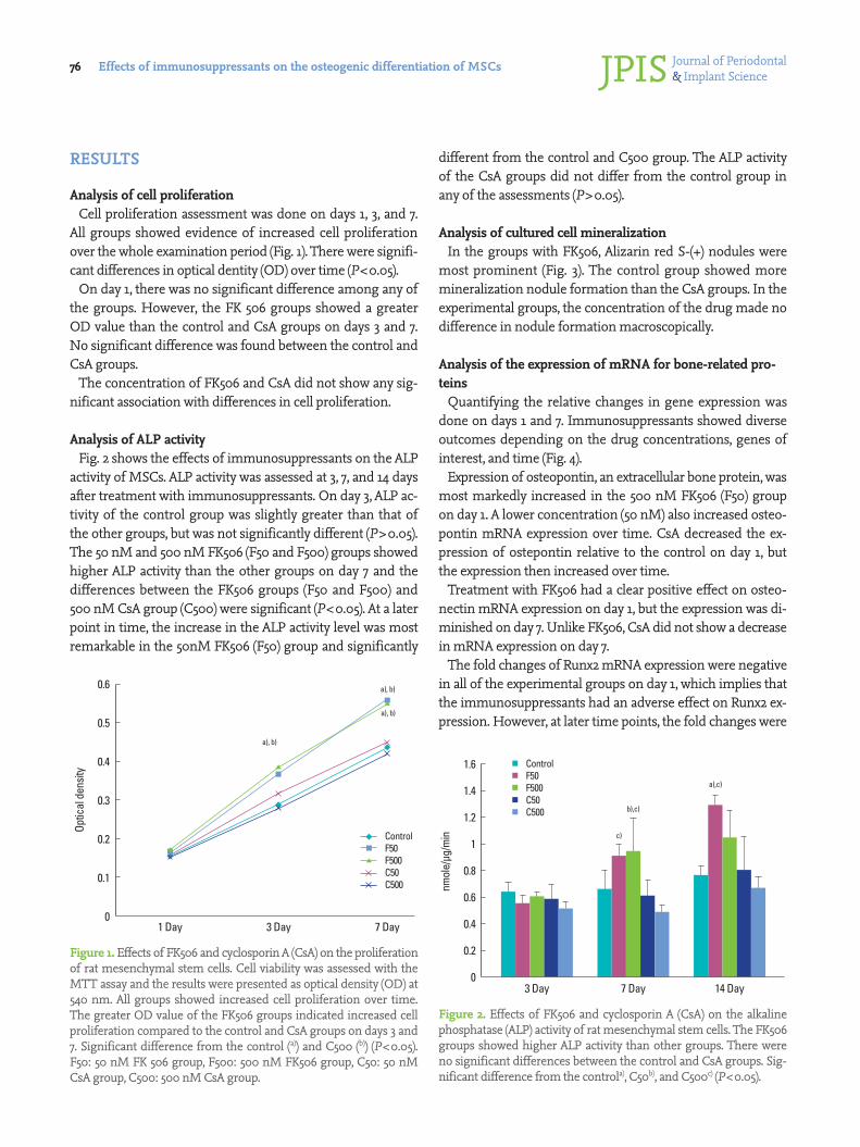
Analysis of cell proliferationCell proliferation assessment was done on days 1, 3, and 7.
All groups showed evidence of increased cell proliferation over the whole examination period (Fig. 1). There were signifi-cant differences in optical dentity (OD) over time (P<0.05).
On day 1, there was no significant difference among any of the groups. However, the FK 506 groups showed a greater OD value than the control and CsA groups on days 3 and 7. No significant difference was found between the control and CsA groups.
The concentration of FK506 and CsA did not show any sig-nificant association with differences in cell proliferation.
Analysis of ALP activityFig. 2 shows the effects of immunosuppressants on the ALP
activity of MSCs. ALP activity was assessed at 3, 7, and 14 days after treatment with immunosuppressants. On day 3, ALP ac-tivity of the control group was slightly greater than that of the other groups, but was not significantly different (P>0.05). The 50 nM and 500 nM FK506 (F50 and F500) groups showed higher ALP activity than the other groups on day 7 and the differences between the FK506 groups (F50 and F500) and 500 nM CsA group (C500) were significant (P<0.05). At a later point in time, the increase in the ALP activity level was most remarkable in the 50nM FK506 (F50) group and significantly
different from the control and C500 group. The ALP activity of the CsA groups did not differ from the control group in any of the assessments (P>0.05).
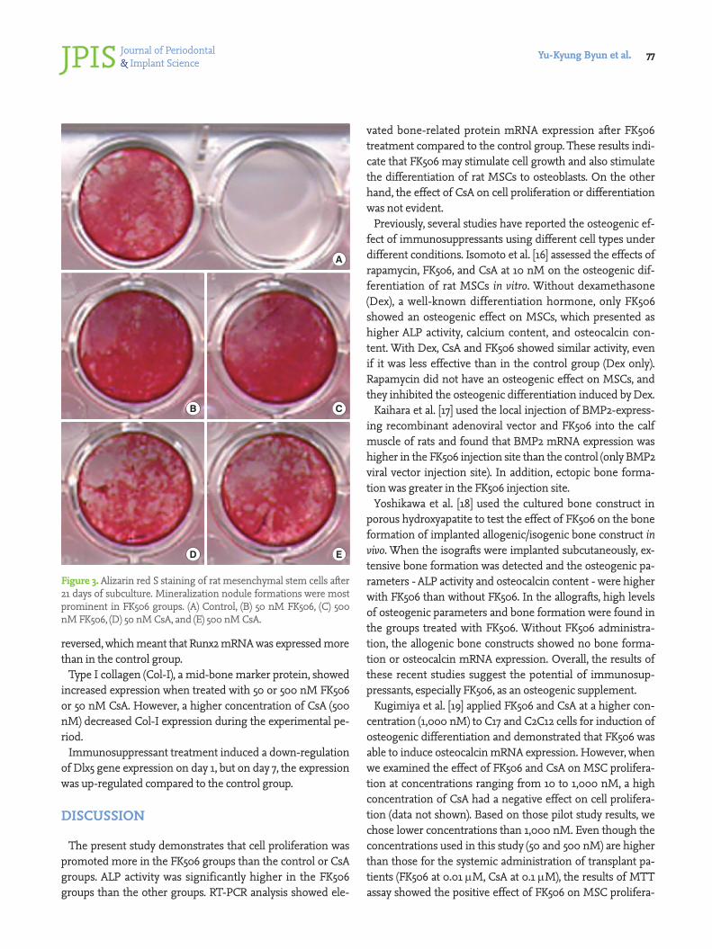
Analysis of cultured cell mineralizationIn the groups with FK506, Alizarin red S-(+) nodules were
most prominent (Fig. 3). The control group showed more mineralization nodule formation than the CsA groups. In the experimental groups, the concentration of the drug made no difference in nodule formation macroscopically.
Analysis of the expression of mRNA for bone-related pro-teins
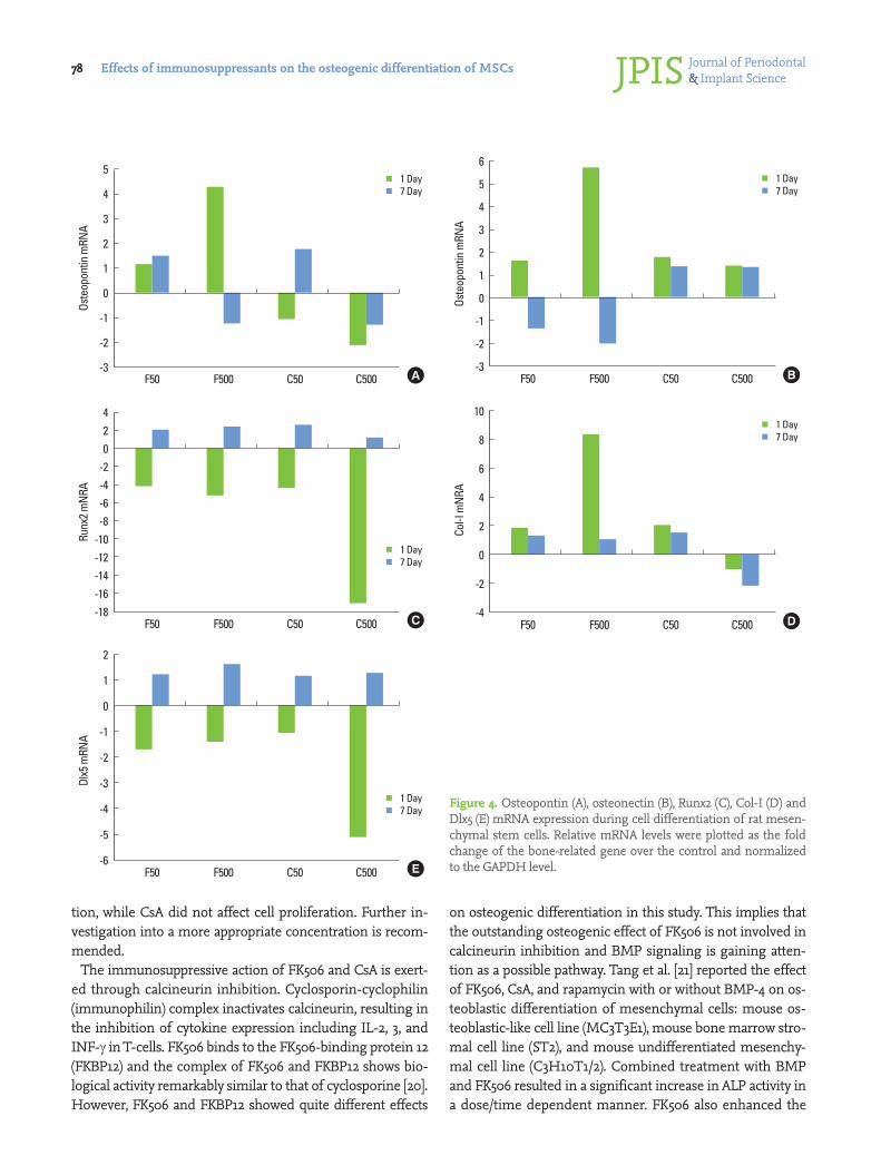
Quantifying the relative changes in gene expression was done on days 1 and 7. Immunosuppressants showed diverse outcomes depending on the drug concentrations, genes of interest, and time (Fig. 4).
Expression of osteopontin, an extracellular bone protein, was most markedly increased in the 500 nM FK506 (F50) group on day 1. A lower concentration (50 nM) also increased osteo-pontin mRNA expression over time. CsA decreased the ex-pression of ostepontin relative to the control on day 1, but the expression then increased over time.
Treatment with FK506 had a clear positive effect on osteo-nectin mRNA expression on day 1, but the expression was di-minished on day 7. Unlike FK506, CsA did not show a decrease in mRNA expression on day 7.
The fold changes of Runx2 mRNA expression were negative in all of the experimental groups on day 1, which implies that the immunosuppressants had an adverse effect on Runx2 ex-pression. However, at later time points, the fold changes were
Figure 1. Effects of FK506 and cyclosporin A (CsA) on the proliferation of rat mesenchymal stem cells. Cell viability was assessed with the MTT assay and the results were presented as optical density (OD) at 540 nm. All groups showed increased cell proliferation over time. The greater OD value of the FK506 groups indicated increased cell proliferation compared to the control and CsA groups on days 3 and 7. Significant difference from the control (a)) and C500 (b)) (P<0.05). F50: 50 nM FK 506 group, F500: 500 nM FK506 group, C50: 50 nM CsA group, C500: 500 nM CsA group.
0.6
0.5
0.4
0.3
0.2
0.1
0
Optic
al d
ensit
y
1 Day 3 Day 7 Day
a), b)
a), b)
a), b)
ControlF50F500C50C500
Figure 2. Effects of FK506 and cyclosporin A (CsA) on the alkaline phosphatase (ALP) activity of rat mesenchymal stem cells. The FK506 groups showed higher ALP activity than other groups. There were no significant differences between the control and CsA groups. Sig-nificant difference from the controla), C50b), and C500c) (P<0.05).
1.6
1.4
1.2
1
0.8
0.6
0.4
0.2
0
nmol
e/μg
/min
3 Day 7 Day
c)
b),c)
14 Day
a),c)
ControlF50F500C50C500
Journal of Periodontal& Implant ScienceJPIS Yu-Kyung Byun et al. 77
reversed, which meant that Runx2 mRNA was expressed more than in the control group.
Type I collagen (Col-I), a mid-bone marker protein, showed increased expression when treated with 50 or 500 nM FK506 or 50 nM CsA. However, a higher concentration of CsA (500 nM) decreased Col-I expression during the experimental pe-riod.
Immunosuppressant treatment induced a down-regulation of Dlx5 gene expression on day 1, but on day 7, the expression was up-regulated compared to the control group.
DISCUSSION
The present study demonstrates that cell proliferation was promoted more in the FK506 groups than the control or CsA groups. ALP activity was significantly higher in the FK506 groups than the other groups. RT-PCR analysis showed ele-
vated bone-related protein mRNA expression after FK506 treatment compared to the control group. These results indi-cate that FK506 may stimulate cell growth and also stimulate the differentiation of rat MSCs to osteoblasts. On the other hand, the effect of CsA on cell proliferation or differentiation was not evident.
Previously, several studies have reported the osteogenic ef-fect of immunosuppressants using different cell types under different conditions. Isomoto et al. [16] assessed the effects of rapamycin, FK506, and CsA at 10 nM on the osteogenic dif-ferentiation of rat MSCs in vitro. Without dexamethasone (Dex), a well-known differentiation hormone, only FK506 showed an osteogenic effect on MSCs, which presented as higher ALP activity, calcium content, and osteocalcin con-tent. With Dex, CsA and FK506 showed similar activity, even if it was less effective than in the control group (Dex only). Rapamycin did not have an osteogenic effect on MSCs, and they inhibited the osteogenic differentiation induced by Dex.
Kaihara et al. [17] used the local injection of BMP2-express-ing recombinant adenoviral vector and FK506 into the calf muscle of rats and found that BMP2 mRNA expression was higher in the FK506 injection site than the control (only BMP2 viral vector injection site). In addition, ectopic bone forma-tion was greater in the FK506 injection site.
Yoshikawa et al. [18] used the cultured bone construct in porous hydroxyapatite to test the effect of FK506 on the bone formation of implanted allogenic/isogenic bone construct in vivo. When the isografts were implanted subcutaneously, ex-tensive bone formation was detected and the osteogenic pa-rameters - ALP activity and osteocalcin content - were higher with FK506 than without FK506. In the allografts, high levels of osteogenic parameters and bone formation were found in the groups treated with FK506. Without FK506 administra-tion, the allogenic bone constructs showed no bone forma-tion or osteocalcin mRNA expression. Overall, the results of these recent studies suggest the potential of immunosup-pressants, especially FK506, as an osteogenic supplement.
Kugimiya et al. [19] applied FK506 and CsA at a higher con-centration (1,000 nM) to C17 and C2C12 cells for induction of osteogenic differentiation and demonstrated that FK506 was able to induce osteocalcin mRNA expression. However, when we examined the effect of FK506 and CsA on MSC prolifera-tion at concentrations ranging from 10 to 1,000 nM, a high concentration of CsA had a negative effect on cell prolifera-tion (data not shown). Based on those pilot study results, we chose lower concentrations than 1,000 nM. Even though the concentrations used in this study (50 and 500 nM) are higher than those for the systemic administration of transplant pa-tients (FK506 at 0.01 μM, CsA at 0.1 μM), the results of MTT assay showed the positive effect of FK506 on MSC prolifera-
A
B C
D E
Figure 3. Alizarin red S staining of rat mesenchymal stem cells after 21 days of subculture. Mineralization nodule formations were most prominent in FK506 groups. (A) Control, (B) 50 nM FK506, (C) 500 nM FK506, (D) 50 nM CsA, and (E) 500 nM CsA.
Journal of Periodontal& Implant ScienceJPISEffects of immunosuppressants on the osteogenic differentiation of MSCs78
tion, while CsA did not affect cell proliferation. Further in-vestigation into a more appropriate concentration is recom-mended.
The immunosuppressive action of FK506 and CsA is exert-ed through calcineurin inhibition. Cyclosporin-cyclophilin (immunophilin) complex inactivates calcineurin, resulting in the inhibition of cytokine expression including IL-2, 3, and INF-γ in T-cells. FK506 binds to the FK506-binding protein 12 (FKBP12) and the complex of FK506 and FKBP12 shows bio-logical activity remarkably similar to that of cyclosporine [20]. However, FK506 and FKBP12 showed quite different effects
on osteogenic differentiation in this study. This implies that the outstanding osteogenic effect of FK506 is not involved in calcineurin inhibition and BMP signaling is gaining atten-tion as a possible pathway. Tang et al. [21] reported the effect of FK506, CsA, and rapamycin with or without BMP-4 on os-teoblastic differentiation of mesenchymal cells: mouse os-teoblastic-like cell line (MC3T3E1), mouse bone marrow stro-mal cell line (ST2), and mouse undifferentiated mesenchy-mal cell line (C3H10T1/2). Combined treatment with BMP and FK506 resulted in a significant increase in ALP activity in a dose/time dependent manner. FK506 also enhanced the
Figure 4. Osteopontin (A), osteonectin (B), Runx2 (C), Col-I (D) and Dlx5 (E) mRNA expression during cell differentiation of rat mesen-chymal stem cells. Relative mRNA levels were plotted as the fold change of the bone-related gene over the control and normalized to the GAPDH level.
5
4
3
2
1
0
-1
-2
-3
Oste
opon
tin m
RNA
1 Day7 Day
F50 F500 C50 C500 A
420
-2-4-6-8
-10-12-14-16-18
Runx
2 mN
RA
1 Day7 Day
F50 F500 C50 C500 C
10
8
6
4
2
0
-2
-4
Col-I
mN
RA
1 Day7 Day
F50 F500 C50 C500 D
6
5
4
3
2
1
0
-1
-2
-3
Oste
opon
tin m
RNA
1 Day7 Day
F50 F500 C50 C500 B
2
1
0
-1
-2
-3
-4
-5
-6
Dlx5
mRN
A
1 Day7 Day
F50 F500 C50 C500 E
Journal of Periodontal& Implant ScienceJPIS Yu-Kyung Byun et al. 79
positive effects of BMP on ALP and osteocalcin mRNA. How-ever, osteocalcin expression was not affected by treatment with FK506 alone, without BMP. Kugimiya et al. [19] found that FK506 rapidly induced the phosphorylation of the BMP-dependent Smads, and the induction was blocked by Smad6; the activation of the Smads by FK506 is attenuated by FKBP12, and the binding of FKBP12 to the BMP receptors is suppressed by FK506. They suggested the hypothesis that the osteogenic signal induced by FK506 might be associated with the activa-tion of the BMP receptors, probably by separating them from FKBP12.
In this study, most of the bone-related genes were expressed more in the FK506 groups than the control. Osteopontin, os-teonectin, and Col-I genes were expressed more than in the control on day 1, but the intensity of expression had dimin-ished by day 7. Runx2 and Dlx5 gene expression were reduced in the early stage, but at a later time, the levels of these genes were expressed more than in the control. A low concentra-tion of CsA was also associated with an elevated level of os-teonectin and a Col-I gene expression like FK506 on day 1, but a high concentration of CsA reduced the Runx2 and Dlx5 gene expression more severelyon day 1. Also, in 500nM CsA (C500) group, osteopontin and the Col-I gene were expressed less than in the control during the investigated period. On the other hand, osteocalcin genes were not detected in any group including the control group (data not shown). Osteocalcin is a bone-specific marker protein, which is generally a gene of interest in osteoblast differentiation studies. It has been re-ported that osteocalcin mRNA is not detected in less than 12 days during the proliferation and maturation stage, and was robustly expressed in the mineralization stage [22]. Osteocal-cin gene expression is associated with the mineralization of osteoblasts and is considered a terminal marker of osteoblast differentiation. In this regard, our result that osteocalcin ex-pression was not observed during the 7-day culture may be the result of relatively short-term exposure to the target drugs.
Recently, our group showed that ex vivo BMP-2 gene deliv-ery using human gingival fibroblasts promotes bone regen-eration in rats [23]. In the study, FK506 was successfully used as an immunosuppressant to diminish the immune response of rats to human cells. In addition, under FK506 coverage, considerable bone generation was observed even in the non-grafted control group. Therefore, although evidence on whether FK506 increases bone healing is still conflicting, it might be beneficial for bone regeneration in allotransplanta-tion and/or xenotransplantation cases.
In conclusion, our results suggest that FK506 stimulates the osteoblastic differentiation of rat MSCs based on ALP activity and bone-specific gene expression. Therefore, FK506 might improve the results of bone regeneration in xenogenic or al-
logenic stem cell transplantation to treat bone defects. In ad-dition, FK506 would be preferred over CsA in such cases.
CONFLICT OF INTEREST
No potential conflict of interest relevant to this article was reported.
ACKNOWLEDGEMENTS
This research was supported by the Bio & Medical Technol-ogy Development Program of the National Research Foun-dation (NRF) funded by the Korean government (MEST) (No. 2011-0027790).
REFERENCES
1. Otto WR, Wright NA. Mesenchymal stem cells: from ex-periment to clinic. Fibrogenesis Tissue Repair 2011;4:20.
2. Caplan AI. Adult mesenchymal stem cells for tissue engi-neering versus regenerative medicine. J Cell Physiol 2007; 213:341-7.
3. Nauta AJ, Westerhuis G, Kruisselbrink AB, Lurvink EG, Willemze R, Fibbe WE. Donor-derived mesenchymal stem cells are immunogenic in an allogeneic host and stimulate donor graft rejection in a nonmyeloablative set-ting. Blood 2006;108:2114-20.
4. Penninga L, Moller CH, Gustafsson F, Steinbruchel DA, Gluud C. Tacrolimus versus cyclosporine as primary im-munosuppression after heart transplantation: systematic review with meta-analyses and trial sequential analyses of randomised trials. Eur J Clin Pharmacol 2010;66:1177-87.
5. Vincenti F, Jensik SC, Filo RS, Miller J, Pirsch J. A long-term comparison of tacrolimus (FK506) and cyclosporine in kidney transplantation: evidence for improved allograft survival at five years. Transplantation 2002;73:775-82.
6. Epstein S. Post-transplantation bone disease: the role of immunosuppressive agents and the skeleton. J Bone Miner Res 1996;11:1-7.
7. Kino T, Hatanaka H, Hashimoto M, Nishiyama M, Goto T, Okuhara M, et al. FK-506, a novel immunosuppressant isolated from a Streptomyces. I. Fermentation, isolation, and physico-chemical and biological characteristics. J An-tibiot (Tokyo) 1987;40:1249-55.
8. Kino T, Hatanaka H, Miyata S, Inamura N, Nishiyama M, Yajima T, et al. FK-506, a novel immunosuppressant iso-lated from a Streptomyces. II. Immunosuppressive effect of FK-506 in vitro. J Antibiot (Tokyo) 1987;40:1256-65.
9. Kulak CA, Borba VZ, Kulak Junior J, Campos DJ, Shane E. Post-transplantation osteoporosis. Arq Bras Endocrinol
Journal of Periodontal& Implant ScienceJPISEffects of immunosuppressants on the osteogenic differentiation of MSCs80
Metabol 2010;54:143-9.10. Rodino MA, Shane E. Osteoporosis after organ transplan-
tation. Am J Med 1998;104:459-69.11. Stein E, Ebeling P, Shane E. Post-transplantation osteopo-
rosis. Endocrinol Metab Clin North Am 2007;36:937-63; viii.12. Cvetkovic M, Mann GN, Romero DF, Liang XG, Ma Y, Jee
WS, et al. The deleterious effects of long-term cyclospo-rine A, cyclosporine G, and FK506 on bone mineral me-tabolism in vivo. Transplantation 1994;57:1231-7.
13. Fukunaga J, Yamaai T, Yamachika E, Ishiwari Y, Tsujigiwa H, Sawaki K, et al. Expression of osteoclast differentiation factor and osteoclastogenesis inhibitory factor in rat os-teoporosis induced by immunosuppressant FK506. Bone 2004;34:425-31.
14. Krocker D, Perka C, Tuischer J, Funk J, Tohtz S, Buttgereit F, et al. Effects of tacrolimus, cyclosporin A and sirolimus on MG63 cells. Transpl Int 2006;19:563-9.
15. Lee KW, Yook JY, Son MY, Kim MJ, Koo DB, Han YM, et al. Rapamycin promotes the osteoblastic differentiation of human embryonic stem cells by blocking the mTOR pathway and stimulating the BMP/Smad pathway. Stem Cells Dev 2010;19:557-68.
16. Isomoto S, Hattori K, Ohgushi H, Nakajima H, Tanaka Y, Takakura Y. Rapamycin as an inhibitor of osteogenic dif-ferentiation in bone marrow-derived mesenchymal stem cells. J Orthop Sci 2007;12:83-8.
17. Kaihara S, Bessho K, Okubo Y, Sonobe J, Kawai M, Iizuka T. Simple and effective osteoinductive gene therapy by local injection of a bone morphogenetic protein-2-expressing
recombinant adenoviral vector and FK506 mixture in rats. Gene Ther 2004;11:439-47.
18. Yoshikawa T, Nakajima H, Yamada E, Akahane M, Dohi Y, Ohgushi H, et al. In vivo osteogenic capability of cultured allogeneic bone in porous hydroxyapatite: immunosup-pressive and osteogenic potential of FK506 in vivo. J Bone Miner Res 2000;15:1147-57.
19. Kugimiya F, Yano F, Ohba S, Igawa K, Nakamura K, Kawa-guchi H, et al. Mechanism of osteogenic induction by FK506 via BMP/Smad pathways. Biochem Biophys Res Commun 2005;338:872-9.
20. Liu J, Farmer JD Jr, Lane WS, Friedman J, Weissman I, Sch-reiber SL. Calcineurin is a common target of cyclophilin-cyclosporin A and FKBP-FK506 complexes. Cell 1991;66: 807-15.
21. Tang L, Ebara S, Kawasaki S, Wakabayashi S, Nikaido T, Takaoka K. FK506 enhanced osteoblastic differentiation in mesenchymal cells. Cell Biol Int 2002;26:75-84.
22. Owen TA, Aronow M, Shalhoub V, Barone LM, Wilming L, Tassinari MS, et al. Progressive development of the rat os-teoblast phenotype in vitro: reciprocal relationships in ex-pression of genes associated with osteoblast proliferation and differentiation during formation of the bone extra-cellular matrix. J Cell Physiol 1990;143:420-30.
23. Shin JH, Kim KH, Kim SH, Koo KT, Kim TI, Seol YJ, et al. Ex vivo bone morphogenetic protein-2 gene delivery us-ing gingival fibroblasts promotes bone regeneration in rats. J Clin Periodontol 2010;37:305-11.