Embed Size (px)
Citation preview
EFFECTS OF HYPOXIA EXPOSURE ON HUMAN
HIPPOCAMPAL ASTROCYTES CULTURES
NURUL ATIKAH BINTI M. NOR NAZLI
UNIVERSITI SAINS MALAYSIA
2017
EFFECTS OF HYPOXIA EXPOSURE ON HUMAN
HIPPOCAMPAL ASTROCYTES CULTURES
By
NURUL ATIKAH BINTI M. NOR NAZLI
Thesis submitted in partial fulfilment of the requirements for the degree of
Master of Neuroscience
JUNE 2017
KESAN PENDEDAHAN HIPOKSIA TERHADAP KULTUR SEL
ASTROSIT HIPPOCAMPUS MANUSIA
Oleh
NURUL ATIKAH BINTI M. NOR NAZLI
Tesis diserahkan untuk memenuhi sebahagian keperluan bagi
Ijazah Sarjana Neurosains
JUN 2017
ii
Acknowledgement
First of all, I am grateful to The Almighty Allah S.W.T as I have completed my research
project delightfully. I take this opportunity to express my profound gratitude and deep
regards to my project supervisor, Prof Dato Dr Jafri Malin Abdullah for his exemplary
guidance, monitoring and constant encouragement throughout the course of this
thesis. Many thanks to my co-supervisor, Dr Sangu Muthuraju for his patience in guide
me and teach me new skills throughout the laboratory work. The blessing, help and
guidance given by them time to time shall carry me a long way in the journey of life on
which I am about to embark.
I also take this opportunity to express a deep sense of gratitude to INP coordinator,
Assoc Prof. M, Muzaimi and lecturers for their cordial support, valuable information
and guidance, which helped me in completing this task through various stages. Besides
that, without cooperation of staff members of neuroscience laboratory and central
research laboratory, this research may not be completed. With that, I bid thousand
thanks. Special notes of appreciation for laboratory mates my friends especially Nor
Aqilah, Chuang Huei Gau, Shazlan, Ainun, Salihah and Mazira for their endless help
and guidance.
Last but not least, a million thanks and appreciations to my beloved parents, M Nor
Nazli and Nor Surianty Soo and my family for their constant encouragement and
support in every ways from the start until the end of the projects. To all others who
helped me directly and indirectly, thank you.
iii
TABLE OF CONTENTS
Page No.
ACKNOWLEDGEMENT
TABLE OF CONTENTS
LIST OF TABLES
LIST OF FIGURES
LIST OF SYMBOLS AND ABBREVIATION
ABSTRACT
ii
iii
vii
viii
x
xi
CHAPTER 1: INTRODUCTION 1
CHAPTER 2: LITERATURE REVIEW
2.1 Hypoxia
2.2 Different type of hypoxia condition
2.2.1 Maternal Hypoxia
2.2.2 Perinatal Hypoxia
2.2.3 Cerebral ischemia
2.2.4 Obstructive Sleep Apnea (OSA)
2.2.5 High altitudes sickness
2.3 Vulnerability of brain to hypoxic
5
7
7
9
11
12
13
14

iv
2.4 Hypoxia in different species
2.5 Morphological and types of astrocytes cells
2.6 Functions of astrocytes
2.6.1 The brain micro-architecture
2.6.2 Extracellular homeostasis in brain
2.6.3 Removal of Glutamate
2.6.4 Glutamatergic neurotransmission
2.6.5 Metabolic support
2.6.6 Synaptogenesis
2.6.7 Neuronal-glial signalling
2.6.8 Gliotransmission
2.7 Intermediate filament (IFs) of astrocytes and its potential marker,
GFAP
2.8 Changes of astrocyte cells after hypoxia exposure
2.9 Consequences of Hypoxia Exposure to Astrocytes cells
2.9 Hypothesis
2.10 Rational of the studies
2.11 Objectives
15
17
23
23
24
25
25
26
27
28
29
31
33
35
35
36
CHAPTER 3: METHODOLOGY
3.1 Study design
37
v
3.2 Culture procedures and conditions
3.3 Cell culture
3.3.1 Human Hippocampal Astrocytes
3.3.2 Complete Growth Medium for Astrocyte Cell line
3.3.3 Retrieving of cells from frozen storage
3.3.4 Maintaining cell growth and subculturing the cells
3.3.5 Cryopreservation
3.4 Hypoxia exposure
3.5 Cell viability assay using Trypan blue dye exclusion
3.6 Glial Fibrillary Acidic Protein (GFAP) Immunofluorescence Assay
3.7 Hypoxia Inducible Factor 1-α staining
3.8 Reverse Transcription Polymerase Chain Reaction (RT-PCR) and
Electrophoresis
3.8.1 TRIZOL RNA isolation
3.8.2 cDNA Synthesis Reaction
3.8.3 PCR amplification
3.8.4 Separation of DNA Fragments via Electrophoresis
3.9 Statistical analysis
37
39
39
39
40
40
41
42
43
45
46
47
47
49
50
52
53
vi
CHAPTER 4: RESULT
4.1 Cell culture characteristics and morphology
4.2 Trypan blue dye exclusion for the cell viability
4.3 Portray structural of astrocyte by GFAP staining
4.4 Hypoxia-Inducible factor staining in confirming the death of astrocyte
cells
4.5 Molecular analysis of astrocyte cells for control, acute hypoxia and
chronic hypoxia
54
56
59
62
65
CHAPTER 5: DISCUSSSION 67
CHAPTER 6: CONCLUSION 76
REFERENCES 77
APPENDICES 102
vii
LIST OF TABLES
Table No. Page
Table 2.1 Types of astrocytes
20
Table 4.1
Cell viability after expose cells to chronic
hypoxia (3% Oxygen)
55
Table 4.2
Cell viability after expose cells to different
oxygen level in 15minutes
57
viii
LIST OF FIGURES
Figure No. Page
Figure 2.1 Four major types of astrocytes cells 22
Figure 3.1 Flowchart of research methods 37
Figure 3.2 Hypoxia chamber C374 model was used 41
Figure 3.3 Flowchart of Trypan Blue Assay 43
Figure 4.1 Culturing human hippocampal astrocyte cells line 54
Figure 4.2 Cell viability after expose cells to chronic hypoxia
(3% Oxygen)
56
Figure 4.3 Cell viability after expose cells to different oxygen
level in 15minutes
57
Figure 4.4 Staining human hippocampal astrocyte cell line using
the GFAP
60
Figure 4.5 The mean of GFAP intensity for astrocyte cells in
different oxygen concentration
61
Figure 4.6
HIF staining human hippocampal astrocyte cell line
63
Figure 4.7 The mean of HIF-1α intensity for astrocyte cells in 64
ix
different oxygen concentration
Figure 4.8 Molecular analysis involving GAPDH, GFAP, HIF-
1αand Bc12 in control, acute and chronic hypoxia
group.
66
Figure 5.1 Summarize the effect of chronic hypoxia to human
hippocampal astrocytes cells
75
x
LIST OF SYMBOLS AND ABBREVIATION
HhA Human Hippocampal Astrocyte
AM Astrocyte media
AGS Astrocyte growth supplement
CNS Central nervous system
IFs Intermediate Filaments
P/S solution Penicillin/ Streptomycin solution
DMSO Dimethyl sulphoxide
GFAP Glial Fibriallary Acidic Protein
HIF-1 Hypoxia Inducible Factor
FITC Fluorescein Isothiocyanate
GAPDH Glyceraldehyde 3-phosphate dehydrogenase
Bcl2 B-cell lymphoma 2
RT-PCR Reverse Transcription Polymerase chain reaction
et al. (et alia); and others
xi
Abstrak
KESAN PENDEDAHAN HIPOKSIA TERHADAP KULTUR SEL
ASTROSIT HIPPOCAMPUS MANUSIA
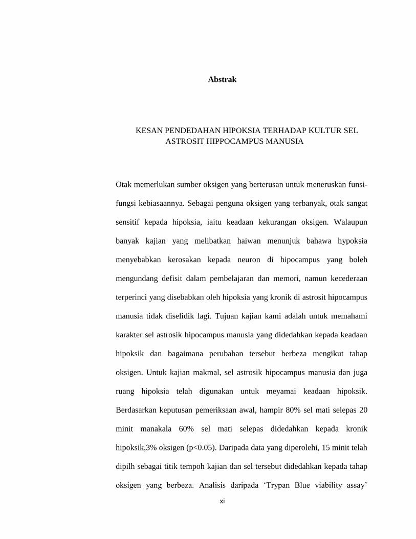
Otak memerlukan sumber oksigen yang berterusan untuk meneruskan funsi-
fungsi kebiasaannya. Sebagai penguna oksigen yang terbanyak, otak sangat
sensitif kepada hipoksia, iaitu keadaan kekurangan oksigen. Walaupun
banyak kajian yang melibatkan haiwan menunjuk bahawa hypoksia
menyebabkan kerosakan kepada neuron di hipocampus yang boleh
mengundang defisit dalam pembelajaran dan memori, namun kecederaan
terperinci yang disebabkan oleh hipoksia yang kronik di astrosit hipocampus
manusia tidak diselidik lagi. Tujuan kajian kami adalah untuk memahami
karakter sel astrosik hipocampus manusia yang didedahkan kepada keadaan
hipoksik dan bagaimana perubahan tersebut berbeza mengikut tahap
oksigen. Untuk kajian makmal, sel astrosik hipocampus manusia dan juga
ruang hipoksia telah digunakan untuk meyamai keadaan hipoksik.
Berdasarkan keputusan pemeriksaan awal, hampir 80% sel mati selepas 20
minit manakala 60% sel mati selepas didedahkan kepada kronik
hipoksik,3% oksigen (p<0.05). Daripada data yang diperolehi, 15 minit telah
dipilh sebagai titik tempoh kajian dan sel tersebut didedahkan kepada tahap
oksigen yang berbeza. Analisis daripada „Trypan Blue viability assay‟
xii
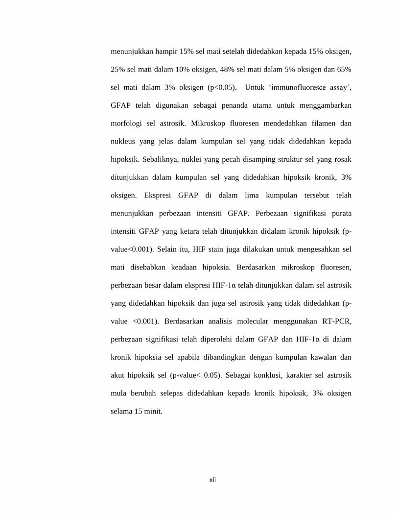
menunjukkan hampir 15% sel mati setelah didedahkan kepada 15% oksigen,
25% sel mati dalam 10% oksigen, 48% sel mati dalam 5% oksigen dan 65%
sel mati dalam 3% oksigen (p<0.05). Untuk „immunofluoresce assay‟,
GFAP telah digunakan sebagai penanda utama untuk menggambarkan
morfologi sel astrosik. Mikroskop fluoresen mendedahkan filamen dan
nukleus yang jelas dalam kumpulan sel yang tidak didedahkan kepada
hipoksik. Sebaliknya, nuklei yang pecah disamping struktur sel yang rosak
ditunjukkan dalam kumpulan sel yang didedahkan hipoksik kronik, 3%
oksigen. Ekspresi GFAP di dalam lima kumpulan tersebut telah
menunjukkan perbezaan intensiti GFAP. Perbezaan signifikasi purata
intensiti GFAP yang ketara telah ditunjukkan didalam kronik hipoksik (p-
value<0.001). Selain itu, HIF stain juga dilakukan untuk mengesahkan sel
mati disebabkan keadaan hipoksia. Berdasarkan mikroskop fluoresen,
perbezaan besar dalam ekspresi HIF-1α telah ditunjukkan dalam sel astrosik
yang didedahkan hipoksik dan juga sel astrosik yang tidak didedahkan (p-
value <0.001). Berdasarkan analisis molecular menggunakan RT-PCR,
perbezaan signifikasi telah diperolehi dalam GFAP dan HIF-1α di dalam
kronik hipoksia sel apabila dibandingkan dengan kumpulan kawalan dan
akut hipoksik sel (p-value< 0.05). Sebagai konklusi, karakter sel astrosik
mula berubah selepas didedahkan kepada kronik hipoksik, 3% oksigen
selama 15 minit.
xiii
Abstract
EFFECTS OF HYPOXIA EXPOSURE ON HUMAN HIPPOCAMPAL
ASTROCYTES CULTURES
The brain requires a continuous supply of oxygen to perform its normal
function. Being the largest consumer of oxygen, it is especially sensitive to
hypoxia, a condition in which brain receives reduced oxygen. Despite many
animal studies reported that hypoxia caused neuronal damage in
hippocampus which could deficits learning and memory, the exact damage
caused by chronic hypoxia on human hippocampal astrocyte has not been
analysed yet. Our aim for this study is to understand the characterization of
human hippocampal astrocyte following hypoxia exposure and how the
changes varied according to different concentration levels of oxygen. For the
laboratory work, human hippocampal astrocytes cell line and hypoxia
chamber were used in mimicking the hypoxic condition. Based on the
preliminary screening, almost 80% of cell death occurred after 20 min and
60% cell death occurred in 15 min after exposed to chronic hypoxia, 3% of
oxygen level (p<0.05). From the data gained, 15 minutes was chosen as the
time point and the cells were exposed to different oxygen percentage.
Analysis from Trypan blue viability assay showed about 15% of cells were
xiv
dead in 15% oxygen, 25% dead cells in 10% oxygen, 48% dead cells in 5%
oxygen and 65% dead cells in 3% oxygen (p<0.05). For the
immunofluorescence assay, a reliable marker Glial Fibriallary Acidic
Protein (GFAP) was used in order to portray the architecture and
morphology of astrocytes cells. Fluorescence scanning microscope revealed
a filamentous and clear nucleas appearance in a control. In contrast, the
rupture nuclei along with no rigid structure of cell were displayed in chronic
hypoxia group, the 3% oxygen exposure. The expression of GFAP among
the five groups showed different intensity of GFAP. The significant different
of the mean intensity was clearly showed in the chronic hypoxia group (p-
value<0.001). Along with that, the HIF-1 staining was performed to confirm
the cell death due to hypoxia exposure. Based on the fluorescence
microscope viewed, different expression of HIF-1α were displayed in all
exposed astrocyte cells (p-value<0.001). In the molecular analysis using RT-
PCR, there were significant changes of GFAP and HIF-1α in chronic
hypoxia exposed cells when compared to control and acute hypoxia exposed
cells (p-value< 0.05). To conclude, changes of the morphology of astrocyte
cells are seen after 15 exposed to chronic hypoxia, 3% oxygen.
1
CHAPTER 1: INTRODUCTION
The brain requires a continuous supply of oxygen to perform its normal function.
Regardless not performing mechanical work like skeletal muscle or heart, human brain
is consider as one of the most metabolically active organ in the body. The brain utilizes
25% of the body‟s total oxygen consumption and expends about 3.5 ml of oxygen per
100 g of brain tissue per minute. The value needed for brain to function well in period
of wakefulness remains constant even in sleep even though the rate of blood flow
increased during sleep (Harris et al, 2012; Morselli et al, 2012; Hajjawi, 2014). As the
largest consumer to oxygen, the brain needs oxygen desperately because it is important
substrate to finely tune with signalling activities and cognitive functions (Ivanisevic
and Siuzdak, 2015).
The brain also highly sensitivity to any significant changes in its environment,
thus reduction of oxygen level may interfere with its optimal function. In general,
hypoxia can be defined as reduction of oxygen level in cells or tissue of the body
(Pighin et al, 2012). There are several critical components are involved in elucidate the
hypoxia exposure, namely the rate of hypoxia occurrence, duration of hypoxia
exposure, the present of reoxygenation and the severity of the hypoxic stimulus
(Dempsey and Morgan, 2015).
2
In human brain, hippocampus is considered as one of the highly sensitive
regions to hypoxia. Exposure to hypoxia would trigger several disastrous effects on the
central nervous system (CNS), especially on the neurological and physiological
aspects. Reduction of oxygen may also cause alteration in the brain structure (Ando et
al, 2013; Mateika et al, 2015). For the molecular level, hypoxia condition may lead to
elevation of free radical generation, oxidative stress and increased the level of L-type
calcium channels (Barhwal et al., 2009; Hota et al, 2012). Other than that, hypoxia
exposure may cause apoptosis or necrosis in hippocampal neurons which lead to
impairment in learning and memory functions (Chiu et al, 2012). There are also
growing body of evidences have clearly reported that alteration in oxygen level may
lead to neurodegerative disorders and impaired cognitive functions in terms of learning
and memory (Zhang et al., 2012; Smith et al., 2013 ).
Over the last few decades, the functions of astrocytes have been acknowledged
widely in contributing to many essential functions in CNS. Apart from its numerous
numbers that occupy the brain, astrocytes not only play a part as a number one
supporting cell that provides its architectural structure as well as essential in anti-
oxidant defence and inflammatory response. Besides, astrocyte cells support neuronal
activity via astrocytic glycogen, induce neurogenesis from neural stem cells in the
adult brain and acts as source of neural stem cells (DiNuzzo et al, 2012; Sirko et al,
2013).
3
Classically, astrocytes can be divided into two types according to their
anatomical location and cellular morphology. Protoplasmic astrocytes dominant in
grey matter while fibrous astrocytes located in all white matter (Sun and Jakobs,
2012). To characterise these specialized subtypes of astrocytes cells, several cells can
be out lined such as Bergmann glia of the cerebellum and Muller glia of retina (Heller
and Rusakov, 2015).
Astrocyte cells are involved in regulation of brain pathologies from acute lesions
such as stroke and stress to chronic neurodegenerative processes such as Alzheimer
diseases, Parkinson diseases and psychiatric diseases such as Schizophrenia and
Bipolar disorder. Asides from its numerous essential functions in supporting neurons,
they also involve in various activation programmes, which are important for; limiting
the areas of damage, producing neuro-immune responses and for the post-insult
remodelling and recovery of neural function (Kettenmann and Verkhratsky, 2011).
Activation of astrocytes cell, also known as astrogliosis arise as part of the
response of the CNS to critical situation like neurotrauma, brain injury, ischemic
damage and neurodegenerative diseases (Sofroniew and Vinters, 2010; Parpura et al,
2012). Regardless of its origin, the hallmark of reactive astrocyte is inflation of GFAP
expression, vimentin and nestin. Generally, GFAP, vimentin and nestin can be
categorized as building blocks of intermediate filaments (IFs), which form the
cytoskeleton along with microtubules and actin filaments (Oberheim et al, 2012).
4
Immunohistochemical techniques that enable the detection of specific molecular
markers at the single-cell level are essential tools for identifying and characterizing
cells in healthy and pathological condition (Sofroniew and Vinters, 2010; Duraiyan et
al, 2012). The astroglial component of gliosis is characterised by the accumulation of
glial filaments, of which GFAP is the major constituent. Besides that, GFAP gene
activation and protein induction appear to play a critical role in astroglial cell
activation (O‟Callaghan and Sriram, 2005; Yang and Wang, 2015). For decades, the
expression of GFAP has been considered as the most reliable marker to
immunohistochemically identify the astrocytes, even though not all astrocytes in the
healthy brain express GFAP. GFAP also is not immunohistochemically detectable in
all normal astrocytes, its expression exhibiting both regional and local variability.
However, the use of antibodies to GFAP in histological studies has firmly established
the existence of reactive gliosis as a dominant response to many different types of
brain injuries (O‟Callaghan and Sriram, 2005; Sun and Jakobs, 2012).
.Aside from GFAP marker, other astrocytes markers such as S100ß and
glutamine synthetase have similar shortcomings (Sofroniew and Vinters, 2010
Oberheim et al, 2012). Recently, the aldehyde dehydrogenase 1 family, memer LI
(Aldh 1L1, also known as 10-formyltetrahydrofolate dehydrogenase (FDH), was
suggested as a pan-astrocyte marker used on transcriptome gene profiling and in situ
hybridization (Cahoy et al, 2008; Sun et al, 2017).
5
CHAPTER 2 : LITERATURE REVIEW
2.1 Hypoxia
In the 21st century, the common causes of the death amongst average age men are heart
infarction, stroke and cancer. There are many reasons on how these disorders conquer
the human body, not only from environmental factors and lifestyle habits but also
genetic predisposition. However, they all share a common feature in which the
limitation of oxygen availability participates in the development of these pathological
conditions. In molecular context, cells are able to cope with these threatening
conditions like hypoxia conditions as they can trigger adaptive response to hypoxic
conditions but their response are depending on the type of hypoxic condition either
acute or chronic (Sjöberg and Singer, 2013; Kumar and Choi, 2015;).
Oxygen is the most vital element in maintaining the homeostasis and ensures
efficiency of human body systems. Lacking of this chemical element or disruption of
balance between its supply and demand may cause disparity changes. A condition
where oxygen availability is limited can be described as hypoxia (Sjöberg and Singer,
2013; Nakazawa, 2016). In detail, hypoxia can be defined as the deficiency in the
bioavailability of oxygen to the tissues of the body (Loiacono, et aI., 2010). Many
situations where oxygen is lacking
6
not lead to death of the organism, or even cause damage but in extreme reduction of
brain oxygen may lead to neuronal death. Deficiency of oxygen condition has been
reported to trigger free radical generation and depletion of antioxidant status, thus
leading to oxidative damage of vital cellular components (Rahal et al, 2014).
The brain is considered as the most hypoxia-sensitive organs because of its
demand for a high oxygen supply, whereas the skeletal muscle is amongst the most
hypoxia-tolerant. The brain, an organ with high metabolic rate along with a rich store
of polyunsaturated fatty acids is count as the most sensitive organ and a vulnerable
target to oxidative damage (Kalogeris et al, 2012). Indeed, the brain also has been
categorized as one of the critical targets of stressors and act as the central organ which
is responsible for stress responses, determining the adaptive or maladaptive
responsiveness to various condition either acute or chronic. The architecture and
structural function of the brain may disrupt due to its correspondent to the stressful
events cause by stress, brain injury, oxygen deprivation or glucose alteration (Solaini
et al, 2010; McEwen et al, 2012).
7
2.2 Different types of hypoxia condition
Brain needs approximately 20% of the oxygen consumed by the human body. The
great demand of this oxygen is needed to produce ATP. ATP productions are required
in order to maintain the membrane potentials which are necessary for electrical
signalling of synaptic and action potentials (Harris et al., 2012). Interruption of this
supply for more than a few minutes among most of vertebrates including human, may
leads to irreversible neurological damage and neurological diseases. However, some
studies reported that during mild hypoxia of short duration, the brain develops
powerful neuroprotective and adaptive mechanisms that allow it to maintain normal
physiological conditions (Rybnikova et al, 2012).
2.2.1 Maternal Hypoxia
Fetal stress such as hypoxia, malnutrition or excess glucocorticoids have a long lasting
impact to the fetus development especially on developing brain. Exposure over a
longer period to a pregnant mother may alter the fetal brain‟s ontogeny, organization,
structure and functions (Harris & Seckl, 2011; Gonzalez-Rodriguez, 2014).
Historically, Kingdom and Kaufmann in 1997 have suggested that hypoxic pregnancy
condition can be divided into three; pre-placental hypoxia, uteroplacental hypoxia and
post-placental hypoxia. Preplacental hypoxia can be described as a condition where
both mother and her fetus would be in hypoxic condition. Usually pregnant mothers
who live at high-altitude or having cyanotic maternal heart disease would experience
this kind of condition.
8
Meanwhile, uteroplacental hypoxia is a condition where the maternal oxygenation
is normal but the utero-placental circulation is impaired and the example where the
situation implies is preeclampsia or placenta insufficiency. The post –placental
hypoxia is a condition where only the fetus is in hypoxic condition. Lower level of
oxygen during pregnancy is a one of common hostile environment associated with
abnormal system of the pregnant mother which causes high risk to the fetus. Because
the placenta helps to exchange oxygen, nutrients and waste between the mother and the
offspring, therefore malfunction of placenta also may express acute and chronic effects
on the developing fetus and drive to intrauterine growth restriction (IUGR), asphyxia,
multiorgan failure and premature delivery (Herrera et al, 2014). Besides that,
numerous animal studies have disclosed that maternal hypoxia affects the
organogenesis of brain and heart (Tong et al, 2011; Davis et al, 2012).
2.2.2 Perinatal Hypoxia
Fetus in the utero does not undergo respiration process but imbalance of gas exchange
due to defect of umbilical or uterine blood flow will lead to fetal asphyxia. The
condition also can be described where there are low oxygen level in fetal blood and
tissue decrease while the carbon dioxide level are high. The simultaneous changes of
oxygen and carbon dioxide may lead to consequence of hypoxia condition which the
injury effect to the fetal is depend on the time duration, intensity and occurrence rate of
the insult (Baburamani et al, 2012; Thornton et al, 2012).
9
Hypoxia is considered as one of the most common causes of neonatal brain injury.
However, it still remains to be well elucidated that why some brain regions are more
sensitive in certain condition compare to other regions. Besides that, as the infant
matures or after experience the insult, the susceptible of some brain regions are
changed which may be due to on metabolic reserves and physiological adjustments
(Rey-Santano et al, 2011). Post mortem studies pointed out that critical events such as
hypoxic-ischemic and asphyxic episodes during pregnancy could lead to brain injury,
morbidity and mortality. Cognitive impairment, delay in developmental progress,
epilepsy, motor deficits and cerebral palsy also may occur due to intrauterine asphyxia
(Glass et al., 2009; Baburamani et al, 2012).
One of the most common of brain damages among neonatal resulting from a
shortage of oxygen or blood flow to the tissues is Infant Hypoxic-ischemic
encephalopathy (HIE) (Stoll and Kliegman, 2007; Castillo and Chiang 2014). This
major contributor to neonatal death and morbidity can be acute or subacute brain
injury due to asphyxia and may occur prior, during or after birth. This kind of hypoxia-
ischemia can be caused by placental insufficiency or infection, which also often an
indication for preterm delivery by caesarean section (Goldenberg et al, 2008; Castillo
and Chiang 2014). Epidemiological studies have showed that about 15%–20% of HIE
cases died during the neonatal period and 30% of those who survive would develop
neurological deficits and long-term neurodevelopmental disabilities in later life
including, mental retardation, visual motor dysfunction, cerebral palsy and epilepsy
neurodevelopmental disorders (Eghbalian and Monsef, 2008; Dauglass-Escobar and
Weiss, 2012). As study of the pathophysiology of perinatal HIE is difficult to conduct
10
in human, a neonatal animal model known as Vannucci model has been used in
numerous studies in mimicking the condition. In this model, 7-day postnatal rats
experienced a unilateral common carotid ligation followed by systemic hypoxia in 8%
oxygen balanced with nitrogen environment. The insult produces permanent
hypoxicischemic brain damage limited to the cerebral hemisphere ipsilateral to the
carotid artery occlusion (Vannuci and Vannuci, 2005; Riljak et al, 2016). Despite the
advances in the last two decades in research of cellular processes and molecular
mechanism underlying HIE, hypothermia could be the only effective treatment (Wu
and Gonzalez, 2015).
2.2.3 Cerebral ischemia
The brain has homeostatic mechanisms which that can cope with mild ischemic
attacks. However a severe ischemia overwhelms the mechanism resulting in cell death
in smaller or larger parts of the brain. Cells that are experienced the ischemic
condition may die within minutes or display delayed vulnerability depending on how
early reperfusion is initiated, metabolic and ionic homeostasis can return and cell
survival maintained. Basically cerebral ischemia disrupts several aspects of
physiological, biochemical,genetics and molecular in human body which lead to
malfunction of cellular integrity. Then, the alteration of the cellular mechanism may
cause several consequences include imbalance of ionic level, glutamate excitatory,
calcium overload and oxidative stress (Kalogeriset al, 2012; Bretón and Rodríguez,
2012).
11
There are two common types related to cerebral ischemia; global ischemia and
focal ischemia. When cerebral blood flows (CBF) is reduced throughout most or all of
the brain, this type of condition is known as Global ischemia. Meanwhile focal
ischemia is explained by a reduction in blood flow to a very distinct, specific brain
region (Lee et al, 2016).
2.2.4 Obstructive Sleep Apnea (OSA)
Sleep-disordered breathing is highly prevalent and it composes of several types which
includes primary snoring, upper airway resistance syndrome, obstructive sleep apnea
(OSA), central sleep apnea, and obesity-hypoventilation syndrome. The most common
is the Obstructive sleep apnea (OSA), a disorder that is characterised by repetitive
complete or partial obstruction of the upper airway during sleep which leads to
decreased of air flow and snoring. Repeated apneic and hypopneic events during sleep
would lead to intermittent hypoxemia, hypercania, cortical and sympathetic nervous
system arousal and sleep fragmentation (Yadav et al, 2013; Baril et al, 2015). OSA is
commonly related to diminish neurocognitive function, neuropsychological
impairments and cardiovascular morbidities. OSA would affect the sleep
fragmentation which later reduced quality of life, impaired work-performance and also
increased risk of motor vehicle accidents in most cases (Yadav et al, 2013; Garvey et
al 2015).
However, in a worst case scenario, this type of sleep disorder may change
directly to several part of brain function especially on cognitive function and cause the
neurobehavioral consequences (Beebe, 2011).
12
Previous studies have been focused to investigate the specific cognitive functions and
some have attempted to identify a “pattern” of cognitive dysfunctions in OSA.
Executive functioning, a set of mental skills which include essential function in
planning, initiation, execution of goal-oriented behavior and mental flexibility, is
another affected domain. Indeed, several studies insisted that it is the most prominent
area of cognitive impairment in untreated sleep-disordered breathing which can be
found both in adults and children (Zimmerman et al, 2012; Olaithe et al, 2015; Krysta
et al, 2017).
2.2.5 High altitudes sickness
As the altitudes location getting higher, the barometric pressure is reduce, thus less
oxygen is inhaled. This kind of situation where oxygen availability is restricted may
lead to imbalance of oxygen in brain tissue and cause cerebral damage, neurological
deficits and cognitive dysfunctions. Today‟s increasing popularity and ability to travel
rapidly to high altitudes exposed millions of people to acute mountain sickness (AMS),
a most common condition occur in those that go too high and too fast. Typically, AMS
will be experienced by non-acclimatized mountaineers or high-altitudes training‟s
athletes within 6-12 hours of arrival to altitudes above 2,500m (Bärtsch and Swenson,
2013; Netzer et al, 2013). The symptoms include dizziness, anorexia, nausea,
vomiting, fatigue and insomnia.
13
AMS also is considered as mild High-altitudes cerebral edema (HACE), a severe
case that may face by the climbers above 4,000 m. HACE is characterized by altered
consciousness, ataxia, or both in a subject with AMS (Bailey et al, 2009; Netzer et al,
2013). HACE can progress quickly, usually from mild ataxia to a coma, with death
occurring within hours. If remains untreated, HACE can cause brain herniation from
unchecked cerebral edema.
Besides that, a rapid ascent to high altitudes also can lead to potentially fatal
consequences, known as High-altitude pulmonary edema (HAPE). HAPE is one of the
causes of most death related to high altitudes. HAPE also hard to diagnose earlier as it
showed common symptoms for climbers such as shortness of breath, tachypnea,
tachycardia, reduced arterial saturation, fatigue, and cough. The onset of HAPE is
usually delayed and typically occurs 2–4 days after arrival at altitude (Darosa et al,
2012; Derby and deWeber, 2010).
2.3 Vulnerability of brain to hypoxic
Brain tissues continuously demand oxygen even in inactive state, unlike most other
tissue in human body. Even though its weigh is less than 2% of body mass, but it still
needs an impressive oxygen supply as the small tissue mass is required to support the
high rate of adenosine tri-phosphate (ATP) production in order to maintain an
electrically active state for the continual transmission of neuronal signals.
14
Corresponding to anoxia, a condition of zero oxygen will cause the ATP fall
drastically within minutes which lead to highly destructive consequences. Other
complications that made the scenario worst include stroke, head trauma, brain injury
and others (Mergenthaler et al, 2013). The injuries continue to worse and become
irreversible in prolonged exposure except re-oxygenation is restored. Mainly necrosis
is the reason of the acute cell death occurs but hypoxic condition also trigger delayed
apoptosis (Chavez-Valdez et al, 2012).
Among different parts of brain, the most susceptible regions to hypoxic insult
are the cerebral cortex, hippocampus and sub-ventricular zone. To be specific,
hippocampus has been indicated to be more vulnerable to hypoxia stress compared
with the cerebellum and cortex (Jai et al, 2013). In the hippocampus it was reported
that a reducing essential elements like oxygen induced neuronal death in the CA1.
Fetal brain injury due to maternal hypoxia may associate with inflammation and cause
imbalance of protein or hormones. Exposure of chronic hypoxia cause elevation of
lactate:pyruvate ratio and decrease of the GSH:GSSG ratio, a favorable pro-oxidant
state. Besides that, there were arising of expression levels of some pro-inflammatory
and several pro-apoptotic proteins including Bax, Bcl-2 and p53 (Guo et al, 2010;
Wang et al, 2016).
15
2.4 Hypoxia in different species
In mammals or human specifically, brain function is vulnerable to the effects of
hypoxia and in some condition, it can be irreversibly impaired by even brief periods of
low oxygen supply. In biological aspects, the cellular ATP demands of most
mammalian cells and tissues remained constant even though the oxygen supply is
reduced. This kind of situation may lead to energetic deficit that can be made up for
only by activation of anaerobic ATP supply pathways, the Pasteur Effect. However,
because of the rapid depletion of fermentable substrate together with the accumulation
of deleterious end-products, this anaerobic pathway unable to cope with the pre-
existing energy demands. The failure in fulfil the oxygen demand resulting in necrosis
and cell death (Thornton et al, 2012; Hagberg et al, 2014)
However, in certain vertebrate species, they acquired to cope with brain
hypoxia as the availability of oxygen is limited in their natural environments. Four
model hypoxia tolerant species includes freshwater turtles that can survive several
months trapped in frozen-over lakes, coolest burrows of arctic ground squirrels
hibernated at extremely low rates during winter, seals and whales diving mammals that
can undertake breath-hold dives up to several hours without signs of deleterious
hypoxia effects and naked mole-rats that faced hypoxia condition in their entire life as
they lives completely in underground (Schneuer et al, 2012; Larson et al, 2014).
16
These remarkable specializations of brain physiology shown by these species
were essential for them to survive acute or chronic episodes of hypoxia. To be specific,
these species are able to adapt the hypoxia condition because of their body
morphologies, habitat and utilization of dormancy (Schneuer et al, 2012; Jonz et al,
2015). Body system acquired by these species may be differ from human but deep
understanding on how it co-operate in order for hypoxia survival are really important
as it can lead to better appreciation of how nervous systems are adapted for life in
specific ecological niches. Besides that, analyses on these systems may become crucial
elements that can be applied in therapy for neurological conditions such as stroke and
epilepsy (Larson et al, 2014; Jonz et al, 2015).
2.5 Morphological and types of astrocytes cells
Glial cells were first described by Virchow in the middle of the nineteenth century as
a merely supportive structural element of the nervous system. The astrocytes comprise
a heterogeneous family of morphologically and functionally distinct cells whose
structural plasticity is maintained mostly by a filamentous network consisting mainly
of vimentin and GFAP (Yang and Wang 2015). Astrocytes, a star-shaped cells are
distributed throughout the brain and spinal cord. The term astrocytes itself derived
from combination Latin word for stars, (astra, singular astrum) and cyte, which is in
turn derived from the Greek word kytos, meaning vessel.
17
Historically, in 1858 Rudolf Virchow was the first researcher who proposed that
neuroglia comprised the connective tissue of the brain and the cellular elements
complete them. Later, the present of astrocyte in the CNS was described by Camillo
Golgi who then furthers wider the concepts that these cells are the “glue” of the brain.
In 1893, Michael von Lenhossek introduced the term „Astrocyte‟. After that, Koliker
and Andriezen categorized astrocyte cells into two; fibrous and protoplasmic
astrocytes. Then, the extraordinary pleomorphism of astrocyte cells was visualized by
Ramón y Cajal (Kimelberg and Nedergaard, 2010; Oberheim et al, 2012; Chaboub and
Deneen, 2013).
Morphology of astrocytes is diverse in character. Numerous studies related to
these diverse neuroglial cells have shown that these cells are not uniform and have
many unequivocal definitions (Kimelberg and Nedergaard, 2010; Lee and MacLean,
2015). Some astrocytes do portray a star-like shape with several stem processes
originating from soma. Meanwhile, some of them do not display the star-like shape
and not contact brain capillaries. In addition, not all of them express the glial fibrillary
acidic protein (Hewett, 2009; Placone et al, 2015). Glial fibrillary acidic protein
(GFAP) is one of the major astroglial intermediate filaments beside vimentin which are
form of cytoskeleton that expressed the structural of astrocyte architecture. It also
considered as one of the specific marker in differentiate astrocyte cell from others
(Placone et al 2015).
18
Classically, these cells are divided into two; protoplasmic and fibrous type
depending on their morphology and their location in central nervous system (CNS)
(Privat and Rataboul 1986). Protoplasmic astrocytes are commonly found in grey
matter while the fibrous astrocyte dominated the white matter (Placone et al, 2014).
Protoplasmic astrocytes have numerous complex fine processes which contact blood
vessels and form a „perivascular‟ endfeet and some of them send processes to the pial
surface to form the „subpial‟ endfeet. These types of astrocytes also form multiple
contacts with neurones. In contrast to protoplasmic, the processes of fibrous astrocytes
are long and less complex. Several perivascular or subpial endfeet are established and
numerous extensions were sending through these fibrous processes in order to contact
axons at nodes of Ranvier (Oberheim et al, 2012; Placone et al, 2014).
Larger number of astrocytes has made these cells as the most important
supporting cell in brain. These cells exclusively tile the entire CNS and provide many
important functions in order for brain to function well. In respond to all forms of CNS
insults, reactive astrogliosis will be triggered which is consider as a pathological
hallmark of CNS structural lesions (Sofroniew and Vinters, 2010; Parpura et al, 2012).
Neuropathologist such as Carl Frommann, Franz Nissl, Alois Alzheimer and Pio del
Rio-Hortega recognized the pathological potential of astrocyte cells at the end of the
19th
to the beginning of the 20th
centuries. In spite of that, the particular about
neuroglia and astroglia remains incomplete because of a long-lasting prevalence of
neurocentric views in neurology and neuropathology.
19
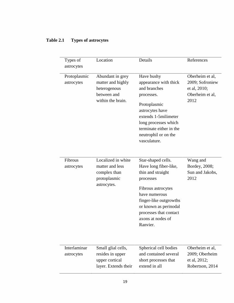
Table 2.1 Types of astrocytes
Types of
astrocytes
Location Details References
Protoplasmic
astrocytes
Abundant in grey
matter and highly
heterogenous
between and
within the brain.
Have bushy
appearance with thick
and branches
processes.
Protoplasmic
astrocytes have
extends 1-5milimeter
long processes which
terminate either in the
neutrophil or on the
vasculature.
Oberheim et al,
2009; Sofroniew
et al, 2010;
Oberheim et al,
2012
Fibrous
astrocytes
Localized in white
matter and less
complex than
protoplasmic
astrocytes.
Star-shaped cells.
Have long fiber-like,
thin and straight
processes
Fibrous astrocytes
have numerous
finger-like outgrowths
or known as perinodal
processes that contact
axons at nodes of
Ranvier.
Wang and
Bordey, 2008;
Sun and Jakobs,
2012
Interlaminar
astrocytes
Small glial cells,
resides in upper
upper cortical
layer. Extends their
Spherical cell bodies
and contained several
short processes that
extend in all
Oberheim et al,
2009; Oberheim
et al, 2012;
Robertson, 2014
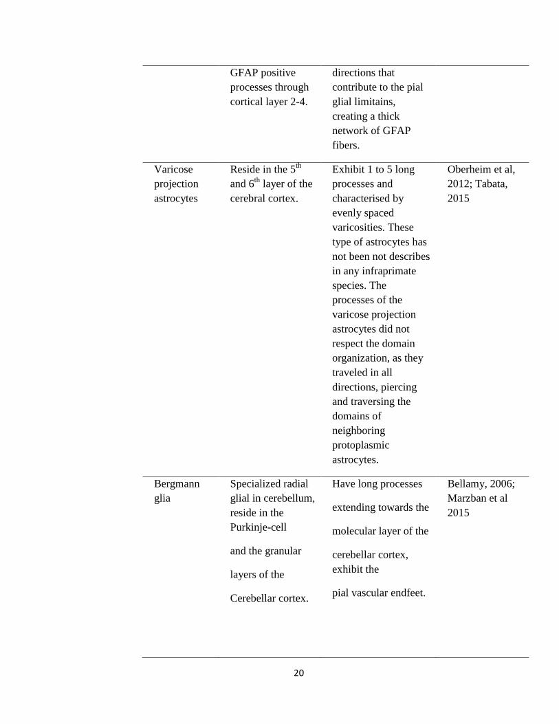
20
GFAP positive
processes through
cortical layer 2-4.
directions that
contribute to the pial
glial limitains,
creating a thick
network of GFAP
fibers.
Varicose
projection
astrocytes
Reside in the 5th
and 6th
layer of the
cerebral cortex.
Exhibit 1 to 5 long
processes and
characterised by
evenly spaced
varicosities. These
type of astrocytes has
not been not describes
in any infraprimate
species. The
processes of the
varicose projection
astrocytes did not
respect the domain
organization, as they
traveled in all
directions, piercing
and traversing the
domains of
neighboring
protoplasmic
astrocytes.
Oberheim et al,
2012; Tabata,
2015
Bergmann
glia
Specialized radial
glial in cerebellum,
reside in the
Purkinje-cell
and the granular
layers of the
Cerebellar cortex.
Have long processes
extending towards the
molecular layer of the
cerebellar cortex,
exhibit the
pial vascular endfeet.
Bellamy, 2006;
Marzban et al
2015
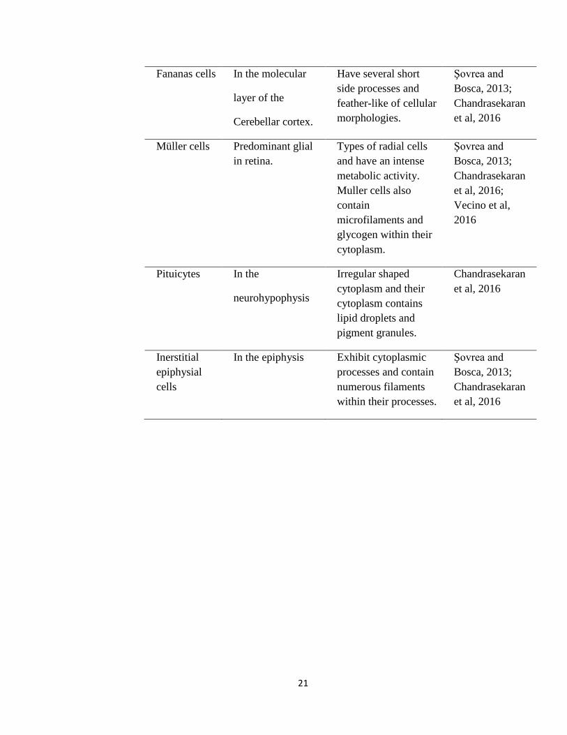
21
Fananas cells In the molecular
layer of the
Cerebellar cortex.
Have several short
side processes and
feather-like of cellular
morphologies.
Şovrea and
Bosca, 2013;
Chandrasekaran
et al, 2016
Müller cells Predominant glial
in retina.
Types of radial cells
and have an intense
metabolic activity.
Muller cells also
contain
microfilaments and
glycogen within their
cytoplasm.
Şovrea and
Bosca, 2013;
Chandrasekaran
et al, 2016;
Vecino et al,
2016
Pituicytes In the
neurohypophysis
Irregular shaped
cytoplasm and their
cytoplasm contains
lipid droplets and
pigment granules.
Chandrasekaran
et al, 2016
Inerstitial
epiphysial
cells
In the epiphysis Exhibit cytoplasmic
processes and contain
numerous filaments
within their processes.
Şovrea and
Bosca, 2013;
Chandrasekaran
et al, 2016
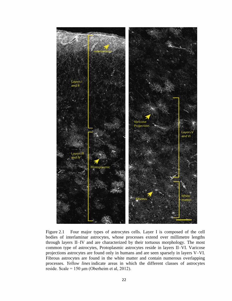
22
Figure 2.1 Four major types of astrocytes cells. Layer I is composed of the cell
bodies of interlaminar astrocytes, whose processes extend over millimetre lengths
through layers II–IV and are characterized by their tortuous morphology. The most
common type of astrocytes, Protoplasmic astrocytes reside in layers II–VI. Varicose
projections astrocytes are found only in humans and are seen sparsely in layers V–VI.
Fibrous astrocytes are found in the white matter and contain numerous overlapping
processes. Yellow lines indicate areas in which the different classes of astrocytes
reside. Scale = 150 μm (Oberheim et al, 2012).
23
2.6 Functions of astrocytes
2.6.1 The brain micro-architecture
In mammalian brain, astrocytes play a role as a definer for the parenchyma specifically
in its micro-architecture structure. Through the process “tiling”, it divided the grey
matter into relatively independent structural units. The protoplasmic type of astrocytes
engages their own position and constructs the micro-anatomical domains within the
limits of their processes. The membrane of the astrocyte within these domains, not just
covers synapses and neuronal membranes, but also sends processes to coat the wall of
neighbouring blood vessel with their endfeet. This kind of complex form consists of
astrocyte-neuron-blood vessel is known as neurovascular unit (Freeman, 2010; Jakobs,
2014; Muoio et al, 2014).
2.6.2 Extracellular homeostasis in brain
Astrocytes also play a duty as one of the gate keepers in brain as it controls
concentrations of ions, neurotransmitters and metabolites and also regulate the
movement of water. It also induces and ensures the stabilization of neuronal synapses
(Clarke and Barres, 2013). One of the recognized functions of astrocytes in order to
maintain the homeostasis in brain is controlling the level of K+ concentration. The
K+ concentration arise from its resting state as there are present of neuronal activity.
The accumulation of K+ concentration level in extracellular space modulates may
initiate epileptic seizures (Florence et al, 2012; Molofsky et al, 2012).
24
There are two common mechanisms that were used by astrocytes to remove
this excess extracellular K+
(Scemes and Spray, 2012). One of the mechanisms is a
passive mechanism known as „spatial buffering‟. The mechanism work by
redistributed within the astrocyte or the coupled astrocytes network after reuptake the
K+
at the higher concentration and then released at sites with lower concentration.
Besides that, astrocytes can discard the excess K+
by increase the pump activities. As
the Na+/K
+-ATPase activity increases, the intracellular K
+ will be increased. The glial
syncytia and aquaporine channels expressed in astrocytes also play a role in water
homeostasis in the brain (Scemes and Spray, 2012; Hertz et al, 2013).
2.6.3 Removing of Glutamate
Glutamate, major excitatory neurotransmitter in brain will act as a powerful neurotoxin
when it releases more than required amounts or being excess for a long time. This
neurotoxin may trigger neuronal cell death in numerous acute or chronic brain lesions.
Function of astrocyte in taking up transmitter was first described by Ernesto Lugaro,
an Italian psychiatrist in 1907 (Kettenmann and Verkhratsky, 2008). Astrocytes
remove a huge amount of glutamate from extracellular space, about 80% of the
glutamate released while neurons take the remaining 20%. Astrocyte cells remove the
glutamate through excitatory amino acid transporters (EAAT) which present in five
types in human brain but two expressed exclusively in astrocyte; EAAT1 and EAAT2
(Hayashi and Yasui 2015; Kinoshita et al 2016).