Embed Size (px)
Citation preview
572 J. Agric. Food Chem. 1982, 30, 572-580
even the relatively high concentrations found in the no. 4 and no. 12 samples will be lower. ACKNOWLEDGMENT
We are indebited to Montedison, Ravit, SIPCAM, and BASF Agritalia for their courtesy in furnishing analytical samples, to Mariangela Lostia Fantola for her valuable technical assistance, and to Consorzio di Bonifica della Sardegna Meridionale for the grape spraying collaboration. LITERATURE CITED Aten, C. F.; Bourke, J. B. J. Agric. Food Chem. 1977,25, 1428. Cabras, P.; Diana, P.; Meloni, M.; Pirisi, F. M. Riu. Vitic. Enol.
Cabras, P.; Meloni, M.; Perra, M.; Pirisi, F. M. J . Chromatogr.
EEC Council Directive Nov 23, 1976, 76/895 EEC no. 340.
1979a, 32, 3.
1979b, 180, 184.
Hoodless, R. A.; Sidwell, J. A.; Skinner, J. C.; Treble, R. D. J.
Huglin, P.; Julliard, B. Prog. Agric. Vitic. 1959, 76, 15. Lawrence, J. F.; Turton, D. J . Chromatogr. 1978, 159, 207. Ripley, B. D.; Cox, D. F.; Wiebe, J.; Frank, R. J. Agnc. Food Chem.
Seiber, J. N. J. Chromatogr. 1974, 94, 151. Sparacino, C. M.; Hines, J. W. J. Chromatogr. Sei. 1976,14,549. Worthing, C. R., Ed. “The Pesticide Manual”, 6th ed.; British Crop
Chromatogr. 1978, 166, 279.
1978, 26, 134.
Protection Council: Croydon, England, 1979.
Received for review August 11,1980. Revised manuscript received October 21,1981. Accepted January 28,1982. This work has been supported by a grant from Echological Branch of Regional Gouvernement of Sardinia. It has been also performed within the Oriented Project “Pesticides and Growth Regulators” of CNR (Italian Research Council, Rome), section no. 8.
Effects of Hapten Structure and Bridging Groups on Antisera Specificity in Parat hion Immunoassay Development
Rem0 P. Vallejo, Edward R. Bogus, and Ralph 0. Mumma*
Five conjugates of parathion to bovine serum albumin (antigens) were prepared wherein bridging groups of varying length and structure were used between the hapten and the protein carrier. In three of these conjugates, the bridging group was attached at either the 2- or 3-phenyl position of parathion, allowing the preservation of all parathion determinant groups. Several immunization modes and schedules were also investigated. Antisera obtained from the use of these antigens contained no antibody specificity for free parathion or 4-nitrophenol in radioimmunoassay binding tests. Enzyme-linked immunosorbent assay binding and inhibition experiments demonstrated the presence of antibodies in antisera associated with three of the conjugates. These antibodies preferentially bind the parathion derivatives that contain the bridging group structures ,and the various precursors of these derivatives. Of four immunization regimens used, only the combination of intradermal and intravenous modes produced any anti-hapten antibody activity.
The important advantage of immunoassays for pesticide residues lies in the minimal requirement for sample cleanup. The specificity of the antibody reaction allows the deletion of a good portion of the rigorous sample pu- rification required by more conventional methods such as GLC, HPLC, and colorimetry. The promise of immu- noassays for pesticides was first envisioned by Ercegovich (1971) and further detailed by Hammock and Mumma (1980). Concrete evidence of this potential has been presented in radioimmunoassay (RIA) procedures suc- cessfully developed for parathion (Ercegovich et al., 1977, 1981; Vallejo, 1981), dieldrin (Langone and Van Vunakis, 1975), S-bioallethrin (Wing et al., 1978; Wing and Ham- mock, 1980), and benomyl (Newsome and Shields, 1981), an enzyme-linked immunosorbent assay (ELISA) for parathion (Al-Rubae, 1978), and a fluorescence immu- noassay for the degradation product of benomyl (Lukens et al., 1977).
The parathion RIA was successfully developed by em- ploying an antigen wherein parathion, by reducing its nitro group, was directly conjugated to a carrier protein via diazo
Pesticide Research Laboratory and Graduate Study Center, Department of Entomology and Department of Chemistry, The Pennsylvania State University, University Park, Pennsylvania 16802.
coupling. The assay allowed detection of parathion resi- dues in plant and plasma samples to the 10-ng level, equivalent to 0.1 ppm, without any sample cleanup.
Improvement of the sensitivity of the assay was there- after sought by enhancing the specificity of the antibody response. Bridging groups of varying length and structure were employed between the hapten and carrier protein to render the hapten more immunogenically visible. The structure of the hapten moiety was also manipulated with a particular view to preserving all determinant groups. In this regard, antigens were prepared wherein parathion was conjugated to the carrier protein via a bridging group at- tached to an unsubstituted aryl position, thus avoiding alteration of the nitro or any other functional group. Finally, the relative efficacies of different immunization modes and schedules were investigated. The specificities of the antisera obtained from these experiments were characterized by RIA and ELISA binding and inhibition tests. MATERIALS AND METHODS
Reagents and Equipment. Parathion, 98.5%, was obtained from American Cyanamid Co., Princeton, NJ. Bovine serum albumin (BSA) (crystallized, purified bovine albumin fraction V), rabbit serum albumin (RSA), Freund’s complete and incomplete adjuvant, and per- oxidase-anti-rabbit IgG were purchased from Miles Lab-
@ 1982 American Chemical Society 0021-8561/82/1430-0572$01.25/0
Antisera Specificity in Parathion Immunoassay Development
oratories, Inc., Elkhart, IN. 2-Amino-4-nitropheno1, p - nitrobenzoyl chloride, m-hydroxycinnamic acid, succinic anhydride, and a 25% aqueous solution of glutaric di- aldehyde were obtained from Aldrich Chemical Co., Inc., Milwaukee, WI. Infrared spectra were taken on a Per- kin-Elmer 421 grating spectrophotometer. NMR spectra were obtained on a Varian A-60 NMR spectrometer. UV spectra and visible-region absorbances were measured on a Gilford spectrophotometer, Model 200. Mass spectra were taken on an AEI Model MS-902 mass spectrometer. Radioactivity was quantified by using a Beckman LS8000 liquid scintillation counter. All thin-layer chromatography (TLC) was performed on precoated silica gel 60 F, plates of 0.25" thickness (EM Reagents, MC/B Manufac- turing Chemists, Inc., Cincinnati, OH). Radioimmu- noassay-grade charcoal and dextran (M, 80 000) were ob- tained from Schwarz/Mann, Orangeburg, NY. Phos- phate-buffered saline (PBS), 0.075 M, pH 7.4, was made by mixing 24 mL of 0.15 M KH2POl and 76 mL of 0.15 M Na2HP04, to which was added 100 mL of 0.9% NaC1. ELISA experiments were conducted with a Gilford eia 50 ELISA reader.
Characterization of Antigens. The number of para- thion residues conjugated to each BSA or RSA molecule was obtained by determination of the phosphorus content of the conjugates (Chen et al., 1956). For those antigens where conjugation of the hapten could possibly involve covalent bonding with a free amino nitrogen on the protein, the number of remaining free amines after conjugation was determined by the 2,4,6-trinitrobenzenesulfonic acid (TNBS) method (Habeeb, 1966), in addition to the de- termination of phosphorus content.
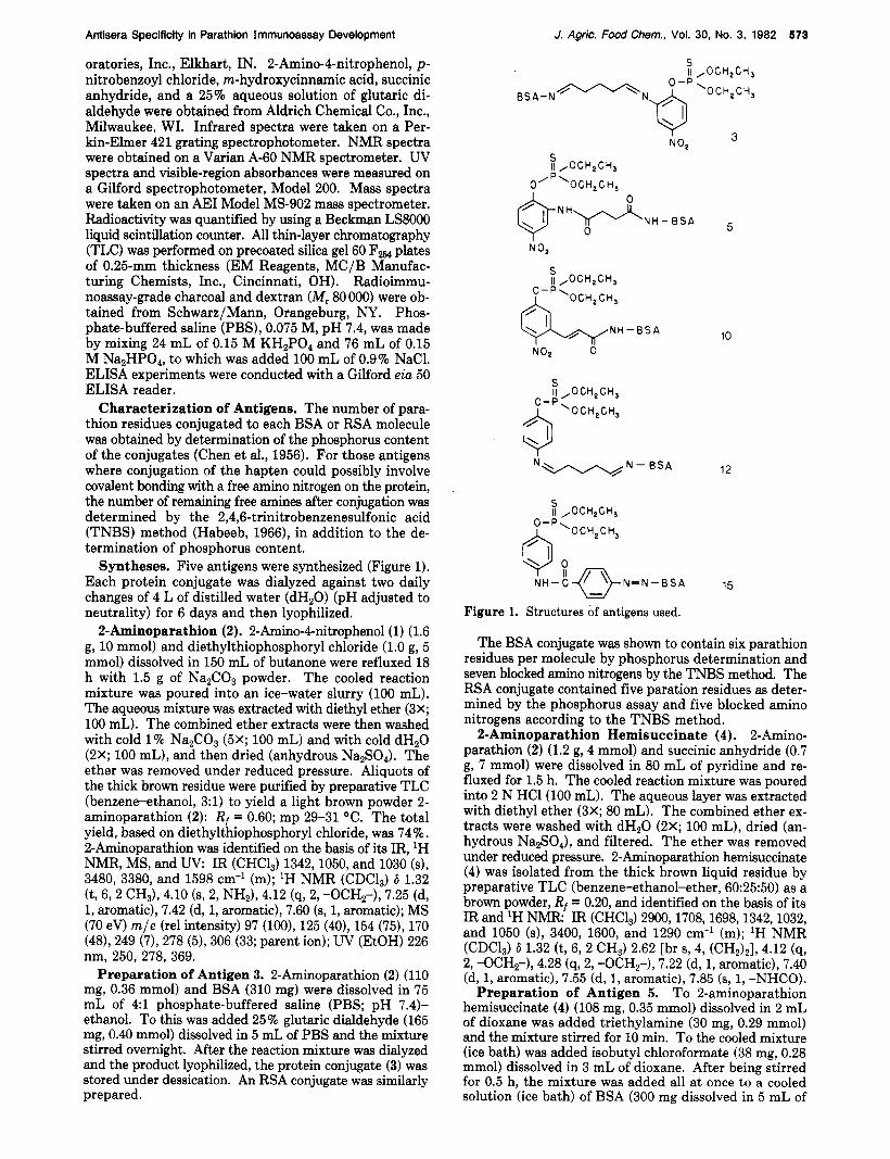
Syntheses. Five antigens were synthesized (Figure 1). Each protein conjugate was dialyzed against two daily changes of 4 L of distilled water (dHzO) (pH adjusted to neutrality) for 6 days and then lyophilized.
2-Aminoparathion (2). 2-Amino-4-nitrophenol (1) (1.6 g, 10 mmol) and diethylthiophosphoryl chloride (1.0 g, 5 mmol) dissolved in 150 mL of butanone were refluxed 18 h with 1.5 g of Na2C03 powder. The cooled reaction mixture was poured into an ice-water slurry (100 mL). The aqueous mixture was extracted with diethyl ether (3X; 100 mL). The combined ether extracts were then washed with cold 1% Na2C03 (5X; 100 mL) and with cold dH20 (2X; 100 mL), and then dried (anhydrous Na,S04). The ether was removed under reduced pressure. Aliquots of the thick brown residue were purified by preparative TLC (benzene-ethanol, 3:l) to yield a light brown powder 2- aminoparathion (2): R, = 0.60; mp 29-31 OC. The total yield, based on diethylthiophosphoryl chloride, was 74%. 2-Aminoparathion was identified on the basis of its IR, 'H
3480, 3380, and 1598 cm-l (m); 'H NMR (CDCl,) 6 1.32
1, aromatic), 7.42 (d, 1, aromatic), 7.60 (9, 1, aromatic); MS (70 eV) m/e (re1 intensity) 97 (loo), 125 (40), 154 (75), 170 (48), 249 (7), 278 (5), 306 (33; parent ion); UV (EtOH) 226 nm, 250, 278, 369.
Preparation of Antigen 3. 2-Aminoparathion (2) (110 mg, 0.36 mmol) and BSA (310 mg) were dissolved in 75 mL of 4:l phosphate-buffered saline (PBS; pH 7.4)- ethanol. To this was added 25% glutaric dialdehyde (165 mg, 0.40 mmol) dissolved in 5 mL of PBS and the mixture stirred overnight. After the reaction mixture was dialyzed and the product lyophilized, the protein conjugate (3) was stored under dessication. An RSA conjugate was similarly prepared.
NMR, MS, and UV IR (CHC13) 1342,1050, and 1030 (s),
(t, 6, 2 CH3), 4.10 (5, 2, NHJ, 4.12 (9, 2, -OCHz-), 7.25 (d,
J. Agric. Food Chem., Vol. 30, No. 3, 1982 573
S Ii ,OCH,GH,
3 7 NO2
N 0,
10
12 N - B S A N w /
Figure 1. Structures of antigens used.
The BSA conjugate was shown to contain six parathion residues per molecule by phosphorus determination and seven blocked amino nitrogens by the TNBS method. The RSA conjugate contained five paration residues as deter- mined by the phosphorus assay and five blocked amino nitrogens according to the TNBS method.
2-Aminoparathion Hemisuccinate (4). 2-Amino- parathion (2) (1.2 g, 4 mmol) and succinic anhydride (0.7 g, 7 mmol) were dissolved in 80 mL of pyridine and re- fluxed for 1.5 h. The cooled reaction mixture was poured into 2 N HCl(lO0 mL). The aqueous layer was extracted with diethyl ether (3X; 80 mL). The combined ether ex- tracts were washed with dHzO (2X; 100 mL), dried (an- hydrous Na2S04), and filtered. The ether was removed under reduced pressure. 2-Aminoparathion hemisuccinate (4) was isolated from the thick brown liquid residue by preparative TLC (benzene-ethanol-ether, 60:25:50) as a brown powder, Rf = 0.20, and identified on the basis of its IR and 'H NMR: IR (CHC1,) 2900,1708,1698,1342,1032, and 1050 (s), 3400, 1600, and 1290 cm-' (m); 'H NMR (CDClJ 6 1.32 (t, 6, 2 CHJ 2.62 [br s, 4, (CH,),], 4.12 (4, 2, -OCH,-), 4.28 (q,2, -OCH,-), 7.22 (d, 1, aromatic), 7.40 (d, 1, aromatic), 7.55 (d, 1, aromatic), 7.85 (s, 1, -NHCO).
To 2-aminoparathion hemisuccinate (4) (108 mg, 0.35 mmol) dissolved in 2 mL of dioxane was added triethylamine (30 mg, 0.29 mmol) and the mixture stirred for 10 min. To the cooled mixture (ice bath) was added isobutyl chloroformate (38 mg, 0.28 mmol) dissolved in 3 mL of dioxane. After being stirred for 0.5 h, the mixture was added all a t once to a cooled solution (ice bath) of BSA (300 mg dissolved in 5 mL of
Preparation of Antigen 5.
574 J. Agric. Food Chem., Vol. 30, No. 3, 1982
dHzO, to which was slowly added 4 mL of dioxane) at pI4 9 (0.1 N NaOH). After being stirred at ice-bath temper- ature for 2 h, the mixture was allowed to react at room temperature overnight. The mixture was dialyzed and lyophilized and the product stored under dessication. The RSA equivalent of this conjugate was similarly prepared.
Phosphorus determination showed the BSA conjugate to contain eight parathion residues per molecule and the TNBS procedure showed seven blocked amino nitrogens on the protein. The RSA conjugate was similarly shown to contain four parathion residues per molecule and five blocked amino nitrogens. 2-Nitro-5-hydroxycinnamic Acid (8). m-Hydroxy-
cinnamic acid (6) (1.64 g, 10 mmol) dissolved in 15 mL of glacial acetic acid was refluxed with 70% HNO, (0.9 g) dissolved in 5 mL of glacial acetic acid for 2 h. The acetic acid was distilled to a volume of 5 mL and the solution brought up to 200 mL with absolute ethanol. After ad- dition of 10 mg of p-toluenesulfonic acid, the mixture was refluxed for 2 h, and then the solvent was removed by distillation, the last 10 mL under reduced pressure. The ethyl ester (7) of the desired nitrogen isomer was purified via preparative column chromatography (benzene-ethanol, 7:3) to yield a light yellow powder, mp 135-137 OC, and identified on the basis of its IR, 'H NMR, MS, and UV: IR (CHCl,) 1720,1342, and 1180 (s), 3220 and 1585 (m), 3560 cm-'; 'H NMR (CDC1,) 6 6.32 (d, 1, CH), 7.11 (9, 1, aromatic), 7.42 (d, 1, aromatic), 8.07 (d, 1, aromatic), 8.12 (d, 1, CH); MS (70 eV) m / e (re1 intensity) 80 (19), 89 (17), 108 (42), 118 (19), 136 (32), 146 (loo), 163 (81), 164 (83), 191 (53), 192 (34), 237 (61, parent ion; UV (EtOH) 260 nm, 310 (ETOH, KOH), 248,293,412. The ethyl ester (0.6 g) was hydrolyzed by refluxing in 0.2 N KOH (40 mL) for 0.5 h, and 2-nitro-5-hydroxycinnamic acid (8) was obtained upon acidification with diluted HCl as a light yellow powder, mp 188-190 OC and used in subsequent reactions.
Preparation of Antigen 10. 2-Nitro-5-hydroxy- cinnamic acid (8) (0.5 g, 2.4 mmol), Na2C03 (0.53 g), and diethylthiophosphoryl chloride (0.9 g, 4.8 mmol) were re- fluxed in 60 mL of butanone for 15 h. The unreacted Na2C03 powder was removed by filtration and then the solvent was removed under reduced pressure. TLC analysis (ether) of the thick light brown residue showed only one spot, Rr = 0.6 (9). Further washings and attempts at purification (distillation and preparative TLC) resulted only in hydrolysis and other degradation products. An aliquot of the residue (0.27 g) dissolved in 2.0 mL of di- oxane was dropped to a cooled solution of BSA in 15 mL of 40% dioxane. The mixture was allowed to stir at ice- bath temperature for 3.5 h while maintaining the pH at 8 with 0.1 N KOH. The mixture was dialyzed and lyo- philized, and the antigen 10 was stored under desiccation. The RSA equivalent of this conjugate was similarly pre- pared.
The BSA conjugate was shown to contain 16 parathion residues per molecule by the phosphorus assay and 13 blocked amino nitrogens by the TNBS method. The RSA conjugate contained 13 parathion residues per molecule and 11 blocked amino nitrogens.
Preparation of Antigen 12. Reduced parathion (11) [ 0,O-diethyl O-@-aminophenyl) thiophosphate] (Ercego- vich et al., 1981) (350 mg, 1.3 mmol) and BSA (600 mg) were dissolved in 75 mL of 2:l PBS (pH 7.4)-dioxane. To this solution was added 25% glutaric dialdehyde (350 mg, 0.9 "01). The light yellow solution turned to light orange in 5 min. The mixture was allowed to stir a t room tem- perature for 3.0 h and then dialyzed. The dialyzed solution was lyophilized and the light orange protein conjugate (12)
Vallejo, Bogus, and Mumma
stored under desiccation. The RSA equivalent of this conjugate was similarly synthesized.
The BSA conjugate was shown to contain 23 parathion residues per molecule by phosphorus determination and 21 blocked amino nitrogens by the TNBS method. The RSA conjugate contained 14 parathion residues and 12 blocked amino nitrogens.
4-Nitro-4'-(0 ,O -diethylphosphorothionyl) benz- anilide (13). Reduced parathion (11) (2.0 g, 8 mmol) and p-nitrobenzoyl chloride (0.6 g, 3 mmol) were dissolved in benzene (40 mL) and refluxed 2.5 h. After the mixture was cooled, the reaction volume was brought up to 100 mL with diethyl ether. The organic phase was washed with cold 1 N HCl(2X; 50 mL), dHzO (1X; 50 mL), cold 1% NaZCO3 (3X; 50 mL), and dHzO (2X; 50 mL). The crude benz- ene-ether solution was shown to contain the desired product (95%; 13) and other impurities, mostly the acid chloride, by TLC analysis (benzene-ethanol, 95:5). This solution was used in the subsequent reaction without further purification.
4-Amino-4'-( 0 ,O -diethylphosphorothionyl)benz- anilide (14). The crude benzene-ether solution (100 mL) of the 4-nitrobenzanilide (13) was stirred at room tem- perature with 2 g of zinc dust suspended in 50 mL of 9:l 1 N acetic acid-concentrated HC1. After 1.5 h, the dark yellow solution had turned colorless. The organic phase was separated and washed with cold 1% Na2C03 (3X; 50 mL), and dH20 (2X; 50 mL) before drying (anhydrous Na2S04). The solvent was removed under reduced pres- sure to yield a thick white oil, which under TLC analysis (benzene-ethanol, 9:l) was shown to be mostly the desired amine (90%). Aliquots of this mixture were purified via preparative TLC. The aminobenzanilide (14) was iden- tified on the basis of its IR, 'H NMR, and MS: IR (CHCI,) 1655,1605,1300,1050,1030, and 960 (s), 3410,3310,2990, and 2450 (m), 3490 cm-' (w); 'H NMR (CDC1,) 6 1.32 (t,
2, -OCH2-), 6.70 (d, 2, aromatic), 7.10 (d, 2, aromatic), 7.59 (d, 2, aromatic), 7.69 (d, 2, aromatic), 7.95 (s, 1, -NHCO); MS (70 eV) m/e (re1 intensity) 92 (46), 120 (57), 148 (1001, 176 (14), 228 (3), 261 (5), 380 (60, parent ion).
Preparation of Antigen 15. To a cooled solution (ice bath) of the aminobenzanilide (14) (240 mg, 0.63 mmol) in 10 mL of THF was added 2 mL of 6 N HC1. NaN02 (120 mg) was added and the cooled mixture stirred for 0.5 h. The excess nitrous acid was then decomposed with urea. The reaction mixture was added to BSA (500 mg) dissolved in borate buffer, pH 8.8 (50 mL). The reaction mixture turned a bright orange within 5 min. After being stirred at ice-bath temperature for 3.5 h, the mixture was stored at 4 "C (refrigerator) overnight and then dialyzed. After lyophilization, the bright orange protein conjugate (15) was stored under desiccation. The RSA equivalent of this conjugate was similarly synthesized.
The BSA conjugate was shown to contain 41 parathion residues by phosphorus determination. Similarly, the RSA conjugate was shown to contain 35 parathion residues.
Preparation of Radiolabeled Tracers. The syntheses of ring-labeled nitr~[~H]phenol and [,H]parathion have been previously reported (Ercegovich et al., 1981). The respective specific activities of ni t r~[~H]phenol and [,H]parathion were 520 and 300 mCi/mmol.
Injection and Bleeding Schedule. Four different modes and schedules of immunization were employed. In all regimens, New Zealand female rabbits, at least 12 weeks of age, were employed, and bleedings were performed through the central ear artery. An initial bleeding before treatment for control sera was performed in each case.
6, 2 CH,), 3.82 (8, 2, NHZ), 4.12 (9, 2, -OCH2-), 4.25 (9,
Antisera Specificity in Parathion Immunoassay Development
(A) Intradermal-Intravenous. The conjugate (0.7 mg) homogenized in 2 mL of a mixture of equal parts of com- plete Freund's adjuvant (CFA) and 0.9% saline was in- troduced intradermally in small aliquots in 30-40 sites on shaven areas of the back and flanks of rabbits. Two weeks later, the animals were boosted with the same amount of antigen in the same manner as the initial injection. A second booster was likewise administered 2 weeks later by using incomplete Freund's adjuvant (IFA) instead of CFA. Subsequent booster injections were given every 2 weeks intravenously into the marginal ear vein by using 2.0 mg of conjugate dissolved in 2.0 mL of saline. The rabbits were bled 8 days after each intravenous booster.
(B) Intradermal. Two levels of antigen were used in this program, 0.5 or 2.5 mg. The conjugate (0.5 or 2.5 mg) was homogenized in 1 mL of a mixture of equal parts of CFA and saline and introduced intradermally in small aliquots in 15-20 sites on shaven areas of the back and flanks. The animals were boosted every 2 weeks after the initial im- munization in the same manner by using IFA instead of CFA. The rabbits were bled 8 days after each booster.
(C) Intradermal with an Increase in the Amount o f Killed Bacteria. The conjugate (0.1-0.3 mg) homogenized in 3 mL of 1:l CFA-saline with 5-10 mg of total killed bacteria (desiccated Mycobacterium tuberculosis, DIFCO Laboratories, Detroit, MI) was introduced intradermally in small aliquots in 50-60 sites on shaven areas of the back and flanks. The animals were boostered with 0.1-0.3 mg of conjugate in 3 mL of 1:l IFA-saline in the same manner after 1 month. The rabbits were bled 8 days after the booster. (D) Intramuscular. Two levels of antigen were used, 1
or 5 mg. The conjugate (1.0 or 5.0 mg) dissolved in 1 mL of 1:l CFA-saline was introduced in four sites into the thigh muscle. The rabbits were boostered similarly by using IFA instead of CFA, after 1 month and every month thereafter. The rabbits were bled 8 days after each booster.
Characterization of Antisera. Immunodiffusion. Established agar gel procedures were adapted with certain modifications as previously described (Ouchterlony, 1958; Campbell et al., 1970; Ercegovich et al., 1981).
Radioimmunoassay. Antisera were screened for the ability to bind radiolabeled parathion by using RIA pro- cedures. Assays were performed in triplicate in 12 X 75 mm disposable glass tubes. To a solution of 200 pL of antiserum dissolved in 750 pL of PBA (pH 7.4) was added 20 pL of the [3H]parathion (approximately 45000 dpm) in ethanol. The mixture was agitated in a Vortex mixer and allowed to stand at room temperature for 4 h and at 4 OC overnight. Separation of the bound and nonbound radioactivity was accomplished by the addition of 500 pL of a stock mixture of dextran-coated charcoal (DCC). The latter was prepared by mixing 10 g of RIA-grade charcoal and 250 mg of dextran in 100 mL of PBS (pH 7.4) with constant stirring. The reaction mixture was agitated in a Vortex mixer, incubated at 25 "C for 15 min, and then centrifuged at 1500g for 10 min. The supernatant, con- taining antibody-bound pesticide, was transferred by pipet to liquid scintillation vials for radioassay. A positive control was run for each set of RIA binding tests, em- ploying antisera with proven parathion-specific antibodies (Ercegovich et al. 1981). The efficiency of each step in the RIA procedure was confirmed by the fact that the expected levels of binding of parathion by these antisera were always attained.
ELISA. (1) Coating of Microtiter Plate Wells. The wells of a microtiter plate (Gilford Laboratories, Inc., Oberlin, OH) were washed with doubly distilled water.
J. Agric. Food Chem., Vol. 30, No. 3, 1982 575
These wells were coated with 10 pg of the RSA conjugate dissolved in 0.3 mL of 0.1 M carbonate buffer (pH 9.6) containing 0.2% sodium azide at 4 "C for 12 h. The wells were then emptied and washed 5 times (99-s incubation) with 0.3 mL of 0.9% saline solution containing 0.05% Tween- 20.
(2) Inhibition of Antisera. Antiserum (1.0 mL) diluted 1 : l O in PBS (pH 7.4) containing 0.2% sodium azide and 0.05% Tween-20 was incubated with different levels of inhibitor dissolved in 40 pL of ethanol for 2 h at 25 "C in separate 12 X 75 mm disposable glass tubes.
(3) Binding of Inhibited Antiserum to Wells. Aliquots (250 mL) of the inhibited antiserum were then introduced into the coated wells and incubated at 37 OC for 3 h. The wells were emptied after incubation and washed with sa- line-Tween-20 solution (4X; 0.3 mL; 99-s incubation).
(4 ) Addition of Enzyme-Conjugate. Diluted (MOO; PBA; pH 7.4) horseradish peroxidase-anti-rabbit IgG en- zyme-antibody complex (250 FL) was introduced into the wells, incubated at 37 OC for 3 h, emptied, and washed with the saline-Tween-20 solution (4X; 0.3 mL; 99-s incubation).
(5) Addition of Enzyme Substrate. The amount of horseradish peroxidase complex bound to the wells was determined by introducing 0.25 mL of the enzyme sub- strate, p-aminosalicylic acid (0.7 mg/mL in 0.005% HzOz, pH 6), into the wells and allowing color to develop for 0.5 h. The reaction was stopped by the addition of 1 N NaOH (50 pL) and the absorbance determined at 450 nm.
RESULTS AND DISCUSSION The successful generation of antisera highly specific
against a small molecule is greatly dependent upon the characteristics of the hapten-protein carrier conjugate. The determinant groups of the small molecule must be preserved. It is realized that one or another functional group in the molecule may have to be used and altered in linking hapten to the carrier. This is where the proper design considerations have to be made.
The hapten's determinant groups must not be masked. Although all or most of the molecule's functional groups may be unaltered, they still could be masked by or lost within the protein tertiary structure. It is not only im- perative that there is a minimum of structure alteration to the hapten but also necessary to ensure that the hapten, unaltered as it is, is visible above the protein carrier mass. It is important that the hapten structure is physically set apart from the carrier's. Any masking caused by the carrier's spatial conformation has as much a deleterious effect on hapten immunogenicity as any chemical struc- tural alteration.
The visibility of the hapten depends largely on the mode of conjugation. A small molecule that is directly covalently linked to some site on a protein may suffer a considerable amount of masking in the region of the hapten nearest the site of linkage. Antisera raised would suffer a lack of specificity for those determinant groups that have been so masked. The most obvious means to avoid this problem would be to physically extend the hapten out in space via bridging groups. The bridging group could prosaically be seen as a stick on which the hapten is pushed out into space.
The effect of extending haptens out in space via bridging groups is best exemplified in the development of an RIA for diphenylhydantoin (Paxton et al., 1976). Two conju- gates were employed as antigens, one containing an acetyl bridging group between hapten and carrier protein and the other, a pentanoyl, both bridges attached to the same site on the hapten's ethyleneurea nucleus. The antigen with the shorter bridging group failed to elicit antibodies that
576 J. Agric. Food Chem., Vol. 30, No. 3, 1982
could recognize free diphenylhydantoin, while the one with the longer bridge produced antibodies of high titer and specificity against the free hapten. Both conjugates made use of bridging groups, and it is noteworthy that one failed to fulfill its desired function and the other, employing a longer bridge, delivered excellent results.
In the original parathion RIA (Ercegovich et al., 1981), parathion was functionalized prior to conjugation by re- ducing its nitro group to an amino function. This con- stituted a major alteration of a potentially important de- terminant group. Furthermore, the reduced parathion structure was directly conjugated to the carrier protein without the mediation of a bridging group.
Improvement in sensitivity and specificity could possibly be attained by using other means of covalently bonding the hapten to the protein, choosing especially those methods which preserve all determinant groups including the nitro function. The extension of the hapten farther away from the protein mass by employing bridging groups of varied size and structure could also result in improve- ment.
Efficacy of Various Immunization Protocols. Sera obtained from bleedings were assayed for antibody titer and specificity by immunodiffusion, RIA and ELISA binding, and inhibition experiments. Of the four im- munization regimens applied, only one, the intradermal- intravenous program (A), yielded any significant antibody activity specific against haptens. In any immunization program using hapten-protein conjugates, it is expected that the first and most easily detectable antibody response would be that directed against the carrier protein. Con- tinued exposure of the animal to the antigen via subse- quent booster injections may then develop antibodies against the hapten in detectable levels. The intradermal (B) and intramuscular (D) schedules both generated de- tectable levels of antibody against the BSA carrier as ev- idenced by precipitin formation as early as after the first booster. Both, however, did not elicit any antibody activity against the haptens even after five or more boosters.
The intradermal program using an increased amount of killed bacteria was even less effective. Although the use of such increased levels of killed bacteria is designed to further stimulate the immune system’s response to anti- genic material, it was nevertheless observed that detectable antibody activity against the protein carrier alone neces- sitated at least three boosters as opposed to one or at most two with the other programs. This delayed response to the antigen may be due to the overwhelming amount of killed bacteria (5-10 mg) as compared to antigen (0.14.3 mg) in the initial injection. Although the efficacious use of this schedule has been reported (Robbins, 19801, high titers of antibodies against capsular polysaccharides con- jugated to large proteins having been obtained after only one booster, the protocol may not be as effective when attempting to generate antibodies against a small hapten (M, -400) conjugated to a relatively small protein like BSA.
The singular success obtained with the intradermal-in- travenous schedule does not necessarily lead to any gen- eralized conclusions regarding the choice of immunization programs for immunoassay development. The fact that the other modes of immunization failed in no way means that, using another antigen, they also will not work. They may, indeed, prove successful with the self-same antigens used if the number of rabbits so inoculated was increased. It may be that, had the population of animals been large enough, a significant number, albeit a minority, may have responded favorably to treatment. As it was, only two
Vallejo, Bogus, and Mumma
Table I . Reaction of Antisera from Rabbits Exposed to Various Conjugatesa conjugate immunodiffusion RIA ELISA
-. - _. 3 5 + + + +
10 12 + + 1 5 + + + + + +
a (+ + ) represents activity against hapten; (-) represents
- - + + -
no activity.
replicate animals were available per treatment per conju- gates, as dictated by budgetary and manpower constraints.
Upon examination, the intradermal-intravenous regimen may actually afford the best mode of exposure of the an- imal to the antigen, especially since aliquots of each in- tradermal dose are spread over a wide area of the shaven back, flanks, and hindquarters of the rabbit. Each in- travenous dose allows the circulatory system to deliver the antigen to an even wider area of lymphatic sites than is allowed via the intradermal route, further stimulating specific antibody response.
Characterization of Antisera. Antisera were exam- ined for antibody titer and specificity by using three dif- ferent immunoassays. The antisera were first tested by using immunodiffusion. In this, the formation of precipitin lines in agar gel plates would reveal antibody activity against the protein carrier; the formation of spurs in the conditions described would be indicative of anti-hapten specificity (Ouchterlony, 1958; Ercegovich et al., 1981).
The antisera were subsequently tested for their ability to bind [3H]parathion and 4-nitr0[~H]phenol. Any an- tiserum with the ability to bind a significant percentage of the radioactivity present would then be fit for use in radioimmunoassays for parathion or 4-nitrophenol. Its specificity for parathion would then be challenged by ex- amining the degree of inhibition that other compounds similar in chemical structure to parathion exercise over the antiserum’s ability to bind the radiolabeled tracer. Sim- ilarly, any antisera that bind 4-nitr0[~H]phenol would undergo inhibition tests.
Antisera which do not bind the radiolabeled tracers are not necessarily devoid of anti-hapten activity. They may be able to recognize the parathion derivative employed in the conjugation to a protein. Such parathion derivatives may include all or part of the bridging groups used in conjugation. The fact that parathion has been altered, functibnalized, a d condensed with various bridging groups may disallow any recognition of the unaltered parathion structure by the antibodies which may, in reality, require that the alterations, functionalizations, or bridging groups be present before any antihapten activity is demonstrated.
An antiserum which does not bind the radiolabeled tracers but shows the capability to bind the RSA-hapten conjugate in the ELISA test manifests antibody specificity against the hapten. Although parathion specificity can be ruled out if such antisera cannot bind [3H]parathion, there still is the question of what hapten structures the anti- bodies are specific to. This can be resolved by determining the degree of inhibition on ELISA color formation effected by the different parathion derivatives used in conjugation. Those derivatives that do inhibit color formation in ELISA are then determined to be those structures the antibodies are specifically against.
A summary of the results obtained from testing antisera obtained with the different antigens in immunodiffusion, RIA binding, and ELISA binding tests is presented in Table I. This table indicates only whether the test in
Antisera Specificity in Parathion Immunoassay Development J. Agric. Food Chem., Vol. 30, No. 3, 1982 577
S
Figure 2.
I, ,
O-P-(OEl12
2 5 1 13 - HO.r/J-koH
I us /
OH / /
5 -
L
N O2 / P
e - p a r a t h i o n
I I I 1 I I 50 2 5 0 500 1000 2500 5000
NG INHIBITOR
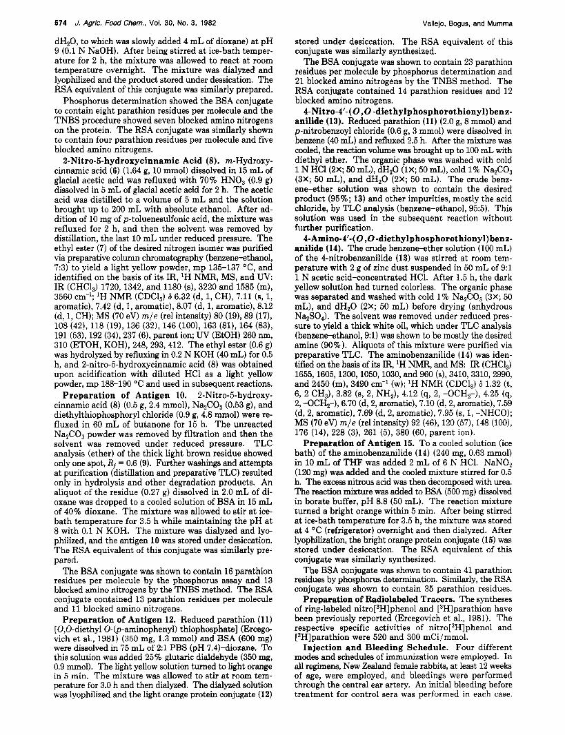
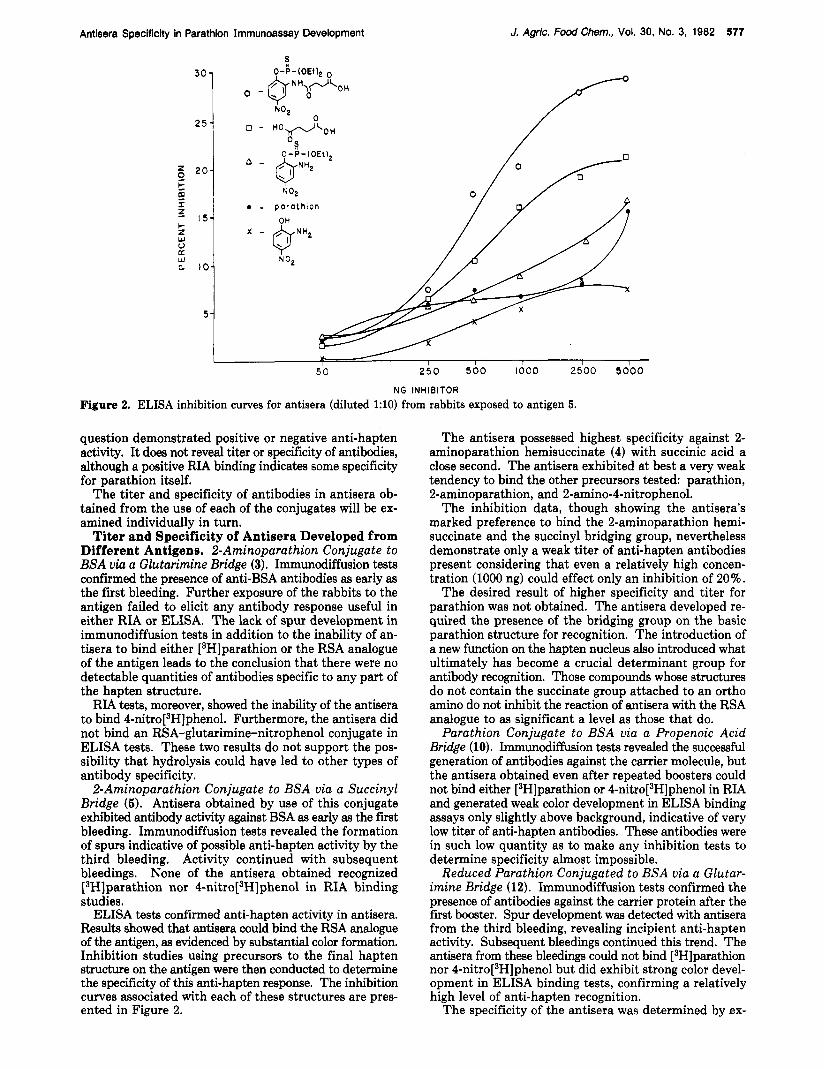
ELISA inhibition curves for antisera (diluted 1:lO) from rabbits exposed to antigen 5.
question demonstrated positive or negative anti-hapten activity. It does not reveal titer or specificity of antibodies, although a positive RIA binding indicates some specificity for parathion itself.
The titer and specificity of antibodies in antisera ob- tained from the use of each of the conjugates will be ex- amined individually in turn.
Titer and Specificity of Antisera Developed from Different Antigens. 2-Aminoparathion Conjugate to BSA via a Glutarimine Bridge (3). Immunodiffusion tests conf i ied the presence of anti-BSA antibodies as early as the first bleeding. Further exposure of the rabbits to the antigen failed to elicit any antibody response useful in either RIA or ELISA. The lack of spur development in immunodiffusion tests in addition to the inability of an- tisera to bind either [3H]parathion or the RSA analogue of the antigen leads to the conclusion that there were no detectable quantities of antibodies specific to any part of the hapten structure.
RIA testa, moreover, showed the inability of the antisera to bind 4-nitr0[~H]phenol. Furthermore, the antisera did not bind an RSA-glutarimine-nitrophenol conjugate in ELISA tests. These two results do not support the pos- sibility that hydrolysis could have led to other types of antibody specificity.
2-Aminoparathion Conjugate to BSA via a Succinyl Bridge (5). Antisera obtained by use of this conjugate exhibited antibody activity against BSA as early as the first bleeding. Immunodiffusion tests revealed the formation of spurs indicative of possible anti-hapten activity by the third bleeding. Activity continued with subsequent bleedings. None of the antisera obtained recognized [3H]parathion nor 4-nitr0[~H]phenol in RIA binding studies.
ELISA tests confirmed anti-hapten activity in antisera. Resulta showed that antisera could bind the RSA analogue of the antigen, as evidenced by substantial color formation. Inhibition studies using precursors to the final hapten structure on the antigen were then conducted to determine the specificity of this anti-hapten response. The inhibition curves associated with each of these structures are pres- ented in Figure 2.
The antisera possessed highest specificity against 2- aminoparathion hemisuccinate (4) with succinic acid a close second. The antisera exhibited at best a very weak tendency to bind the other precursors tested: parathion, 2-aminoparathion, and 2-amino-4-nitrophenol.
The inhibition data, though showing the antisera’s marked preference to bind the 2-aminoparathion hemi- succinate and the succinyl bridging group, nevertheless demonstrate only a weak titer of anti-hapten antibodies present considering that even a relatively high concen- tration (1000 ng) could effect only an inhibition of 20%.
The desired result of higher specificity and titer for parathion was not obtained. The antisera developed re- quired the presence of the bridging group on the basic parathion structure for recognition. The introduction of a new function on the hapten nucleus also introduced what ultimately has become a crucial determinant group for antibody recognition. Those compounds whose structures do not contain the succinate group attached to an ortho amino do not inhibit the reaction of antisera with the RSA analogue to as significant a level as those that do.
Parathion Conjugate to BSA via a Propenoic Acid Bridge (10). Immunodiffusion tests revealed the successful generation of antibodies against the carrier molecule, but the antisera obtained even after repeated boosters could not bind either [3H]parathion or 4-nitr0[~H]phenol in RIA and generated weak color development in ELISA binding assays only slightly above background, indicative of very low titer of anti-hapten antibodies. These antibodies were in such low quantity as to make any inhibition tests to determine specificity almost impossible.
Reduced Parathion Conjugated to BSA via a Glutar- imine Bridge (12). Immunodiffusion tests confirmed the presence of antibodies against the carrier protein after the f i s t booster. Spur development was detected with antisera from the third bleeding, revealing incipient anti-hapten activity. Subsequent bleedings continued this trend. The antisera from these bleedings could not bind [3H]parathion nor 4-nitr0[~H]phenol but did exhibit strong color devel- opment in ELISA binding tests, confirming a relatively high level of anti-hapten recognition.
The specificity of the antisera was determined by ex-
578 J. Agric. Food Chem., Vol. 30, No. 3, 1982 Vallejo, Bogus, and Mumma
0 - H
6 C _I
5 I O 2 0 5 0 250 560 1000 2500 5600 N G INHIBITOR
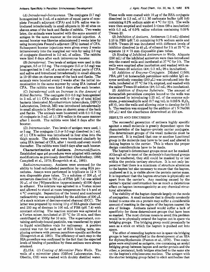
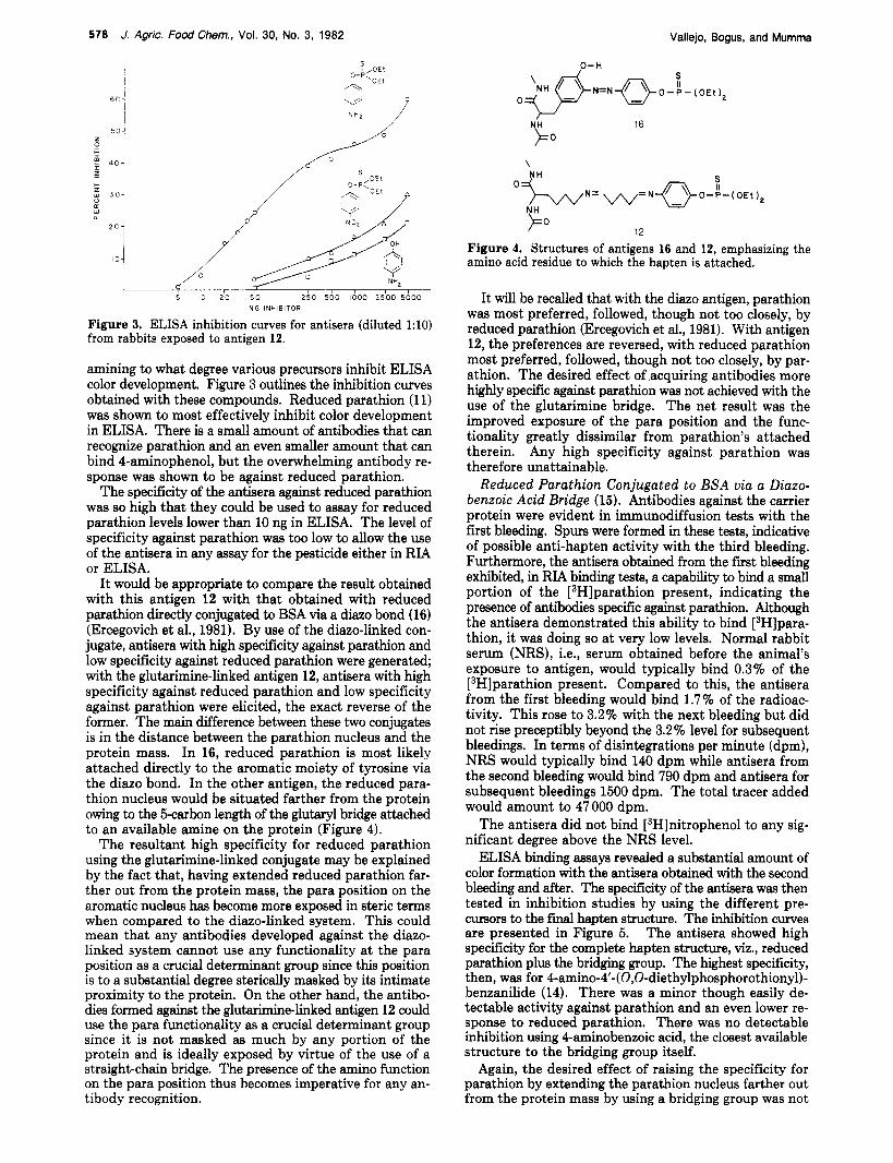
Figure 3. ELISA inhibition curves for antisera (diluted 1:lO) from rabbits exposed to antigen 12.
amining to what degree various precursors inhibit ELISA color development. Figure 3 outlines the inhibition curves obtained with these compounds. Reduced parathion (1 1) was shown to most effectively inhibit color development in ELISA. There is a small amount of antibodies that can recognize parathion and an even smaller amount that can bind 4-aminophenol, but the overwhelming antibody re- sponse was shown to be against reduced parathion.
The specificity of the antisera against reduced parathion was so high that they could be used to assay for reduced parathion levels lower than 10 ng in ELISA. The level of specificity against parathion was too low to allow the use of the antisera in any assay for the pesticide either in RIA or ELISA.
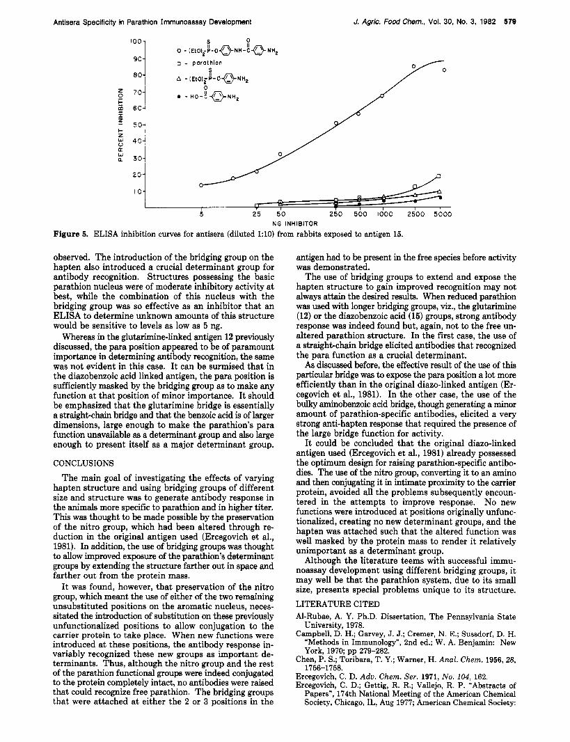
It would be appropriate to compare the result obtained with this antigen 12 with that obtained with reduced parathion directly conjugated to BSA via a diazo bond (16) (Ercegovich et al., 1981). By use of the diazo-linked con- jugate, antisera with high specificity against parathion and low specificity against reduced parathion were generated; with the glutarimine-linked antigen 12, antisera with high specificity against reduced parathion and low specificity against parathion were elicited, the exact reverse of the former. The main difference between these two conjugates is in the distance between the parathion nucleus and the protein mass. In 16, reduced parathion is most likely attached directly to the aromatic moiety of tyrosine via the diazo bond. In the other antigen, the reduced para- thion nucleus would be situated farther from the protein owing to the &carbon length of the glutaryl bridge attached to an available amine on the protein (Figure 4).
The resultant high specificity for reduced parathion using the glutarimine-linked conjugate may be explained by the fact that, having extended reduced parathion far- ther out from the protein mass, the para position on the aromatic nucleus has become more exposed in steric terms when compared to the diazo-linked system. This could mean that any antibodies developed against the diazo- linked system cannot use any functionality a t the para position as a crucial determinant group since this position is to a substantial degree sterically masked by its intimate proximity to the protein. On the other hand, the antibo- dies formed against the glutarimine-linked antigen 12 could use the para functionality as a crucial determinant group since i t is not masked as much by any portion of the protein and is ideally exposed by virtue of the use of a straight-chain bridge. The presence of the amino function on the para position thus becomes imperative for any an- tibody recognition.
\
12
Figure 4. Structures of antigens 16 and 12, emphasizing the amino acid residue to which the hapten is attached.
It will be recalled that with the diazo antigen, parathion was most preferred, followed, though not too closely, by reduced parathion (Ercegovich et al., 1981). With antigen 12, the preferences are reversed, with reduced parathion most preferred, followed, though not too closely, by par- athion. The desired effect of acquiring antibodies more highly specific against parathion was not achieved with the use of the glutarimine bridge. The net result was the improved exposure of the para position and the func- tionality greatly dissimilar from parathion's attached therein. Any high specificity against parathion was therefore unattainable.
Reduced Parathion Conjugated to BSA via a Diazo- benzoic Acid Bridge (15). Antibodies against the carrier protein were evident in immunodiffusion tests with the first bleeding. Spurs were formed in these tests, indicative of possible anti-hapten activity with the third bleeding. Furthermore, the antisera obtained from the first bleeding exhibited, in RIA binding testa, a capability to bind a small portion of the [3H]parathion present, indicating the presence of antibodies specific against parathion. Although the antisera demonstrated this ability to bind [3H]para- thion, it was doing so at very low levels. Normal rabbit serum (NRS), i.e., serum obtained before the animal's exposure to antigen, would typically bind 0.3% of the [3H]parathion present. Compared to this, the antisera from the first bleeding would bind 1.7% of the radioac- tivity. This rose to 3.2% with the next bleeding but did not rise preceptibly beyond the 3.2% level for subsequent bleedings. In terms of disintegrations per minute (dpm), NRS would typically bind 140 dpm while antisera from the second bleeding would bind 790 dpm and antisera for subsequent bleedings 1500 dpm. The total tracer added would amount to 47 000 dpm.
The antisera did not bind [3H]nitrophenol to any sig- nificant degree above the NRS level.
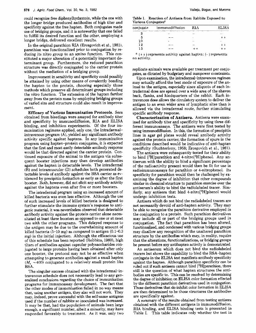
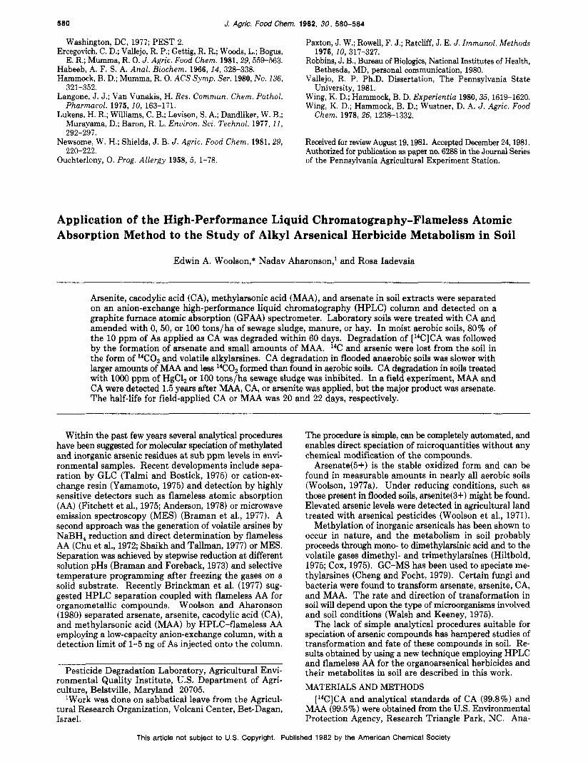
ELISA binding assays revealed a substantial amount of color formation with the antisera obtained with the second bleeding and after. The specificity of the antisera was then tested in inhibition studies by using the different pre- cursors to the f i i hapten structure. The inhibition curves are presented in Figure 5. The antisera showed high specificity for the complete hapten structure, viz., reduced parathion plus the bridging group. The highest specificity, then, was for 4-amino-4'- (0,O-diethylphosphorothiony1)- benzanilide (14). There was a minor though easily de- tectable activity against parathion and an even lower re- sponse to reduced parathion. There was no detectable inhibition using 4-aminobenzoic acid, the closest available structure to the bridging group itself.
Again, the desired effect of raising the specificity for parathion by extending the parathion nucleus farther out from the protein mass by using a bridging group was not
Antisera Specificity in Parathion Immunoassay Development
S 0 0 - LEt0,fC-OQN"-;QN"2
o - p a r a t h i o n 90
J. Agric. Food Chem., Vol. 30, No. 3, 1982 570
- m n I
I , l- w 1
I I T I I I I 5 25 50 250 500 1000 2500 5000
NG INHIBITOR
Figure 5. ELISA inhibition curves for antisera (diluted 1:lO) from rabbits exposed to antigen 15.
observed. The introduction of the bridging group on the hapten also introduced a crucial determinant group for antibody recognition. Structures possessing the basic parathion nucleus were of moderate inhibitory activity at best, while the combination of this nucleus with the bridging group was so effective as an inhibitor that an ELISA to determine unknown amounts of this structure would be sensitive to levels as low as 5 ng.
Whereas in the glutarimine-linked antigen 12 previously discussed, the para position appeared to be of paramount importance in determining antibody recognition, the same was not evident in this case. It can be surmised that in the diazobenzoic acid linked antigen, the para position is sufficiently masked by the bridging group as to make any function at that position of minor importance. It should be emphasized that the glutarimine bridge is essentially a straighbchain bridge and that the benzoic acid is of larger dimensions, large enough to make the parathion's para function unavailable as a determinant group and also large enough to present itself as a major determinant group.
CONCLUSIONS The main goal of investigating the effects of varying
hapten structure and using bridging groups of different size and structure was to generate antibody response in the animals more specific to parathion and in higher titer. This was thought to be made possible by the preservation of the nitro group, which had been altered through re- duction in the original antigen used (Ercegovich et al., 1981). In addition, the use of bridging groups was thought to d o w improved exposure of the parathion's determinant groups by extending the structure farther out in space and farther out from the protein mass.
I t was found, however, that preservation of the nitro group, which meant the use of either of the two remaining unsubstituted positions on the aromatic nucleus, neces- sitated the introduction of substitution on these previously unfunctionalized positions to allow conjugation to the carrier protein to take place. When new functions were introduced at these positions, the antibody response in- variably recognized these new groups as important de- terminants. Thus, although the nitro group and the rest of the parathion functional groups were indeed conjugated to the protein completely intact, no antibodies were raised that could recognize free parathion. The bridging groups that were attached at either the 2 or 3 positions in the
antigen had to be present in the free species before activity was demonstrated.
The use of bridging groups to extend and expose the hapten structure to gain improved recognition may not always attain the desired results. When reduced parathion was used with longer bridging groups, viz., the glutarimine (12) or the diazobenzoic acid (15) groups, strong antibody response was indeed found but, again, not to the free un- altered parathion structure. In the first case, the use of a straight-chain bridge elicited antibodies that recognized the para function as a crucial determinant.
As discussed before, the effective result of the use of this particular bridge was to expose the para position a lot more efficiently than in the original diazo-linked antigen (Er- cegovich et al., 1981). In the other case, the use of the bulky aminobenzoic acid bridge, though generating a minor amount of parathion-specific antibodies, elicited a very strong anti-hapten response that required the presence of the large bridge function for activity.
It could be concluded that the original diazo-linked antigen used (Ercegovich et al., 1981) already possessed the optimum design for raising parathion-specific antibo- dies. The use of the nitro group, converting it to an amino and then conjugating it in intimate proximity to the carrier protein, avoided all the problems subsequently encoun- tered in the attempts to improve response. No new functions were introduced at positions originally unfunc- tionalized, creating no new determinant groups, and the hapten was attached such that the altered function was well masked by the protein mass to render it relatively unimportant as a determinant group.
Although the literature teems with successful immu- noassay development using different bridging groups, it may well be that the parathion system, due to its small size, presents special problems unique to its structure. LITERATURE CITED Al-Rubae, A. Y. Ph.D. Dissertation, The Pennsylvania State
University, 1978. Campbell, D. H.; Garvey, J. J.; Cremer, N. E.; Sussdorf, D. H.
"Methods in Immunology", 2nd ed.; W. A. Benjamin: New York, 1970; pp 279-282.
Chen, P. S.; Toribara, T. Y.; Warner, H. Anal. Chem. 1956,28,
Ercegovich, C. D. Adv. Chem. Ser. 1971, No. 104, 162. Ercegovich, C. D.; Gettig, R. R.; Vallejo, R. P. "Abstracts of
Papers", 174th National Meeting of the American Chemical Society, Chicago, IL, Aug 1977; American Chemical Society:
1756-1758.
580 J. Agric. Food Chem. 1982, 30, 580-584
Washington, DC, 1977; PEST 2. Ercegovich, C. D.; Vallejo, R. P.; Gettig, R. R.; Woods, L.; Bogus,
E. R.; Mumma, R. 0. J. Agric. Food Chem. 1981,29,559-563. Habeeb, A. F. S. A. Anal. Biochem. 1966,14, 328-338. Hammock, B. D.; Mumma, R. 0. ACS Symp. Ser. 1980, No. 136,
Langone, J. J.; Van Vunakis, H. Res. Commun. Chem. Pathol. Pharmacol. 1975, 10, 163-171.
Lukens, H. R.; Williams, C. B.; Levison, S. A.; Dandliker, W. B.; Murayama, D.; Baron, R. L. Enuiron. Sci. Technol. 1977, 11,
Newsome, W. H.; Shields, J. B. J . Agric. Food Chem. 1981, 29,
Ouchterlony, 0. Prog. Allergy 1958, 5, 1-78.
321-352.
292-297.
220-222.
Paxton, J. W.; Rowell, F. J.; Rat.cliff, J. E. J. Immunol. Methods
Robbins, J. B., Bureau of Biologics, National Institutes of Health,
Vallejo, R. P. Ph.D. Dissertation, The Pennsylvania State
Wing, K. D.; Hammock, B. D. Experientia 1980,35,1619-1620. Wing, K. D.; Hammock, B. D.; Wustner, D. A. J . Agric. Food
1976, 10, 317-327.
Bethesda, MD, personal communication, 1980.
University, 1981.
Chem. 1978,26, 1238-1332.
Received for review August 19,1981. Accepted December 24,1981. Authorized for publication as paper no. 6288 in the Journal Series of the Pennsylvania Agricultural Experiment Station.
Application of the High-Performance Liquid Chromatography-Flameless Atomic Absorption Method to the Study of Alkyl Arsenical Herbicide Metabolism in Soil
Edwin A. Woolson,* Nadav Aharonson,' and Rosa Iadevaia
Arsenite, cacodylic acid (CA), methylarsonic acid (MAA), and arsenate in soil extracts were separated on an anion-exchange high-performance liquid chromatography (HPLC) column and detected on a graphite furnace atomic absorption (GFAA) spectrometer. Laboratory soils were treated with CA and amended with 0, 50, or 100 tons/ha of sewage sludge, manure, or hay. In moist aerobic soils, 80% of the 10 ppm of As applied as CA was degraded within 60 days. Degradation of [14C]CA was followed by the formation of arsenate and small amounts of MAA. 14C and arsenic were lost from the soil in the form of 14C02 and volatile alkylarsines. CA degradation in flooded anaerobic soils was slower with larger amounts of MA4 and less 14C02 formed than found in aerobic soils. CA degradation in soils treated with 1000 ppm of HgCl, or 100 tons/ha sewage sludge was inhibited. In a field experiment, MAA and CA were detected 1.5 years after MAA, CA, or arsenite was applied, but the major product was arsenate. The half-life for field-applied CA or MAA was 20 and 22 days, respectively.
Within the past few years several analytical procedures have been suggested for molecular speciation of methylated and inorganic arsenic residues at sub ppm levels in envi- ronmental samples. Recent developments include sepa- ration by GLC (Talmi and Bostick, 1975) or cation-ex- change resin (Yamamoto, 1975) and detection by highly sensitive detectors such as flameless atomic absorption (AA) (Fitchett et al., 1975; Anderson, 1978) or microwave emission spectroscopy (MES) (Braman et al., 1977). A second approach was the generation of volatile arsines by NaBH, reduction and direct determination by flameless AA (Chu et al., 1972; Shaikh and Tallman, 1977) or MES. Separation was achieved by stepwise reduction at different solution pHs (Braman and Foreback, 1973) and selective temperature programming after freezing the gases on a solid substrate. Recently Brinckman et al. (1977) sug- gested HPLC separation coupled with flameless AA for organometallic compounds. Woolson and Aharonson (1980) separated arsenate, arsenite, cacodylic acid (CA), and methylarsonic acid (MAA) by HPLC-flameless AA employing a low-capacity anion-exchange column, with a detection limit of 1-5 ng of As injected onto the column.
Pesticide Degradation Laboratory, Agricultural Envi- ronmental Quality Institute, U.S. Department of Agri- culture, Belstville, Maryland 20705.
'Work was done on sabbatical leave from the Agricul- tural Research Organization, Volcani Center, Bet-Dagan, Israel.
The procedure is simple, can be completely automated, and enables direct speciation of microquantities without any chemical modification of the compounds.
Arsenate(5+) is the stable oxidized form and can be found in measurable amounts in nearly all aerobic soils (Woolson, 1977a). Under reducing conditions, such as those present in flooded soils, arsenite(3+) might be found. Elevated arsenic levels were detected in agricultural land treated with arsenical pesticides (Woolson et al., 1971).
Methylation of inorganic arsenicals has been shown to occur in nature, and the metabolism in soil probably proceeds through mono- to dimethylarsinic acid and to the volatile gases dimethyl- and trimethylarsines (Hiltbold, 1975; Cox, 1975). GC-MS has been used to speciate me- thylamines (Cheng and Focht, 1979). Certain fungi and bacteria were found to transform arsenate, arsenite, CA, and MAA. The rate and direction of transformation in soil will depend upon the type of microorganisms involved and soil conditions (Walsh and Keeney, 1975).
The lack of simple analytical procedures suitable for speciation of arsenic compounds has hampered studies of transformation and fate of these compounds in soil. Re- sults obtained by using a new technique employing HPLC and flameless AA for the organoarsenical herbicides and their metabolites in soil are described in this work. MATERIALS AND METHODS
[14C]CA and analytical standards of CA (99.8%) and MA4 (99.5%) were obtained from the U.S. Environmental Protection Agency, Research Triangle Park, NC. Ana-
This article not subject to US. Copyright. Published 1982 by the American Chemical Society