Embed Size (px)
Citation preview
AC
D
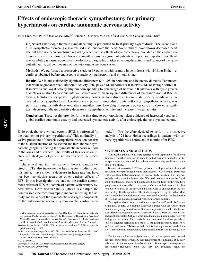
Effects of endoscopic thoracic sympathectomy for primaryhyperhidrosis on cardiac autonomic nervous activity
Jorge Cruz, MD, PhD,a,c Joao Sousa, MD,b,c Antonio G. Oliveira, MD, PhD,d and Luis Silva-Carvalho, MD, PhDe,f
Objective: Endoscopic thoracic sympathectomy is performed to treat primary hyperhidrosis. The second and
third sympathetic thoracic ganglia excised also innervate the heart. Some studies have shown decreased heart
rate but have not been conclusive regarding other cardiac effects of sympathectomy. We studied the cardiac au-
tonomic effects of endoscopic thoracic sympathectomy in a group of patients with primary hyperhidrosis. Heart
rate variability is a simple, noninvasive electrocardiographic marker reflecting the activity and balance of the sym-
pathetic and vagal components of the autonomous nervous system.
Methods: We performed a prospective study in 38 patients with primary hyperhidrosis with 24-hour Holter re-
cordings obtained before endoscopic thoracic sympathectomy and 6 months later.
Results: We found statistically significant differences (P<.05) in both time and frequency domains. Parameters
that evaluate global cardiac autonomic activity (total power, SD of normal R-R intervals, SD of average normal R-
R intervals) and vagal activity (rhythm corresponding to percentage of normal R-R intervals with cycle greater
than 50 ms relative to previous interval, square root of mean squared differences of successive normal R-R in-
tervals, high-frequency power, high-frequency power in normalized units) were statistically significantly in-
creased after sympathectomy. Low-frequency power in normalized units, reflecting sympathetic activity, was
statistically significantly decreased after sympathectomy. Low-/high-frequency power ratio also showed a signif-
icant decrease, indicating relative decrease in sympathetic activity and increase in vagal activity.
Conclusion: These results provide, for the first time to our knowledge, clear evidence of increased vagal and
global cardiac autonomic activity and decreased sympathetic activity after endoscopic thoracic sympathectomy.
Acquired Cardiovascular Disease Cruz et al
Endoscopic thoracic sympathectomy (ETS) is performed for
the treatment of primary hyperhidrosis.1 This minimally in-
vasive procedure of thoracic sympathetic resection consists
of the bilateral ablation of the second and third thoracic sym-
pathetic ganglia, affecting the sympathetic nervous outflow
to the arms and elsewhere. The results of this operation in-
clude a high degree of patient satisfaction with the proce-
dure.1-6
The second and third sympathetic thoracic ganglia ex-
cised in ETS also innervate the heart. A few studies about
this subject have shown a decrease in heart rate but have
not been conclusive regarding other cardiac effects of
ETS. In this investigation, we studied the cardiac autono-
mous effects of ETS in a sample of patients with primary hy-
perhidrosis. Heart rate variability (HRV) is a simple and
noninvasive electrocardiographic marker reflecting the ac-
tivity and balance of the sympathetic and vagal components
of the autonomous nervous system on the cardiac sinus
From the Service of Cardiothoracic Surgerya and Cardiology,b Hospital de Santa Ma-
ria; Instituto Cardiovascular de Lisboac; the Department of Biostatistics, Faculty of
Medical Sciences, Universidade Nova de Lisboad; Instituto de Fisiologia, Faculdade
de Medicina de Lisboae; and Instituto de Medicina Molecular,f Lisbon, Portugal.
Received for publication Feb 19, 2008; revisions received June 7, 2008; accepted for
publication July 6, 2008.
Address for reprints: Jorge Cruz, MD, PhD, Avenida Guerra Junqueiro No. 11-4�,
Esquerdo 1000-166 Lisboa, Portugal (E-mail: [email protected]).
J Thorac Cardiovasc Surg 2009;137:664-9
0022-5223/$36.00
Copyright � 2009 by The American Association for Thoracic Surgery
doi:10.1016/j.jtcvs.2008.07.021
664 The Journal of Thoracic and Cardiovascular Su
node.7-11 We therefore decided to perform a prospective
analysis of 24-hour Holter recordings in patients with pri-
mary hyperhidrosis before and 6 months after ETS.
MATERIALS AND METHODSConsecutive patients who were referred to our department for bilateral
thoracic sympathectomy for primary hyperhidrosis were included in this
prospective study. None of the patients were receiving medication at the
time of the study.
Bilateral video-assisted ETS was performed with general anesthesia. All
operations were performed by the same surgeon (J.C.), who had experience
with more than 400 operations at the time of study initiation. The lung was
excluded with a double-lumen tube. We used two incisions on the fourth
and fifth intercostal spaces, and in all cases the second and third sympathetic
ganglia were destroyed by electrocautery. One chest drain was left in place
for about 8 hours after the operation. Patients were discharged from the hos-
pital the day after the operation. The study was approved by the Lisbon Med-
ical School ethics committee and was performed according the Declaration
of Helsinki. All patients provided written, informed consent.
Twenty-four–hour Holter recordings were performed 2 weeks before and
6 months after ETS. A 3-channel Spacelab Burdick with the Holter Vision
Premier analysis system (Cardiac Science Corporation, Seattle, Wash) was
used. The recordings were started systematically at 4:00 PM and were discon-
nected 24 hours later. Subjects were asked to maintain routine daily activity
during the recording. Each variable was analyzed for three periods as
follows: daytime period from 6 AM to 10 PM, nighttime period from 10 PM
to 6 AM, and entire 24-hour period. In this investigation we studied the
HRV in the time and the frequency domains and the arrhythmic changes after
ETS.
The frequency domain analysis was based on a nonparametric method,
the fast Fourier transformation, that transformed the individual R-R inter-
vals stored in the computer into bands with different spectral frequencies:
the ultralow-frequency band, the very low–frequency band, the
rgery c March 2009
Cruz et al Acquired Cardiovascular Disease
AC
D
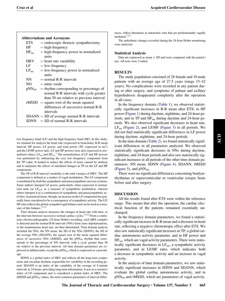
Abbreviations and AcronymsETS ¼ endoscopic thoracic sympathectomy
HF ¼ high-frequency
HFnu ¼ high-frequency power in normalized
units
HRV ¼ heart rate variability
LF ¼ low-frequency
LFnu ¼ low-frequency power in normalized
units
NN ¼ normal R-R intervals
NO ¼ nitric oxide
pNN50 ¼ rhythm corresponding to percentage of
normal R-R intervals with cycle greater
than 50 ms relative to previous interval
rMSSD ¼ square root of the mean squared
differences of successive normal R-R
intervals
SDANN ¼ SD of average normal R-R intervals
SDNN ¼ SD of normal R-R intervals
low-frequency band (LF) and the high frequency band (HF). In this study,
we retained for analysis the heart rate (expressed in beats/min); R-R mean
interval; HF power, LF power, and total power (TP, expressed in ms2)
and the LF/HF power ratio. LF and HF powers were also expressed as nor-
malized values, LFnu and HFnu.7 The normalization of LF and HF powers
was performed by subtracting the very low–frequency component from
the TP value. It tended to reduce the effects of noise caused by artifacts
and to minimize the effects of unrelated changes in TP on the LF and HF
components.
The TP of R-R interval variability is the total variance of HRV. The HF
component is defined as a marker of vagal modulation. The LF component
is modulated by both the sympathetic and parasympathetic nervous systems.
Some authors interpret LF power, particularly when expressed in normal-
ized units (as LFnu), as a measure of sympathetic modulation, whereas
others interpret it as a combination of sympathetic and parasympathetic ac-
tivities. In practical terms, though, an increase in the LF component has gen-
erally been considered to be a consequence of sympathetic activity. The LF/
HF ratio reflects the global sympathovagal balance and can be used as a mea-
sure of this balance.13,14
Time domain analysis measures the changes in heart rate with time, or
the intervals between successive normal cardiac cycles.15,16 From a contin-
uous electrocardiographic (24-hour Holter) recording, each QRS complex
is detected and the normal R-R intervals (NNs) from sinus depolarizations,
or the instantaneous heart rate, are then determined. Time domain analysis
included the NNs, the NN mean, the SD of the NNs (SDNN), the SD of
the average NNs (SDANN), the square root of the mean squared differ-
ences of successive NNs (rMSSD), and the pNN50 rhythm that corre-
sponds to the percentage of NN intervals with a cycle greater than 50
ms relative to the previous interval. All time domain parameters are ex-
pressed in milliseconds, except the pNN50, which is expressed as a percent-
age.
SDNN is a global index of HRV and reflects all the long-term compo-
nents and circadian rhythms responsible for variability in the recording pe-
riod. SDANN is an index of the variability of the average of 5-minute
intervals in 24 hours, providing long-term information. It acts as a sensitive
index of LF component and is considered a global index of HRV. The
rMSSD and pNN50 values, the most common parameters of interval differ-
The Journal of Thoracic and C
ences, reflect alterations in autonomic tone that are predominantly vagally
mediated.7,12
The arrhythmic changes recorded during the 24-hour Holter monitoring
were analyzed.
Statistical AnalysisData are expressed as mean � SD and were compared with the paired t
test. All tests were 2-tailed.
RESULTSThe study population consisted of 28 female and 10 male
patients with an average age of 27.5 years (range 15–42
years). No complications were recorded in any patient dur-
ing or after surgery, and symptoms of palmar and axillary
hyperhidrosis disappeared completely after the operation
in all cases.
In the frequency domain (Table 1), we observed statisti-
cally significant increases in R-R mean after ETS; in HF
power (Figure 1) during daytime, nighttime, and 24-hour pe-
riods; and in TP and HFnu during daytime and 24-hour pe-
riods. We also observed significant decreases in heart rate,
LFnu (Figure 2), and LF/HF (Figure 3) in all periods. We
did not find statistically significant differences in LF power
during daytime, nighttime, and 24-hour periods.
In the time domain (Table 2), we found statistically signif-
icant differences in all parameters analyzed. We observed
statistically significant decreases in NNs during daytime,
nighttime, and 24-hour periods and also saw statistically sig-
nificant increases in all periods of the other time domain pa-
rameters: NN mean, SDNN (Figure 4), SDANN, rMSSD
(Figure 5), and pNN50.
There were no significant differences concerning bradyar-
rhythmias or supraventricular or ventricular ectopic beats
before and after surgery.
DISCUSSIONAll the results found after ETS were within the reference
range. This means that after the operation, the cardiac elec-
trical function of the patients remained essentially un-
changed.
In the frequency domain parameters, we found a statisti-
cally significant increase in R-R mean and a decrease in heart
rate, reflecting a negative chronotropic effect after ETS. We
also saw statistically significant increases in TP, a global car-
diac autonomous activity parameter, and in HF power and
HFnu, which are vagal activity parameters. There were statis-
tically significant decreases in LFnu, a sympathetic activity
parameter, and in LF/HF ratio, which indicates both
a decrease in sympathetic activity and an increase in vagal
activity.
In the analysis of time domain parameters, we saw statis-
tically significant increases in SDNN and SDANN, which
evaluate the global cardiac autonomous activity, and in
pNN50 and rMSSD, which are parameters of vagal activity.
ardiovascular Surgery c Volume 137, Number 3 665

Acquired Cardiovascular Disease Cruz et al
AC
D
TABLE 1. Frequency domain results
Difference
Baseline (mean ± SD) 6 mo (mean ± SD) Mean 95% Confidence interval P value
R-R mean (ms)
24 h 828.9 � 130.8 913.3 � 129.8 84.4 55.3–113.6 <.001
Daytime 800.4 � 130.9 890.2 � 133.4 89.8 62.6–117.0 <.001
Nighttime 885.1 � 155.3 971.7 � 146.1 86.6 39.6–133.6 .001
Heart rate (beats/min)
24 h 75.3 � 10.6 68.0 � 9.4 �7.4 �9.7 to�5.0 <.001
Daytime 77.8 � 10.8 69.6 � 10.0 �8.2 �10.4 to�6.0 <.001
Nighttime 70.3 � 13.0 63.6 � 9.6 �6.8 �10.5 to�3.1 .001
Total power (ms2)
24 h 6545.5 � 8397.6 7934.1 � 8257.1 1388.6 82.3–2694.9 .038
Daytime 6781.5 � 9962.4 8286.0 � 10959.1 1504.6 658.5–2350.6 .001
Nighttime 6404.7 � 7616.4 7851.4 � 7857.7 1446.7 535.1–4428.5 .329
Low-frequency power (ms2)
24 h 1833.4 � 1468.1 1633.0 � 1420.7 �200.4 �531.3–130.4 .225
Daytime 1836.2 � 1687.5 1737.7 � 1472.1 �98.4 �445.5–248.6 .565
Nighttime 1783.3 � 1598.8 1522.7 � 1497.3 �260.6 �813.4–292.1 .342
Low-frequency power
in normalized units
24 h 63.4 � 13.0 53.6 � 15.7 �9.8 �13.4 to�6.2 <.001
Daytime 66.3 � 13.2 57.9 � 16.3 �8.3 �12.3 to�4.4 <.001
Nighttime 58.3 � 14.8 46.8 � 17.8 �11.4 �17.1 to�5.8 <.001
High-frequency power (ms2)
24 h 1184.2 � 1210.8 1717.7 � 1805.2 533.5 196.7–870.2 .003
Daytime 1060.3 � 1252.7 1581.2 � 1636.6 520.9 251.8–790.0 .001
Nighttime 1308.3 � 1360.0 1958.2 � 2303.9 649.9 21.2–1278.7 .043
High-frequency power
in normalized units
24 h 34.2 � 11.4 40.8 � 13.6 6.6 3.2–10.0 <.001
Daytime 30.9 � 11.5 38.8 � 14.3 7.9 4.2–11.6 <.001
Nighttime 39.6 � 13.5 43.4 � 14.8 3.8 �1.5–9.1 .152
Low-/high-frequency power ratio
24 h 3.3 � 1.8 2.3 � 1.8 �0.9 �1.3 to�0.5 <.001
Daytime 3.7 � 2.0 2.7 � 2.3 �1.0 �1.5 to�0.5 <.001
Nighttime 2.6 � 1.6 1.8 � 1.4 �0.7 �1.3 to�0.2 .008
In summary, in this investigation we found statistically
significant differences in both time and frequency domains.
The parameters that evaluate the global cardiac autonomous
activity (TP, SDNN, SDANN) and the vagal activity
(pNN50, rMSSD, HF power, HFnu) had statistically signifi-
cant increases after ETS. LFnu, which reflects the sympa-
thetic activity, had a statistically significant decrease after
ETS. The significant decrease in LF/HF ratio also indicates
a relative decrease in sympathetic activity and an increase in
vagal activity. These results provide clear and consistent ev-
idence pointing toward increases in vagal and global cardiac
autonomous activities after ETS.
Noppen and colleagues,8 Tygesen and associates,9 Tedor-
iya and colleagues,10 Wettervik and coworkers,11 Abraham
and associates,17 and Drott and colleagues18 had previously
studied the cardiac effects of ETS in patients with primary
hyperhidrosis, and they reported an increase of R-R mean
666 The Journal of Thoracic and Cardiovascular Sur
and a decrease in heart rate, confirming the negative chrono-
tropic effect of ETS. Abraham et and colleagues,17 who also
studied HRV with 24-hour Holter recordings, demonstrated
statistically significant decreases in heart rate, LF power, and
LF/HF ratio but did no find any differences in all other pa-
rameters studied. They concluded that a decrease of sympa-
thetic activity was observed, but without any difference in
vagal activity. Tedoriya and colleagues10 found a signifi-
cantly decrease only in LF/HF ratio in the head-up tilt posi-
tion after surgery and concluded that the influences of ETS
on the cardiac autonomic nerve system were of a lesser de-
gree at rest, but the response to sympathetic stimulation was
suppressed after surgery. Wilkund and coworkers19 verified
that the patients with primary hyperhidrosis had a tendency
toward higher power in the LF and HF components than
seen in control subjects in the upright position. After sympa-
thectomy, LF power was reduced, but HF power was
gery c March 2009

Cruz et al Acquired Cardiovascular Disease
AC
D
unchanged. At follow-up, LF power remained at a lower
level, but now HF power was reduced. They concluded
that patients with palmar hyperhidrosis have sympathetic
hyperactivity but with a compensatory high parasympathetic
activity. Sympathectomy results in an initial sympathovagal
imbalance with a parasympathetic predominance, which is
restored on a long-term basis. The results of these investiga-
tions revealed a decrease in sympathetic activity but failed to
show an increase in vagal activity. Why these investigators
were not able to show the decrease in vagal activity decrease
in our study can be explained, in our opinion, by our selec-
tion of 24-hour Holter recordings in this investigation and
the larger number of cases studied (38 patients) than in the
other studies (between 10 and 13 patients).
In summary, the articles discussed here consistently
showed a negative chronotropic effect, suggesting a decrease
in sympathetic activity, but failed to show an increase in va-
gal activity.8-11,17-19 In contrast, our investigation demon-
strated that ETS in patients with primary hyperhidrosis
causes increases in vagal and cardiac autonomic activities
and a decrease in sympathetic activity. As far as we know,
this is the first time that these results have been presented
in such a clear way.
These results confirm that the second and third sympa-
thetic thoracic ganglia excised in ETS do influence the au-
tonomous cardiac activity. Because of the anatomic
variability of the cardiac sympathetic efferent innervations
of second and third thoracic ganglia that usually are rare,
however, it is difficult to explain these results as a conse-
quence of cardiac adrenergic efferent section.20-22 We there-
fore can consider the hypothesis that the effects we have
found are a consequence of cardiac adrenergic afferent sec-
tion. In fact, Malliani,23 Ruscone and coworkers,24 and
Bishop and associates25 demonstrated in experimental
works the importance of cardiac sympathetic afferents in
the autonomic modulation of the heart. Foreman,26 in studies
in rabbits, also demonstrated that the second and third tho-
0
500
1000
1500
2000
2500
3000
Daytime* Night-time*24-hours*
HF
Pow
er (m
s2 )
FIGURE 1. High-frequency power (HF) before (open circles) and after
(closed circles) sympathectomy. Mean and 95% confidence interval; aster-
isk indicates P< .05.
The Journal of Thoracic and
racic ganglia received an important number of sympathetic
afferents. We do not know if the human second and third
thoracic ganglia also have a superior number of sympathetic
afferents, but we can hypothesize that the observed cardiac
effects in HRV analysis are probably not the result of sym-
pathetic efferent nerve section but rather of the cardiac affer-
ent nerves of the second and third thoracic ganglia.
Another hypothesis that we have formulated to explain
our results is the relationship between ETS and increased ni-
tric oxide (NO) activity.27,28 The sympatholytic activity of
NO, with an adrenergic decrease and a vagal activity in-
crease, has been demonstrated.29,30 Chowdhary and co-
workers29 studied the effects of NO in healthy people and
confirmed that this substance increases cardiac vagal activ-
ity. Although the study of Chowdhary and coworkers29
was performed in healthy people, if these effects can be gen-
eralized to patients with primary hyperhidrosis, an increase
in NO activity after ETS could, in our opinion, explain the
higher vagal activity. These hypothesis seems consistent,
0
10
20
30
40
50
60
70
80
Daytime* Night-time*24-hours*
LF n
u (m
s2 )
FIGURE 2. Low-frequency power in normalized units (LFnu) before (open
circles) and after (closed circles) sympathectomy. Mean and 95% confi-
dence interval; asterisk indicates P< .05.
0
1
2
3
4
5
Daytime* Night-time*24-hours*
LF /
HF
FIGURE 3. Low-/high-frequency (LF/HF) ratio before (open circles) and
after (closed circles) sympathectomy. Mean and 95% confidence interval;
asterisk indicates P< .05.
Cardiovascular Surgery c Volume 137, Number 3 667

Acquired Cardiovascular Disease Cruz et al
AC
D
and we therefore believe in the importance of pursuing the
investigation regarding NO activity in cardiac autonomic
control before and after ETS in patients with primary hyper-
hidrosis.
It is important to underline that our study population was
composed of patients who already had an autonomic dys-
function disorder. We do not know whether primary hyper-
hidrosis is a systemic or a local disease. As we stated before,
TABLE 2. Time domain results
Difference
Baseline (mean ± SD) 6 months (mean ± SD) Mean 95% Confidence interval P value
Normal R-R intervals
24 h 4243 � 565.1 3725.1 � 608.6 �518.1 �731.1 to�305.0 <.0001
Daytime 4391.6 � 605.5 3847.3 � 682.7 �544.3 �726.9 to�361.6 <.0001
Nighttime 3953.8 � 570.2 3624.6 � 504.6 �329.2 �555.8 to�102.6 .0060
Mean of normal R-R intervals (ms)
24 h 814.6 � 111.7 892.0 � 115.4 77.4 48.5–106.2 <.0001
Daytime 779.0 � 97.5 843.4 � 121.4 64.4 38.6–90.2 <.0001
Nighttime 879.5 � 153.9 968.0 � 131.3 88.4 43.3–133.6 .0004
SD of normal R-R intervals (ms)
24 h 91.8 � 26.6 100.7 � 28.6 8.9 4.1–13.9 .0010
Daytime 96.3 � 27.9 104.3 � 30.1 8.1 3.7–12.2 .0007
Nighttime 85.5 � 27.4 96.0 � 29.3 10.5 2.8–18.2 .0093
SD index of normal R-R intervals (ms)
24 h 72.6 � 23.2 79.8 � 24.8 7.3 3.1–11.4 .0014
Daytime 75.4 � 24.8 81.9 � 26.7 6.5 2.6–10.4 .0021
Nighttime 69.7 � 22.6 77.2 � 24.5 8.3 2.2–14.3 .0090
Mean squared differences of successive
normal R-R intervals (ms)
24 h 49.6 � 21.4 60.0 � 26.8 10.5 5.4–15.6 .0002
Daytime 49.0 � 21.4 58.6 � 26.3 9.6 4.9–14.3 .0003
Nighttime 51.5 � 23,4 63.5 � 30.9 12.0 4.6–19.5 .0026
pNN50* (%)
24 h 18.2 � 13.2 25.4 � 15.3 7.2 4.4–10.0 <.0001
Daytime 17.1 � 12.6 22.8 � 15.1 5.7 3.3–8.2 <.0001
Nighttime 20.6 � 15.9 30.0 � 18.9 9.4 4.6–14.3 .0005
SD of average normal R-R intervals (ms)
24 h 47.2 � 12.1 52.8 � 14.2 5.7 2.5–8.9 .0012
Daytime 50.6 � 12.9 56.3 � 15.0 5.7 2.1–9.4 .0030
Nighttime 40.9 � 13.84 46.3 � 15.3 5.4 0.8–10.0 .0227
*Rhythm corresponding to percentage of normal R-R intervals with cycle greater than 50 ms relative to previous interval.
Daytime* Night-time*24-hours*0
20
40
60
80
100
120
SDN
N (m
s)
FIGURE 4. SD of normal R-R intervals (SDNN) before (open circles) and
after (closed circles) sympathectomy. Mean and 95% confidence interval;
asterisk indicates P< .05.
Daytime* Night-time*24-hours*0
20
40
60
80
rMSS
D (m
s)
FIGURE 5. Square root of mean squared differences of successive normal
R-R intervals (rMSSD) before (open circles) and after (closed circles) sym-
pathectomy. Mean and 95% confidence interval; asterisk indicates P<.05.
668 The Journal of Thoracic and Cardiovascular Surgery c March 2009
Cruz et al Acquired Cardiovascular Disease
AC
D
however, in our opinion, sweating in patients with primary
hyperhidrosis apparently has no homeostatic function.
This means that our sample has specific clinical characteris-
tics, which makes us believe that the observed cardiac ef-
fects in patients with primary hyperhidrosis undergoing
ETS would probably not be observed in healthy individuals.
CONCLUSIONSIn this prospective study, we demonstrated with statisti-
cally significant results in both time and frequency domain
analyses a decrease in sympathetic activity, an increase in
vagal activity, and increase in R-R variability 6 months after
ETS in patients with primary hyperhidrosis.
References1. Krasna MJ. Thoracoscopic sympathectomy: a standardized approach to therapy
for hyperhidrosis. Ann Thorac Surg. 2008;85:S764-7.
2. Cruz J, Caldeira J, Cravino J. Simpaticectomia Toracica Video-assistida no trata-
mento da hiperhidrose palmar e axilar. Rev Port CCT e Vasc. 2002;9:149-52.
3. Lin CC, Mo LR, Lee LS. Thoracoscopic T2 sympathetic block by clipping. A bet-
ter and reversible operation for treatment of hyperhidrosis palmaris: experience
with 326 cases. Eur J Surg Suppl. 1998;S580:13.
4. Singh B, Shaik AS, Moodley J, Ramdial P, Rajaruthnam P. Limited thoracoscopic
ganglionectomy for primary hyperhidrosis. S Afr J Surg. 2002;40:50-3.
5. Han PP, Gottfried ON, Kenny KJ, Dickman CA. Biportal thoracoscopic sympa-
thectomy: surgical techniques and clinical results for the treatment of hyperhidro-
sis. Neurosurgery. 2002;50:306-11.
6. Lin TS, Huang LC, Wang NP, Chang CC. Endoscopic thoracic sympathetic block
by clipping for palmar and axillary hyperhidrosis in children and adolescents.
Pediatr Surg Int. 2001;17:535-7.
7. Task Force of the European Society of Cardiology and the North American Society
of Pacing and Electrophysiology. Heart rate variability: standards of measurement,
physiological interpretation, and clinical use. Circulation. 1996;93:1043-65.
8. Noppen M, Dendale P, Hagers Y, Herregodts P, Vincken W, D’Haens J. Changes
in cardiocirculatory autonomic function after thoracoscopic upper dorsal sympa-
thicolysis for essential hyperhidrosis. J Auton Nerv Syst. 1996;60:115-20.
9. Tygesen H, Claes G, Drott C, Emanuelsson H, Lomsky M, Lurje L, et al. Effects
of endoscopic transthoracic sympathicotomy on heart rate variability in severe an-
gina pectoris. Am J Cardiol. 1997;79:1447-52.
10. Tedoriya T, Sakagami S, Ueyama T, Thompson L, Hetzer R. Influences of bilat-
eral endoscopic transthoracic sympathicotomy on cardiac autonomic nervous ac-
tivity. Eur J Cardiothorac Surg. 1999;15:194-8.
11. Wettervik C, Claes G, Drott C. Endoscopic transthoracic sympathicotomy for se-
vere angina. Lancet. 1995;345:97-8.
The Journal of Thoracic and C
12. Sztajzel J. Heart rate variability: a noninvasive electrocardiographic method to
measure the autonomic nervous system. Swiss Med Wkly. 2004;134:514-22.
13. Malliani A, Lombardi F, Pagani M. Power spectrum analysis of heart rate
variability: a tool to explore neural regulatory mechanisms. Br Heart J. 1994;
71:1-2.
14. Eckberg DL. Sympathovagal balance. Circulation. 1997;96:3224-32.
15. Malik M. Measurement of heart rate variability. In: Malik M, Camm J, eds. Heart
rate variability. Armonk (NY): Futura; 1995:33-132.
16. Kleiger RE, Stein PK, Bosner MS, Rottman JN. Time domain measurements of
heart rate variability. Cardiol Clin. 1992;10:487-98.
17. Abraham P, Berthelot J, Victor J, Saumet J, Picqet J, Enon B. Holter changes
resulting from right-sided and bilateral infrastellate upper thoracic sympathec-
tomy. Ann Thorac Surg. 2002;74:2076-81.
18. Drott C, Claes G, Gothberg G, Paszkowski P. Cardiac effects of endoscopic elec-
trocautery of the upper thoracic sympathetic chain. Eur J Surg Suppl. 1994;572:
65-70.
19. Wiklund U, Koskinen LO, Niklasson U, Bjerle P, Elfversson J. Endoscopic
transthoracic sympathicotomy affects the autonomic modulation of heart
rate in patients with palmar hyperhidrosis. Acta Neurochir (Wien). 2000;
142:691-6.
20. Jeffrey PE, Terence HW. Sympathetic nerve pathways to the human heart and
their variations. Am J Anat. 1969;124:149-62.
21. Janes R, Christopher B, David AH, David EJ, David AM, Andrew A. Anat-
omy of human extrinsic cardiac nerves and ganglia. Am J Cardiol. 1986;57:
299-309.
22. Ellison JP, Williams TH. Sympathetic nerve pathways to the human heart, and
their variations. Am J Anat. 1969;124:149-62.
23. Malliani A. General concepts and hypotheses in the study of cardiovascular neural
regulation. In: Malliani A, ed. Principles of cardiovascular neural regulation in
health and disease. Norwell (MA): Kluwer Academic; 2000:1-29.
24. Ruscone T, Lombardi F, Malfatto G, Malliani A. Attenuation of baroreceptive
mechanisms by cardiovascular sympathetic afferent fibers. Am J Physiol. 1987;
253:787-91.
25. Bishop VS, Lombardi F, Malliani A, Pagani M, Recordati G. Reflex sympathetic
tachycardia during intravenous infusion in chronic spinal cats. Am J Physiol.
1976;230:25-9.
26. Foreman R. Spinal cord neuronal regulation of the cardiovascular system. In:
Armour J, Ardell J, eds. Neurocardiology. New York: Oxford University Press
(USA); 1994:245-76.
27. Lepori M, Sartori C, Duplain D, Nicod P, Scherrer U. Sympathectomy potentiates
the vasoconstrictor response to nitric oxide synthase inhibition in humans. Cardi-
ovasc Res. 1999;43:739-43.
28. Charkoudian N, Eisenach JH, Atkinson JL, Fealey RG, Joyner MJ. Effects of
chronic sympathectomy on locally mediated cutaneous vasodilatation in humans.
J Appl Physiol. 2001;92:685-90.
29. Chowdhary S, Vaile J, Fletcher J, Ross H, Coote J, Townend N. Nitric oxide and
cardiac autonomic control in humans. Hypertension. 2000;36:264-9.
30. Chowdhary S, Townend N. Role of nitric oxide in the regulation of cardiovascular
autonomic control. Clin Sci’. 1999;97:5-17.
ardiovascular Surgery c Volume 137, Number 3 669


![Peripheral Sympathectomy for Raynaud's Phenomenon: A ... · sympathectomy [1–3,6,7]. Conventional cervical sym-pathectomy has been used, but the results are disap-pointing. It has](https://img.pdfslide.us/doc/110x75/602abfd7d9da841d876f1382/peripheral-sympathectomy-for-raynauds-phenomenon-a-sympathectomy-1a367.jpg)
![aAR Journal of Anesthesia & Clinical Research … · [1]. Autonomic dysfunction should also be expected. Experimental studies involving unilateral sympathectomy have produced similar](https://img.pdfslide.us/doc/110x75/606cf87713f13167367b615a/aar-journal-of-anesthesia-clinical-research-1-autonomic-dysfunction.jpg)