Embed Size (px)
Citation preview
657
Journal of Environmental Biology, May 2013
Abstract
Organ histopathology and changes in biochemical parameters in fish are good biomarkers of aquatic
pollution. This study is an attempt to assess the effects of dimethoate, an organophosphate
insecticide on the liver of common carp (C. carpio). Healthy individual fish were exposed to 0.40 mg l1 (25%
of 96 hr LC50) concentration of dimethoate, for short term (96 hr). Liver of the exposed fish exhibited
alterations like disruption of regular arrangement of hepatocytes, congestion and rupture of vessels;
hemorrhage, cytoplasmic vacuolization, pyknotic nuclei and necrosis. Biochemical parameters
viz. total liver protein (p<0.001) and liver glycogen (p<0.001) registered a significant decrease and
blood glucose (p<0.001) exhibited significant increase throughout exposure.
Key words
Toxicity, Histopathology, Common carp, Organophosphate, Dimethoate
Publication Info
Paper received:
21 July 2011
Revised received:
12 January 2012
Re-revised received:
08 May 2012
Accepted:
26 July 2012
Introduction
Synthetic pesticides are mostly non selective but
highly effective chemicals (Breckenridge and Stevens,
2008). Their widespread use in agriculture and in health
and hygiene programs is one of the major causes of
aquatic pollution (Cope, 2004). Sometimes pesticides are
directly applied in water bodies but their residues mostly
reach into aquatic ecosystems through surface run off
(Jergentz et al., 2004) and affect the health of non target
organisms including fish. Ecological concerns arising out
of high persistence of organochlorines, have favored
extensive use of organophosphates for controlling
household, agricultural and public health pests due to
their less persistence and high efficacy (Oruc et al., 2006;
Jyothi and Narayan, 1999). This shift has resulted into
increased occurrence of organophosphates into water
bodies causing acute and chronic toxicity to fish fauna
(Aker et al., 2008; Rao et al., 2005; Velmurugan et al.,
2007; Pandey et al., 2009).
Different kind of biological responses can be used
as biomarkers to assess the toxic effects of pollutants.
Histopathology of fish liver is increasingly being utilized as
biomarkers of xenobiotic exposure (Fernandes et al., 2008)
along with biochemical parameters to study the stress
(Rawat et al., 2002). The present work therefore was an
effort to assess the impact of dimethoate on the
histopathology and biochemical parameters on liver of
common carp, Cyprinus carpio.
Materials and Methods
Fish caught from local government hatchery and
carefully packaged into aerated polythene bags were
brought to the laboratory. Fish were treated with 0.05%
KMnO4 for disinfecting and were further transferred into
plastic pools of 500 l capacity for two weeks for
acclimatization to laboratory conditions. The rice bran mixed
with mustard oil cake in the ratio of 2:1 was given to fish as
feed during acclimatization. Water of the pool was changed
daily and dead fish were removed immediately.
Effects of dimethoate (30% EC), an organophosphate pesti-cide on liver of common carp, Cyprinus carpio
Ram Nayan Singh
Department of Zoology, Kamla Nehru Institute of Physical and Social Sciences, Sultanpur- 228 118, India
*Corresponding Author email : [email protected]
© Triveni Enterprises, Lucknow (India ) Journal of Environmental Biology, Vol. 34, 657-661, May 2013
ISSN: 0254-8704CODEN: JEBIDPJournal of Environmental Biology
JEB
Journal Home page : www.jeb.co.in � E-mail : [email protected]
658
Journal of Environmental Biology, May 2013
R.N. Singh
The experiment was conducted under natural
photoperiod and temperature (28-32ºC). Water quality was
measured as per APHA (2005). The temperature of the water
was 23 ± 1.5ºC, pH 7.2 ± 0.4, dissolved oxygen 7.2 ± 0.6 mg
l-1, free carbon dioxide 6.2 ± 0.4 mg l-1 and total hardness as
calcium carbonate 112 ± 3.2 mg l-1.
Based on 96 hr LC50 value 1.60 mg l-1 of dimethoate
(Rogor 30%EC, Rallis India Ltd, Mumbai) for C. carpio
(Singh et al., 2009), a dose of 0.40 mgl-1 (25% of 96 hr LC50)
was selected as sub lethal concentration. Fish (17-22 cm
length, and 50-65 gm weight) were starved for 24 hrs before
starting the experiment. Six fish each were exposed to 0.40
mgl-1 dose for 24, 48, 72 and 96 hr in each set and control
with six fish was run simultaneously for each interval.
Fish were sacrificed at intervals of 24, 48, 72 and 96
hr of exposure. Immediately the blood was collected from
sinus venosus and blood glucose was measured by Accu
Chek glucometer. The liver was removed and a portion fixed
for 24 hr in bouins fluid for histology and other portion
processed for estimating glycogen and total protein. For
estimating liver glycogen and total protein, 100 mg liver
tissue was transferred in to manual ice cooled homogenizers
containing 10% TCA. Glycogen was measured according
to the anthrone method of Van der Vies (1954). Total protein
was measured according to Lowry et al. (1951). For
measuring optical density digital spectrophotometer of MS
Electronics, India (Model, 305) was used. Significance of
the data was analyzed by student’s t – test.
The fixed liver tissues dehydrated in graded series
of alcohol were embedded in paraffin. A sections of 5-6
micron were cut and stained in haematoxylin and eosin. The
slides were examined under light microscope (Olympus CH
20i) and photographed for histopatholgical observations.
Results and Discussion
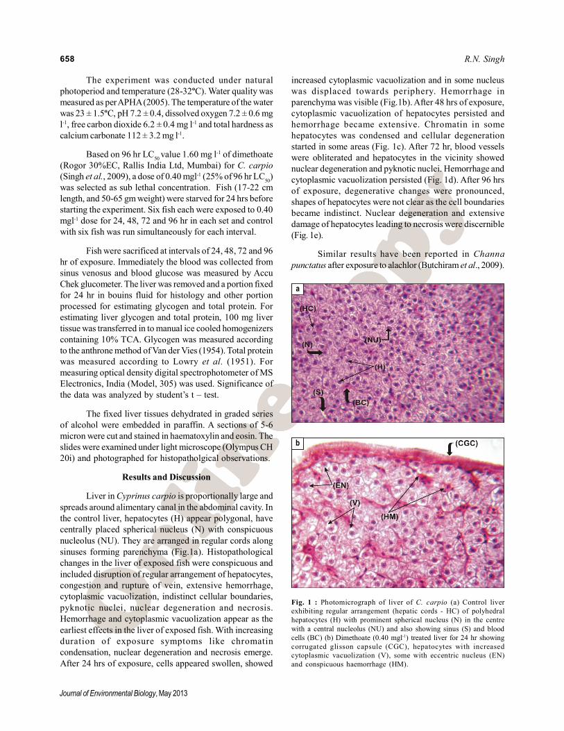
Liver in Cyprinus carpio is proportionally large and
spreads around alimentary canal in the abdominal cavity. In
the control liver, hepatocytes (H) appear polygonal, have
centrally placed spherical nucleus (N) with conspicuous
nucleolus (NU). They are arranged in regular cords along
sinuses forming parenchyma (Fig.1a). Histopathological
changes in the liver of exposed fish were conspicuous and
included disruption of regular arrangement of hepatocytes,
congestion and rupture of vein, extensive hemorrhage,
cytoplasmic vacuolization, indistinct cellular boundaries,
pyknotic nuclei, nuclear degeneration and necrosis.
Hemorrhage and cytoplasmic vacuolization appear as the
earliest effects in the liver of exposed fish. With increasing
duration of exposure symptoms like chromatin
condensation, nuclear degeneration and necrosis emerge.
After 24 hrs of exposure, cells appeared swollen, showed
increased cytoplasmic vacuolization and in some nucleus
was displaced towards periphery. Hemorrhage in
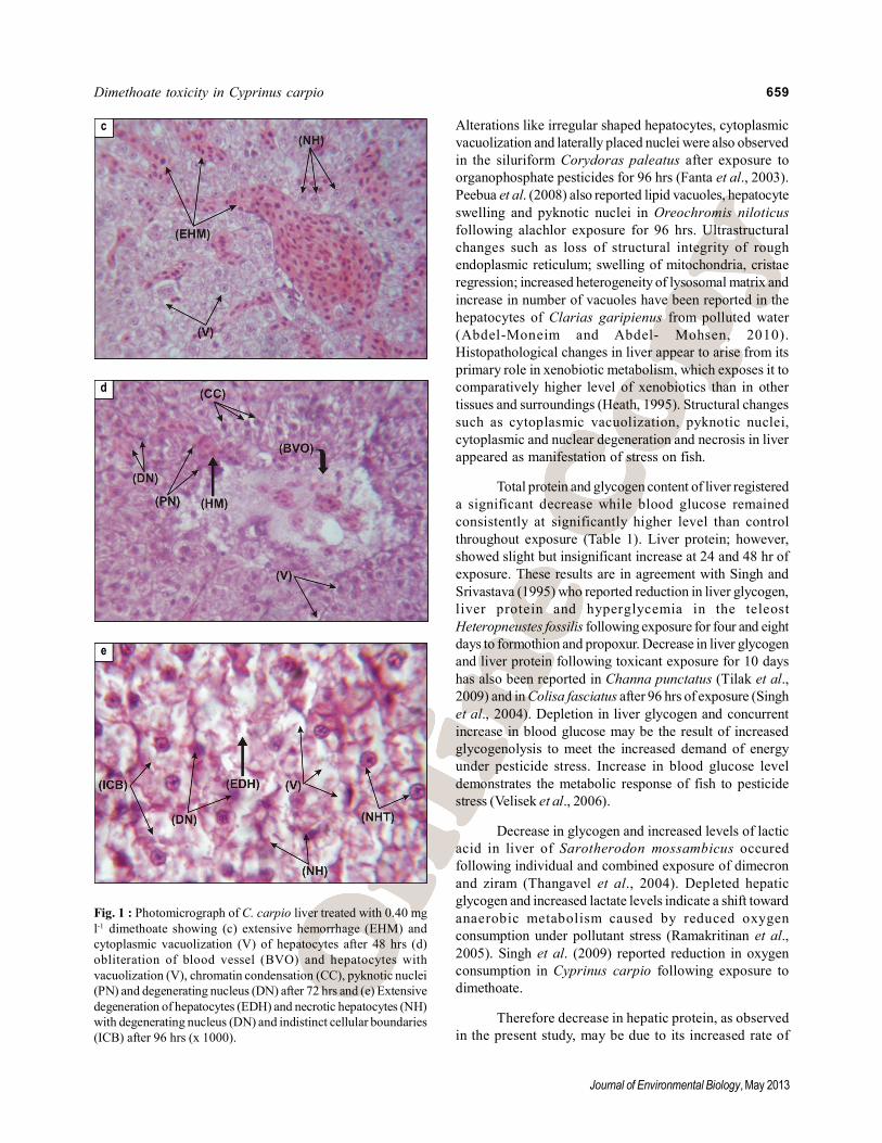
parenchyma was visible (Fig.1b). After 48 hrs of exposure,
cytoplasmic vacuolization of hepatocytes persisted and
hemorrhage became extensive. Chromatin in some
hepatocytes was condensed and cellular degeneration
started in some areas (Fig. 1c). After 72 hr, blood vessels
were obliterated and hepatocytes in the vicinity showed
nuclear degeneration and pyknotic nuclei. Hemorrhage and
cytoplasmic vacuolization persisted (Fig. 1d). After 96 hrs
of exposure, degenerative changes were pronounced,
shapes of hepatocytes were not clear as the cell boundaries
became indistinct. Nuclear degeneration and extensive
damage of hepatocytes leading to necrosis were discernible
(Fig. 1e).
Similar results have been reported in Channa
punctatus after exposure to alachlor (Butchiram et al., 2009).
Fig. 1 : Photomicrograph of liver of C. carpio (a) Control liver
exhibiting regular arrangement (hepatic cords - HC) of polyhedral
hepatocytes (H) with prominent spherical nucleus (N) in the centre
with a central nucleolus (NU) and also showing sinus (S) and blood
cells (BC) (b) Dimethoate (0.40 mgl-1) treated liver for 24 hr showing
corrugated glisson capsule (CGC), hepatocytes with increased
cytoplasmic vacuolization (V), some with eccentric nucleus (EN)
and conspicuous haemorrhage (HM).
a
b
659
Journal of Environmental Biology, May 2013
Dimethoate toxicity in Cyprinus carpio
Alterations like irregular shaped hepatocytes, cytoplasmic
vacuolization and laterally placed nuclei were also observed
in the siluriform Corydoras paleatus after exposure to
organophosphate pesticides for 96 hrs (Fanta et al., 2003).
Peebua et al. (2008) also reported lipid vacuoles, hepatocyte
swelling and pyknotic nuclei in Oreochromis niloticus
following alachlor exposure for 96 hrs. Ultrastructural
changes such as loss of structural integrity of rough
endoplasmic reticulum; swelling of mitochondria, cristae
regression; increased heterogeneity of lysosomal matrix and
increase in number of vacuoles have been reported in the
hepatocytes of Clarias garipienus from polluted water
(Abdel-Moneim and Abdel- Mohsen, 2010).
Histopathological changes in liver appear to arise from its
primary role in xenobiotic metabolism, which exposes it to
comparatively higher level of xenobiotics than in other
tissues and surroundings (Heath, 1995). Structural changes
such as cytoplasmic vacuolization, pyknotic nuclei,
cytoplasmic and nuclear degeneration and necrosis in liver
appeared as manifestation of stress on fish.
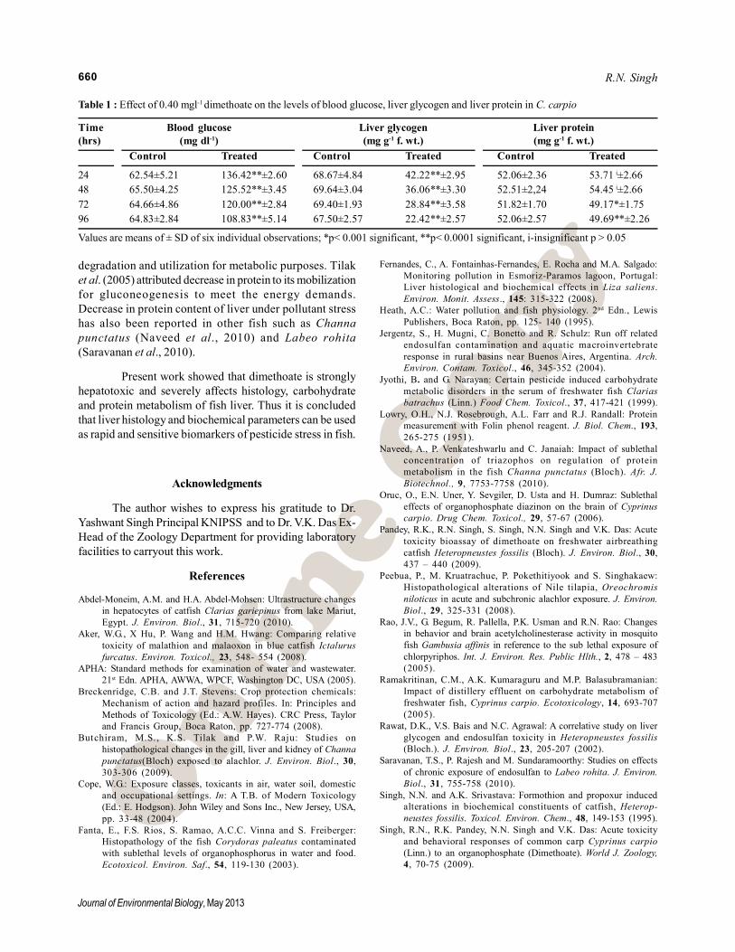
Total protein and glycogen content of liver registered
a significant decrease while blood glucose remained
consistently at significantly higher level than control
throughout exposure (Table 1). Liver protein; however,
showed slight but insignificant increase at 24 and 48 hr of
exposure. These results are in agreement with Singh and
Srivastava (1995) who reported reduction in liver glycogen,
liver protein and hyperglycemia in the teleost
Heteropneustes fossilis following exposure for four and eight
days to formothion and propoxur. Decrease in liver glycogen
and liver protein following toxicant exposure for 10 days
has also been reported in Channa punctatus (Tilak et al.,
2009) and in Colisa fasciatus after 96 hrs of exposure (Singh
et al., 2004). Depletion in liver glycogen and concurrent
increase in blood glucose may be the result of increased
glycogenolysis to meet the increased demand of energy
under pesticide stress. Increase in blood glucose level
demonstrates the metabolic response of fish to pesticide
stress (Velisek et al., 2006).
Decrease in glycogen and increased levels of lactic
acid in liver of Sarotherodon mossambicus occured
following individual and combined exposure of dimecron
and ziram (Thangavel et al., 2004). Depleted hepatic
glycogen and increased lactate levels indicate a shift toward
anaerobic metabolism caused by reduced oxygen
consumption under pollutant stress (Ramakritinan et al.,
2005). Singh et al. (2009) reported reduction in oxygen
consumption in Cyprinus carpio following exposure to
dimethoate.
Therefore decrease in hepatic protein, as observed
in the present study, may be due to its increased rate of
d
c
e
Fig. 1 : Photomicrograph of C. carpio liver treated with 0.40 mg
l-1 dimethoate showing (c) extensive hemorrhage (EHM) and
cytoplasmic vacuolization (V) of hepatocytes after 48 hrs (d)
obliteration of blood vessel (BVO) and hepatocytes with
vacuolization (V), chromatin condensation (CC), pyknotic nuclei
(PN) and degenerating nucleus (DN) after 72 hrs and (e) Extensive
degeneration of hepatocytes (EDH) and necrotic hepatocytes (NH)
with degenerating nucleus (DN) and indistinct cellular boundaries
(ICB) after 96 hrs (x 1000).
660
Journal of Environmental Biology, May 2013
degradation and utilization for metabolic purposes. Tilak
et al. (2005) attributed decrease in protein to its mobilization
for gluconeogenesis to meet the energy demands.
Decrease in protein content of liver under pollutant stress
has also been reported in other fish such as Channa
punctatus (Naveed et al., 2010) and Labeo rohita
(Saravanan et al., 2010).
Present work showed that dimethoate is strongly
hepatotoxic and severely affects histology, carbohydrate
and protein metabolism of fish liver. Thus it is concluded
that liver histology and biochemical parameters can be used
as rapid and sensitive biomarkers of pesticide stress in fish.
Acknowledgments
The author wishes to express his gratitude to Dr.
Yashwant Singh Principal KNIPSS and to Dr. V.K. Das Ex-
Head of the Zoology Department for providing laboratory
facilities to carryout this work.
References
Abdel-Moneim, A.M. and H.A. Abdel-Mohsen: Ultrastructure changes
in hepatocytes of catfish Clarias gariepinus from lake Mariut,
Egypt. J. Environ. Biol., 31, 715-720 (2010).
Aker, W.G., X Hu, P. Wang and H.M. Hwang: Comparing relative
toxicity of malathion and malaoxon in blue catfish Ictalurus
furcatus. Environ. Toxicol., 23, 548- 554 (2008).
APHA: Standard methods for examination of water and wastewater.
21st Edn. APHA, AWWA, WPCF, Washington DC, USA (2005).
Breckenridge, C.B. and J.T. Stevens: Crop protection chemicals:
Mechanism of action and hazard profiles. In: Principles and
Methods of Toxicology (Ed.: A.W. Hayes). CRC Press, Taylor
and Francis Group, Boca Raton, pp. 727-774 (2008).
Butchiram, M.S. , K.S. Tilak and P.W. Raju: Studies on
histopathological changes in the gill, liver and kidney of Channa
punctatus(Bloch) exposed to alachlor. J. Environ. Biol., 30,
303-306 (2009).
Cope, W.G.: Exposure classes, toxicants in air, water soil, domestic
and occupational settings. In: A T.B. of Modern Toxicology
(Ed.: E. Hodgson). John Wiley and Sons Inc., New Jersey, USA,
pp. 33-48 (2004).
Fanta, E., F.S. Rios, S. Ramao, A.C.C. Vinna and S. Freiberger:
Histopathology of the fish Corydoras paleatus contaminated
with sublethal levels of organophosphorus in water and food.
Ecotoxicol. Environ. Saf., 54, 119-130 (2003).
Table 1 : Effect of 0.40 mgl-1 dimethoate on the levels of blood glucose, liver glycogen and liver protein in C. carpio
Time Blood glucose Liver glycogen Liver protein
(hrs) (mg dl-1) (mg g-1 f. wt.) (mg g-1 f. wt.)
Control Treated Control Treated Control Treated
24 62.54±5.21 136.42**±2.60 68.67±4.84 42.22**±2.95 52.06±2.36 53.71 i±2.66
48 65.50±4.25 125.52**±3.45 69.64±3.04 36.06**±3.30 52.51±2,24 54.45 i±2.66
72 64.66±4.86 120.00**±2.84 69.40±1.93 28.84**±3.58 51.82±1.70 49.17*±1.75
96 64.83±2.84 108.83**±5.14 67.50±2.57 22.42**±2.57 52.06±2.57 49.69**±2.26
Values are means of ± SD of six individual observations; *p< 0.001 significant, **p< 0.0001 significant, i-insignificant p > 0.05
Fernandes, C., A. Fontainhas-Fernandes, E. Rocha and M.A. Salgado:
Monitoring pollution in Esmoriz-Paramos lagoon, Portugal:
Liver histological and biochemical effects in Liza saliens.
Environ. Monit. Assess., 145: 315-322 (2008).
Heath, A.C.: Water pollution and fish physiology. 2nd Edn., Lewis
Publishers, Boca Raton, pp. 125- 140 (1995).
Jergentz, S., H. Mugni, C. Bonetto and R. Schulz: Run off related
endosulfan contamination and aquatic macroinvertebrate
response in rural basins near Buenos Aires, Argentina. Arch.
Environ. Contam. Toxicol., 46, 345-352 (2004).
Jyothi, B. and G. Narayan: Certain pesticide induced carbohydrate
metabolic disorders in the serum of freshwater fish Clarias
batrachus (Linn.) Food Chem. Toxicol., 37, 417-421 (1999).
Lowry, O.H., N.J. Rosebrough, A.L. Farr and R.J. Randall: Protein
measurement with Folin phenol reagent. J. Biol. Chem., 193,
265-275 (1951).
Naveed, A., P. Venkateshwarlu and C. Janaiah: Impact of sublethal
concentration of triazophos on regulation of protein
metabolism in the fish Channa punctatus (Bloch). Afr. J.
Biotechnol., 9, 7753-7758 (2010).
Oruc, O., E.N. Uner, Y. Sevgiler, D. Usta and H. Dumraz: Sublethal
effects of organophosphate diazinon on the brain of Cyprinus
carpio. Drug Chem. Toxicol., 29, 57-67 (2006).
Pandey, R.K., R.N. Singh, S. Singh, N.N. Singh and V.K. Das: Acute
toxicity bioassay of dimethoate on freshwater airbreathing
catfish Heteropneustes fossilis (Bloch). J. Environ. Biol., 30,
437 – 440 (2009).
Peebua, P., M. Kruatrachue, P. Pokethitiyook and S. Singhakaew:
Histopathological alterations of Nile tilapia, Oreochromis
niloticus in acute and subchronic alachlor exposure. J. Environ.
Biol., 29, 325-331 (2008).
Rao, J.V., G. Begum, R. Pallella, P.K. Usman and R.N. Rao: Changes
in behavior and brain acetylcholinesterase activity in mosquito
fish Gambusia affinis in reference to the sub lethal exposure of
chlorpyriphos. Int. J. Environ. Res. Public Hlth., 2, 478 – 483
(2005).
Ramakritinan, C.M., A.K. Kumaraguru and M.P. Balasubramanian:
Impact of distillery effluent on carbohydrate metabolism of
freshwater fish, Cyprinus carpio. Ecotoxicology, 14, 693-707
(2005).
Rawat, D.K., V.S. Bais and N.C. Agrawal: A correlative study on liver
glycogen and endosulfan toxicity in Heteropneustes fossilis
(Bloch.). J. Environ. Biol., 23, 205-207 (2002).
Saravanan, T.S., P. Rajesh and M. Sundaramoorthy: Studies on effects
of chronic exposure of endosulfan to Labeo rohita. J. Environ.
Biol., 31, 755-758 (2010).
Singh, N.N. and A.K. Srivastava: Formothion and propoxur induced
alterations in biochemical constituents of catfish, Heterop-
neustes fossilis. Toxicol. Environ. Chem., 48, 149-153 (1995).
Singh, R.N., R.K. Pandey, N.N. Singh and V.K. Das: Acute toxicity
and behavioral responses of common carp Cyprinus carpio
(Linn.) to an organophosphate (Dimethoate). World J. Zoology,
4, 70-75 (2009).
R.N. Singh
661
Journal of Environmental Biology, May 2013
Singh, S.K., P.K. Tripathi, R.P. Yadav, D. Singh and A. Singh: Toxicity
of malathion and carbaryl pesticides: Effects on some
biochemical profiles of the freshwater fish Colisa fasciatus.
Bull. Environ. Contam. Toxicol., 72, 592-599 (2004).
Thangavel, P., H. Nivedhitha and M. Ramaswamy: Comparative
study on individual and combined effects of dimecron and
ziram on carbohydrate metabolites in liver, muscle, heart and
blood of a freshwater teleost, Sarotherodon mossambicus
(Peters). Bull. Environ. Contam. Toxicol ., 72 , 365-372
(2004).
Tilak, K.S., K. Veeraiah and D.K. Rao: The effect of chlorpyrifos,
an organophosphate in acetylcholinesterase activity in
freshwater Fish. J. Environ. Biol., 26, 73-78 (2005).
Tilak, K.S., P. Wilson Raju and M.S. Butchiram: Effects of alachlor
on biochemical parameters of the freshwater fish, Channa
punctatus (Bloch). J. Environ. Biol., 30, 421- 426 (2009).
Van der Vies, J.: Two methods for determination of glycogen in liver.
Biochem. J., 57, 410-416 (1954).
Velisek, J., R. Dobsikova, Z. Svobodova, H. Modra and V. Luskova:
Effect of deltamethrin on the biochemical profile of common
carp (Cyprinus carpio L.) Bull. Environ. Contam. Toxicol., 76,
992-998 (2006).
Velmurugan, B., M. Selvanayagam, E.I. Cengiz and E. Unlu: The
effects of monocrotophos to different tissues of freshwater
fish Cirrhinus mrigala. Bull. Environ. Contam. Toxicol., 78,
450 – 454 (2007).
Dimethoate toxicity in Cyprinus carpio