Embed Size (px)
Citation preview
Effects of Cryogenic Preservationand Storage on the MolecularCharacteristics of Microorganismsin SedimentsC H R I S T I N A N . B R O W ,R E I D O ’ B R I E N J O H N S O N ,M O U Z H O N G X U , R I C H A R D L . J O H N S O N ,A N D H O L L Y M . S I M O N *
Division of Environmental and Biomolecular Systems, OregonHealth and Science University, Portland, Oregon
Received May 13, 2010. Revised manuscript receivedSeptember 14, 2010. Accepted September 16, 2010.
Sediment samples from a large physical-model aquifer andlaboratory-generatedsampleswereusedtosystematicallyassessthe effects of whole-sample freezing on the integrity ofbiomolecules relevant to bioremediation. Impacts of freezingon DNA and RNA were assessed using quantitative polymerasechain reaction (PCR) as well as the community fingerprintingmethod, PCR single-strand conformation polymorphism (PCR-SSCP). We did not observe any significant degradation of a suiteof genes and gene transcripts, including short-lived mRNAtranscripts, from P. putida F1 or from B. subtilis JH642 in single-species samples, or from archaea in enrichment culturesamples that also contained members of diverse bacterialphyla. Similarly, freezing did not change the relative abundanceof dominant phylotypes in enrichment culture samples asmeasured by PCR-SSCP of bacterial 16S rDNA. Additionally,freezing and storage for 5 months at -80 °C did not affect themicrobial community composition of samples from the modelaquifer. Of even greater significance is that freezing and storagedid not affect the relative abundance of 16S rRNA phylotypes,since in vivo rRNA content is often correlated with cellulargrowth rate. Thus, we conclude that cryogenic preservation andstorage of intact sediment samples can be used for accuratemolecular characterization of microbial populations andmay facilitate high-resolution capture of biogeochemicalinterfaces important to bioremediation.
IntroductionSubsurface bioremediation strategies should be based onknowledge of indigenous microbial organisms, including theirmetabolic capabilities and the ways in which they respondto changing environmental conditions (1, 2). Because mostmicroorganisms are not easily cultured in the laboratory,the use of molecular biological tools (MBTs) for genedetection and quantification, community fingerprinting, andgene expression has tremendous potential to improve thedesign, monitoring, and field performance of subsurfaceremediation.
Many studies suggest that 90-99% of bacterial popula-tions, including those that degrade a variety of contaminants,are attached to solid phase materials in both laboratory-
scale column experiments and in the field (3-7). There areadditionally a number of reports documenting the enhance-ment, by particle-attached bacteria, of the rates of dechlo-rination of chlorinated hydrocarbons (3), biodegradation ofaromatic hydrocarbons (5), and other cellular activities(4, 6, 7), compared to rates observed for unattached bacteria.Collection of intact core samples is, therefore, an optimalapproach for accurately characterizing subsurface microbialpopulations.
Furthermore, a mounting body of evidence suggests thatbiodegradation “hotspots” in the subsurface may exist onspatial scales too fine to be adequately captured by con-ventional groundwater sampling (8, 9). For example, thedegradation process may largely be confined to the fringesof contaminant plumes, where overlapping “counter-gradients” of electron donors and acceptors exist. This “plumefringe theory” has been repeatedly validated in both labora-tory (10, 11) and field studies (8, 9, 12, 13), further demon-strating the need for fine-resolution sampling such as thatprovided by sediment coring.
However, the collection and storage of core samples formolecular analyses can be problematic. Whereas watersamples can be filtered on site and stored in extraction bufferor preservation media (RNAlater, etc.) to prevent thedegradation of nucleic acids, it is difficult to separate cellsfrom soil in the field. For this reason, soil samples formolecular analyses are often stored in buffers as well butdoing so with core samples is not practical. Disturbing ordestroying the core would obscure biogeochemical interfacesthat may be relevant to bioremediation. Consequently, thepreservation of biomolecules such as DNA and RNA withinintact core samples is an important issue. This is particularlytrue due to the relatively recent and rapidly increasing useof MBTs by the bioremediation community and is ofheightened concern for the study of gene expression (mes-senger RNA, mRNA). Gene expression, rather than genepresence, is a better indicator of physiological activity becausemRNA molecules are relatively short-lived compared to DNAmolecules. For example, reported in vivo mRNA half-livesfor the reductive dehalogenase genes vcrA and tceA are 4.8and 6.1 h, respectively (14), underscoring the importance ofsample preservation.
Cryogenic preservation is commonly used for environ-mental samples, but the effects of whole-core freezing onthe integrity of biomolecules such as DNA and RNA has notbeen systematically examined. Additionally, cryogenic drilling(freezing the core in situ prior to extraction to the surface)has been used to preserve the in situ macrobiological andphysical characteristics of core samples for at least 30 years(15-18), though its use for molecular biological analysis hasbeen limited.
DNA and RNA become increasingly fragmented assamples degrade. This can lead to reductions in polymerasechain reaction (PCR) amplification efficiencies, increases indetection limits, and even amplification failure. These effectsbecome increasingly pronounced for longer target sequencesas the yield of complete target fragments is greatly reduced(19-21). Additionally, sample degradation can lead to thepreferential amplification of undamaged targets (21), thusaffecting assessments of microbial community structure. Inthe work reported here, we examine the impacts of whole-sample freezing on the integrity of DNA and RNA throughthe detection and quantification of a suite of genes and genetranscripts from Bacteria and Archaea. Single-strain experi-ments using laboratory-generated samples were performedwith both Pseudomonas putida F1 and Bacillus subtilis JH642,
* Corresponding author e-mail: [email protected]; phone: 503-748-1873; fax: 503-748-1273.
Environ. Sci. Technol. 2010, 44, 8243–8247
10.1021/es101641y 2010 American Chemical Society VOL. 44, NO. 21, 2010 / ENVIRONMENTAL SCIENCE & TECHNOLOGY 9 8243
Published on Web 09/30/2010
two bacterial strains with very different cellular properties(e.g., cell wall composition and structure) potentially resultingin different responses to the freezing and thawing processes.PCR single-strand conformation polymorphism (PCR-SSCP)was used to assess the effects of freezing on relativephylogenetic type (phylotype) abundance within morecomplex laboratory samples, as well as in sediment samplestaken from a physical-model aquifer. A subset of frozensamples was also stored at-80 °C before DNA and RNA wereextracted in order to determine the effects of frozen storageon intact samples.
Materials and MethodsLaboratory-Generated Samples. Pseudomonas putida F1 andBacillus subtilis JH642 strains were obtained from Dr. DanArp and Dr. Michiko Nakano, respectively. Cultures of P.putida F1 were grown at room temperature, overnight, in 2×YT medium (22) with shaking at 120 rpm. B. subtilis JH642was grown at 37 °C, overnight, in 2× YT (22) medium withshaking at 150 rpm. An archaeal enrichment culture alsocontaining members of diverse bacterial phyla was grown asdescribed by Simon et al. (23). Each culture was used tocreate a set of three samples (unfrozen, frozen, and frozenwith granular media) consisting of 300 µL of the culture in2 mL microcentrifuge tubes. One of each set of samplescontained 0.5 g of sterile 0.1 mm zirconia/silica beads(Biospec Products, Inc. Bartlesville, OK) to simulate soil. This“simulated core sample” and one of the nonbead-containingsamples were frozen by immersion in liquid nitrogen for10 s.
Sediment Samples from a Large Physical Model Aquifer.Sediment from a large laboratory physical model was usedto assess the effects of whole-sample freezing on DNA andRNA integrity under complex, environmentally realisticconditions. The dimensions of the physical model are ∼8 mlong × 2.5 m high × 0.5 m thick. A toluene plume is introducednear the up-gradient end of the model and results in ananaerobic zone within an otherwise aerobic aquifer. Ground-water flow in the model is approximately 30 cm/d, and themodel has been in continuous operation for more than ayear. Sediment (approximately 50 g) was collected througha port installed through the side of the model aquifer fromwithin the anaerobic toluene-containing zone. The samplewas homogenized and divided into 0.5 g fractions for MBTanalysis with unfrozen samples processed immediately forDNA and RNA and remaining samples frozen by immersionfor 10 s in liquid nitrogen and stored at -80 °C for 5 and 10months for DNA and RNA analysis, respectively.
DNA and RNA Extraction. DNA from laboratory-gener-ated samples was extracted via bead beating for 30 s at aspeed of 5.5 m/s using a Bio101 FastPrep instrument (ThermoFisher Scientific, Waltham, MA) and purified using the WizardSV96 genomic DNA purification kit (Promega Corp., Madison,WI). DNA from model aquifer samples was extracted andpurified using a FastDNA Spin Kit for Soil (MP Biomedicals,Solon, OH). Total RNA (which includes rRNA and mRNA)was isolated as described in Smit et al. (24), except that eachextraction was performed on 0.5 g of sediment as opposedto filters. Total DNA and RNA concentrations were deter-mined fluorometrically using PicoGreen and RiboGreenreagents, respectively (Invitogen Corp., Carlsbad, CA) and aNanoDrop fluorometer (Thermo Scientific, Wilmington, DE).DNA from select laboratory-generated samples was elec-trophoresized and visualized in a 1% agarose gel. Additionally,the quality of RNA extracted from the laboratory-generatedsamples was evaluated using an Agilent 2100 Bioanalyzer(Agilent Technologies, Inc., Santa Clara, CA). All nucleic acidextracts were stored at -80 °C until use.
Effects of Freezing on Quantification of Genes and GeneTranscripts. The effects of freezing on the quantification of
genes and gene transcripts were assessed by quantitativePCR (qPCR) of DNA and RNA recovered from the laboratory-generated samples. Equal amounts of either DNA or RNAwere used from each set of extracts. Total RNA (includingribosomal RNA (rRNA) and messenger RNA (mRNA)) wasconverted to complementary DNA (cDNA) using SuperScriptIII First-Strand Synthesis Supermix (Invitogen Corp., Carls-bad, CA). qPCR was performed in 25 µL reactions in a MyiQreal-time qPCR detection system using iQ SYBR GreenSupermix (BioRad Laboratories Inc., Hercules, CA). Severalindependent master mixes were prepared for each set ofsamples (n ) 10 and 5 for DNA and RNA, respectively) andwere tested in triplicate within a single microtiter plate.
Dilution series spanning 6 orders of magnitude were testedin duplicate for each experiment to ensure that productconcentrations were within the instrument’s linear dynamicrange. Experiments performed with each set of extracts werereplicated using primers targeting the following genes/transcripts: todE (25), todC1 (26), and 16S rRNA (27) for P.putida F1; thrB, pheA, and trpC (28) for B. subtilis JH642; andarchaeal 16S rRNA (29) and amoA for the enrichment culture.Primer sequences and cycling conditions can be found inTable S1, Supporting Information. All primers were addedat a final concentration of 200 nM. qPCR data were analyzedusing Data Analysis for Real-Time PCR (DART-PCR) software(30). DART-PCR was used to calculate fold differences relativeto the unfrozen sample and to assess differences in ampli-fication efficiencies between frozen (both frozen and frozenwith granular media) and unfrozen samples based onobservation of individual-sample reaction kinetics.
Effects of Storage on Quantification of Genes and GeneTranscripts. Storage experiments were conducted usingreplicates of the single-strain simulated core samples de-scribed above (i.e., with granular media). One sample wasprocessed immediately and served as the t ) 0 referencesample. Additional samples were stored at-80 °C for 1 week,2 weeks, or 1 month. At each time point, samples wereremoved from storage, and the DNA and RNA were extractedand purified as described previously. Purified DNA and RNAextracts from each time point were subsequently stored at-80 °C until use in qPCR experiments. After 1 month, equalamounts of either DNA or RNA (n ) 5) from all four timepoints were evaluated by qPCR on the same microtiter plate,ensuring that the results were directly comparable.
Effects of Freezing and Storage on Relative PhylotypeAbundance. PCR-SSCP was used to assess the effects offreezing on the relative abundance of bacterial 16S rDNA(rDNA) and RNA (rRNA) phylotypes recovered from theenrichment culture samples (frozen, unfrozen, and frozenwith granular media). Additionally, PCR-SSCP was used toassess both the effects of whole-sample freezing and storageat -80 °C on the relative abundance of bacterial 16S rDNAand rRNA phylotypes recovered from model aquifer samples.RNA was converted to cDNA as described. DNA and cDNAfrom the enrichment culture samples were PCR amplifiedusing universal bacterial 16S rRNA gene primers 357F (5′-CCT ACG GGA GGC AGC AG-3′) and 5′-phosphorylated 519R(5′-phosphorylation- ACC GCG GCT GCT GGC AC-3′) (31).Cycling parameters consisted of 4 min initial denaturationat 95 °C and 20 touchdown cycles of 30 s denaturation at 95°C, 1 min annealing starting at 66 °C, and 1.5 min extensionat 72 °C. The annealing temperature was decreased 0.5 °Cwith every cycle until a final annealing temperature of 56 °Cwas reached. The touchdown step was followed by 20 cyclesof 30 s denaturation at 95 °C, 1 min annealing at 56 °C, and1.5 min extension at 72 °C, with a final extension of 7 minat 72 °C.
After amplification, PCR products were purified using theWizard SV gel and PCR cleanup system (Promega Corp.,Madison, WI). Purified phosphorylated PCR products were
8244 9 ENVIRONMENTAL SCIENCE & TECHNOLOGY / VOL. 44, NO. 21, 2010
digested (to form single-stranded products), desalted, com-bined with SSCP stop solution (Lonza, Basel, Switzerland),denatured at 95 °C for 3 min, and placed on ice as describedin Sliwinski and Goodman (32). Products were run on a 0.75mm 1× MDE gel (Mutation Detection Enhancement gel,Lonza Group Ltd., Basel, Switzerland) at 300 V for 25 h at 17°C. After electrophoresis, the gel was imaged directly on aTyphoon variable mode imager (GE Healthcare Bio-SciencesCorp., Piscataway, NJ) following staining with GelRed (Bio-tum, Inc., Hayward, CA). Alternatively, it was subsequentlyfound that the use of FAM-labeled 357F with nonphospho-rylated 519R eliminated the need for the digestion to single-stranded DNA and staining steps; thus, this approach wasused in PCR-SSCP experiments with DNA and RNA from themodel aquifer sediment samples.
Gel fingerprint patterns were analyzed using GelComparII software (Applied Maths, Austin, TX), which was used todetects bands, create densitometric profile curves, andcalculate the area under each peak. This information wastransformed into a relative area under each peak (relative tothe sum of the areas under all peaks), thus making possiblethe comparison of independent samples. A similarity matrixof the densitometric curves was calculated on the basis ofpairwise Pearson’s correlations.
Results and DiscussionEffects of Freezing and Storage on Quantification of Genesand Gene Transcripts. Quantification of target genes andgene transcripts from laboratory-generated samples (un-frozen, frozen, and frozen with granular media) was ac-complished by qPCR with equal starting amounts of eitherDNA or RNA, respectively. Fold differences for the two frozensamples relative to the unfrozen sample should be equal to1, barring any effects of DNA or RNA degradation. Ourexperiments indicated 10% variability in replicate fluoro-metric DNA and RNA concentration measurements (datanot shown), thus fold differences of less than 10% were notconsidered to be indicators of sample degradation.
All qPCR dilution series were linear (R2 > 0.98) andconfirmed that concentrations examined were within thelinear dynamic range of the instrument. Fold differences,relative to the unfrozen sample, for genes and gene transcriptsfrom frozen single-species and enrichment culture samplesare shown in Table 1. The results of t tests indicated nostatistically significant differences between target genes orgene transcripts from frozen and unfrozen samples (p > 0.05,n) 10 and 5 for DNA and RNA, respectively) when calculatedto allow for a 10% uncertainty in initial concentration. It isparticularly noteworthy that freezing did not affect therecovery of mRNA molecules examined here (i.e., todC, todE,trpC, thrB, pheA, and amoA gene transcripts), which areparticularly vulnerable to decay, make up only a fraction(≈3%) of the total RNA pool (33), and yet are crucial to geneexpression analysis. Additionally, results obtained targetingarchaeal genes and gene transcripts from the enrichmentculture samples are significant because the enrichmentculture samples represent a more microbially complex, and,therefore, environmentally relevant system. Furthermore, weevaluated individual sample reaction kinetics with DART-PCR software and observed no trends in amplificationefficiency for any target DNA or RNA (e.g., better efficienciesfor unfrozen vs frozen samples). Amplification efficienciesfor all targets from unfrozen and frozen samples werecomparable (p > 0.05, n ) 10 and 5 for DNA and RNA,respectively) and were greater than 94.8% and 88.2% for allDNA and RNA, respectively.
For practical reasons, field-collected cores are likely to bestored frozen until further subcoring and/or processing canbe carried out in a laboratory setting. Results from storageqPCR experiments with both single-strain sets of laboratory-
generated cores are shown in Table 2. Fold differences werecalculated with respect to DNA or RNA from a sample thatwas extracted immediately after freezing (0 day). t tests wereperformed and indicated that no storage time for either strainwas statistically different from the 0 day sample (p > 0.05, n) 5) when allowing for a 10% uncertainty. Furthermore,calculated amplification efficiencies were comparable (p >0.05, n ) 5) among all storage durations for both strains
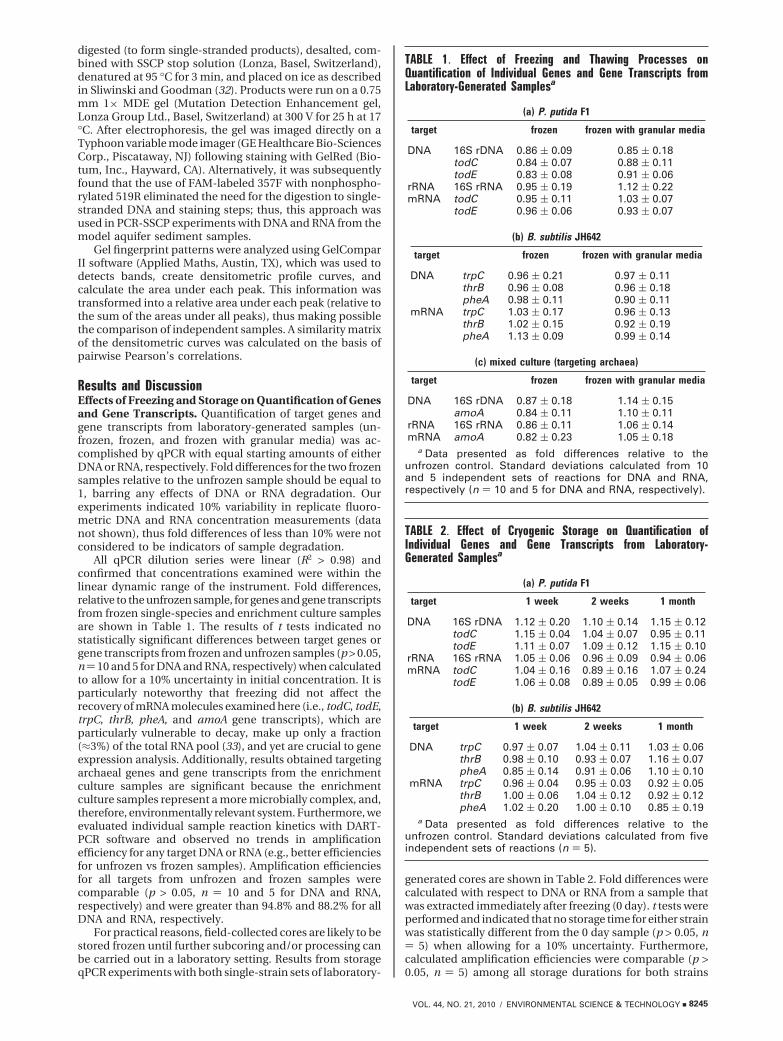
TABLE 1. Effect of Freezing and Thawing Processes onQuantification of Individual Genes and Gene Transcripts fromLaboratory-Generated Samplesa
(a) P. putida F1
target frozen frozen with granular media
DNA 16S rDNA 0.86 ( 0.09 0.85 ( 0.18todC 0.84 ( 0.07 0.88 ( 0.11todE 0.83 ( 0.08 0.91 ( 0.06
rRNA 16S rRNA 0.95 ( 0.19 1.12 ( 0.22mRNA todC 0.95 ( 0.11 1.03 ( 0.07
todE 0.96 ( 0.06 0.93 ( 0.07
(b) B. subtilis JH642
target frozen frozen with granular media
DNA trpC 0.96 ( 0.21 0.97 ( 0.11thrB 0.96 ( 0.08 0.96 ( 0.18pheA 0.98 ( 0.11 0.90 ( 0.11
mRNA trpC 1.03 ( 0.17 0.96 ( 0.13thrB 1.02 ( 0.15 0.92 ( 0.19pheA 1.13 ( 0.09 0.99 ( 0.14
(c) mixed culture (targeting archaea)
target frozen frozen with granular media
DNA 16S rDNA 0.87 ( 0.18 1.14 ( 0.15amoA 0.84 ( 0.11 1.10 ( 0.11
rRNA 16S rRNA 0.86 ( 0.11 1.06 ( 0.14mRNA amoA 0.82 ( 0.23 1.05 ( 0.18
a Data presented as fold differences relative to theunfrozen control. Standard deviations calculated from 10and 5 independent sets of reactions for DNA and RNA,respectively (n ) 10 and 5 for DNA and RNA, respectively).
TABLE 2. Effect of Cryogenic Storage on Quantification ofIndividual Genes and Gene Transcripts from Laboratory-Generated Samplesa
(a) P. putida F1
target 1 week 2 weeks 1 month
DNA 16S rDNA 1.12 ( 0.20 1.10 ( 0.14 1.15 ( 0.12todC 1.15 ( 0.04 1.04 ( 0.07 0.95 ( 0.11todE 1.11 ( 0.07 1.09 ( 0.12 1.15 ( 0.10
rRNA 16S rRNA 1.05 ( 0.06 0.96 ( 0.09 0.94 ( 0.06mRNA todC 1.04 ( 0.16 0.89 ( 0.16 1.07 ( 0.24
todE 1.06 ( 0.08 0.89 ( 0.05 0.99 ( 0.06
(b) B. subtilis JH642
target 1 week 2 weeks 1 month
DNA trpC 0.97 ( 0.07 1.04 ( 0.11 1.03 ( 0.06thrB 0.98 ( 0.10 0.93 ( 0.07 1.16 ( 0.07pheA 0.85 ( 0.14 0.91 ( 0.06 1.10 ( 0.10
mRNA trpC 0.96 ( 0.04 0.95 ( 0.03 0.92 ( 0.05thrB 1.00 ( 0.06 1.04 ( 0.12 0.92 ( 0.12pheA 1.02 ( 0.20 1.00 ( 0.10 0.85 ( 0.19
a Data presented as fold differences relative to theunfrozen control. Standard deviations calculated from fiveindependent sets of reactions (n ) 5).
VOL. 44, NO. 21, 2010 / ENVIRONMENTAL SCIENCE & TECHNOLOGY 9 8245
regardless of the primers used, indicating that storage timedid not affect the integrity of the DNA or RNA.
It is also important to note that freezing and storage didnot result in any trends in either DNA or RNA yields (FigureS1, Supporting Information) from laboratory-generated coresnor was the quality of DNA affected, as visualized byelectrophoresis in a 1% agarose gel (Figure S2, SupportingInformation). Moreover, measurement of RNA using anAgilent 2100 Bioanalyzer indicated that cryogenic preserva-tion did not result in RNA degradation. Specifically, RNAIntegrity Numbers, or RINs, did not change as a function oftime at -80 °C (data not shown).
Effects of Freezing and Storage on the Relative Abun-dance of Bacterial Phylotypes. To further evaluate the effectsof freezing and storage on DNA and RNA from a broad rangeof bacterial phyla (Table S2, Supporting Information), PCR-SSCP was used to generate profiles of the bacterial communitypresent in the enrichment culture samples. Bacterial 16SrRNA genes (rDNA) and gene transcripts (rRNA) from thesuite of enrichment culture samples (unfrozen, frozen, andfrozen with granular media) were profiled in triplicate.Additionally, triplicate bacterial 16S rDNA and rRNA profilesfrom an unfrozen model aquifer sample were compared toprofiles from replicate samples stored at -80 °C for 5 and 10months, respectively.
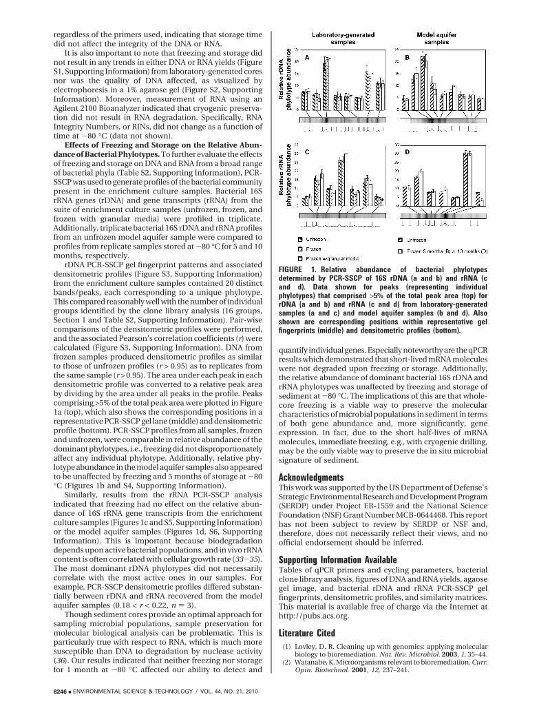
rDNA PCR-SSCP gel fingerprint patterns and associateddensitometric profiles (Figure S3, Supporting Information)from the enrichment culture samples contained 20 distinctbands/peaks, each corresponding to a unique phylotype.This compared reasonably well with the number of individualgroups identified by the clone library analysis (16 groups,Section 1 and Table S2, Supporting Information). Pair-wisecomparisons of the densitometric profiles were performed,and the associated Pearson’s correlation coefficients (r) werecalculated (Figure S3, Supporting Information). DNA fromfrozen samples produced densitometric profiles as similarto those of unfrozen profiles (r > 0.95) as to replicates fromthe same sample (r > 0.95). The area under each peak in eachdensitometric profile was converted to a relative peak areaby dividing by the area under all peaks in the profile. Peakscomprising >5% of the total peak area were plotted in Figure1a (top), which also shows the corresponding positions in arepresentative PCR-SSCP gel lane (middle) and densitometricprofile (bottom). PCR-SSCP profiles from all samples, frozenand unfrozen, were comparable in relative abundance of thedominant phylotypes, i.e., freezing did not disproportionatelyaffect any individual phylotype. Additionally, relative phy-lotype abundance in the model aquifer samples also appearedto be unaffected by freezing and 5 months of storage at -80°C (Figures 1b and S4, Supporting Information).
Similarly, results from the rRNA PCR-SSCP analysisindicated that freezing had no effect on the relative abun-dance of 16S rRNA gene transcripts from the enrichmentculture samples (Figures 1c and S5, Supporting Information)or the model aquifer samples (Figures 1d, S6, SupportingInformation). This is important because biodegradationdepends upon active bacterial populations, and in vivo rRNAcontent is often correlated with cellular growth rate (33-35).The most dominant rDNA phylotypes did not necessarilycorrelate with the most active ones in our samples. Forexample, PCR-SSCP densitometric profiles differed substan-tially between rDNA and rRNA recovered from the modelaquifer samples (0.18 < r < 0.22, n ) 3).
Though sediment cores provide an optimal approach forsampling microbial populations, sample preservation formolecular biological analysis can be problematic. This isparticularly true with respect to RNA, which is much moresusceptible than DNA to degradation by nuclease activity(36). Our results indicated that neither freezing nor storagefor 1 month at -80 °C affected our ability to detect and
quantify individual genes. Especially noteworthy are the qPCRresults which demonstrated that short-lived mRNA moleculeswere not degraded upon freezing or storage. Additionally,the relative abundance of dominant bacterial 16S rDNA andrRNA phylotypes was unaffected by freezing and storage ofsediment at -80 °C. The implications of this are that whole-core freezing is a viable way to preserve the molecularcharacteristics of microbial populations in sediment in termsof both gene abundance and, more significantly, geneexpression. In fact, due to the short half-lives of mRNAmolecules, immediate freezing, e.g., with cryogenic drilling,may be the only viable way to preserve the in situ microbialsignature of sediment.
AcknowledgmentsThis work was supported by the US Department of Defense’sStrategic Environmental Research and Development Program(SERDP) under Project ER-1559 and the National ScienceFoundation (NSF) Grant Number MCB-0644468. This reporthas not been subject to review by SERDP or NSF and,therefore, does not necessarily reflect their views, and noofficial endorsement should be inferred.
Supporting Information AvailableTables of qPCR primers and cycling parameters, bacterialclone library analysis, figures of DNA and RNA yields, agaosegel image, and bacterial rDNA and rRNA PCR-SSCP gelfingerprints, densitometric profiles, and similarity matrices.This material is available free of charge via the Internet athttp://pubs.acs.org.
Literature Cited(1) Lovley, D. R. Cleaning up with genomics: applying molecular
biology to bioremediation. Nat. Rev. Microbiol. 2003, 1, 35–44.(2) Watanabe, K. Microorganisms relevant to bioremediation. Curr.
Opin. Biotechnol. 2001, 12, 237–241.
FIGURE 1. Relative abundance of bacterial phylotypesdetermined by PCR-SSCP of 16S rDNA (a and b) and rRNA (cand d). Data shown for peaks (representing individualphylotypes) that comprised >5% of the total peak area (top) forrDNA (a and b) and rRNA (c and d) from laboratory-generatedsamples (a and c) and model aquifer samples (b and d). Alsoshown are corresponding positions within representative gelfingerprints (middle) and densitometric profiles (bottom).
8246 9 ENVIRONMENTAL SCIENCE & TECHNOLOGY / VOL. 44, NO. 21, 2010

(3) Doong, R.; Chen, T.; Wu, Y. Anaerobic dechlorination of carbontetrachloride by free-living and attached bacteria under variouselectron-donor conditions. Appl. Microbiol. Biotechnol. 1997,47, 317–323.
(4) Haglund, A.; Tornblom, E.; Bostrom, B.; Tranvik, L. Largedifferences in the fraction of active bacteria in plankton,sediments, and biofilm. Microb. Ecol. 2002, 43, 232–241.
(5) Holm, P. E.; Nielsen, P. H.; Albrechtsen, H. J.; Christensen, T. H.Importance of unattached bacteria and bacteria attached tosediment in determining potentials for degradation of xenobioticorganic contaminants in an aerobic aquifer. Appl. Environ.Microbiol. 1992, 58, 3020–3026.
(6) Lehman, R. M.; O’Connell, S. P. Comparison of extracellularenzyme activities and community composition of attached andfree-living bacteria in porous medium columns. Appl. Environ.Microbiol. 2002, 68, 1569–1575.
(7) Lehman, R. M.; Colwell, F. S.; Bala, G. A. Attached and unattachedmicrobial communities in a simulated basalt aquifer underfracture- and porous-flow conditions. Appl. Environ. Microbiol.2001, 67, 2799–2809.
(8) Wilson, R. D.; Thornton, S. F.; Mackay, D. M. Challenges inmonitoring the natural attenuation of spatially variable plumes.Biodegradation 2004, 15, 359–369.
(9) Winderl, C.; Anneser, B.; Griebler, C.; Meckenstock, R. U.;Lueders, T. Depth-resolved quantification of anaerobic toluenedegraders and aquifer microbial community patterns in distinctredox zones of a tar oil contaminant plume. Appl. Environ.Microbiol. 2008, 74, 792–801.
(10) Bauer, R. D.; Maloszewski, P.; Zhang, Y.; Meckenstock, R. U.;Griebler, C. Mixing-controlled biodegradation in a tolueneplume: results from two-dimensional laboratory experiments.J. Contam. Hydrol. 2008, 96, 150–168.
(11) Rees, H. C.; Oswald, S. E.; Banwart, S. A.; Pickup, R. W.; Lerner,D. N. Biodegradation processes in a laboratory-scale ground-water contaminant plume assessed by fluorescence imagingand microbial analysis. Appl. Environ. Microbiol. 2007, 73, 3865–3876.
(12) Mayer, K. U.; Benner, S. G.; Frind, E. O.; Thornton, S. F.; Lerner,D. N. Reactive transport modeling of processes controlling thedistribution and natural attenuation of phenolic compoundsin a deep sandstone aquifer. J. Contam. Hydrol. 2001, 53, 341–368.
(13) Tuxen, N.; Albrechtsen, H.; Bjerg, P. L. Identification of a reactivedegradation zone at a landfill leachate plume fringe using highresolution sampling and incubation techniques. J. Contam.Hydrol. 2006, 85, 179–194.
(14) Lee, P. K. H.; Johnson, D. R.; Holmes, V. F.; He, J.; Alvarez-Cohen, L. Reductive dehalogenase gene expression as a biom-arker for physiological activity of Dehalococcoides spp. Appl.Environ. Microbiol. 2006, 72, 6161–6168.
(15) Everest, F. H.; McLemore, C. E.; Ward, J. F. An improved tri-tubecryogenic gravel sampler. Res. Note: PNW-RN-350; U.S. Depart-ment of Agriculture, Forest Service, Pacific Northwest Forestand Range Experiment Station: Portland, OR, 1980; p 8.
(16) Moser, D. P.; Fredrickson, J. K.; Geist, D. R.; Arntzen, E. V.;Peacock, A. D.; Li, S. W.; Spadoni, T.; McKinley, J. P. Bio-geochemical processes and microbial characteristics acrossgroundwater-surface water boundaries of the Hanford Reachof the Columbia River. Environ. Sci. Technol. 2003, 37, 5127–5134.
(17) Petts, G. E.; Coats, J. S.; Hughes, N. Freeze-sampling method ofcollecting drainage sediments for gold exploration. Trans. Inst.Min. Metall., Sect. B 1991, 100, B28–B32.
(18) Petts, G. E.; Thoms, M. C.; Brittan, K.; Atkin, B. A freeze-coringtechnique applied to pollution by fine sediments in gravel-bedrivers. Sci. Total Environ. 1989, 84, 259–272.
(19) Chung, D. T.; Drabek, J.; Opel, K. L.; Butler, J. M.; McCord, B. R.A study on the effects of degradation and template concentrationon the amplification efficiency of the STR miniplex primer sets.J. Forensic Sci. 2004, 49, 1–8.
(20) Timken, M. D.; Swango, K. L.; Orrego, C.; Chong, M. D.;Buoncristiani, M. R. Quantitation of DNA for forensic DNA typingby qPCR. Final grant report to the California Department ofJustice; 2005.
(21) Piyamongkol, W.; Bermudez, M. G.; Harper, J. C.; Wells, D.Detailed investigation of factors influencing amplificationefficiency and allele drop-out in single cell PCR: implicationsfor preimplantation genetic diagnosis. Mol. Hum. Reprod. 2003,9, 411–420.
(22) Sambrook, J.; Russell, D. W. Molecular Cloning: A LaboratoryManual; 3rd ed.; CSHL Press: Cold Spring Harbor, NY, 2001.
(23) Simon, H. M.; Jahn, C. E.; Bergerud, L. T.; Sliwinski, M. K.;Weimer, P. J.; Willis, D. K.; Goodman, R. M. Cultivation ofmesophilic soil crenarchaeotes in enrichment cultures fromplant roots. Appl. Environ. Microbiol. 2005, 71, 4751–4760.
(24) Smith, M. W.; Herfort, L.; Tyrol, K.; Suciu, D.; Campbell, V.;Crump, B. C.; Peterson, T. D.; Zuber, P.; Baptista, A. M.; Simon,H. M. Seasonal changes in bacterial and archaeal gene expressionpatterns across salinity gradients of the Columbia River coastalmargin. PLoS ONE 2010, in press.
(25) Hendrickx, B.; Junca, H.; Vosahlova, J.; Lindner, A.; Ruegg, I.;Bucheli-Witschel, M.; Faber, F.; Egli, T.; Mau, M.; Schlomann,M.; Brennerova, M.; Brenner, V.; Pieper, D. H.; Top, E. M.;Dejonghe, W.; Bastiaens, L.; Springael, D. Alternative primersets for PCR detection of genotypes involved in bacterial aerobicBTEX degradation: distribution of the genes in BTEX degradingisolates and in subsurface soils of a BTEX contaminatedindustrial site. J Microbiol. Methods 2006, 64, 250–265.
(26) Kabir, S.; Rajendran, N.; Urushigawa, Y.; Itoh, K. Interferenceof contaminating DNA in the quantification of a toluene-inducedtod gene in Pseudomonas putida. J. Biosci. Bioeng. 2003, 96,250–256.
(27) Johnsen, K.; Enger, O.; Jacobsen, C. S.; Thirup, L.; Torsvik, V.Quantitative selective PCR of 16S ribosomal DNA correlateswell with selective agar plating in describing populationdynamics of indigenous Pseudomonas spp. in soil hot spots.Appl. Environ. Microbiol. 1999, 65, 1786–1788.
(28) Kanno, J.; Aisaki, K.; Igarashi, K.; Nakatsu, N.; Ono, A.; Kodama,Y.; Nagao, T. “Per cell” normalization method for mRNAmeasurement by quantitative PCR and microarrays. BMCGenomics 2006, 7, 64.
(29) Francis, C. A.; Roberts, K. J.; Beman, J. M.; Santoro, A. E.; Oakley,B. B. Ubiquity and diversity of ammonia-oxidizing archaea inwater columns and sediments of the ocean. Proc. Natl. Acad.Sci. U.S.A. 2005, 102, 14683–14688.
(30) Peirson, S. N.; Butler, J. N.; Foster, R. G. Experimental validationof novel and conventional approaches to quantitative real-timePCR data analysis. Nucleic Acids Res. 2003, 31, e73.
(31) Lane, D. J. In Nucleic Acid Techniques in Bacterial Systematics;Stackebrandt, E., Goodfellow, M., Eds.; Wiley, John & Sons,Incorporated: Chichester, NY, 1991.
(32) Sliwinski, M. K.; Goodman, R. M. Spatial heterogeneity ofcrenarchaeal assemblages within mesophilic soil ecosystemsas revealed by PCR-single-stranded conformation polymor-phism profiling. Appl. Environ. Microbiol. 2004, 70, 1811–1820.
(33) Lamond, A. I. The control of stable RNA synthesis in bacteria.Trends Biochem. Sci. 1985, 10, 271–274.
(34) Binder, B. J.; Liu, Y. C. Growth rate regulation of rRNA contentof a marine Synechococcus (Cyanobacterium) strain. Appl.Environ. Microbiol. 1998, 64, 3346–3351.
(35) Wagner, R. The regulation of ribosomal RNA synthesis andbacterial cell growth. Arch. Microbiol. 1994, 161, 100–109.
(36) Burlage, R. S.; Atlas, R.; Stahl, D.; Geesey, G.; Sayler, G. Techniquesin microbial ecology; Oxford University Press US: New York,1998.
ES101641Y
VOL. 44, NO. 21, 2010 / ENVIRONMENTAL SCIENCE & TECHNOLOGY 9 8247