Embed Size (px)
Citation preview

EFFECTS OF C-TERMINAL MODIFICATIONS OF GEC1 AND GABARAP, TWO MICROTUBULES-ASSOCIATED PROTEINS, ON KAPPA OPIOID RECEPTOR EXPRESSION
Chongguang Chen, Yulin Wang, Peng Huang and Lee-Yuan Liu-Chen Department of Pharmacology, Temple University School of Medicine, Philadelphia, PA, USA
Running head: GEC1-KOPR interaction Address correspondence to: Dr. Lee-Yuan Liu-Chen, Department of Pharmacology, Temple University School of Medicine, 3420 North Broad Street, Philadelphia, PA 19140, USA Tel: 1-215-707-4188; Fax:
1-215-707-7068; E-Mail: [email protected] We demonstrated previously that GEC1, a member of the microtubules-associated protein (MAP) family, bound to the human kappa opioid receptor (hKOPR) and promoted hKOPR cell surface expression by facilitating its trafficking along the secretory pathway. GABAA receptor-associated protein (GABARAP), a GEC1 analogue, also enhanced KOPR expression, but to a lesser extent. The MAP family proteins undergo cleavage of their C-terminal residue(s) and the exposed conserved glycine forms conjugates with phosphatidylethanolamine, which associate with membranes. Here, we examined whether such modifications were required for GEC1 and GABARAP to enhance hKOPR expression. When transiently transfected into CHO or Neuro2A cells, GEC1 and GABARAP were cleaved at the C-termini. G116A mutation alone or combined with deletion of K117 in GEC1 (GEC1-A) or L117 in GABARAP (GABARAP-A) blocked their C-terminal cleavage, indicating that the conserved G116 is necessary for C-terminal modification. The two GEC1 mutants enhanced hKOPR expression to similar extents as the wild-type GEC1; however, the two GABARAP mutants did not. Immunofluorescence studies showed that HA-GEC1, HA-GEC1-A and HA-GABARAP were distributed in a punctate manner and co-localized with KOPR-EGFP in the Golgi apparatus, while HA-GABARAP-A did not. Pull-down assay of GST-KOPR-C tail with HA-GEC1 or HA-GABARAP revealed that GEC1 had stronger association with KOPR-C tail than GABARAP. These results suggest that because of its stronger binding for hKOPR, GEC1 is able to be recruited by hKOPR sufficiently without membrane association via
its C-terminal modification; however, due to its weaker affinity for the hKOPR, GABARAP appears to require C-terminal modifications to enhance KOPR expression. INTRODUCTION
Kappa opioid receptor (KOPR) is one of the three major types of opioid receptors mediating effects of opioid drugs and endogenous peptides. Effects of KOPR activation in vivo include antinociception (especially for visceral chemical pain), antipruritic, water diuresis and psychotomimetic effects (1). The KOPR agonist nalfurafine (TRK-820) is used clinically in Japan for the treatment of uremic pruritus in kidney dialysis patients (2). KOPR antagonists may be useful for curbing cocaine craving and as anti-anxiety drugs (3,4). In addition, it has been proposed that KOPR agonists may be useful in treating mania, antagonists as anti-depressants and partial agonists for the management of bipolar disorder (5).
We have demonstrated that the protein glandular epithelial cell 1 (GEC1) interacts directly with the C-terminal domain of the KOPR by hydrophobic interactions (6,7). The interaction increases cell surface expression of the KOPR by enhancing the conversion of the glycosylated intermediates to fully glycosylated forms of the receptor, indicating facilitation of trafficking from the endoplasmic reticulum to Golgi to plasma membranes (7).
GEC1 was first cloned as an early estrogen-induced mRNA from guinea-pig endometrial glandular epithelial cells (8). Its deduced amino acid sequences are completely conserved across the several species cloned to date, except the orangutan. GEC1 is widely
1
http://www.jbc.org/cgi/doi/10.1074/jbc.M111.230896The latest version is at JBC Papers in Press. Published on March 9, 2011 as Manuscript M111.230896
Copyright 2011 by The American Society for Biochemistry and Molecular Biology, Inc.
by guest on February 27, 2020http://w
ww
.jbc.org/D
ownloaded from

distributed in mouse and human tissues (9-11). GEC1 is abundant in the central nervous system and is expressed throughout the rat brain (11,12). Two other names have been used for GEC1: GABAA receptor-associated protein like 1 (GABARAPL1) (9) and Apg8L (12).
GEC1 belongs to the family of microtubules-associated proteins (MAPs). Other members of this family include GABAA receptor-associated protein (GABARAP) (13), Golgi-associated ATPase enhancer of 16 kDa (GATE-16) (also named GABARAPL2) (14) and the yeast protein Atg8 (previously named apg8/aut7) (15). All four are 117-amino acid proteins. Light chain 3 of microtubules-associated protein 1 (MAP1-LC3) is a less similar member of the family (16). The identities of amino acid sequence of GEC1 to its analogues are: GABARAP (86%), GATE-16 (61%), Atg8 (55%) and LC3 (~30%).
Proteins of this family have been shown to play important roles in two biological functions: intracellular protein transport and autophagy. GATE-16, GABARAP and GEC1 are involved in intracellular protein transport by enhancing vesicle fusion (7,13,14,17-19). GATE-16 is involved in intra-Golgi transport (14). GABARAP interacts with GABAA and AT1 angiotensin II receptors and transient receptor potential vanilloid 1 (TRPV1) and promotes their cell surface expression (13,18,20,21). Atg8 and LC3 are essential for autophagy and GABARAP and GATE16 are also shown to be involved. During autophagy, an evolutionarily highly conserved process occurring under nutrient deprivation conditions, cytoplasmic components and intracellular organelles are engulfed by autophagosomes (double-membrane-bound compartments) and transported into lysosomes or vacuoles for degradation (22,23). The process involves a series of biochemical reactions similar to ubiquitination (24,25). The residues C-terminal to glycine (Gly116 of GABARAP, GATE16 or Atg8 and Gly120 of LC3) are cleaved in cytoplasm by Atg4 in yeast or its orthologue to yield form I. Forms I are then modified sequentially by Atg7 (E1-like activating enzyme) and Atg3 (E2-like conjugating enzyme), resulting in covalent conjugations with phosphatidylethanolamine (PE) in membranes of
autophagosomes, which are described named forms II. Under conditions of starvation, forms II of Atg8 and LC3 are enriched to promote autophagic activities (16,24,26). Currently, the Form II of LC-3 is widely accepted as a marker of preautophagosomes and autophagosomes in mammalian cells (12,27,28). Although it is not clear if GEC1 is involved in autophagy, GEC1 was shown to undergo similar processes (12,29). Whether such C-terminal processing is needed for intracellular transport is unclear.
We demonstrated previously that both GEC1 and GABARAP promoted KOPR expression (6,7). In this study, we investigated if the C-terminal processing of GEC1 and GABARAP played a role in the observed enhancement in KOPR expression. We compared two GEC1 mutants and two GABARAP mutants with their wild-type counterparts, respectively, in biochemical processing and effects on hKOPR expression. For both proteins, one mutant has G116A substitution and the other one has deletion of K117 in GEC1 and L117 in GABARAP besides G116A mutation (Fig.1).
EXPERIMENTAL PROCEDURES Materials [15, 16-3H]-Diprenorphine (~56Ci/mmol) was purchased from PerkinElmer Life and Analytical Sciences (Boston, MA). Naloxone was from Sigma-Aldrich (Sigma-Aldrich, St. Louis, MO). The following antibodies were used: rabbit anti-FLAG polyclonal antibody (F7425, Sigma-Aldrich, St. Louis, MO); mouse monoclonal anti-HA (HA.11, Covance, Princeton, NJ); mouse monoclonal anti-V5 (Invitrogen, Carlsbad, CA); mouse monoclonal anti-c-Myc (9E12, Santa Cruz Biotechnology, CA); HRP-conjugated goat anti-rabbit IgG and HRP-conjugated goat anti-mouse IgG (Cell Signaling, Danvers, MA). Rabbit anti-GEC1 antibody (PA629p) was generated and purified as described in our previous publication (7,11). SuperSignal West Pico Chemiluminescent and GelCode Blue stain reagents were from Pierce (Rockford, IL). Cell media (DMEM/F-12, 1:1), Opti-MEM I reduced serum, fetal bovine serum (FBS), and Lipofectamine 2000 transfection reagent were acquired from Invitrogen (Carlsbad,
2
by guest on February 27, 2020http://w
ww
.jbc.org/D
ownloaded from

CA). Small interfering RNAs (siRNAs) against mouse GEC1 and GABARAP genes (siGEC1 and siGABARAP, respectively) were purchased from Santa Cruz Biotechnology (Santa Cruz, CA), the negative siRNA control (siControl) was from Qiagen Inc. (Cat. No. 1027281, Valencia, CA). Glutathione-S-transferase (GST) fusion system was from Novagen (GE healthcare Life Sciences, Piscataway, NJ). Cell lines A clonal CHO cell line stably expressing the FLAG-hKOPR was generated previously (30,31), and the Bmax value of FLAG-hKOPR was ~1.9 pmol/mg protein (32). Two clonal Neuro 2A cell lines expressing FLAG-hKOPR or 3HA-hKOPR were similarly established expressing ~1 pmol/mg protein of hKOPR. All cells were cultured in 10-cm culture dishes or 6-well plates in DMEM/F-12 medium supplemented with 10% FBS, 0.2 mg/ml geneticin in a humidified atmosphere consisting of 5% CO2 and 95% air at 37C. Generation of cDNA constructs For C-terminal processing and receptor binding experiments, full-length GEC1 and GABARAP cDNAs and their mutants were inserted into the EcoRI/XhoI sites of the vector pcDNA3.1 / Hygro(+) (Invitrogen). HA epitope was added 5’ to the initiation codon and c-myc epitope immediately 5’ to the stop codon of the cDNAs in the same vector for expression of HA- or HA- and -myc tagged proteins. V5-GABARAP-myc and V5-GABARAP-AL-myc were gifts from Dr. Richard W. Olson of University of California Los Angeles School of Medicine (33). For immunofluorescence microscopy and protein pull-down assays, GEC1 and GABARAP and their mutants were inserted into SalI/XhoI sites of pCMV-HA vector (Clontech). hKOPR-EGFP was constructed into pLenti6/V5-TOPO vector (Invitrogen) and used as a regular CMV promoter-driven expression plasmid. Figure 1 shows the cDNA constructs used in this study. For Glutathione-S-transferase (GST) fusion proteins, KOPR-C tail (D334-V380) (KCT), DOPR-C tail (D322-A372) (DCT), HA-epitope tagged GEC1 (HA-GEC1) and GABARAP (HA-GABARAP) were inserted into BamHI/XhoI site of pGEX-4T-1 vector.
Transient transfection of GEC1, GABARAP, their mutants and siRNAs Lipofectamine-mediated DNA transfection experiments were performed by following the manufacturer’s protocol with some modifications. Twenty-four hours before transfection, 1.8~2.0 million CHO-FLAG-hKOPR cells were seeded on each 10-cm cell culture dish. Transfection was carried out with 30 l of Lipofectamine 2000, 10 g of the cDNA constructs or the blank plasmid vector (control) and 6 ml of Opti-MEM medium per 10-cm dish. At 16 hours after transfection, medium was replaced by 10 ml of Opti-MEM. Thirty hours following transfection, cells were harvested for receptor binding and western blot experiments. GEC1 and GABARAP knock-down experiments were performed with Neuro 2A cells expressing 3HA-hKOPR similarly as above except that siGEC1, siGABARAP and siControl were used. For immunofluorescence microscopy, HEK293 cells were cultured on cover-slips placed in 12-well plate at 2x10⁵ cells/well for 24 hours. Cells were then co-transfected with 50ng of hKOPR-EGFP (h-EGFP) and 25ng of HA-tagged constructs as indicated in Fig.8 with Lipofectamine 2000. Twenty four hours after transfection, cells were fixed in 4% paraformaldehyde in PBS buffer for 15min. Immunofluorescence was performed by incubating cells with both mouse anti-HA (1/1000) and rabbit anti-giantin (1/1000) antibodies overnight at 4C and then the secondary antibodies Texas Red conjugated anti-mouse IgG (1/1000) and Alexa-350 conjugated anti-rabbit IgG (1/1000) at room temp for 1 hour. Images were acquired using a Nikon TE300 fluorescence microscope and a 60x oil objective lens and a Magnifire digital camera. NIH Image and Adobe Photoshop were used for imaging processing. Care has been taken to avoid over expression of the HA-GEC1 and its analogues. Transfection of 25 ng each of HA-GEC1 and its analogues was determined empirically so that the HA-GEC1 expressed at a level matched that of endogenous GEC1 (data not shown). [3H]Diprenorphine binding to hKOPR in Intact Cells
3
by guest on February 27, 2020http://w
ww
.jbc.org/D
ownloaded from
Intact cell binding was performed as described previously (7). Briefly, 100,000 or 200,000 cells/tube was incubated with 1 nM [3H]diprenorphine in PBS buffer for 1 h. Naloxone (10 M) was used to define the nonspecific binding for total receptors, whereas dynorphin (1-17) (1M) was used to define nonspecific binding for cell surface receptors. KaleidaGragh program (Synergy Software) was used for data processing. Protein pull-down assay GST-KCT, GST-DCT, GST-HA-GEC1 and GST-HA-GABARAP proteins were prepared and bound to glutathione-Sepharose 4B beads as described previously (7). GST-HA-GEC1 and GST-HA-GABARAP bound to glutathione-Sepharose 4B beads were treated with biotinylated thrombin to produce HA-GEC1 and HA-GABARAP. After thrombin was removed by use of Streptavidin-Agarose, the purities of HA-GEC1 and HA-GABARAP were determined to be >90 % by SDS-PAGE and GelCode Blue staining. The protein concentrations of purified HA-GEC1 and HA-GABARAP were determined by BCA reagent. DOPR-C tail was chosen as negative control against KOPR-C tail for their similarity in size and pI value (51 aa/47 aa and pI 9.21/pI 9.66). The GST-DCT also provides better separation in SDS-PAGE than GST from HA-GEC1 and HA-GABARAP, which is critical for reliable quantitation of bound GEC1 or GABARAP. Four concentrations of HA-GEC1 or HA-GABARAP (0.5ml of 10g/ml, 20g/ml, 40g/ml, 80g/ml) and 10l (20g proteins) of Sepharose 4B-GST-KCT or -GST-DCT in TBS-T++ buffer (20mM Tris, pH7.4, 0.2M NaCl, 2mM MgCl2, 1mM DTT and 0.1% Tween 20) were incubated at 4oC overnight on a rotating rack. An aliquot (5l) was taken from each concentration of HA-GEC1 or HA-GABARAP and diluted 50-fold in 2X Laemmli sample buffer and used as the loading control in immunoblotting (see below). The incubation mixtures were washed 5 x 5min with pre-cooled TBS-T++ buffer buffer by centrifugation and re-suspension and the buffer aspirated. Thirty l of 2X Laemmli sample buffer were then added to gel beads to dissociate bound proteins. The
samples were heated at 60 oC for 10min and then 20l each was loaded onto SDS-PAGE. Immunoblotting was performed with the anti-HA mAb (1:10000) and the protein bands were quantified with OptiQuant program as described previously (7). The linear relationship between intensities (DLU) and the amounts of HA-GEC1/HA-GABARAP was determined empirically to be within 5-80ng (Fig. 9B) and the incubation concentrations were adjusted so that the intensities of bound proteins were within this linear range. Ten ng, 20ng, 40ng and 80ng of HA-GEC1 and HA-GABARAP were included in each immunoblotting as the loading controls and were also used to calculate bound proteins in the same data set (Fig. 9B,C). SDS-PAGE and Immunoblotting Cells were harvested using Versene buffer, solubilized in 2X Laemmli sample buffer, and subjected to Tricine-SDS-PAGE in 10% or 12% separating gel as described previously (6,7). The separated protein bands were transferred to Immobilon-P polyvinylidene difluoride membranes on which immunoblotting was carried out with primary antibodies indicated in figure legend, horseradish peroxidase-linked secondary antibody, and SuperSignal West Pico Chemiluminescent reagents. Antibodies were used at following dilutions: rabbit anti-FLAG polyclonal antibody, 1:2000; mouse monoclonal anti-HA, 1:4000 unless indicated otherwise; mouse monoclonal anti-V5, 1:5000; mouse monoclonal anti-c-Myc, 1:2000; horseradish peroxidase-linked secondary antibodies, 1:10,000. The protein bands were visualized and digitalized with Fuji LAS-1000 Plus gel documentation system (Fuji Film, Tokyo, Japan).
RESULTS The role of the glycine 116 (G116) in the C-terminal processing of GEC1
When HA-GEC1-myc, HA-GEC1AK-myc or HA-GEC1A-myc was transiently transfected into CHO-FLAG-hKOPR cells, all the expressed proteins were recognized by antibody against the HA epitope (the upper panel of Fig. 2A). Although HA-GEC1-myc, HA-GEC1AK-myc or HA-GEC1A-myc had
4
by guest on February 27, 2020http://w
ww
.jbc.org/D
ownloaded from
almost identical calculated molecular mass, HA-GEC1-myc migrated faster than the other two, yielding similar relative molecular weight (Mr) as HA-GEC1. However, HA-GEC1AK-myc and HA-GEC1A-myc had higher Mr’s than HA-GEC1AK and HA-GEC1A, respectively. In addition, HA-GEC1AK-myc and HA-GEC1A-myc, but not HA-GEC1-myc, were recognized by anti-c-myc antibody (Fig. 2A, lower panel). These findings indicate loss of c-myc from HA-GEC1-myc due to cleavage at C-terminus of GEC1, but not from HA-GEC1AK-myc and HA-GEC1A-myc. Thus, C-terminal modifications do occur to GEC1, perhaps similar to Atg8, for which the G116 is required (24,25). Similar results were also obtained in blank Neuro2A cells (Fig. 2B), indicating that the cleavage at the C-terminus of GEC1 does not depend on cell types or KOPR expression. Effect of G116A mutation on GEC1-induced increase of hKOPR expression
Following transient transfection, GEC1A and GEC1AK enhanced total and cell surface hKOPR expression to similar extents as the wild-type GEC1, as determined by both radioligand binding and immunoblotting (Fig. 3). Thus, C-terminal modification is not required for GEC1 to enhance expression of the hKOPR. In addition, compared to the vector control, GEC1, GEC1A and GEC1AK did not change the ratio of cell surface to total receptors, indicating that all three proteins did not act differentially on intracellular or cell surface receptors and that both mutants act in similar manners as the wildtype. Moreover, GEC1AK enhanced hKOPR expression with a time course similar to that of GEC1 (Fig. 4).
These results are different from those of Chen et al. (33) that C-terminal modification is required for GABARAP to enhance cell surface expression of the subunit of the GABAA receptor. We have shown previously that GABARAP interacts with the KOPR and enhances KOPR levels, but to a lesser extent than GEC1 (6). We, therefore, examined if C-terminal modification was required for GABARAP to enhance KOPR expression. The role of G116 in the C-terminal processing of GABARAP
Two GABARAP constructs, V5-GABARAP-myc and V5-GABARAP-AL-myc (G116A mutant), were transfected into CHO cells (Fig. 5A) or Neuro 2A cells (Fig. 5B) and immunoblotting was performed with antibodies against V5 and c-myc. While anti-V5 antibody recognized both proteins (upper panel, Fig. 5A, B), anti-c-myc antibody only recognized the G116A mutant (lower panel, Fig. 5A, B). In addition, V5-GABARAP-myc yielded lower Mr than V5-GABARAP-AL-myc (upper panel, Fig. 5A, B). These results demonstrate that similar to GEC1, the C-terminus of the wildtype GABARAP, but not the G116A mutant, is cleaved, and that the G116 of GABARAP is required for its C-terminal processing. Effect of G116A mutation on GABARAP-induced hKOPR expression
GABARAP, GABARAP-AL (G116A mutant) and GABARAP-A (containing G116A mutation and L117 deletion) were examined along with GEC1 for their effects on expression of hKOPR (Fig. 6). GABARAP transfection increased both total and cell surface hKOPR expression by more than 80% without changing the % of total receptors on cell surface. In contrast, GABARAP-AL and GABARAP-A failed to show any increase. These results demonstrate that, unlike GEC1, GABARAP requires C-terminal cleavage to enhance hKORP expression. GABARAP did not alter the % of total receptors on cell surface, similar to GEC1. The degree of hKOPR increase induced by GABARAP was lower than that of GEC1, consistent with our previous findings (6) Distribution of GEC1, GABARAP and their mutants in cells and relationship to hKOPR distribution
We used HEK293 cells for localization studies instead of CHO cells because CHO cells have much larger nuclei and very limited cytosol space, making it difficult to visualize subcellular organelles. Cells were co-transfected transiently with KOPR-EGFP (green) and HA-tagged GEC1, GEC1A, GEC1-F60A GABARAP, or GABARA-A. We showed previously that GEC1-F60A did not interact with hKOPR and did not enhance hKOPR expression (6). Immunofluorescence was performed (Fig. 7) for
5
by guest on February 27, 2020http://w
ww
.jbc.org/D
ownloaded from
HA-GEC1 and analogues with anti-HA antibodies (red) and giantin with giantin antibodies (blue), a marker for the Golgi apparatus. Staining of HA-GEC1, HA-GEC1A and HA-GABARAP was punctate in cells, and co-localized with intracellular KOPR-EGFP and giantin. In contrast, HA-GABARAP-A and HA-GEC1F60A had a diffused pattern and did not show significant co-localization with KOPR-EGFP or giantin. These results suggest that the localization of GEC1 and GABARAP with KOPR is required for them to enhance receptor expression.
It should be noted that cells transiently transfected with KOPR-EGFP were used for immunofluorescence studies, whereas stably transfected cells were used for the studies in which KOPR quantitation was involved. The reason for these choices is that transiently transfected cells have a much larger intracellular pool of receptor precursors than stably transfected cells, which is especially true when examined within 24 h after transfection (the condition used in experiments for Fig. 7). The high levels of intracellular receptor precursors facilitated studies on possible spatial differences displayed by GEC1, GABARA and their mutants and their co-localization of the hKOPR in the Golgi (Fig. 7). Effect of GEC1 and GABARAP knock-down on hKOPR expression
Both CHO cells and Neuro 2A cells express GEC1 and GABARAP endogenously, which were readily detectable by immunoblotting with the GEC1 antibody PA629p (Fig. 8B). We have shown previously that PA629p has significant cross-reactivity with GABARAP and that GABARAP has a lower relative molecular weight than GEC1 in SDS-PAGE (11). Since siRNAs of the mouse, but not Chinese hamster, origin are readily available, Neuro 2A cells (mouse) expressing 3HA-hKOPR were used. Transfection of siGEC1 or siGABARAP reduced their targeting protein levels, compared with siControl (Fig. 8B, bottom panel). Importantly, knock-down of GEC1 or GABARAP decreased the total and cell surface hKOPR expression as shown by receptor binding (Fig. 8A) and immunoblot (Fig. 8B). Note that the GEC1 siRNA showed
significant cross-reactivity to GABARAP and vice versa. Interaction with KOPR-C tail: comparison between GEC1 and GABARAP Pull-down techniques were employed for these experiments, using GST-KOPR-C tail or GST-DOPR-C tail (as the control) coupled to glutathione Sepharose and HA-GEC1 and HA-GABARAP purified from GST fusion products. This combination allowed reproducible results. As shown in Fig. 9, at 40g/ml and 80g/ml, HA-GEC1 had significantly more binding to GST-KCT than HA-GABARAP. In contrast, DOPR-C tail did not bind HA-GEC1
It should be noted that the binding has not reached saturation because of technical limitations. To reach saturation, the amount of GST-KOPR-C-tail or –DOPR-C tail beads has to be very low and the amount we used was at the limit of reproducible pipetting. In addition, the staining density of HA-GEC1 or HA-GABARAP has to be within the linear detection range. Therefore, instead of a ‘saturation curve’, here we presented data from low ‘ligand’ concentrations. The results, nevertheless, revealed that GEC1 binds to KOPR-C tail at higher affinity than GABARAP (Fig. 9C). DISCUSSION
In this study, we found that C-terminal
cleavage of GEC1 is not required for its effects in enhancing expression of the hKOPR; however, GABARAP requires C-terminal cleavage to have the same effect. In addition, we demonstrated that GEC1 had stronger interaction with KOPR-C-tail than GABARAP, which may account for the observed difference. Similarities and differences between GEC1 and GABARAP
The C-termini of GEC1 and GABARAP were cleaved and the G116A mutation blocked this process, indicating that the Gly116 residue is critical for C-terminal processing. Our results are in agreement with the observations of Tanida et al. (27,29) and Chen et al. (33) and support the notion that the C-terminal processing is universal for all the GEC1 analogues identified to date. In addition, we found that GEC1,
6
by guest on February 27, 2020http://w
ww
.jbc.org/D
ownloaded from

GEC1A as well as GEC1AK were equally effective in promoting KOPR expression. In contrast, GABARAP increased KOPR expression, while GABARAP-A and GABARAP-AL did not. Therefore, the C-terminal processing is not necessary for GEC1 to enhance KOPR expression, but is required for GABARAP to have the same effect. Our results on GABARAP are similar to those of Chen et al. (33) on the subunit of the GABAA receptor, but different from those of Alam et al. (36) on angiotension II type 1A receptor (see below).
The marked difference between GEC1 and GABARAP in their requirement of C-terminal cleavage for promoting KORP expression was unexpected. C-terminal modification by the Atg4B-Atg7-Atg3 system defines a common biochemical pathway for this family of proteins and presumably leads to similar functional consequences. In addition to and/or independent of these shared features, there may be factors that set diverging points for members of the family. We demonstrated that both wildtype GEC1 and GABARAP enhanced KOPR expression with GABARAP having a lower degree of increases (Fig. 6) (also (6)), which may reflect lower affinities for the hKOPR. This notion is supported by the data from quantitative analysis of the pull-down assay that HA-GEC1 had significantly stronger association with the KOPR-C-tail than HA-GABARAP (Fig. 9C). In addition, using yeast two-hybrid technique, we showed previously that on QDO plates, the colony forming units were 83±10 and 66±5 (n=3, P<0.05, Student’s t test) for interaction of KOPR-C-tail with GEC1(38-117) and GABARAP(38-117), respectively (7), indicating that GABARAP(38-117) had weaker binding to the KOPR C-tail than GEC1(38-117). Intact GEC1 or GABARAP did not work in yeast two-hybrid, most likely because of their strong association with microtubules via the N-terminal domains (6,7).
Deletion of Gly116 and Lys117 did not show any effect on direct interaction of GEC1 with KOPR C-tail in yeast two-hybrid assay (6). In addition, based on our GEC1 model (6) and crystal structures of GABARAP (37-39), Gly116 is unlikely to contribute to any differences in their binding surface. Therefore, we postulate
that their ‘intrinsic’ affinities to KOPR are independent of the C-terminal processing.
We have demonstrated that the interaction between hKOPR C-tail and GEC1 is mediated by direct contacts between the kinked hydrophobic fragment in hKOPR C-tail (containing Phe345, Pro346, and Met350) and the curved hydrophobic surface in GEC1 around the S2 beta-strand formed by Tyr49, Val51, Leu55, Thr56, Val57, Phe60, and Ile64 (6). Despite the high amino acid sequence identity between GABARAP and GATE16, there are a few differences in their X-ray crystal structures (39,40). The crystal structure of GEC1 has not been resolved; however, it is likely to be similar to that of GABARAP, with some subtle differences. These differences may contribute to lower affinity of GABARAP for the hKOPR C-tail and thus lower levels of enhancement in hKOPR expression.
We postulate that two independent factors may contribute to enhancing effects of GEC1 and analogues on hKOPR cell surface expression: (1) the affinity of GEC1 and analogues for the hKOPR; (2) C-terminal modifications, which facilitate association of GEC1 and analogues with intracellular membranes, where hKOPR to be exported resides. If the affinity is sufficiently high, as in the case of GEC1, adequate amounts of GEC1 will interact with hKOPR even without C-terminal modifications and thus enhance hKOPR expression. If the affinity for the hKOPR C-tail is not high enough, as in the case of GABARAP, enhanced membrane association with C-terminal modifications is needed to have full effects. Our immunofluorescence results (Fig. 7) support this notion. HA-GEC1, HA-GABARAP and their G116A mutants co-transfected with KOPR-EGFP showed remarkably different staining patterns. HA-GEC1 and HA-GABARAP and HA-GEC1A had punctate staining in the cytoplasmic space, which co-localized significantly with KOPR-EGFP in the Golgi apparatus, whereas GABARAP-A had diffuse distribution, with little co-localization with KOPR-EGFP. In addition, the GEC1F60A mutant, which had substantially lower binding to the KOPR C-tail and had no enhancing effect on KOPR expression (6), showed diffuse staining and a low level of co-localization with KOPR
7
by guest on February 27, 2020http://w
ww
.jbc.org/D
ownloaded from
8
even though this mutant most likely undergoes C-terminal modification. It is conceivable that higher intrinsic affinity of GEC1 to KOPR alone allows GEC1A to be recruited sufficiently to Golgi by KOPR where it enhances transport. In contrast, without the benefit of membrane association via phosphatidylethanolamine conjugation, GABARAP-A fails to be recruited sufficiently by KOPR due to its lower binding affinity to the receptor. Whether this inference is valid remains to be examined. Recent studies on effects of GABARAP on trafficking of different receptors have yielded different results. Chen et al. (33) have shown that cleavage at C-terminus in GABARAP is required for GABARAP-mediated GABAA trafficking and enhanced GABA-induced currents. Alam et al. (36), however, demonstrated that the wild-type GABARAP and its G116A mutant were equally effective in stimulating surface expression and signaling activity of angiotensin II type 1A receptor. The two findings may seem contradictory to each other but can be explained by the different affinities of GABARAP for the 2 subunit of the GABAA receptor and for the angiotensin II type 1A receptor. In addition, C-terminal modification of GABARAP is required for enhancing surface expression of the KOPR, but not angiotensin II type 1A receptor, suggesting that interactions of GABARAP and its analogues with receptors are related to their affinity for the receptor, more specifically the fragment of the receptor, but independent of whether the receptor is coupled to G proteins or not. Redundant functions of GEC1 and GABARAP
Both GEC1 and GABARAP have been shown to bind to membrane-bound proteins. GEC1 has been shown to interact with GABAA receptor (35), KOPR (7) and AT1 angiotensin II receptor (20) and enhance cell surface expression of the KOPR (7). GABARAP is associated with and increases cell surface expression of GABAA receptor (13,18), KOPR (6), Na+-dependent Pi cotransporter, NaPi-IIa
(41), AT1 angiotensin II receptor (20) and TRPV1 (21). In addition, GABARAP also binds transferrin receptor (42). Because of the high degree of identity, GABARAP and GEC1 are likely to have overlapping functions. The findings that GABAA receptor, KOPR and AT1 angiotensin II receptor interact with both GEC1 and GABARAP (6,13,20,35) support this notion. In GABARAP-null mice, there is no change in GABAA receptor levels or membrane clustering (43), most likely due to the redundant functions between the two proteins.
Possible molecular mechanisms of GEC1- or GABARAP-promoted trafficking
We have shown that GEC1 facilitates transport of the KOPR from ER to Golgi to plasma membranes. However, the molecular mechanisms of this facilitation are not clear. Both GEC1 and GABARAP bind tubulin and NSF (6,7,17,34,35); therefore, their functions are related to transport along microtubules. Since they are not motor proteins like the anterograde transport protein kinesin and thus do not move vesicles actively along microtublues. Cook et al. (20) postulated that GABARAP binds microtubules and vesicular cargo such as the AT1 angiotensin II receptor and, by doing so, stabilizes the kinesin-vesicle complex on microtubules as kinesin moves the cargo forward. In addition, the GABARAP binding stabilizes vesicle cargo on microtubules after one kinesin molecule dissociates from microtubules and before another one associates. It is conceivable that GEC1 acts in a similar manner to facilitate anterograde transport of the KOPR, which remains to be studied. CONCLUSION
Our finding that C-terminal
modifications are not required for GEC1 to enhance KOPR expression indicates that this function is independent of the possible involvement of GEC1 in autophagy, and clearly demonstrates that this family of proteins has two very distinct functions.
by guest on February 27, 2020http://w
ww
.jbc.org/D
ownloaded from

References
1. Liu-Chen, L.-Y. (2004) Life Sci. 75, 511-536 2. Wikstrom, B., Gellert, R., Ladefoged, S. D., Danda, Y., Akai, M., Ide, K., Ogasawara, M.,
Kawashima, Y., Ueno, K., Mori, A., and Ueno, Y. (2005) J.Am.Soc.Nephrol. 16, 3742-3747 3. Knoll, A. T., Meloni, E. G., Thomas, J. B., Carroll, F. I., and Carlezon, W. A., Jr. (2007)
J.Pharmacol.Exp.Ther. 323, 838-845 4. Beardsley, P. M., Howard, J. L., Shelton, K. L., and Carroll, F. I. (2005)
Psychopharmacol.(Berlin) 183, 118-126 5. Carlezon, W. A., Jr., Beguin, C., Dinieri, J. A., Baumann, M. H., Richards, M. R., Todtenkopf, M.
S., Rothman, R. B., Ma, Z., Lee, D. Y., and Cohen, B. M. (2006) J.Pharmacol.Exp.Ther. 316, 440-447
6. Chen, Y., Chen, C., Kotsikorou, E., Lynch, D. L., Reggio, P. H., and Liu-Chen, L. Y. (2009) J.Biol.Chem. 284, 1673-1685
7. Chen, C., Li, J. G., Chen, Y., Huang, P., Wang, Y., and Liu-Chen, L. Y. (2006) J.Biol.Chem. 281, 7983-7993
8. Pellerin, I., Vuillermoz, C., Jouvenot, M., Ordener, C., Royez, M., and Adessi, G. L. (1993) Mol.Cell Endocrinol. 90, R17-R21
9. Xin, Y., Yu, L., Chen, Z., Zheng, L., Fu, Q., Jiang, J., Zhang, P., Gong, R., and Zhao, S. (2001) Genomics 74, 408-413
10. Nemos, C., Mansuy, V., Vernier-Magnin, S., Fraichard, A., Jouvenot, M., and Delage-Mourroux, R. (2003) Brain Res.Mol.Brain Res. 119, 216-219
11. Wang, Y., Dun, S. L., Huang, P., Chen, C., Chen, Y., Unterwald, E. M., Dun, N. J., Van Bockstaele, E. J., and Liu-Chen, L. Y. (2006) Neuroscience 140, 1265-1276
12. Hemelaar, J., Lelyveld, V. S., Kessler, B. M., and Ploegh, H. L. (2003) J.Biol.Chem. 278, 51841-51850
13. Wang, H., Bedford, F. K., Brandon, N. J., Moss, S. J., and Olsen, R. W. (1999) Nature 397, 69-72 14. Sagiv, Y., Legesse-Miller, A., Porat, A., and Elazar, Z. (2000) EMBO J. 19, 1494-1504 15. Lang, T., Schaeffeler, E., Bernreuther, D., Bredschneider, M., Wolf, D. H., and Thumm, M.
(1998) EMBO J. 17, 3597-3607 16. Kabeya, Y., Mizushima, N., Ueno, T., Yamamoto, A., Kirisako, T., Noda, T., Kominami, E.,
Ohsumi, Y., and Yoshimori, T. (2000) EMBO J. 19, 5720-5728 17. Kittler, J. T., Rostaing, P., Schiavo, G., Fritschy, J. M., Olsen, R., Triller, A., and Moss, S. J.
(2001) Mol.Cell Neurosci. 18, 13-25 18. Leil, T. A., Chen, Z. W., Chang, C. S., and Olsen, R. W. (2004) J.Neurosci. 24, 11429-11438 19. Chen, Z. W. and Olsen, R. W. (2007) J.Neurochem. 100, 279-294 20. Cook, J. L., Re, R. N., deHaro, D. L., Abadie, J. M., Peters, M., and Alam, J. (2008) Circ.Res.
102, 1539-1547 21. Lainez, S., Valente, P., Ontoria-Oviedo, I., Estevez-Herrera, J., Camprubi-Robles, M., Ferrer-
Montiel, A., and Planells-Cases, R. (2010) FASEB J. 22. Shintani, T. and Klionsky, D. J. (2004) Science 306, 990-995 23. Yoshimori, T. (2004) Biochem.Biophys.Res.Commun. 313, 453-458 24. Ichimura, Y., Kirisako, T., Takao, T., Satomi, Y., Shimonishi, Y., Ishihara, N., Mizushima, N.,
Tanida, I., Kominami, E., Ohsumi, M., Noda, T., and Ohsumi, Y. (2000) Nature 408, 488-492 25. Kabeya, Y., Mizushima, N., Yamamoto, A., Oshitani-Okamoto, S., Ohsumi, Y., and Yoshimori,
T. (2004) J.Cell Sci. 117, 2805-2812 26. Kirisako, T., Ichimura, Y., Okada, H., Kabeya, Y., Mizushima, N., Yoshimori, T., Ohsumi, M.,
Takao, T., Noda, T., and Ohsumi, Y. (2000) J.Cell Biol. 151, 263-276 27. Tanida, I., Komatsu, M., Ueno, T., and Kominami, E. (2003) Biochem.Biophys.Res.Commun. 300,
637-644
9
by guest on February 27, 2020http://w
ww
.jbc.org/D
ownloaded from
28. Tanida, I., Sou, Y. S., Ezaki, J., Minematsu-Ikeguchi, N., Ueno, T., and Kominami, E. (2004) J.Biol.Chem. 279, 36268-36276
29. Tanida, I., Sou, Y. S., Minematsu-Ikeguchi, N., Ueno, T., and Kominami, E. (2006) FEBS J. 273, 2553-2562
30. Li, J., Li, J.-G., Chen, C., Zhang, F., and Liu-Chen, L.-Y. (2002) Mol.Pharmacol. 61, 73-84 31. Li, J.-G., Chen, C., and Liu-Chen, L.-Y. (2002) J.Biol.Chem. 277, 27545-27552 32. Zhang, F., Li, J., Li, J.-G., and Liu-Chen, L.-Y. (2002) J.Pharmacol.Exp.Ther. 302, 1184-1192 33. Chen, Z. W., Chang, C. S., Leil, T. A., and Olsen, R. W. (2007) J.Neurosci. 27, 6655-6663 34. Wang, H. and Olsen, R. W. (2000) J.Neurochem. 75, 644-655 35. Mansuy, V., Boireau, W., Fraichard, A., Schlick, J. L., Jouvenot, M., and Delage-Mourroux, R.
(2004) Biochem.Biophys.Res.Commun. 325, 639-648 36. Alam, J., Deharo, D., Redding, K. M., Re, R. N., and Cook, J. L. (2010) Regul.Pept. 159, 78-86 37. Bavro, V. N., Sola, M., Bracher, A., Kneussel, M., Betz, H., and Weissenhorn, W. (2002) EMBO
Rep. 3, 183-189 38. Coyle, J. E., Qamar, S., Rajashankar, K. R., and Nikolov, D. B. (2002) Neuron 33, 63-74 39. Knight, D., Harris, R., McAlister, M. S., Phelan, J. P., Geddes, S., Moss, S. J., Driscoll, P. C., and
Keep, N. H. (2002) J.Biol.Chem. 277, 5556-5561 40. Paz, Y., Elazar, Z., and Fass, D. (2000) J.Biol.Chem. 275, 25445-25450 41. Reining, S. C., Gisler, S. M., Fuster, D., Moe, O. W., O'Sullivan, G. A., Betz, H., Biber, J., Murer,
H., and Hernando, N. (2009) Am.J.Physiol Renal Physiol 296, F1118-F1128 42. Green, F., O'Hare, T., Blackwell, A., and Enns, C. A. (2002) FEBS Lett. 518, 101-106 43. O'Sullivan, G. A., Kneussel, M., Elazar, Z., and Betz, H. (2005) Eur.J.Neurosci. 22, 2644-2648 FOOTNOTES Acknowledgments: This study was supported by NIH grants R01 DA17302 and P30 DA13429. The abbreviations used are: Atg8, yeast autophagy protein 8; GEC1, glandular epithelial cell 1, also named GABAA receptor-associated protein like 1, GABARAPL1; GABARAP, GABAA receptor-associated protein; GATE16, Golgi-associated ATPase enhancer of 16 KDa; KOPR, opioid receptor; KCT, opioid receptor C tail; DCT, opioid receptor C tail; LC3, light chain 3 of MAP 1A/1B; MAPs , microtubule-associated proteins. FIGURE LEGENDS Figure 1. (A) Amino acid sequence comparison among GEC1 and its three analogues GABARAP, GATE16 and Atg8. The residue(s) C-terminal to the conserved glycine (equivalent to Gly116 of GEC1) of these analogues are cleaved and the conserved glycine becomes the C-terminus. Under starvation conditions, the glycine is conjugated to phosphotidylethanolamine. (B) cDNA constructs used in this study. cDNA constructs of the mutants and wildtypes of GEC1 and GABARAP shown are inserted into the pcDNA3.1 or pCMV-HA mammalian expression vector. Figure 2. (A) GEC1 undergoes C-terminal cleavage, but GEC1AK and GEC1A do not. Each construct was transfected into CHO cells with Lipofectamine. Forty hours later, cells were collected, dissolved in SDS loading buffer and loaded onto 12% SDS-PAGE (4x10⁵ cells/lane) and immunoblotting was performed with the antibodies indicated. (B) HA-GEC1-myc and the HA-GEC1AK-myc were transfected into Neuro 2A cells and immunoblotted in a similar manner as in (A). Note that C-terminal cleavage of HA-GEC1-myc was incomplete, suggesting less robust Atg4B activity in Neuro 2A cells. The figures shown are results of one of three independent experiments.
10
by guest on February 27, 2020http://w
ww
.jbc.org/D
ownloaded from
Figure 3. C-terminal cleavage is not required for GEC1 to enhance total and cell surface KOPR expression. CHO cells stably expressing FLAG-hKOPR were transfected as described in Fig. 2 and cells were harvested 40 h later. (A) KOPR binding was performed with 1 nM [3H]diprenorphine on intact cells using 10 M naloxone and 1 M dynorphin to define nonspecific binding for total and cell surface receptors, respectively. The results are presented as mean ± sem of 5 experiments. ***p<0.0005, compared with Vector group by one-way ANOVA followed by Dunnett’s post-hoc test. (B) Aliquots of each transfected cells from (A) were immunoblotted with antibodies indicated. Each lane was loaded with 20 g of proteins. The blot represents one of the 4 experiments performed. Figure 4. GEC1 and GEC1AK affect KOPR expression with similar time courses. Transfection and intact cell binding were performed as described in Fig. 3. KOPR binding was performed with ~1 nM [3H]diprenorphine on intact cells using 10 M naloxone to define nonspecific binding. Each value is mean ± sem of 3 or 4 experiments. ***p<0.0005, **p<0.005, *p<0.05, compared with the vector control group at same time point by one-way ANOVA followed by Dunnett’s post-hoc test. Figure 5. The C-terminus of the wildtype GABARAP, but not the G116A mutant (GABARAP-AL), is cleaved, similar to GEC1, in CHO cells (A) and in Neuro 2A cells (B). The experiment was performed similarly as described in Fig. 2 legend. The blots represent one of the three experiments. Figure 6. Contrary to GEC1, the C-terminal cleavage is essential for GABARAP to enhance KOPR total and cell surface expression. Experiments were carried out as described in Fig. 3 legend. (A) Each value is mean ± sem of 4 experiments. (B) The immunoblot represents one of the three experiments. *p<0.05, ***p<0.0005 compared with the vector control by one-way ANOVA followed by Dunnett’s post-hoc test. Figure 7. Co-localization of KOPR with GEC1, GEC1-A, GABARAP, but not with GABARAP-A and GEC1F60A. HEK293 cells were cultured on cover-slips for 24 h and then co-transfected with KOPR-EGFP (h-EGFP) and HA-tagged constructs as indicated with Lipofectamine 2000. Twenty-four h after transfection, cells were fixed in 4% paraformaldehyde. Immunofluorescence was performed with mouse anti-HA antibodies followed by Texas red conjugated anti-mouse IgG for wildtype and mutant GEC1 / GABARAP (red) and rabbit anti-giantin antibody and then Alexa-350 conjugated anti-rabbit IgG for and the Golgi marker giantin (blue). Images shown are KOPR-EGFP (green, the first column) co-expressed with HA-GEC1 (A), HA-GEC1-A (B), HA-GABARAP (C), HA-GABARAP-A (D) and HA-GEC1F60A (E) (red, the second column). The Golgi marker giantin is shown in the third column (blue). Merged images are shown in the 4th column. These images are representatives of at least 50 images per row from 4 independent experiments. Figure 8. Knock-down of endogenous GEC1 or GABARAP reduces both total and cell surface KOPR expression in Neuro 2A cells. siRNAs targeting mouse genes gec1 (siGEC1) or gabarap (siGABARAP) were transfected into Neuro 2A cells stably expressing 3HA-hKOPR. siControl,a non-targeting siRNA, was used as the negative control. Thirty hours after transfection, cells were collected for receptor binding and immunoblotting assays. (A) Total and cell surface receptor binding was conducted with 1 nM [3H]diprenorphine using 10 M naloxone and 1 M dynorphin to define nonspecific binding for total and cell surface receptors, respectively. The results are presented as mean ± sem of 4 independent experiments. ***p<0.0005, *p<0.05, compared with siControl group by one-way ANOVA followed by Dunnett’s post-hoc test. (B) Aliquots of transfected cells from (A) were immunoblotted with antibodies indicated. Each lane was loaded with 20 g of proteins. The blot represents one of the 3 experiments performed.
11
by guest on February 27, 2020http://w
ww
.jbc.org/D
ownloaded from
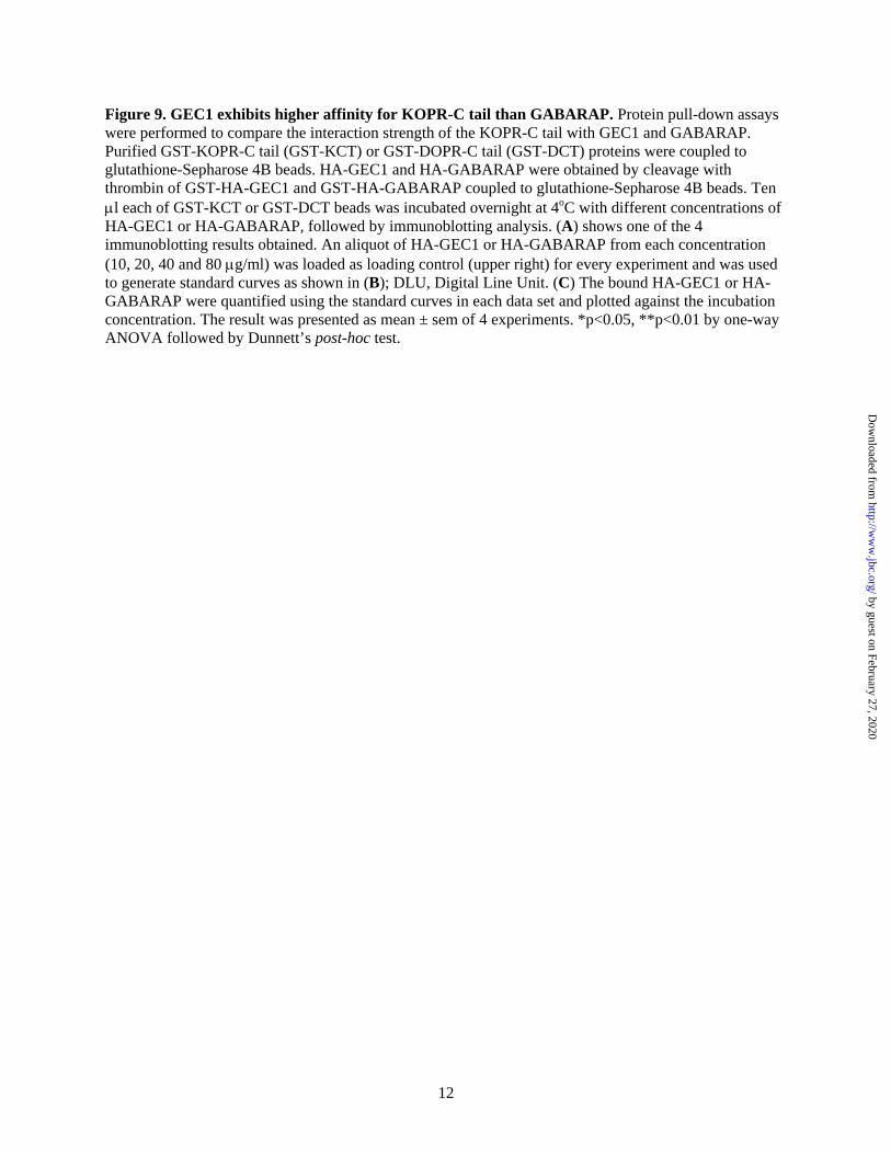
Figure 9. GEC1 exhibits higher affinity for KOPR-C tail than GABARAP. Protein pull-down assays were performed to compare the interaction strength of the KOPR-C tail with GEC1 and GABARAP. Purified GST-KOPR-C tail (GST-KCT) or GST-DOPR-C tail (GST-DCT) proteins were coupled to glutathione-Sepharose 4B beads. HA-GEC1 and HA-GABARAP were obtained by cleavage with thrombin of GST-HA-GEC1 and GST-HA-GABARAP coupled to glutathione-Sepharose 4B beads. Ten l each of GST-KCT or GST-DCT beads was incubated overnight at 4oC with different concentrations of HA-GEC1 or HA-GABARAP, followed by immunoblotting analysis. (A) shows one of the 4 immunoblotting results obtained. An aliquot of HA-GEC1 or HA-GABARAP from each concentration (10, 20, 40 and 80 g/ml) was loaded as loading control (upper right) for every experiment and was used to generate standard curves as shown in (B); DLU, Digital Line Unit. (C) The bound HA-GEC1 or HA-GABARAP were quantified using the standard curves in each data set and plotted against the incubation concentration. The result was presented as mean ± sem of 4 experiments. *p<0.05, **p<0.01 by one-way ANOVA followed by Dunnett’s post-hoc test.
12
by guest on February 27, 2020http://w
ww
.jbc.org/D
ownloaded from
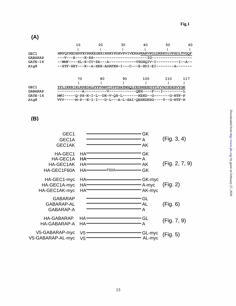
Fig.1 Fig.1
(A)
(B)
GKGEC1
AKGEC1AKAGEC1A
GKHA-GEC1 HAAHA-GEC1A HAHAAKHA-GEC1AK HA
GK-mycHA-GEC1-myc HAA-mycHAAK-mycHA
GLGABARAPALGABARAP-AL
GL-mycV5-GABARAP-myc V5V5
HA-GEC1A-mycHA-GEC1AK-myc
V5-GABARAP-AL-myc
GLHA-GABARAPAHA-GABARAP-A
HAHA
GKHA-GEC1F60A HA F60A
AGABARAP-A
AL-myc
(Fig. 3, 4)
(Fig. 2, 7, 9)
(Fig. 2)
(Fig. 6)
(Fig. 7, 9)
(Fig. 5)
13
by guest on February 27, 2020http://w
ww
.jbc.org/D
ownloaded from
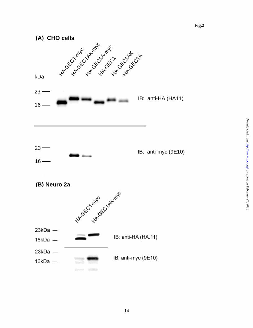
Fig.2
16
23
kDa
16
23
IB: anti-HA (HA11)
IB: anti-myc (9E10)
HA-GEC1-
myc
HA-GEC1A
K-myc
HA-GEC1A
-myc
HA-GEC1
HA-GEC1A
KHA-G
EC1A
(A) CHO cells
(B) Neuro 2a
14
by guest on February 27, 2020http://w
ww
.jbc.org/D
ownloaded from
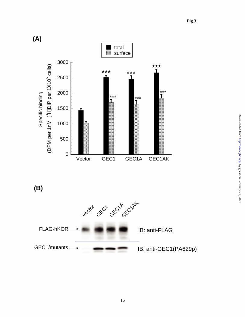
Fig.3
0
500
1000
1500
2000
2500
3000
Vector GEC1 GEC1A GEC1AK
totalsurface
Spe
cific
bin
ding
(DP
M p
er 1
nM
[3H
]DIP
per
1X
105 c
ells
)
*** ******
*** ******
(A)
(B)
GEC1/mutants
FLAG-hKOR
Vecto
r
GEC1
GEC1AK
GEC1A
IB: anti-FLAG
IB: anti-GEC1(PA629p)
15
by guest on February 27, 2020http://w
ww
.jbc.org/D
ownloaded from
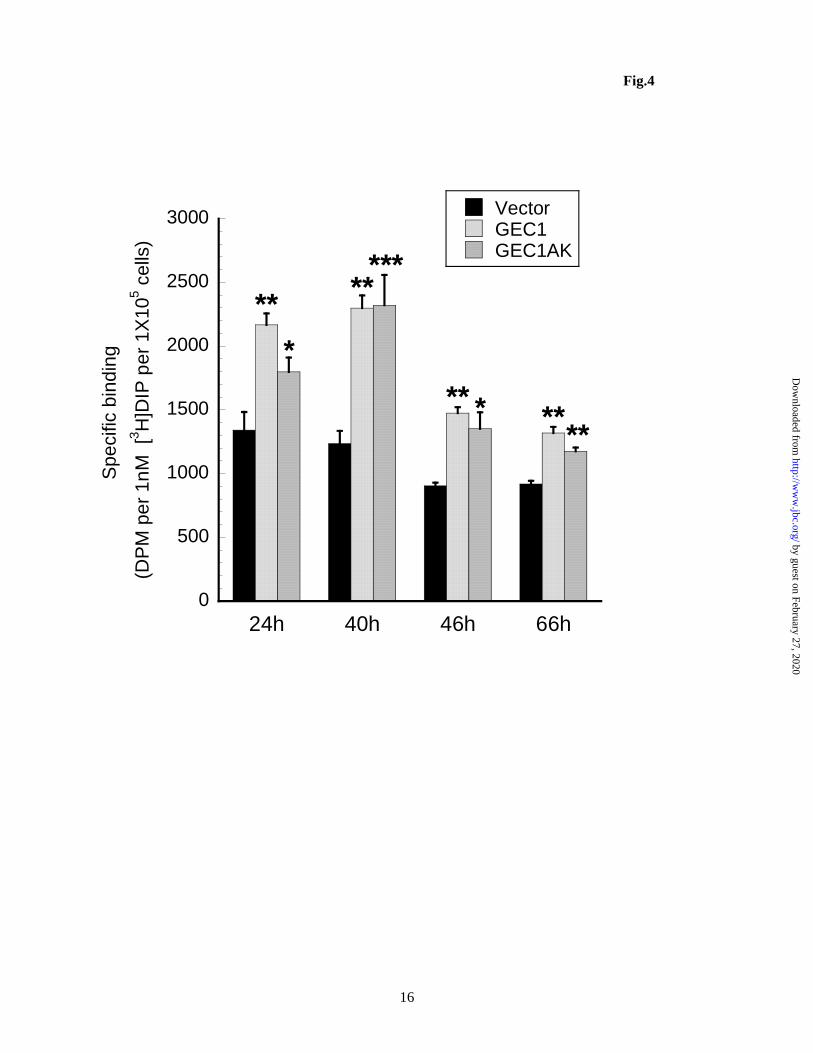
Fig.4
0
500
1000
1500
2000
2500
3000
24h 40h 46h 66h
VectorGEC1GEC1AK
Spe
cific
bin
ding
(DP
M p
er 1
nM
[3
H]D
IP p
er 1
X10
5 c
ells
)
*
*
****
****
***
**
16
by guest on February 27, 2020http://w
ww
.jbc.org/D
ownloaded from
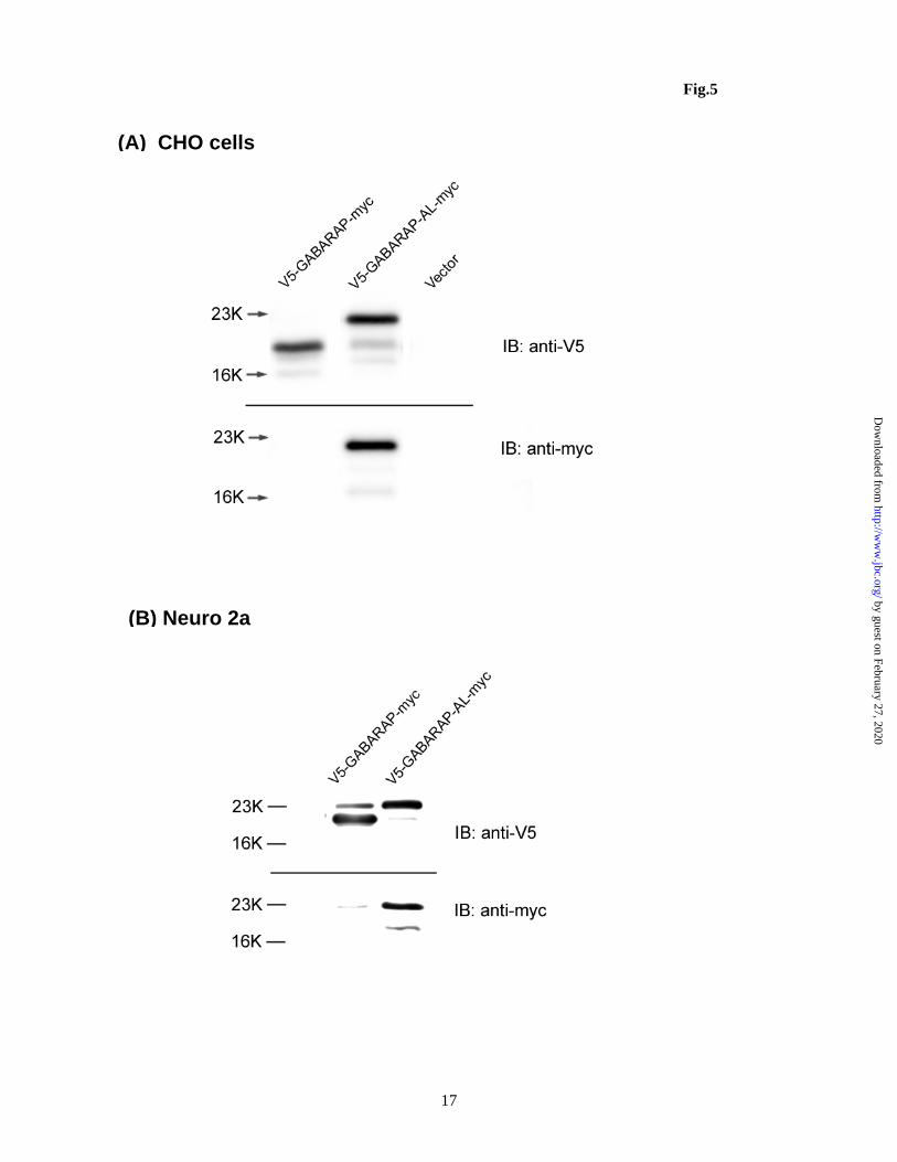
Fig.5
(A) CHO cells
(B) Neuro 2a
17
by guest on February 27, 2020http://w
ww
.jbc.org/D
ownloaded from
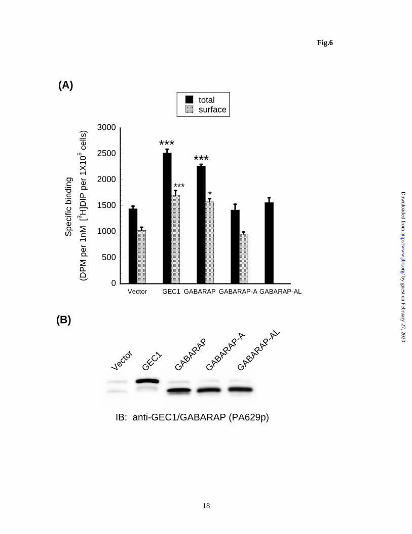
Fig.6
0
500
1000
1500
2000
2500
3000
Vector GEC1 GABARAP GABARAP-A GABARAP-AL
totalsurface
Spe
cific
bin
ding
(DP
M p
er
1nM
[3H
]DIP
per
1X
105 c
ells
)
******
****
(A) (B)
Vecto
r
GEC1
GABARAP
GABARAP-AL
GABARAP-A
IB: anti-GEC1/GABARAP (PA629p)
18
by guest on February 27, 2020http://w
ww
.jbc.org/D
ownloaded from

Fig.7
B
D
C
A
E
19
by guest on February 27, 2020http://w
ww
.jbc.org/D
ownloaded from
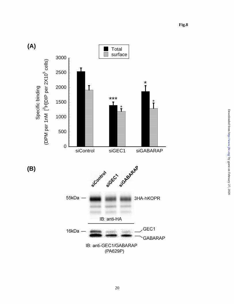
Fig.8 (A)
(B)
0
500
1000
1500
2000
2500
3000
siControl siGEC1 siGABARAP
Totalsurface
Spe
cific
bin
ding
(DP
M p
er 1
nM [
3H
]DIP
per
2X
105 c
ells
)
***
*
**
20
by guest on February 27, 2020http://w
ww
.jbc.org/D
ownloaded from
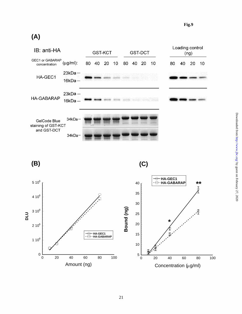
Fig.9
21
(A)
(B) (C)
5
10
15
20
25
30
35
40
0 20 40 60 80 100
HA-GEC1HA-GABARAP
Bo
un
d (
ng
)
Concentration (g/ml)
*
**
0
1 106
2 106
3 106
4 106
5 106
0 20 40 60 80 100
HA-GEC1HA-GABARAP
DL
U
Amount (ng)
by guest on February 27, 2020http://w
ww
.jbc.org/D
ownloaded from
Chongguang Chen, Yulin Wang, Peng Huang and Lee-Yuan Liu-Chenmicrotubules-associated proteins, on kappa opioid receptor expression
Effects of C-terminal modifications of GEC1 and GABARAP, two
published online March 9, 2011J. Biol. Chem.
10.1074/jbc.M111.230896Access the most updated version of this article at doi:
Alerts:
When a correction for this article is posted•
When this article is cited•
to choose from all of JBC's e-mail alertsClick here
by guest on February 27, 2020http://w
ww
.jbc.org/D
ownloaded from