Embed Size (px)
Citation preview
Effects of acute inflammation on plasma retinol, retinol-binding protein, and its mRNA in the liver and kidneys of vitamin A-sufficient rats
Francisco J. Rosales,*it Steven J. Ritter,t Reza Zolfaghari,**t John Edgar Smith,? and A. Catharine Rossl,**t
Department of Biochemistry,* Medical College of Pennsylvania, Philadelphia, PA 19129, and Department of Nutrition,+ Pennsylvania State University, University Park, PA 16802
Abstract The acute inflammatory response to tissue injury and infection is associated with low concentrations of plasma retinol and its specific transport proteins, retinol-binding pro- tein (RBP) and transthyretin (TTR). To examine the kinetics and mechanism of hyporetinemia, we have induced acute inflammation with lipopolysaccharide (LPS, from Pseudo- monas aeruginosa) in rats with adequate stores of vitamin A. Twenty-four h after treatment with LPS (50 pg i.p. per 100 g body weight) or saline and food withdrawal, plasma retinol equalled 0.72 +_ 0.06 pmol/L (mean f SEM) in five LPS- treated rats versus 1.35 k 0.1 pmol/L in five saline-treated rats (P < 0.01). Plasma, liver, and kidney RBP and T T R concentra- tions were also significantly reduced, but liver and kidney retinol concentrations did not differ between treatment groups. The relative abundance of RBP mRNA in liver (LPS treatment compared to saline treatment) was reduced as early as 12 h (0.44 t 0.15, n = 4 pairs, P < 0.02), and continued to be reduced at 24 h (0.57 k 0.12, n = 5 pairs, P < 0.02). In the kidney this ratio did not change significantly due to LPS treatment. The relative abundance of cellular retinol-binding protein (CRBP) mRNA in liver and kidney also was not af- fected by LPS treatment. I We infer from these data that inflanimation-induced hyporetinemia results from a 'reduc- tion in the hepatic synthesis of RBP and secretion of the retinol-RBP complex. Moreover, the results imply that plasma retinol concentration is a poor indicator of vitamin A status during inflammation.-Rosales, F. J., S. J. Ritter, R. Zol- Faghari, J. E. Smith, and A. C. Ross. Effects of acute inflani- mation on plasma retinol, retinol-binding protein, and its inRNA in the liver and kidneys of vitamin A-sufficient rats.). Lipid Res. 1996. 37: 962-971.
Supplementary key words endotoxin inflammatory response lipopolysacrharide RNase protection assay cellular retinol-binding prorein
Retinol (vitamin A alcohol) is transported in plasma by a specific transport protein, retinol-binding protein [(RBP), for review see references 1,2] that is synthesized primarily by liver parenchymal cells and secreted as the holo-RBP complex (3). In plasma, RBP (21 kDa) circu- lates in association with a larger plasma protein, trans-
thyretin (TTR, 52 kDa) (2). Numerous studies have demonstrated tight homeostatic control of plasma reti- nol and RBP concentrations, but the mechanisms that control them are still not well understood. Retinol is known to recirculate between the plasma compartment, liver, and extrahepatic tissues several times before being irreversibly degraded (4). Although the mRNA for RBP is expressed at the highest level in liver, it is also found at lower relative abundance in the kidney (5-10% com- pared to liver), adipose tissue, and in several other organs (5); this distribution suggests that numerous organs could be involved in RBP synthesis and retinol homeostasis.
Hyporetinemia is observed in several pathophysi- ological conditions including a primary deficiency of vitamin A, protein-energy malnutri tion, certain liver and kidney diseases, and in the absence of TI'R (6-8). Hy- poretinemia has also been reported to accompany infec- tions or inflammatory diseases (9). For example, hy- poretinemia (which, in children, is generally defined as a plasma retinol concentration of I 0.7 pmol/L) has been reported during the acute phase of measles or syncytial virus infections in children in the United States where vitamin A status is presumed to be adequate (10,
Hyporetinemia also has been reported in children in developing countries, where vitamin A status is more likely to be low, in association with acute diarrhea (12), measles (1 3), malaria (14), or multiple morbidities (15). Infection-associated hyporetinemia appears to be unre- lated to the infectious agent, or age and gender of the
11).
Abbreviations: RBP, retinol-binding protein; TTK. transthyretin;
'To whom correspondence should he addressed. IPS, lipopolysaccharide; CKBP, cellular retinol-binding prorein.
962 Journal of Lipid Research Volume 37, 1996
by guest, on May 16, 2018
ww
w.jlr.org
Dow
nloaded from
patient, but is most severe during the acute phase of infection and usually is associated with a poor prognosis. Neither the cause of infection-induced hyporetinemia nor its relationship to vitamin A status is well under- stood. Some investigators have considered a low plasma retinol concentration during inflammation to be an indicator of acute vitamin A depletion (10,ll). A plasma retinol concentration below 0.7 pmol/L is a recom- mended indicator of poor vitamin A nutriture in popu- lation studies ( 16), because concentrations below this level are usually associated with clinical manifestations of vitamin A deficiency (xerophthalmia) (17). Thus, infection-associated hyporetinemia is of concern both because it is hypothesized to precipitate an acute defi- ciency of retinol during infection and because it may confound the interpretation of plasma retinol levels in assessing vitamin A status (18).
Several mechanisms have been proposed to explain infection-induced hyporetinemia, including an increase in renal clearance and urinary excretion of retinol and RBP (19), an increase in zinc utilization and a sub- sequent reduction in liver protein synthesis (20), and a transient redistribution of plasma retinol into the ex- travascular space (21). Alternatively, it has been sug- gested that plasma retinol concentrations decrease as a result of the acute inflammatory response (22). The acute inflammatory response is a physiological conse- quence to tissue injury (e.g., infection) characterized by the immediate secretion of pro-inflammatory cytokines and their subsequent effect on the metabolism of liver, among other tissues. Besides alterations in carbohydrate and lipid metabolism (23-26), the synthesis of certain liver proteins is enhanced (positive acute phase pro- teins), while that of others is inhibited (negative acute phase proteins). Previously, Schreiber et al. (27) used turpentine oil to induce inflammation in rats and stud- ied the hepatic synthesis of positive and negative acute phase proteins. This investigation showed compensa- tory changes in protein synthesis between positive and negative acute phase proteins during the acute phase of inflammation, while the overall rate of liver protein synthesis was maintained. For most acute phase pro- teins, changes in synthesis were associated with altera- tions in the levels of mRNA. In the case of RBP and TTR, their respective mRNA levels were reduced (27). How- ever, the consequence of inflammation on retinol trans- port was not studied.
As a comprehensive approach to understanding the kinetics and the mechanism of inflammation-induced hyporetinemia, we have used a pathogen-free model in which inflammation is induced with lipopolysaccharide (LPS) in rats having adequate stores of vitamin A. This model avoids the confounding effects of pathogens or preexisting vitamin A deficiency. We have determined
the effects of inflammation on plasma and tissue levels of retinol, RBP, and TI'R, and the liver and kidney levels of RBP and cellular retinol-binding protein (CRBP) "As. The results of these studies provide evidence that hyporetinemia is related to a reduction in the synthesis of RBP by liver, and that hyporetinemia may occur during inflammation even when tissue vitamin A reserves are substantial.
MATERIALS AND METHODS
Materials Chemical reagents were from Fisher Chemical (Pitts-
burgh, PA), Sigma (St. Louis, MO), or Promega (Madi- son, WI) and, when appropriate, were of molecular biology grade. RNases were purchased from Boehringer Mannheim (Indianapolis, IN), restriction enzymes, po- lymerases, and DNases from Promega (Madison, WI) and [a-S*P]UTP (3000 Ci/mmol) from DuPont New England Nuclear (Boston, MA). The antisense RBP mRNA probe was synthesized from a 548 bp DNA insert of the rat RBP gene, and that for the antisense CRBP probe from a 286 bp DNA insert of the rat CRBP gene, both generously provided by Dr. Dianne R. Soprano (Temple University School of Medicine, Philadelphia, PA). A Factin probe was synthesized from a linearized pTRI-P-Actin-125-rat DNA (Ambion, Inc., Austin, TX). The LPS used was derived from P. uerugznosu, F-D type 1, (List Biological Laboratories, Inc., Campbell, CA). Buffers and solutions were prepared as recommended in Sambrook, Fritsch, and Maniatis (28).
Animals and experimental designs Pathogen-free rats of the Lewis or Sprague-Dawley
strains were purchased from Charles River Breeding Laboratories (Kingston, NY). All experimental proto- cols were in compliance with the Guide for the Care and Use of Laboratory Animals and were approved by the Animal Care and Use Committee of the Medical College of Pennsylvania and Pennsylvania State University. In the first experiment, 12 male Lewis rats weighing 300-350 g were housed in individual plastic cages with free access to water and a vitamin A-sufficient diet (Purina rodent chow, Ralston Purina, St. Louis, MO) in a room maintained at 22°C with a 12 h light-12 h dark cycle. Six rats were allocated to the LPS-treatment group, and six to the saline-treated control group. After an initial blood sample (-0.5 mL) was collected by venipuncture in heparinized syringes from a tail vein for determination of plasma retinol, a single dose of LPS was injected i.p. in unanesthetized rats. The lyophilized LPS had been reconstituted in sterile saline to provide
Rosales et al. Mechanism of inflammation-induced hyporetinemia 963
by guest, on May 16, 2018
ww
w.jlr.org
Dow
nloaded from
a dose of 50 pg of LPS/lOO g of body weight in a 1-mL bolus. Sequential bleedings were conducted by venipuncture for determination of plasma retinol con- centrations at 2, 5, 8, 24, and 72 h post-dosing. During this time, the rats were observed for possible signs of endotoxic shock (e.g., shivering or lethargy).
For the second, third, and fourth experiments, male Sprague-Dawley rats weighing between 250 to 300 g were housed in pairs in plastic cages and maintained under conditions similar to those in experiment 1. At time zero in the second experiment, rectal temperature was determined, 6 cm from the anal opening, with a tele-thermometer and a rectal temperature probe for small animals (Yellow Spring Instruments Co., Inc., Yellow Spring, OH), and an initial blood sample was obtained as previously described. Then, one rat in each pair (n = 5 pairs) was injected with LPS and the other with saline. Subsequently, rectal temperature was monil tored every hour for the first 7 h, and at 12 and 24 h, while small blood samples were obtained at 7 and 12 h. At the end of the experiment, 24 h after administration of LPS, rats were killed by CO2 asphyxiation and 5 mL of blood was drawn from the inferior vena cava. The livers and kidneys were excised, blotted, immediately frozen on solid COS, and stored at -70°C until they could be processed. In experiments 3 and 4, the same design was followed except that, after rats were treated with LPS or saline, their food was withheld for the duration of the 6, 12, or 24h experiments. Blood samples and tissues were collected at the time the animals were euthanized.
Tissue retinol analysis The concentrations of total retinol in plasma and in
liver and kidney homogenates were determined after tissue saponification by high performance liquid chro- matography using trimethylmethoxyphenyl-retinol as an internal standard (29).
Tissue RBP and l T R analysis The concentrations of RBP and TTR in plasma and
homogenates of liver and kidney were determined by sensitive and specific radioimmunoassays, as described previously by Smith, Muto, and Goodman (30).
RNA isolation and analysis Total RNA was isolated from the liver and kidney
tissues of individual rats using 4 mol/L guanidine salts (31). The concentration of RNA in each sample was determined by spectrophotometry at 260 nm and its quality was assessed by the ratio of absorbance at 260 nm:280 nm, and by examining the fluorescence of ethidium bromide-treated RNA after agarose/formal- dehyde gel electrophoresis (32).
Liver and kidney RBP, CRBP and pactin mRNAs were quantified by an RNase protection assay, essen- tially as described by Soprano et al. (33). The specific radioactivity of the cRNA probes was adjusted to com- pensate for differences in their size, and their integrity after labeling was determined by electrophoresis in a 5% polyacrylamide/8 M urea gel. For determination of RBP, CRBP and p-actin mRNAs, 2 pg of liver total RNA and 20 pg of kidney total RNA from each LPS- or saline- treated rat were dried in a Speed Vac (Savant Instru- ments Inc., Farmingdale, NY). After solution hybridiza- tion (with RBP alone in experiment 2 or simultaneously with the three probes in experiment 4), denatured RNA samples were loaded onto 5% polyacryIamide/8 M urea gels and each gel, for liver and kidney samples, was run for 60 min at 400 mV. The position of the 32P-labeled bands was determined by autoradiography at -70°C using Fuji Medical X-ray film (Fuji Photo Film Co., LTD., Japan) and intensifying screens (Fisher Scientific, Pius- burgh, PA). The results were quantified by counting the total radioactivity of excised bands using the Cerenkov radiation method (without scintillant). In experiment 3, hepatic RBP mRNA from rats killed at 6 and 12 h post-treatment was determined in a single Northern blot analysis. RNA samples (10 pg of total liver RNA) from individual rats were fractionated by electrophoresis on formaldehyde-denaturing agarose gels, and these frac- tionated samples were transferred to Nytran membrane (Schleicher 8c Schuell, Inc., Keene, NH) (32). After hybridization to [32P]dCTP-labeled RBP probe, the membrane was washed and then exposed to X-ray film (Eastman Kodak Co., Rochester, NY) at -70°C. The relative intensities of autoradiographic signals, which were within a linear response range, were quantified by scanning with a ScanMaker IIG densitometer (Microtek Lab, Inc., Redondo Beach, CA) standardized with an NIH freeware computer program (Image 1.56).
Statistical analysis Values are given as the mean k SEM. Comparisons
between sample means were done with Student's t-test, and a two-tailed P-value < 0.05 was considered statisti- cally significant. In some experiments, the relative dif- ference for a specific variable was calculated for each pair of rats as the ratio of the LPS-treated rat to its respective control, and the significance of the ratio's difference from one was determined with a one-sample t-test.
RESULTS
Effect of inflammation on plasma retinol In experiment 1, the kinetics of plasma retinol con-
centration after LPS treatment was determined in Lewis
964 Journal of Lipid Research Volume 37, 1996
by guest, on May 16, 2018
ww
w.jlr.org
Dow
nloaded from
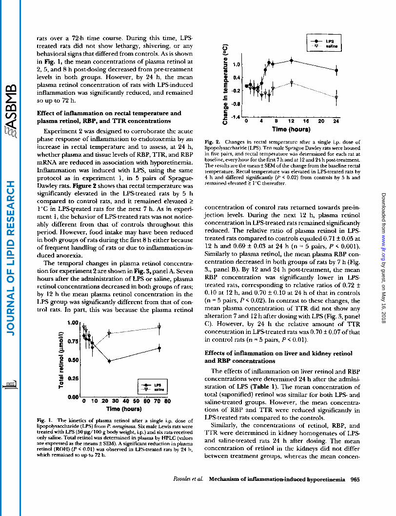
rats over a 72-h time course. During this time, LPS- treated rats did not show lethargy, shivering, or any behavioral signs that differed from controls. As is shown in Fig. 1, the mean concentrations of plasma retinol at 2, 5, and 8 h post-dosing decreased from pre-treatment levels in both groups. However, by 24 h, the mean plasma retinol concentration of rats with LPS-induced inflammation was significantly reduced, and remained so up to 72 h.
Effect of inflammation on rectal temperature and plasma retinol, RBP, and TTR concentrations
Experiment 2 was designed to corroborate the acute phase response of inflammation to endotoxemia by an increase in rectal temperature and to assess, at 24 h, whether plasma and tissue levels of RBP, ITR, and RBP mRNA are reduced in association with hyporetinemia. Inflammation was induced with LPS, using the same protocol as in experiment 1, in 5 pairs of Sprague- Dawley rats. Figure 9 shows that rectal temperature was significantly elevated in the LPStreated rats by 5 h compared to control rats, and it remained elevated 2 1'C in LPS-Teated rats for the next 7 h. As in experi- ment 1, the behavior of LPS-treated rats was not notice- ably different from that of controls throughout this period. However, food intake may have been reduced in both groups of rats during the first 8 h either because of frequent handling of rats or due to inflammation-in- duced anorexia.
The temporal changes in plasma retinol concentra- tion for experiment 2 are shown in Fig. 3, panel A. Seven hours after the administration of LPS or saline, plasma retinol concentrations decreased in both groups of rats; by 12 h the mean plasma retinol concentration in the LPS group was significantly different from that of con- trol rats. In part, this was because the plasma retinol
- o.oo'O 10 20 30'40 50 60 70 80
Time (hours)
Fig. I. The kinetics of plasma retinol after a single i.p. dose of lipopolysaccharide (LPS) from P. amginma. Six male Lewis rats were treated with LPS (50 Clg/lOO g body weight, i.p.) and six rawreceived only saline. Total retinol was determined in plasma by HPLC (values are expressed as the means f SEM). A significant reduction in plasma retinol (ROH) ( P < 0.01) was observed in LPS-treated rats by 24 h, which remained so up to 72 h.
0 -0.81 9
I
r" SF -1.4 0 0 4 8 12 16 20 24
Time (hours)
Fig. 2. Changes in rectal temperature after a single i.p. dose of lipopolysaccharide (LPS). Ten male Sprague Dawley rats were housed in five pairs, and rectal temperature was determined for each rat at baseline, every hour for the first 7 h and at 12 and 24 h post-treatment. The results are the mean f SEM of the change from the baseline rectal temperature. Rectal temperature was elevated in LPStreated rats by 4 h and differed significantly ( P < 0.02) from controls by 5 h and remained elevated 2 1 'C thereafter.
concentration of control rats returned towards pre-in- jection levels. During the next 12 h, plasma retinol concentration in US-treated rats remained significantly reduced. The relative ratio of plasma retinol in LPS- treated rats compared to controls equaled 0.71 f 0.05 at 12 h and 0.69 2 0.03 at 24 h (n = 5 pairs, P < 0.001). Similarly to plasma retinol, the mean plasma RBP con- centration decreased in both groups of rats by 7 h (Fig. 3., panel B). By 12 and 24 h post-treatment, the mean RBP concentration was significantly lower in LPS- treated rats, corresponding to relative ratios of 0.72 _+
0.10 at 12 h, and 0.70 k 0.10 at 24 h of that in controls (n = 5 pairs, P < 0.02). In contrast to these changes, the mean plasma concentration of l T R did not show any alteration 7 and 12 h after dosing with LPS (Fig. 3, panel C). However, by 24 h the relative amount of 'ITR concentration in LPStreated rats was 0.70 k 0.07 of that in control rats (n = 5 pairs, P < 0.01).
Effects of inflammation on liver and kidney retinol and RBP concentrations
The effects of inflammation on liver retinol and RBP concentrations were determined 24 h after the admini- stration of LPS (Table 1). The mean concentration of total (saponified) retinol was similar for both LPS- and saline-treated groups. However, the mean concentra- tions of RBP and TTR were reduced significantly in LPS-treated rats compared to the controls.
Similarly, the concentrations of retinol, RBP, and TTR were determined in kidney homogenates of LPS and saline-treated rats 24 h after dosing. The mean concentration of retinol in the kidneys did not differ between treatment groups, whereas the mean concen-
Rosales et al. Mechanism of Mammation-induced hyporetinemia 965
by guest, on May 16, 2018
ww
w.jlr.org
Dow
nloaded from
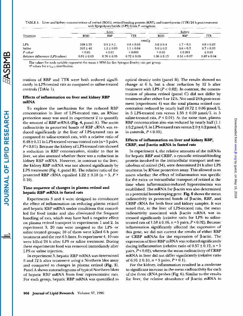
TABLE 1. Liver and kidney concentrations of retinol (ROH), retinol-binding protein (RBP), and transthyretin (TTR) 24 h post-treatment with lipopolysaccharide (LPS) from P. aeruginosa
Lker Kidney Treatment ROH RBP TTR ROH RBP TTR
nmovg LPS 538 f 29 0.9fO.l 0.8 f 0.04 5.6 f 0.4 1.7 f 0.1 0.6 * 0.03 Saline 593 f 42 1.2 f 0.03 1.1 f 0.04 5.0 f 0.2 3.6 * 0.3 0.7 f 0.03 P value" 0.05 < 0.01 < 0.001 0.05 5 0.001 5 0.01 Relative difference (LPS:saline) 0.91 f 0.03 0.76 f 0.05 0.72 f 0.05 1.06 f 0.13 0.51 f 0.07 0.82 f 0.04
The values for each variable represent the mean f SEM for five Sprague-Dawley rats per group. "Pvalues for a tH.u,2 distribution.
trations of RBP and TTR were both reduced signifi- cantly in LPS-treated rats as compared to saline-treated controls (Table 1).
Effects of inflammation on liver and kidney RBP mRNA
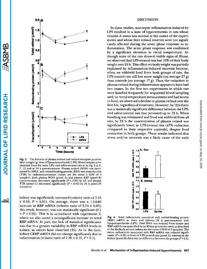
To explore the mechanism for the reduced RBP concentration in liver of LPS-treated rats, an RNase protection assay was used in experiment 2 to quantify the amount of RBP mRNA (Fig. 4, panel A). The mean radioactivity in protected bands of RBP cRNA was re- duced significantly in the liver of LPS-treated rats as compared to saline-treated rats, with a relative ratio of 0.48 f 0.11 in LPS-treated versus control rats (n = 5 pairs, P < 0.01). Because the kidney of LPS-treated rats showed a reduction in RBP concentration, similar to that in liver, we also assessed whether there was a reduction in kidney RBP mRNA. However, in contrast to the liver, the kidney RBP mRNA was not altered significantly by LPS treatment (Fig. 4, panel B). The relative ratio of the protected RBP cRNA equalled 1.22 k 0.18 (n = 5, P 0.10).
Time sequence of changes in plasma retinol and hepatic RBP mRNA in fasted rats
Experiments 3 and 4 were designed to corroborate the effect of inflammation on reducing plasma retinol and hepatic RBP mRNA under conditions that control- led for food intake and also eliminated the frequent handling of rats, which may have had a negative effect on plasma retinol transport in experiments 1 and 2. In experiment 3, 20 rats were assigned to the LPS- or saline-treated groups; 10 of them were killed 6 h post- treatment and the rest 6 h later. In experiment 4,lO rats were killed 24 h after LPS or saline treatment. During these experiments food was removed immediately after LPS or saline injection.
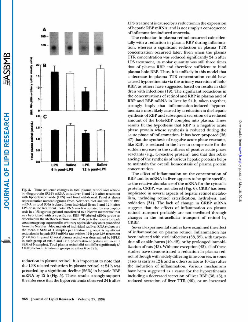
In experiment 3, hepatic RBP mRNA was determined 6 and 12 h after treatment using a Northern blot assay and compared to changes in plasma retinol (Fig. 5). Panel A shows autoradiograms of typical Northern blots of hepatic RBP mRNA from four representative rats. For each group, hepatic RBP mRNA was quantified as
optical density units (panel B). The results showed no change at 6 h, but a clear reduction by 12 h after treatment with LPS (P < 0.02). In contrast, the concen- tration of plasma retinol (panel C) did not differ by treatment after either 6 or 12 h. Not until 24 h post-treat- ment (experiment 4) was the total plasma retinol con- centration reduced by nearly half (0.72 f 0.06 ymol/L in 5 LPS-treated rats versus 1.35 f 0.09 pmol/L in 5 saline-treated rats, P < 0.01). At the same time, plasma RBP concentration also was reduced by nearly half ( 1 . 1 f 0.2 pmol/L in LPS-treated rats versus 2.0 & 0.2 pmol/L in controls, P < 0.01).
Effects of inflammation on liver and kidney RBP, CRBP, and p-actin mRNA in fasted rats
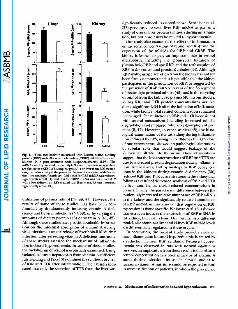
In experiment 4, the relative amounts of the mRNAs for hepatic RBP and CRBP, a cytosolic retinoid-binding protein involved in the intracellular transport and me- tabolism of retinol (34), were determined 24 h after LPS treatment by RNase protection assay. This allowed us to assess whether the effect of inflammation was specific for the extra- or intracellular transport of retinol at the time when inflammation-induced hyporetinemia was established. The mRNA for pactin was also determined as a potential housekeeping gene. Fig. 6 shows the mean radioactivity in protected bands of p-actin, RBP, and CRBP cRNA for both liver and kidney samples. It was noted that, in the liver of LPS-treated rats, the mean radioactivity associated with p-actin mRNA was in- creased significantly (relative ratio for LPS- to saline- treated rats of 1.58 f 0.16, n = 5 pairs, P < 0.02). Because inflammation Significantly affected the expression of this gene, we did not correct the results of either RBP or CRBP mRNAs for the expression of p-actin. The expression of liver RBP mRNA was reduced significantly during inflammation (relative ratio of 0.57 f 0.12, n = 5 pairs, P < 0.02), whereas the mean radioactivity of CRBP mRNA in liver did not differ significantly (relative ratio of 0.91 f 0.10, n = 5 pairs, P
For the kidney, inflammation resulted in a moderate to significant increase in the mean radioactivity for each of the three cRNA probes (Fig. 6). Similar to the results for liver, the relative abundance of pactin mRNA in
0.1).
966 Journal of Lipid Research Volume 37, 1996
by guest, on May 16, 2018
ww
w.jlr.org
Dow
nloaded from
DISCUSSION
= c. 0.5 -
0.0 1
' * [C
O L 0 ' 4 ' li ' 12 ' 16 ' 20 ' 24
Tlme (hours)
Fig. 3. The kinetics of plasma retinol and retinol transport proteins after a single i.p. dose of lipopolysaccharide (LPS). Blood samples were obtained from the same LPS- and saline-treated rats as in Fig. 2 at 0, 7, 12 and at 24 h post-treatment. Plasma retinol (ROH) was deter- mined by HPLC and retinol binding-protein (RBP) and transthyretin ('ITR) by radioimmunoassays (values are the mean f SEM of 5 samples). Both plasma ROH (panel A) and plasma RBP (panel B) concentrations decreased significantly (P I 0.02) by 12, and plasma 'ITR (panel C) decreased significantly (P < 0.02) by 24 h post-LPS treatment.
kidney was significantly increased (relative ratio of 1.55 f 0.10, P < 0.01). On average, there was a 1.5-fold increase in RBP mRNA (relative ratio of 2.54 +_ 0.60); this result, however, was not statistically significant (0.1 < P < 0.05). This is in accordance with experiment 2, where we also noted a nonsignificant increase in renal RBP mRNA. In part, the lack of statistical significance was due to a greater variability in RBP mRNA levels in kidney, as others have observed (35). As in the liver, kidney CRBP mRNA did not change significantly due to inflammation (relative ratio of 1.30 f 0.16, P 0.1).
In these studies, non-septic inflammation induced by LPS resulted in a state of hyporetinemia in rats whose vitamin A status was normal at the outset of the experi- ments and whose liver retinol reserves were not signifi- cantly affected during the acute phase response to in- flammation. The acute phase response was confirmed by a significant elevation in rectal temperature. Al- though none of the rats showed visible signs of illness, we observed that LPS-treated rats lost 10% of their body weight over 24 h. This effect on body weight was partially explained by inflammation-induced anorexia because, when we withheld foad from both groups of rats, the LPS-treated rats still lost more weight (on average 27 g) than controls (on average 17 g). Thus, the reduction in plasma retinol during inflammation appears to have had two causes. In the first two experiments in which rats were handled frequently for sequential blood sampling and/or rectal temperature measurement and had access to food, we observed a declirle in plasma retinol over the first 8 h, regardless of treatment. However, by 12 h there was a statistically significant difference between the LPS- and saline-treated rats that persisted up to 72 h. When handling was minimized and food was withheld from all rats, by 24 h the concentration of plasma retinol was significantly lower in LPS-treated rats (47% reduction compared to their respective controls), despite food restriction in both groups. These results indicated that stress and/or anorexia was a likely cause of the early
1.2
d
m a 0.0 1
T
LPS Saline
Fig. 4. Total radioactivity associated with retinol-binding protein (RBP) mRNA in livers and kidneys 24 h post-treatment with lipopolysaccharide (LPS). Total RNA was isolated from tissues and RBP mRNA was quantified by an RNase protection assay as described in the Methods section (values are the mean f SEM of 5 samples). The mean radioactivity associated with RBP mRNA was reduced signifi- cantly (P < 0.02) in livers of LPStreated rats (panel A), whereas in the kidney (panel Bf there was no difference between the groups (P 0.5).
Rosales et al. Mechanism of inflammation-induced hyporetinemia 967
by guest, on May 16, 2018
ww
w.jlr.org
Dow
nloaded from
A =- m F - .
'8s-
I-
- . .
T
= "[C A
li LPS satin0 LPS saline 6 h port-LPS 12 h p0St-L-
Fig. 5. Time sequence changes in total plasma retinol and retinol- binding-protein (RBP) mRNA in rat liver 6 and 12 h after treatment with lipopolysaccharide (LPS) and food withdrawal. Panel A shows representative autoradiograms from Northern blot analysis of RBP mRNA in total RNA isolated from individual liven 6 and 12 h after LPS or saline treatment. Total RNA was fractionated by electropho- resis in a 1% agarose gel and transferred to a Nytran membrane that was hybridized with a specific rat RBP J2P-labeled cDNA probe as described in the Methods section. Panel B depicts the results for each treatment group expressed in arbitrary optical density units quantified from the Northern blot analysis of individual rat liver RNA (values are the mean f SEM of 4 samples per treatment group). A significant reduction in hepatic RBP mRNA was evident 12 h post-LPS treatment ( P < 0.02). In panel C, total plasma retinol was determined by HPLC in each group of rats 6 and 12 h post-treatment (values are mean f SEM of 5 samples). Total plasma retinol did not differ significantly ( P > 0.05) between treatment groups at either 6 or 12 h.
reduction in plasma retinol. It is important to note that the LPS-related reduction in plasma retinol at 24 h was preceded by a significant decline (56%) in hepatic RBP mRNA by 12 h (Fig. 5). These results strongly support the inference that the hyporetinemia observed 24 h after
968 Journal of Lipid Research Volume 37, 1996
LPS treatment is caused by a reduction in the expression of hepatic RBP mRNA, and is not simply a consequence of inflammation-induced anorexia.
The reduction in plasma retinol occurred coinciden- tally with a reduction in plasma RBP during inflamma- tion, whereas a significant reduction in plasma TTR concentration occurred later. Even when the plasma TTR concentration was reduced significantly 24 h after LPS treatment, its molar quantity was still three times that of plasma RBP and therefore sufficient to bind plasma holo-RBP. Thus, it is unlikely in this model that a decrease in plasma TI'R concentration could have caused hyporetinemia via the urinary excretion of holo- RBP, as others have suggested based on results in chil- dren with infections (19). The significant reductions in the concentrations of retinol and RBP in plasma and of RBP and RBP mRNA in liver by 24 h, taken together, strongly imply that inflammation-induced hyporet- inemia is most likely caused by a reduction in the hepatic synthesis of RBP and subsequent secretion of a reduced amount of the holo-RBP complex into plasma. These results fit the hypothesis that RBP is a negative acute phase protein whose synthesis is reduced during the acute phase of inflammation. It has been proposed (36, 37) that the synthesis of negative acute phase reactants, like RBP, is reduced in the liver to compensate for the sudden increase in the synthesis of positive acute phase reactants (e.g., C-reactive protein), and that this rebal- ancing of the synthesis of various hepatic proteins helps to maintain the overall homeostasis of plasma protein concentration.
The effect of inflammation on the concentration of RBP and its mRNA in liver appears to be quite specific, as the relative abundance of the mRNA for the cytosolic protein, CRBP, was not altered (Fig. 6). CRBP has been implicated in several aspects of hepatic retinol metabo- lism, including retinol esterification, hydrolysis, and oxidation (34). The lack of change in CRBP mRNA suggests that the effects of inflammation on plasma retinol transport probably are not mediated through changes in the intracellular transport of retinol by CRBP.
Several experimental studies have examined the effect of inflammation on plasma retinol. Inflammation has been induced with viral infections (38,39), with turpen- tine oil or skin bums (40-42), or by prolonged immobi- lization of rats (43). With one exception (42), all of these studies have demonstrated a reduction in plasma reti- nol, although with widely differing time courses, in some cases as early as 12 h and in others as late as 10 days after the induction of inflammation. Various mechanisms have been suggested as a cause for the hyporetinemia including a decreased secretion of liver RBP (38,43), a reduced secretion of liver l T R (40), or an increased
by guest, on May 16, 2018
ww
w.jlr.org
Dow
nloaded from
Kidney -
- 1 h
f A
LPS s 8 l l n LPS saline
Fig. 6. Total radioactivity associated with pactin, retinol-binding protein (RBP) and cellular retinol-binding (CRBP) mRNA in livers and kidneys 24 h post-treatment with lipopolysaccharide (LPS). The mRNAs were quantified by a multiple RNase protection assay (values are the mean f SEM of 5 samples/group). For liver from LPStreated rats, the radioactivity in the protected fragment associated with p-actin was increased significantly ( P < 0.01). that for RBP mRNA was reduced significantly (P < 0.01). and that for CRBP mRNA was not affected ( P
0.1). For kidney from LPS-treated rats, pactin mRNA was increased significantly (P < 0.01).
utilization of plasma retinol (38, 39, 41). However, the results of some of these studies may have been con- founded by simultaneously inducing vitamin A defi- ciency and by viral infections (38,39), or by varying the amounts of dietary protein (43) or vitamin A (41, 42). Although these studies have provided valuable informa- tion on the intestinal absorption of vitamin A during viral infection or on the release of liver holo-RBP during infection after refeeding vitamin A-deficient rats, none of these studies assessed the mechanism of inflamma- tion-induced hyporetinemia. In some of these studies, the metabolism of retinol was partially examined. Using isolated cultured hepatocytes from vitamin A-sufficient rats, Felding and Fex (40) examined the synthesis in vitro of RBP and TTR after inflammation. Their results indi- cated that only the secretion of TTR from the liver was
significantly reduced. As noted above, Schreiber et al. (27) previously assessed liver RBP mRNA as part of a study of overall liver protein synthesis during inflamma- tion, but not how it may be related to hyporetinemia.
Our study also examined the effect of inflammation on the renal concentrations of retinol and RBP and the expression of the mRNAs for RBP and CRBP. The kidney is known to play an important role in retinol metabolism, including the glomerular filtration of plasma holo-RBP and apo-RBP, and the reabsorption of RBP in the convoluted proximal tubules (44). Although RBP synthesis and secretion from the kidney has not yet been firmly demonstrated, it is plausible that the kidney participates in the production of RBP, as suggested by the presence of RBP mRNA in cells of the S3 segment of the straight proximal tubules (45), and in the recycling of retinol from the kidney to plasma (46). In our studies, kidney RBP and TTR protein concentrations were re- duced significantly 24 h after the induction of inflamma- tion, while kidney total retinol concentration remained unchanged. The reduction in RBP and TTR is consistent with several mechanisms including increased tubular degradation and impaired tubular reabsorption of pro- teins (2, 47). However, in other studies (48), the histo- logical examination of the rat kidney during inflamma- tion induced by LPS, using 5- to 10-times the LPS dose of our experiments, showed no pathological alterations of tubular cells that would suggest leakage of the glomerular filtrate into the urine. These observations suggest that the low concentrations of RBP and TTR are due to increased protein degradation during inflamma- tion. Alternatively, and by analogy to RBP concentra- tions in the kidneys during vitamin A deficiency (30), reduced RBP and TTR concentrations in the kidney may follow as a result of decreased synthesis of RBP and TTR in liver and, hence, their reduced concentrations in plasma. Finally, the paradoxical difference between the moderately increased relative abundance of RBP mRNA in the kidney and the significantly reduced abundance of RBP mRNA in liver confirm that regulation of RBP expression is tissue specific. Whitman et al. (35) showed that estrogen induces the expression of RBP mRNA in rat kidney, but not in liver. Our results, in a different model, also show that liver and kidney RBP mRNA levels are differentially regulated in these organs.
In conclusion, the present study provides evidence that inflammation-induced hyporetinemia is caused by a reduction in liver RBP synthesis. Because hyporet- inemia was observed in rats with normal vitamin A reserves, an implication from these results is that plasma retinol concentration is a poor indicator of vitamin A status during infection. Its use in clinical studies to measure vitamin A nutriture could be expected to lead to misclassification of patients, in whom the prevalence
Rosales et aZ. Mechanism of inflammation-induced hyporetinemia 969
by guest, on May 16, 2018
ww
w.jlr.org
Dow
nloaded from
or severity of vitamin A deficiency may be overesti- mated. We recommend that an independent measure of inflammation, such as C-reactive protein, be used to aid in the interpretation of plasma retinol concentration in populations where infections may be prevalent.
We thank Dr. Elizabeth Gardner for instructions on sample collection and determination of retinol by HPLC. This work was supported by PHS postdoctoral fellowship DK 091 10 to FJR and grant DK 46869 to ACR, and by funds from the Howard Heinz Endowment. Manuscript received 21 August 1995 and in revised form 29January 1996.
REFERENCES
1. Soprano, D. R., and W. S. Blaner. 1994. Plasma retinol- binding protein. In The Retinoids: Biology, Chemistry and Medicine. M. B. Spom, A. B. Roberts, and D. S. Goodman, editors. Raven Press, New York. 257-281.
2. Goodman, D. S. 1984. Plasma retinol-binding protein. In The Retinoids. Vol. 2. M. B. Sporn, A. B. Roberts, and D. S. Goodman, editors. Academic Press, Inc., New York.
3. Suhara, A., M. Kato, and M. Kanai. 1990. Ultrastructural localization of plasma retinol-binding protein in rat liver. J. Lipid Res. 31: 1669-1681.
4. Blomhoff, R., M. H. Green, 1. B. Green. T. Bere. and R.
42-88.
5.
6.
7.
8.
9.
10.
1 1 .
12.
Norum. 1991. Vitamin A metabolism: new peApectives on absorption, transport, and storage. Physiol. Rev. 71:
Soprano, D. R., K. J. Soprano, and D. S. Goodman. 1986. Retinol-binding protein messenger RNA levels in the liver and in extrahepatic tissues of the rat. J. Lipid Res. 27:
Smith, F. R., and D. S. Goodman. 1971. The effects of diseases of the liver, thyroid, and kidneys on the transport of vitamin A in human p1asma.J. Clin. Invest. 5 0 2426-2436. Smith, F. R., D. S. Goodman, M. S. Zaklama, M. K. Gabr, S. E. Maraghy, and V. N. Patwardhan. 1973. Serum vitamin A, retinol-binding protein, and prealbumin concentrations in proteincalorie malnutrition. I. A functional defect in hepatic retinol release. Am.J. Clin. Nutr. 26: 973-98 1. Episkopou, V., S. Maeda, S. Nishiguchi, K. Shimada, G. A. Gaitanaris, M. Gottesman, and E. J. Robertson. 1993. Disruption of the transthyretin gene results in mice with depressed levels of plasma retinol and thyroid hormone. Proc. Natl. Acad. Sci. USA. 90: 2375-2379. West, K. P., Jr., G. R. Howard, and A. Sommer. 1989. Vitamin A and infection: public health implications. Annu. Rev. Nutr. 9: 63-86. Butler, J. C., P. L. Havens, A. L. Sowell, D. L. Huff, D. E. Peterson, S. E. Day, M. J. Chusid, R. A. Bennin, R. Circo, and J. P. Davis. 1993. Measles serverity and serum retinol (vitamin A) concentration among children in the United States. Pediatrics. 91: 1176-1181. Neuzil, K. M., W. C. Gruber, F. Chytil, M. T. Stahlman, B. Engelhardt, and B. S. Graham. 1994. Serum vitamin A levels in respiratory syncytial virus infection. J. Pediatr.
Salazar-Lindo, E., M. Salazar, and J. 0. Alvarez. 1993. Association of diarrhea and low serum retinol in Peruvian children. Am.J Clin. Nutr. 58: 110-113.
95 1-990.
166- 171.
124: 433-436.
13. Hussey, G. D., and M. Klein. 1990. A randomized, con- trolled trial of vitamin A in children with severe measles. N. Engl. J. Med. 323: 160-164.
14. Samba, C., P. Galan, R. Luzeau, and 0. Amadee- Manesme. 1990. Vitamin A deficiency in pre-school age Congolese children during malarial attacks. Part 1: Utili- sation of the impression cytology with transfer in an equatorial country. Int. J. Vitam. Nutr. Res. 60: 215-223.
15. Filteau, S. M., S. S. Morris, R. A. Abboti, A. M. Tomkins, B. R. Kirkwood, P. Arthur, D. A. Ross, J. 0. Gyapong, and J. G. Raynes. 1993. Influence of morbidity on serum retinol of children in a community-based study in north- ern Ghana. Am. J. Clin. Nutr. 58: 192-197.
16. Underwood, B. A. 1994. Hypovitaminosis A: interna- tional programmatic issues.J Nutr. 124: 1467s- 1472s.
17. Natadisastra, G., J. R. Wittpenn, Muhilal, K. P. West, Jr., L. Mele, and A. Sommer. 1988. Impression cytology: a practical index of vitamin A status. Am. J. Clin. Nutr. 4 8
18. Kagan, B. M., and E. Kaiser. 1955. Vitamin A metabolism in infection. J. Nutr. 57: 277-286.
19. Stephensen, C. G., J. 0. Alvarez, J. Kohatsu, R. Hard- meier, J. I. Kennedy, Jr., and R. B. Gammon, Jr. 1994. Vitamin A is excreted in the urine during acute infection. Am. J. Clin. Nutr. 60: 388-392.
20. Coutsoudis, A., H. M. Coovadia, M. Broughton, R. T. Salisbury, and I. Elson. 199 1. Micronutrient utilisation during measles treated with vitamin A or placebo. Znt. J. Vitam. Nutr. Res. 61: 199-204.
21. Thurnham, D. I., and R. Singkamani. 1991. The acute phase response and vitamin A status in malaria. Trans. R. SOC. Trop. Med. Hyg. 85: 194-199.
22. Rosales, F. J., and C. Kjolhede. 1992. Low vitamin A during measles. Am. J. Dis. Child. 146: 1133-1 134.
23. Romanosky, A. J., G. J. Bagby, E. L. Bockman, and J. J. Spitzer. 1980. Free fatty acid utilization by skeletal muscle after endotoxin administration. Am. J. Physiol. 239:
24. Spolarics, Z., and J. J. Spitzer. 1993. Augmented glucose use and pentose cycle activity in hepatic endothelial cells after in vivo endotoxemia. Hepatology. 17: 615-620.
25. Lang, C. H., G. J. Bagby, C. Dobrescu, A. Ottlakan, and J. J. Spitzer. 1992. Sepsis- and endotoxin-induced increase in organ glucose uptake in leukocyte-depleted rats. Am. J. Physiol. 263: R1324-R1332.
26. Feingold, K. R., I. Hardardottir, R. Memon, E. J. T. Krul, A. H. Moser, J. M. Taylor, and C. Grunfeld. 1993. Effect of endotoxin on cholesterol biosynthesis and distribution in serum lipoproteins in Syrian hamsters.J. Lipid Res. 3 4
27. Schreiber, G., A. Tsykin, A. R. Aldred, T. Thomas, W-P. Fung, P. W. Dickson, T. Cole, H. Birch, F. A. DeJong, and J. Milland. 1989. The acute phase response in the rodent. Ann. NYAcad. Sci. 557: 61-86.
28. Sambrook, J., E. F. Fritsch, and T. Maniatis. 1989. Molecu- lar Cloning. A Laboratory Manual. 2nd ed. Cold Spring Harbor Laboratory Press. Cold Spring, New York. B9-Bl4.
29. Ross, A. C. 1986. Separation and quantitation of retinyl esters and retinol by high-performance liquid chromatog- raphy. Methods Enzymol. 123: 68-74.
30. Smith, J. E., Y. Muto, and D. S. Goodman. 1975. Tissue distribution and subcellular localization of retinol-bind- ing protein in normal and vitamin Adeficient rats. J. Lipid
695-70 1.
E391-E395.
2147-2 158.
R P S . 16: 318-323.
970 Journal of Lipid Research Volume 37, 1996
by guest, on May 16, 2018
ww
w.jlr.org
Dow
nloaded from
31. Zolfaghari, R., X. Chen, and E. A. Fisher. 1993. Simple method for extracting RNA from cultured cells and tissue with guanidine salts. Clin. Chem. 39: 1408-141 1.
32. Fourney, R. M., J. Migakoshi, R. S. 111 Day, and M. C. Paterson. 1988. Northern blotting: efficient RNA staining and transfer. Focus. 10: 5-7.
33. Soprano, D. R., M. L. Wyatt, J. L. Dkon, K. J. Soprano, and D. S. Goodman. 1988. Retinol-binding protein syn- thesis and secretion by the rat visceral yolk sac. J. Biol. Chem. 263 2934-2938.
34. Ross, A. C. 1993. Cellular metabolism and activation of retinoids: roles of the cellular retinoid-binding proteins.
35. Whitman, M. M., D. C. Harnish, K. J. Soprano, and D. R. Soprano. 1990. Retinol-binding protein mRNA is induced by estrogen in the kidney but not the liver.]. Lipid Res.
36. Peterson, P. A., S. F. Nilsson, L. Ostberg, L. Rask, and A. Vahlquist. 1974. Aspects of the metabolism of retinol- binding protein and retinol. In Vitamins and Hormones. Advances in Research and Applications. Vol. 32. R. S. Harris, P. L. Munson, E. Diczfalusy and J. Glover, editors. Academic Press, New York. 181-214.
37. Aldred, A. R., and G. Schreiber. 1993. The negative acute phase proteins. In Acute Phase Proteins. Molecular Biol- ogy, Biochemistry, and Clinical Applications. A. Mackiewicz, I. Kushner, and H. Bauman, editors. CRC Press, Boca Raton, FL. 21-37.
38. Sijtsma, S. R., C. E. West, J. H. W. M. Rombout, and A. J. van der Zijpp. 1989. Effect of Newcastle disease virus infection on vitamin A metabolism in chickens.J Nutr.
39. West, C. E., S. R. Sijtsma, B. Kouwenhoven, J. H. W. M. Rombout, and A. J. van der Zijpp. 1991. Epithelia-damag-
FASEB J. 7: 317-327.
31: 1483-1490.
119: 940-947.
40.
41.
42.
43.
44.
45.
46.
47.
48.
ing virus infections affect vitamin A status in chickens. J. Nutr. 122: 333-339. Felding, P., and G. Fex. 1985. Rates of synthesis of preal- bumin and retinol-binding protein during acute inflam- mation in the rat. Acta Physiol. Scand. 123: 477-483. Kanda, Y., N. Yamamoto, and Y. Yoshino. 1990. Utiliza- tion of vitamin A in rats with inflammation. Biochim. Biophys. Acta. 1034: 337-341. Kuroiwa, K., 0. Trocki, J. W. Alexander, J. Tchervenkov, S. Inoue, and J. L. Nelson. 1990. Effect of vitamin A in enteral formulae in burned guinea-pigs. Burns. 1 6 265-272. Takase, S., T. Coda, H. Yokogoshi, and T. Hoshi. 1991. Effects of various dietary protein contents on vitamin A status of rats exposed to prolonged immobilization through suspension. J. Nutr. Sci. 37: 443-452. Kato, M., K. Kato, and D. S. Goodman. 1984. Immunocyte chemical studies on the localization of plasma and cellular retinol-binding proteins and of transthyretin (prealbumin) in rat liver and kidney.J Cell BioZ. 9 8 1696-1707. Makover, A., D. R. Soprano, M. L. Wyatt, and D. S. Goodman. 1989. Localization of retinol-binding protein messenger RNA in the rat kidney and in perinephric fat tissue.]. Lipid Res. 30: 171-180. Green, M. H., L. Uhl, and J. Balmer Green. 1985. A multi- compartmental model of vitamin A kinetics in rats with marginal liver vitamin A stores. J. Lipid Res. 2 6 806-818. Glover, J., C. Jay, and G. H. White. 1974. Distribution of retinol-binding protein in tissues. In Vitamins and Hor- mones. Advances in Research and Applications. Vol. 32. R. S. Harris, P. L. Munson, E. Diczfalusy and J. Glover, editors. Academic Press, New York. 215-235. Ideura, T., A. Yoshimura, M. Shirai, T. Taira, and S. Koshikawa. 1993. Endotoxin-induced acute tubular ne- crosis in cirrhotic rats. Scand. J. Urol. Nephrol. 27: 433-439.
Rosales et al. Mechanism of inflammation-induced hyporetinemia 97 1
by guest, on May 16, 2018
ww
w.jlr.org
Dow
nloaded from