Embed Size (px)
Citation preview
Proc. Nati. Acad. Sci. USAVol. 88, pp. 8362-8366, October 1991Medical Sciences
Effects of injected Alzheimer f3-amyloid cores in rat brain(ubiqulitin/Alz-50 andgen/lipofuscin)
SALLY A. FRAUTSCHY*t, ANDREW BAIRD*, AND GREG M. COLEO*Department of Molecular and Cellular Growth Biology, The Whittier Institute for Diabetes and Endocrinology, 9894 Genesee, La Jolla, CA 92037; andtDepartment of Neurosciences, University of California at San Diego School of Medicine, La Jolla, CA 92093
Communicated by Roger Guillemin, June 10, 1991
ABSTRACT Although amyloid deposits have long beenknown to accumulate in Alzheimer disease (AD) brain, theirorigin and significance remain speculative. Because of the lackof an in vivo model where amyloid deposits can be induced, therelationship ofthe extracellular f-amnyloid deposits to otherADpathology has never been directly investigated. Therefore, weinjected SDS-isolated amyloid cores into rat cortex and hippo-campus. Similarly isolated lipofuscin fractions from controlhuman brains were injected on the contralateral side. Ratswere perfused and brains were examined immunohichemi-cally at 2 days, 7 days, and 1 month after injection. Alz-50, amonoclonal antibody against abnonrally phosphorylated tauproteins, stained neurons along the cortical needle track at 2but not 7 days after injection of either amyloid or lipofuscin.At 1 month, however, ubiquitin, Alz-50 antigen, and silver-positive structures were observed only in response to amyloid.In 7 of 10 animals, there was considerable neuronal loss in thehippocampal layers. In each instance, these effects were in theimmediate vicinity of 13-protein immunoreactive material.Marked neuronal loss was never observed at any time afterlipofuscin I jection. These results indicate a neuronal responseto amyloid. When preparations of mature plaque amyloidisolated from the AD brain are injected into the rat brain, theyexert neurotoxic effects and induce antigens found in the ADbrain.
Alzheimer disease (AD) is characterized by the presence ofneuritic plaques and neurofibrillary tangles, lesions that areaccompanied by synaptic and neuronal loss. The core of theplaque consists of a unique 4- to 5-kDa A3 or A4 peptide (1, 2)and other proteins (3-5). The P protein is a cleavage productof a larger amyloid precursor protein (APP) (6, 7). Depositsin the neuropil are often found compacted in plaque coressurrounded by a halo of dystrophic (8, 9) or sproutingneurites. This configuration suggests that amyloid eitherderives from the neurites or exerts trophic and/or toxiceffects upon them. Ultrastructural studies ofAD brain showthat amyloid fibrils are often adjacent to profiles of degen-erating neurites (10).Recent studies by several investigators have shown that
conditioned medium from cells transfected with cDNA en-coding the 13 protein (and C-terminal regions of APP) and 13protein solubilized in acetonitrile are toxic to cultured neu-rons (8, 11, 12). Further, 13 protein potentiates the toxicity ofglutamate in cultured cortical neurons (13). In contrast totoxicity (12), f3 protein has also been reported to exertneurotrophic effects (12, 15). Whether insoluble 13 proteinexerts similar effects is unclear. Soluble ,3 protein and relatedfragments have not been found in AD brain (16). Becauseinsoluble 13-protein deposits are abundant in AD brain, wetested the hypothesis that insoluble amyloid cores from AD
brain might produce AD-related pathology when injected intorodent brain.
METHODSPlaque Core and Lipofuscin Preparations. SDS-insoluble
amyloid cores were isolated from AD brain (17). Analogouscontrol brain fractions contained principally lipofuscin. Theplaque-rich fractions were further purified by sorting on aFACStarILUS cell sorter (Becton Dickinson) (17).
Plaque core and lipofuscin fractions were washed in phos-phate-buffered saline, sterilized with 30, 50, and 70% ethanol,washed twice, and resuspended in sterile saline to provide-'300 cores (or an equivalent wet weight of the lipofuscinfraction, -0.1 pug of protein) in a 3-1d injection volume.
Animals and Surgery. Male and female Sprague-Dawleyrats (4-18 months old, 250-390 g, n = 14) were anesthetizedand placed in a stereotaxic instrument (David Kopf, Tujunga,CA), and ,8-amyloid cores and control fractions were injectedwith a 27-gauge Hamilton syringe. The f3-amyloid cores werevortex mixed vigorously immediately prior to infusion at 1jul/2 min into two depths using coordinates from the Paxinosbrain atlas (18): in the cortex (1.8 mm ML, -2.80 mm APBregma 1.2 DV dura) and in the hippocampus (3.2 mm DVdura). An equal volume and concentration of lipofuscin wasinjected at the same rate on the contralateral side to providea matched control. Then 2 days (n = 2), 1 week (n = 2), and1 month (n = 10) after injection of the j3-amyloid corepreparation or lipofuscin, the rats were anesthetized andperfused using the pH-shift method; brains were excised andpostfixed in a 4% (wt/vol) paraformaldehyde/10%o (wt/vol)sucrose solution (19).Immunohistochemistry. Brains were frozen in dry ice and
cryosectioned (20 Am). When sections were examined forCongo red birefringence and autofluorescence, the ,3-amyloidcore and lipofuscin-bearing regions were readily identified.Sections were immunostained for ubiquitin (UBQ) (20) andAlz-50 antigen (8, 24) (P. Davies, Albert Einstein College ofMedicine, New York) and counterstained with Congo red(21) or with hematoxylin and eosin. An antiserum [anti-13-(14-24)] raised to a synthetic peptide from P protein [P-(14-24), HQKLVFFAEDVC] (22) was used to detect f3-amyloidin rat brains. Immunolabeled sections were processed anddeveloped with diaminobenzidine using the Elite Vectastainkit (Vector Laboratories). For double labeling ofamyloid andUBQ, a sequential double-immunoperoxidase procedure wasemployed (23). Silver staining was performed by the modifiedBielschowsky method (9).
RESULTSIsolation of (3-Amyloid Cores and Lipofuscin from Human
Brain. As shown in Fig. 1, the anti-e-(14-24) selectively
Abbreviations: AD, Alzheimer disease; APP, amyloid precursorprotein; UBQ, ubiquitin.TTo whom reprint requests should be addressed.
8362
The publication costs of this article were defrayed in part by page chargepayment. This article must therefore be hereby marked "advertisement"in accordance with 18 U.S.C. §1734 solely to indicate this fact.
Dow
nloa
ded
by g
uest
on
May
2, 2
020
Proc. Natl. Acad. Sci. USA 88 (1991) 8363
A %.
I*-N f
t4
it
FIG. 1. Immunostaining for 8 ]patient with AD. Anti-,-(14-24) latbrain (A), and the labeling was abcfree P-(14-24) peptide (B) (Bar =
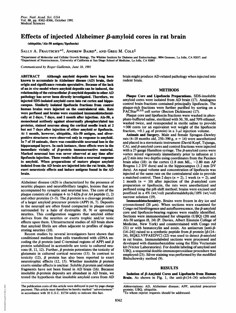
stains amyloid cores in humastaining is blocked by the corre(Fig. 1B). This serum was usedlocate cores after injection. Amfive AD brains and lipofuscintwo normal human brains in thrcores stained with anti-13 proteiireported (17), the principal elipofuscin, which was not stainpurity of the gradient-isolateddetermining the percentage of(among the total number of objphase-contrast, or autofluores4further purified by cell sorting (of the birefringent profiles shotconfiguration (Fig. 2 B-D) at
A22
'I
L
c
d
- z ^ anti-f-(14-24). SDS-isolated cores have been shown to con-.i>e4if tain a1-antichymotrypsin and heparan sulfate proteoglycan.
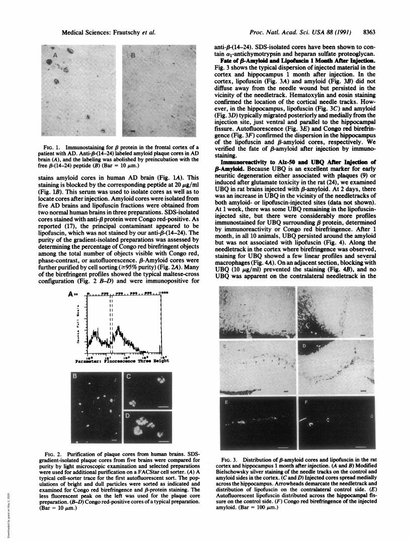
Fate of 3-Amyloid and Lipofusdn 1 Month After hnjection.Fig. 3 shows the typical dispersion of injected material in thecortex and hippocampus 1 month after injection. In the
Yes cortex, lipofuscin (Fig. 3A) and amyloid (Fig. 3B) did notdiffuse away from the needle wound but persisted in thevicinity of the needletrack. Hematoxylin and eosin stainingconfirmed the location of the cortical needle tracks. How-ever, in the hippocampus, lipofuscin (Fig. 3C) and amyloid(Fig. 3D) typically migrated posteriorly and medially from theinjection site, just ventral and parallel to the hippocampalfissure. Autofluorescence (Fig. 3E) and Congo red birefrin-gence (Fig. 3F) confirmed the dispersion in the hippocampusof the lipofuscin and (-amyloid cores, respectively. We
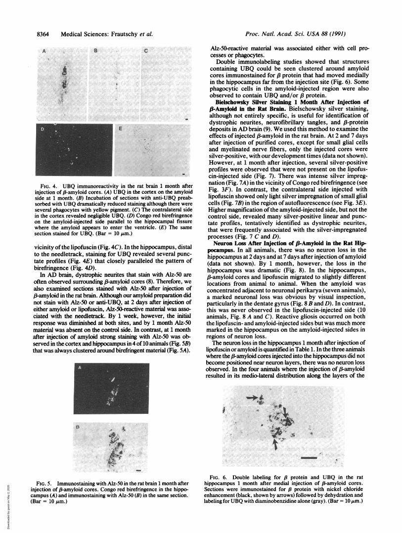
protein in the frontal cortex of a verified the fate of 3-amyloid after injection by immuno-beled amyloid plaque cores in AD staining.)lished by preincubation with the s mvwreacvity to Alz 50 md UBQAfteralncig of10 AM.) P-Amyoid. Because UBQ is an excellent marker for earlyin AD brain (Fig. LA). This neuritic degeneration either associated with plaques (9) or
sponding peptide at 20 ,ug/mlr induced after glutamate toxicity in the rat (24), we examinedito isolate cores as well as to ..UBQ in rat brains injected with f-amyloid. At 2 days, thereIyloid coreswerei solatedfrom was an increase in UBQ in the vicinity of the needletracks offractions were obtained from both amyloid- or lipofuscin-injected sites (data not shown).
eepreparations. SDS-isolated At 1 week, there was some UBQ remaining in the lipofuscin-newerepCongo r ed-positive.dAs injected site, but there were considerably more profileswecongotared-positivto As immunostained for UBQ surrounding P protein, determinedlontaminant appeared to be by immunoreactivity or Congo red birefringence. After 1ied by our anti-/3-(14-24). The month, in all 10 animals, UBQ persisted around the amyloidpreparations was assessed by but was not associated with lipofuscin (Fig. 4). Along theIongo red birefringent objects needletrack in the cortex where birefringence was observed,jects visible with Congo red, staining for UBQ showed a few linear profiles and several
e-95% purity)(Fig. 2A). Many macrophages (Fig. 4A). On an adjacent section, blocking with.95%puriy)(ig. A). any UBQ (10 jug/ml) prevented the staining (Fig. 4B), and nowed the typical maltese-cross UBQ was apparent on the contralateral needletrack in thend were immunopositive for
a.. F. . 143. 5. 5*9
I III II 1a1. *I'1111
Paametr F rsenc t t04Paramete: Fluorescnce Three Height
FIG. 2. Purification of plaque cores from human brains. SDS-gradient-isolated plaque cores from five brains were compared forpurity by light microscopic examination and selected preparationswere used for additional purification on a FACStar cell sorter. (A) Atypical cell-sorter trace for the first autofluorescent sort. The pop-ulations of bright and dull particles were sorted as indicated andexamined for Congo red birefringence and 13-protein staining. Theless fluorescent peak on the left was used for the plaque corepreparation. (B-D) Congo red-positive cores ofa typical preparation.(Bar = 10 Am.)
C
,. __I
~~~~~~~~~~~~~~~~~~~~~~~~~4>~~~~~~~~~~~~~~ " T
FIG. 3. Distribution of P-amyloid cores and lipofuscin in the ratcortex and hippocampus 1 month after injection. (A and B) ModifiedBielschowsky silver staining of the needle tracks on the control andamyloid sides in the cortex. (C and D) Injected cores spread mediallyacross the hippocampus. Arrowheads demarcate the needletrack anddistribution of lipofuscin on the contralateral control side. (E)Autofluorescent lipofuscin distributed across the hippocampal fis-sure on the control side. (F) Congo red birefringence of the injectedamyloid. (Bar = 100 Am.)
Medical Sciences: Frautschy et al.
.-VI
I
6:.
A:.
:. .: .110
I
I
__j
,k.. 11-
4 :, &,.
Dow
nloa
ded
by g
uest
on
May
2, 2
020
8364 Medical Sciences: Frautschy et al.
A B C
-I.4
E
FIG. 4. UBQ immunoreactivity in the rat brain 1 month afterinjection of 8-amyloid cores. (A) UBQ in the cortex on the amyloidside at 1 month. (B) Incubation of sections with anti-UBQ preab-sorbed with UBQ dramatically reduced staining although there wereseveral phagocytes with yellow pigment. (C) The contralateral sidein the cortex revealed negligible UBQ. (D) Congo red birefringenceon the amyloid-injected side parallel to the hippocampal fissurewhere the amyloid appears to enter the ventricle. (E) The samesection stained for UBQ. (Bar = 10 ,um.)
vicinity ofthe lipofuscin (Fig. 4C). In the hippocampus, distalto the needletrack, staining for UBQ revealed several punc-tate profiles (Fig. 4E) that closely paralleled the pattern ofbirefringence (Fig. 4D).
In AD brain, dystrophic neurites that stain with Alz-50 areoften observed surrounding P-amyloid cores (8). Therefore, wealso examined sections stained with Alz-50 after injection of(3-amyloid in the rat brain. Although our amyloid preparation didnot stain with Alz-50 or anti-UBQ, at 2 days after injection ofeither amyloid or lipofuscin, Alz-50-reactive material was asso-ciated with the needletrack. By 1 week, however, the initialresponse was diminished at both sites, and by 1 month Alz-50material was absent on the control side. In contrast, at 1 monthafter injection of amyloid strong staining with Alz-50 was ob-served in the cortex and hippocampus in 4 of 10 animals (Fig. 5B)that was always clustered around birefringent material (Fig. 5A).
I
B
FIG. 5. Immunostaining with Alz-50 in the rat brain 1 month afterinjection of (-amyloid cores. Congo red birefringence in the hippo-campus (A) and immunostaining with Alz-50 (B) in the same section.(Bar = 10 Am.)
Alz-50-reactive material was associated either with cell pro-cesses or phagocytes.Double immunolabeling studies showed that structures
containing UBQ could be seen clustered around amyloidcores immunostained for ,8 protein that had moved mediallyin the hippocampus far from the injection site (Fig. 6). Somephagocytic cells in the amyloid-injected region were alsoobserved to contain UBQ and/or / protein.
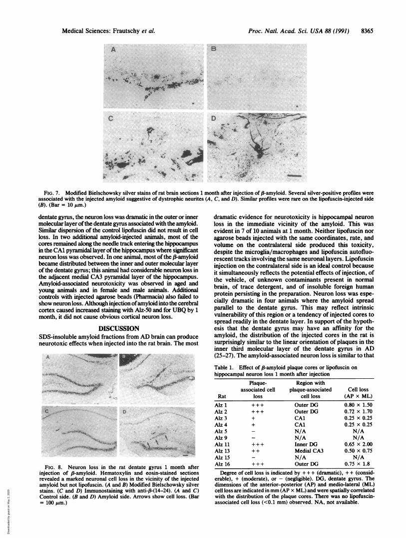
Bielschowsky Silver Staining 1 Month After Injection of3-Amyloid in the Rat Brain. Bielschowsky silver staining,although not entirely specific, is useful for identification ofdystrophic neurites, neurofibrillary tangles, and 8-proteindeposits in AD brain (9). We used this method to examine theeffects of injected f3-amyloid in the rat brain. At 2 and 7 daysafter injection of purified cores, except for small glial cellsand myelinated nerve fibers, only the injected cores weresilver-positive, with our development times (data not shown).However, at 1 month after injection, several silver-positiveprofiles were observed that were not present on the lipofus-cin-injected side (Fig. 7). There was intense silver impreg-nation (Fig. 7A) in the vicinity ofCongo red birefringence (seeFig. 3F). In contrast, the contralateral side injected withlipofuscin showed only light silver impregnation of small glialcells (Fig. 7B) in the region of autofluorescence (see Fig. 3E).Higher magnification of the amyloid-injected side, but not thecontrol side, revealed many silver-positive linear and punc-tate profiles, tentatively identified as dystrophic neurites,that were frequently associated with the silver-impregnatedprocesses (Fig. 7 C and D).Neuron Loss After Injection of fi-Amyloid in the Rat Hip-
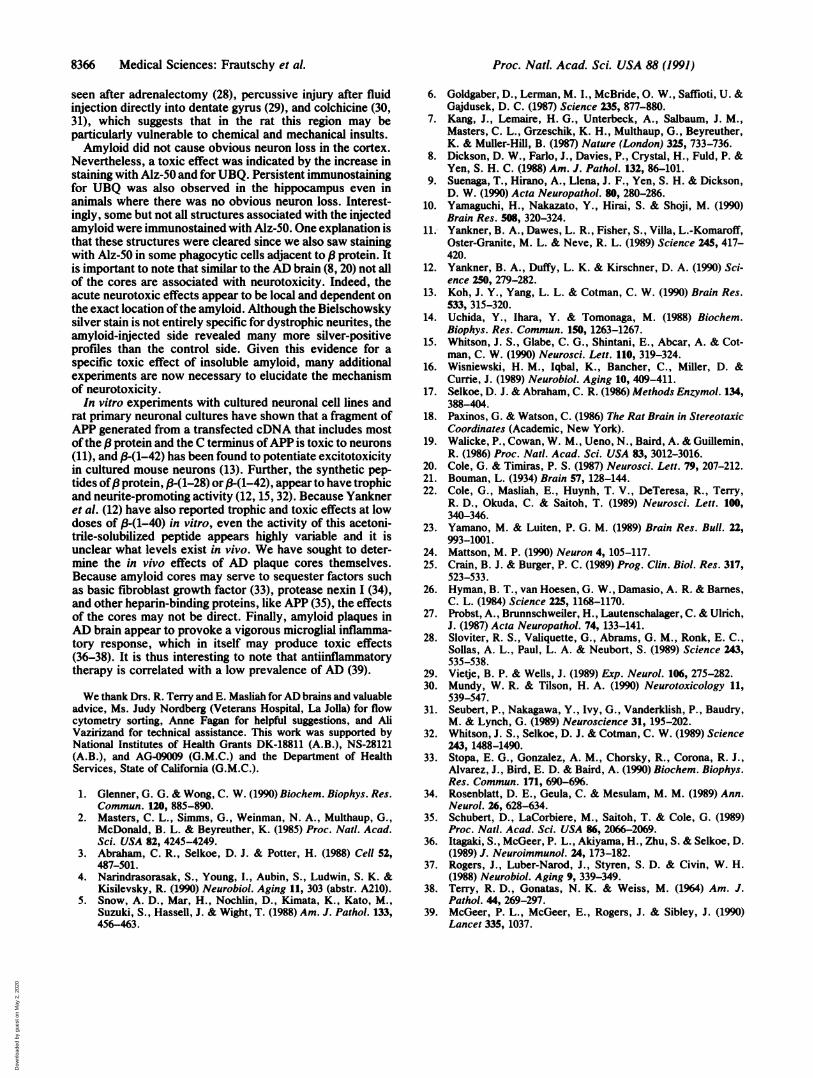
pocampus. In all animals, there was no neuron loss in thehippocampus at 2 days and at 7 days after injection ofamyloid(data not shown). By 1 month, however, the loss in thehippocampus was dramatic (Fig. 8). In the hippocampus,/3-amyloid cores and lipofuscin migrated to slightly differentlocations from animal to animal. When the amyloid wasconcentrated adjacent to neuronal perikarya (seven animals),a marked neuronal loss was obvious by visual inspection,particularly in the dentate gyrms (Fig. 8 B and D). In contrast,this was never observed in the lipofuscin-injected side (10animals, Fig. 8 A and C). Reactive gliosis occurred on boththe lipofuscin- and amyloid-injected sides but was much moremarked in the hippocampus on the amyloid-injected sides inregions of neuron loss.The neuron loss in the hippocampus 1 month after injection of
lipofuscin oramyloid is quantified in Table 1. In the three animalswhere the ,-amyloid cores injected into the hippocampus did notbecome positioned near neuron layers, there was no neuron lossobserved. In the four animals where the injection of (-amyloidresulted in its medio-lateral distribution along the layers of the
0..
. . -._
* ,<<~~~~~~'));r
FIG. 6. Double labeling for f8 protein and UBQ in the rathippocampus 1 month after medial injection of (8-amyloid cores.Sections were immunostained for 8 protein with nickel chlorideenhancement (black, shown by arrows) followed by dehydration andlabeling for UBQ with diaminobenzidine alone (gray). (Bar = 10 gm.)
Proc. NatL Acad Sci. USA 88 (1991)
v
,-? A.14, -.-
. I
I
Dow
nloa
ded
by g
uest
on
May
2, 2
020
Medical Sciences: Frautschy et al.
A B
.. -L
4,,'4
D wb ..(-dl
.4
,. ...
FIG. 7. Modified Bielschowsky silver stains of rat brain sections 1 month after injection of 3-amyloid. Several silver-positive profiles wereassociated with the injected amyloid suggestive of dystrophic neurites (A, C, and D). Similar profiles were rare on the lipofuscin-injected side(B). (Bar = 10 ,Lm.)
dentate gyrms, the neuron loss was dramatic in the outer or innermolecular layer ofthe dentate gyrms associated with the amyloid.Similar dispersion of the control lipofuscin did not result in cellloss. In two additional amyloid-injected animals, most of thecores remained along the needle track entering the hippocampusin the CA1 pyramidal layer ofthe hippocampus where significantneuron loss was observed. In one animal, most ofthe ,-Bamyloidbecame distributed between the inner and outer molecular layerof the dentate gymus; this animal had considerable neuron loss inthe adjacent medial CA3 pyramidal layer of the hippocampus.Amyloid-associated neurotoxicity was observed in aged andyoung animals and in female and male animals. Additionalcontrols with injected agarose beads (Pharmacia) also failed toshow neuron loss. Although injection ofamyloid into the cerebralcortex caused increased staining with Alz-50 and for UBQ by 1month, it did not cause obvious cortical neuron loss.
DISCUSSIONSDS-insoluble amyloid fractions from AD brain can produceneurotoxic effects when injected into the rat brain. The most
A <w .
. ~ ~ ~
4,S-'.F Cit w,_
If .. ~. Ad 1 -.
,-O
FIG. 8. Neuron loss in the rat dentate gyrus 1 month afterinjection of ,B-amyloid. Hematoxylin and eosin-stained sectionsrevealed a marked neuronal cell loss in the vicinity of the injectedamyloid but not lipofuscin. (A and B) Modified Bielschowsky silverstains. (C and D) Immunostaining with anti-,B-(14-24). (A and C)Control side. (B and D) Amyloid side. Arrows show cell loss. (Bar= 100 Jm.)
dramatic evidence for neurotoxicity is hippocampal neuronloss in the immediate vicinity of the amyloid. This wasevident in 7 of 10 animals at 1 month. Neither lipofuscin noragarose beads injected with the same coordinates, rate, andvolume on the contralateral side produced this toxicity,despite the microglia/macrophages and lipofuscin autofluo-rescent tracks involving the same neuronal layers. Lipofuscininjection on the contralateral side is an ideal control becauseit simultaneously reflects the potential effects of injection, ofthe vehicle, of unknown contaminants present in normalbrain, of trace detergent, and of insoluble foreign humanprotein persisting in the preparation. Neuron loss was espe-cially dramatic in four animals where the amyloid spreadparallel to the dentate gyrms. This may reflect intrinsicvulnerability of this region or a tendency of injected cores tospread readily in the dentate layer. In support of the hypoth-esis that the dentate gyrms may have an affinity for theamyloid, the distribution of the injected cores in the rat issurprisingly similar to the linear orientation of plaques in theinner third molecular layer of the dentate gyrus in AD(25-27). The amyloid-associated neuron loss is similar to that
Table 1. Effect of ,-amyloid plaque cores or lipofuscin onhippocampal neuron loss 1 month after injection
Plaque- Region withassociated cell plaque-associated Cell loss
Rat loss cell loss (AP x ML)Alz 1 +++ Outer DG 0.80 x 1.50Alz 2 +++ Outer DG 0.72 x 1.70Alz 3 + CA1 0.25 x 0.25Alz 4 + CA1 0.25 x 0.25Alz 5 - N/A N/AAlz9 - N/A N/AAlz 11 +++ Inner DG 0.65 x 2.00Alz 13 + + Medial CA3 0.50 x 0.75Alz 15 - N/A N/AAlz 16 +++ Outer DG 0.75 x 1.8
Degree of cell loss is indicated by + + + (dramatic), + + (consid-erable), + (moderate), or - (negligible). DG, dentate gyrus. Thedimensions of the anterior-posterior (AP) and medio-lateral (ML)cell loss are indicated in mm (AP x ML) and were spatially correlatedwith the distribution of the plaque cores. There was no lipofuscin-associated cell loss (<0.1 mm) observed. NA, not available.
Proc. Nati. Acad. Sci. USA 88 (1991) 8365
C
I
I .,I*
, . . I
",...
0.00 -k
-IL x. *
.-O ..--
---mv..'J..ov ,., .,.
!:
.$ . -0.,4. q. .
..0..
W
Dow
nloa
ded
by g
uest
on
May
2, 2
020
8366 Medical Sciences: Frautschy et al.
seen after adrenalectomy (28), percussive injury after fluidinjection directly into dentate gyrms (29), and colchicine (30,31), which suggests that in the rat this region may beparticularly vulnerable to chemical and mechanical insults.Amyloid did not cause obvious neuron loss in the cortex.
Nevertheless, a toxic effect was indicated by the increase instaining with Alz-50 and for UBQ. Persistent immunostainingfor UBQ was also observed in the hippocampus even inanimals where there was no obvious neuron loss. Interest-ingly, some but not all structures associated with the injectedamyloid were immunostained with Alz-50. One explanation isthat these structures were cleared since we also saw stainingwith Alz-50 in some phagocytic cells adjacent to ,8 protein. Itis important to note that similar to the AD brain (8, 20) not allof the cores are associated with neurotoxicity. Indeed, theacute neurotoxic effects appear to be local and dependent onthe exact location of the amyloid. Although the Bielschowskysilver stain is not entirely specific for dystrophic neurites, theamyloid-injected side revealed many more silver-positiveprofiles than the control side. Given this evidence for aspecific toxic effect of insoluble amyloid, many additionalexperiments are now necessary to elucidate the mechanismof neurotoxicity.
In vitro experiments with cultured neuronal cell lines andrat primary neuronal cultures have shown that a fragment ofAPP generated from a transfected cDNA that includes mostof the ( protein and the C terminus ofAPP is toxic to neurons(11), and ,8-(1-42) has been found to potentiate excitotoxicityin cultured mouse neurons (13). Further, the synthetic pep-tides of( protein, P-(1-28) or 3-(1-42), appear to have trophicand neurite-promoting activity (12, 15, 32). Because Yankneret al. (12) have also reported trophic and toxic effects at lowdoses of ,B-(1-40) in vitro, even the activity of this acetoni-trile-solubilized peptide appears highly variable and it isunclear what levels exist in vivo. We have sought to deter-mine the in vivo effects of AD plaque cores themselves.Because amyloid cores may serve to sequester factors suchas basic fibroblast growth factor (33), protease nexin I (34),and other heparin-binding proteins, like APP (35), the effectsof the cores may not be direct. Finally, amyloid plaques inAD brain appear to provoke a vigorous microglial inflamma-tory response, which in itself may produce toxic effects(36-38). It is thus interesting to note that antiinflammatorytherapy is correlated with a low prevalence of AD (39).
We thank Drs. R. Terry and E. Masliah forAD brains and valuableadvice, Ms. Judy Nordberg (Veterans Hospital, La Jolla) for flowcytometry sorting, Anne Fagan for helpful suggestions, and AliVazirizand for technical assistance. This work was supported byNational Institutes of Health Grants DK-18811 (A.B.), NS-28121(A.B.), and AG-09009 (G.M.C.) and the Department of HealthServices, State of California (G.M.C.).
1. Glenner, G. G. & Wong, C. W. (1990) Biochem. Biophys. Res.Commun. 120, 885-890.
2. Masters, C. L., Simms, G., Weinman, N. A., Multhaup, G.,McDonald, B. L. & Beyreuther, K. (1985) Proc. Natl. Acad.Sci. USA 82, 4245-4249.
3. Abraham, C. R., Selkoe, D. J. & Potter, H. (1988) Cell 52,487-501.
4. Narindrasorasak, S., Young, I., Aubin, S., Ludwin, S. K. &Kisilevsky, R. (1990) Neurobiol. Aging 11, 303 (abstr. A210).
5. Snow, A. D., Mar, H., Nochlin, D., Kimata, K., Kato, M.,Suzuki, S., Hassell, J. & Wight, T. (1988) Am. J. Pathol. 133,456-463.
6. Goldgaber, D., Lerman, M. I., McBride, 0. W., Saffioti, U. &Gajdusek, D. C. (1987) Science 235, 877-880.
7. Kang, J., Lemaire, H. G., Unterbeck, A., Salbaum, J. M.,Masters, C. L., Grzeschik, K. H., Multhaup, G., Beyreuther,K. & Muller-Hill, B. (1987) Nature (London) 325, 733-736.
8. Dickson, D. W., Farlo, J., Davies, P., Crystal, H., Fuld, P. &Yen, S. H. C. (1988) Am. J. Pathol. 132, 86-101.
9. Suenaga, T., Hirano, A., Llena, J. F., Yen, S. H. & Dickson,D. W. (1990) Acta Neuropathol. 80, 280-286.
10. Yamaguchi, H., Nakazato, Y., Hirai, S. & Shoji, M. (1990)Brain Res. 508, 320-324.
11. Yankner, B. A., Dawes, L. R., Fisher, S., Villa, L.-Komaroff,Oster-Granite, M. L. & Neve, R. L. (1989) Science 245, 417-420.
12. Yankner, B. A., Duffy, L. K. & Kirschner, D. A. (1990) Sci-ence 250, 279-282.
13. Koh, J. Y., Yang, L. L. & Cotman, C. W. (1990) Brain Res.533, 315-320.
14. Uchida, Y., Ihara, Y. & Tomonaga, M. (1988) Biochem.Biophys. Res. Commun. 150, 1263-1267.
15. Whitson, J. S., Glabe, C. G., Shintani, E., Abcar, A. & Cot-man, C. W. (1990) Neurosci. Lett. 110, 319-324.
16. Wisniewski, H. M., Iqbal, K., Bancher, C., Miller, D. &Currie, J. (1989) Neurobiol. Aging 10, 409-411.
17. Selkoe, D. J. & Abraham, C. R. (1986) Methods Enzymol. 134,388-404.
18. Paxinos, G. & Watson, C. (1986) The Rat Brain in StereotaxicCoordinates (Academic, New York).
19. Walicke, P., Cowan, W. M., Ueno, N., Baird, A. & Guillemin,R. (1986) Proc. Nat!. Acad. Sci. USA 83, 3012-3016.
20. Cole, G. & Timiras, P. S. (1987) Neurosci. Lett. 79, 207-212.21. Bouman, L. (1934) Brain 57, 128-144.22. Cole, G., Masliah, E., Huynh, T. V., DeTeresa, R., Terry,
R. D., Okuda, C. & Saitoh, T. (1989) Neurosci. Lett. 100,340-346.
23. Yamano, M. & Luiten, P. G. M. (1989) Brain Res. Bull. 22,993-1001.
24. Mattson, M. P. (1990) Neuron 4, 105-117.25. Crain, B. J. & Burger, P. C. (1989) Prog. Clin. Biol. Res. 317,
523-533.26. Hyman, B. T., van Hoesen, G. W., Damasio, A. R. & Barnes,
C. L. (1984) Science 225, 1168-1170.27. Probst, A., Brunnschweiler, H., Lautenschalager, C. & Ulrich,
J. (1987) Acta Neuropathol. 74, 133-141.28. Sloviter, R. S., Valiquette, G., Abrams, G. M., Ronk, E. C.,
Sollas, A. L., Paul, L. A. & Neubort, S. (1989) Science 243,535-538.
29. Vietje, B. P. & Wells, J. (1989) Exp. Neurol. 106, 275-282.30. Mundy, W. R. & Tilson, H. A. (1990) Neurotoxicology 11,
539-547.31. Seubert, P., Nakagawa, Y., Ivy, G., Vanderklish, P., Baudry,
M. & Lynch, G. (1989) Neuroscience 31, 195-202.32. Whitson, J. S., Selkoe, D. J. & Cotman, C. W. (1989) Science
243, 1488-1490.33. Stopa, E. G., Gonzalez, A. M., Chorsky, R., Corona, R. J.,
Alvarez, J., Bird, E. D. & Baird, A. (1990) Biochem. Biophys.Res. Commun. 171, 690-696.
34. Rosenblatt, D. E., Geula, C. & Mesulam, M. M. (1989) Ann.Neurol. 26, 628-634.
35. Schubert, D., LaCorbiere, M., Saitoh, T. & Cole, G. (1989)Proc. Nat!. Acad. Sci. USA 86, 2066-2069.
36. Itagaki, S., McGeer, P. L., Akiyama, H., Zhu, S. & Selkoe, D.(1989) J. Neuroimmunol. 24, 173-182.
37. Rogers, J., Luber-Narod, J., Styren, S. D. & Civin, W. H.(1988) Neurobiol. Aging 9, 339-349.
38. Terry, R. D., Gonatas, N. K. & Weiss, M. (1964) Am. J.Pathol. 44, 269-297.
39. McGeer, P. L., McGeer, E., Rogers, J. & Sibley, J. (1990)Lancet 335, 1037.
Proc. NatL Acad. Sci. USA 88 (1991)
Dow
nloa
ded
by g
uest
on
May
2, 2
020