Embed Size (px)
Citation preview

Research ArticleEffect of Fiber Post-Resin Matrix Composition on BondStrength of Post-Cement Interface
Ibtisam O. M. Alnaqbi,1 Haitham Elbishari ,2 and Emad S. Elsubeihi2
1Specialist in Restorative Dentistry, Khorfakkan Hospital, Ministry of Health, Sharjah, UAE2Assistant Professor, Department of Restorative Dentistry, College of Dentistry, Ajman University, Ajman, UAE
Correspondence should be addressed to Haitham Elbishari; [email protected]
Received 26 September 2018; Accepted 13 November 2018; Published 2 December 2018
Academic Editor: Spiros Zinelis
Copyright © 2018 Ibtisam O.M. Alnaqbi et al.,is is an open access article distributed under the Creative Commons AttributionLicense, which permits unrestricted use, distribution, and reproduction in any medium, provided the original work isproperly cited.
Objective. To evaluate the influence of 3 different post-resin matrix systems cemented with dual-cure resin cement in simulatedroot canals made of PMMA acrylic sheet. Methods. 3 types of fiber posts (n � 60) with different resin matrixes divided into 3groups: group 1 cross-linked FRC Postec Plus post (n � 20), group 2 cross-linked Rely X post (n � 20), and group 3 InterpenetratedIPN Everstick post (n � 20). All posts were cemented usingMultilink Automix dual-cure cement. Posts were cemented into acrylicblocks in order to purely test the strength of cement-post interface. After one week storage at 37°C, two sections of 1mm thicknessfrom middle-third were subjected to micro-push-out test at crosshead speed 0.5mm/min. Results. ,e data were analyzed usingone-way analysis of variance (ANOVA).,e variable fiber post-matrix system was found to significantly affect the push-out bondstrength (p< 0.001). Group 2 exhibited that the highest mean push-out bond strength was (5.36 + 2.3MPa), and group 3 showedthe lowest mean push-out (0.41 + 0.4MPa). ,ere was significant difference among the groups regarding the failure mode as chi-square test revealed (p< 0.001). Conclusion. Prefabricated cross-linked posts with epoxy-based matrix demonstrated higher bondstrength than prefabricated cross-linked posts with Bis-GMA-based matrix and posts with semi-IPN matrix when luted withdimethacrylate-based dual-cured resin cement.
1. Introduction
Fiber-reinforced posts have been introduced in the early1990s [1], as an alternative to prefabricated metal posts torestore endodontically treated teeth with excessive loss oftooth structure. Clinical studies have shown that the mostfrequent types of failure seen in endodontically treated teethrestored with fiber-reinforced posts were debonding and lossof retention as well as post fracture, [2–7]. Retention ofadhesively luted fiber-reinforced posts relies on the strengthof the bond between dentine-cement interface on one handand that of the post-cement interface on the other. It isimportant that the bond strength of both interfaces is suf-ficiently strong to withstand stresses during functionalloading. Several studies have investigated the bond strengthof dentine-cement interface [8, 9]; however, much less at-tention has been given in investigating the bond strength
of post-cement interface based on models that excludethe combined “sandwich” dentine-cement-post assembly[10–12].
In general, fiber-reinforced posts consist of prestretchedfibers embedded in a polymer resin matrix. ,e functions ofthe matrix in fiber-reinforced posts is to hold the fiberstogether in the post, as well as interact with functionalmonomers contained in the adhesive materials for successfulbonding of the post to cement resin and composite corematerials [13]. Fibers, on the other hand, provide strengthand stiffness to the post.
Several manufacturers employ epoxy resin as a matrixfor fiber-reinforced posts. Posts with the epoxy resin matrix,however, suffered from poor chemical affinity toward theluting resin because of differences in chemical composition[14, 15]. ,e introduction of posts with dimethacrylate resinmatrix including bisphenol A-glycidyl methacrylate (Bis-
HindawiInternational Journal of DentistryVolume 2018, Article ID 4751627, 11 pageshttps://doi.org/10.1155/2018/4751627

GMA) was seen as an advantage toward improving thechemical bonding between the post matrix and that of theresin cement [16]. Studies, however, have shown that thepolymer matrix of such fiber-reinforced posts was virtuallynonreactive, because the resin has a high degree of con-version and is highly cross linked [16]. ,is has promptedresearchers to explore the options of improving the post-cement bonding through pretreatment of post surfaces withsilane either alone [8, 9, 17, 18] or following surfacesconditioning using chemical [9, 14, 18, 19] or mechanical[20] means with varying degrees of success.
,e introduction of posts with unidirectional continu-ous glass fibers embedded in unpolymerized semi-interpenetrating polymer network (IPN) composed ofpolymethylmethacrylate (PMMA) and Bis-GMA hasrenewed interests in achieving a true chemical bondingbetween the post and luting cements. Studies have shownthat adhesive systems with monomers that have solubilityparameters close to that of PMMA such as Bis-GMA, 2-hydroxyethyl methacrylate (HEMA), and triethylene glycoldimethacrylate (TEGDMA) can penetrate into the linearpolymer phase of posts with an IPNmatrix but not into postswith an epoxymatrix [21].,e aim of this study is to evaluatethe bond strength and mode of failure of fiber-reinforcedposts with different resin matrices. ,e null hypothesis isthat the micro-push-out bond strength and mode of failuredo not vary with the type of the post-resin matrix when lutedwith Bis-GMA-based resin cement.
2. Materials and Methods
2.1. StudyDesignandSelectedMaterials. ,ree types of fiber-reinforced posts with different matrices, namely, (1) cross-linked prefabricated Bis-GMA (FRC Postec Plus, IvoclarVivadent, Schaan, Liechtenstein); (2) cross-linked pre-fabricated epoxy (Rely X Fiber Post, 3M ESPE, Seefeld,Germany); and (3) semi-interpenetrating polymer networkof PMMA and Bis-GMA (GC Everstick, StickTech LTD,Turku, Finland) was tested. All posts were luted using Bis-GMA-based, dimethacrylate dual-cured cement (MultilinkAutomix, Ivoclar Vivadent, Schaan, Liechten) in simulatedroot canals made of PMMA (Yearlong Industrial CompanyLimited, Taiwan) with a custom-prepared post space.Composition of posts and luting cement used in this study isillustrated in Table 1.
,e push-out bond strength of twenty samples of eachcemented post on two sections made from each post wasassessed. Furthermore, the mode of failure of all sectionsfollowing push-out bond strength test was evaluated. Studydesign is demonstrated in Figure 1.
2.2. Fabrication of the Experimental Model. PMMA acrylicsheet (Yearlong Industrial Company Limited, Taiwan)was used to make 60 blocks. ,e blocks made were cubicalin shape with 20mm height and 10mm × 10mm base(Figure 2). ,e blocks were prepared by cutting the sheetwith laser-guided machine (Source Company, Ajman) withhigh speed, good cooling, and low cutting force as
recommended by the manufacturer. Each block was thenpolished using a polishing machine (MetaServ 250 twinGrinder-Polisher, Buehler, USA) with speed 70 rpm tocorrect any deviation in the blocks dimension while pre-paring the blocks.
2.3.Post SpacePreparationandCementation. Space for fiber-reinforced post was prepared in each block with size 1 FRCPostec Plus Reamer (Ivoclar Vivadent, Schaan, Liechten-stein) in a low-speed hand piece mounted on a parallelmilling device (Amann Girrbach, Vorarlberg, Germany). Atthe beginning, each block was placed in the block holderperpendicular to the drilling bur at a 90o angle. ,e blockswere then drilled with intermittent light force at 18,000 rpmspeed to the length of 12mm depth for FRC Postec Plus andRely X fiber post and 10mm for the Everstick post. Coolingwith normal saline was used to reduce heat generationduring the drilling procedure. Post space was then rough-ened using the Hedstrom endodontic file size 45 (Technicaland General Ltd, London, UK) to increase the bond strengthbetween the acrylic block and the cement.,e post space wasirrigated with alcohol to ensure no grease or debris in thepost space walls then dried with paper points. ,is wasfollowed by irrigation with distilled water and dried usingpaper point. Following the cleaning and drying stage, thepatency of the post space was confirmed by trying in dif-ferent types of posts to ensure that each type of the post canbe seated to the full-prepared post space length.
Each block was painted with black color using a paintpen marker in order to prevent light penetration during andafter cementation. Multilink Automix, dual-cure lutingcement (Ivoclar Vivadent, Schaan, Liechten) was used tocement all posts according to manufacturer’s instruction.
,e sample was then placed in a 16.0mm deep custom-made white container with a 4.0mm deep plastic cover,which was covered with aluminum foil to prevent un-desirable exposure to light. A 5mm opening was made onthe cover to allow the curing of the sample. After that, thecement was cured for 60 seconds with LED (light-emittingdiode) curing unit at 600mW/cm2 in high intensity mode(Litex 696, Cordless Led Curing Light, DentAmerica, USA).,e tip of the curing unit was placed close to the coronalsurface of the post according to the manufacturers’ in-struction. Light intensity was confirmed with a visible curinglight meter (Cure Rite, Dentsply Caulk, Milford, USA)before curing each post. After curing, all specimens werestored at 37°C in 100% humidity in an incubator for 1 week.
2.4. Sample Preparation. After 1 week of storage time, acrylicblocks were placed in holder of a saw machine (IsoMet 1000Precision, Buehler, USA) with post long axis perpendicular tothe saw blade disc.,e samples were cut into 1.0mm sectionsat a speed of 350 rpm. Two sections of 1.0mm thicknessrepresenting the middle third of each post were used for thestudy.,e location of the first section was about 5.0mm fromthe coronal end of each post. As the thickness of the cuttingblade was about 0.5mm, the second section used for the studywas about 6.5mm from the coronal end of each post.
2 International Journal of Dentistry

�e coronal surface of each section was marked with awaterproof marker to identify the coronal and apical sur-faces of each section. Any sharp edges in the specimen werethen removed by low-speed polishing disk, and thickness ofall slices was measured by a laser scan micrometer (LaserScan Micrometer, LSM-6200, Mitutoyo, Japan) and by adigital caliper.
Each section was divided into 4 equal segments using apencil, and each quarter was color-coded and numberedto be used later for failure mode analysis. Furthermore, the
lateral surface area of each post space preparation wascalculated. To do this, each section was placed under astereomicroscope with a mounted camera (EZ4HD, Leica,Singapore) at magni�cation of × 8, and a photograph wastaken. Photographs of both coronal and apical sides ofeach section were made. Each image was calibrated, andthe radius of the coronal and apical sides of each postspace was measured using an image analysis system(Image J Software, Java 1.6.0 (64-bits) 1.50i, NationalInstitute of Health, USA). �e lateral surface area of eachsection (Figure 3) was then calculated using the followingformula:
LS � π(R2 + R1) H2 +(R2−R1)2[ ]0.5, (1)
Table 1: Description of �ber posts and luting cement used in this study.
Fiber post Manufacturer andbatch number used Matrix Curing condition
of matrix Fibers Fillers Size andshape
GC Everstick StickTech Ltd.,Turku, Finland
Semiinterpenetratingpolymer network ofPMMA and Bis-GMA
Uncured (to becured by theclinician)
Unidirectional silane-coated E-glass �bers(61.5% by weight)
No �ller 0.9mmCustom
FRC Postec PlusIvoclar Vivadent,
Schaan,Liechtenstein
Dimethacrylates(ethoxylated bisphenol Adimethacrylate, Bis-GMA, and 1,4-butanediol
dimethacrylate)
Cured by themanufacturers E-glass �bers Ytterbium
�uoride0.8–1.5mm
Taper
Rely X Fiberpost
3M ESPE, Seefeld,Germany Epoxy-resin Cured by the
manufacturersS-glass �bers (60–70% by
weight)Zirconia�ller
0.8–1.6mmDoubletapered
MultilinkAutomix lutingcement
Ivoclar Vivadent,Schaan,
Liechtenstein
Base Catalyst(i) Ytterbium tri�uoride (i) Ytterbium tri�uoride
(ii) Ethoxylated bisphenol A dimethacrylate(Bis-EMA)
(ii) Ethoxylated bisphenol A dimethacrylate (Bis-EMA)
(iii) Bisphenol A-glycidyl methacrylate (Bis-GMA) (iii) urethane dimethacrylate (UDMA)
(iv) 2-hydroxyethyl methacrylate (HEMA) (iv) 2-hydroxyethyl methacrylate (HEMA)(v) 2-Dimethylaminoethyl methacrylate
(DMAEMA) (v) Dibenzoyl peroxide
FRC Postec postsN = 20
Push-out test2 sections each post
Total 40 sections
Failure mode analysisN = 40 sections
Eversck postsN = 20
Push-out test2 sections each post
Total 40 sections
Failure mode analysisN = 40 sections
STUDY DESIGN
Fiber reinforced postsN = 60
Luted with Variolink II cement
Rely X postsN = 20
Push-out test2 sections each post
Total 40 sections
Failure mode analysisN = 40 sections
Figure 1: Study design. Figure shows the groups, number of postsin each group, and number of sections analyzed. N � sample size.
PMMA
20 mm
10 mm × 10 mm
Figure 2: Schematic drawing represents shape and dimension ofexperimental acrylic block, which is made of PMMA (poly-methylmethacrylate). Blocks were cubical in shape with 20.0mmheight and 10.0mm × 10.0mm base.
International Journal of Dentistry 3
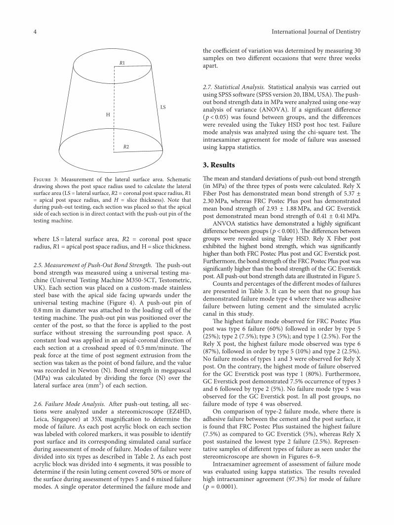
where LS� lateral surface area, R2 � coronal post spaceradius, R1 � apical post space radius, and H� slice thickness.
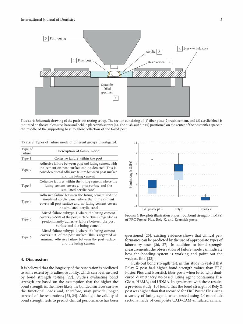
2.5. Measurement of Push-Out Bond Strength. �e push-outbond strength was measured using a universal testing ma-chine (Universal Testing Machine M350-5CT, Testometric,UK). Each section was placed on a custom-made stainlesssteel base with the apical side facing upwards under theuniversal testing machine (Figure 4). A push-out pin of0.8mm in diameter was attached to the loading cell of thetesting machine. �e push-out pin was positioned over thecenter of the post, so that the force is applied to the postsurface without stressing the surrounding post space. Aconstant load was applied in an apical-coronal direction ofeach section at a crosshead speed of 0.5mm/minute. �epeak force at the time of post segment extrusion from thesection was taken as the point of bond failure, and the valuewas recorded in Newton (N). Bond strength in megapascal(MPa) was calculated by dividing the force (N) over thelateral surface area (mm2) of each section.
2.6. Failure Mode Analysis. After push-out testing, all sec-tions were analyzed under a stereomicroscope (EZ4HD,Leica, Singapore) at 35X magni�cation to determine themode of failure. As each post acrylic block on each sectionwas labeled with colored markers, it was possible to identifypost surface and its corresponding simulated canal surfaceduring assessment of mode of failure. Modes of failure weredivided into six types as described in Table 2. As each postacrylic block was divided into 4 segments, it was possible todetermine if the resin luting cement covered 50% or more ofthe surface during assessment of types 5 and 6 mixed failuremodes. A single operator determined the failure mode and
the coe¥cient of variation was determined by measuring 30samples on two di¦erent occasions that were three weeksapart.
2.7. Statistical Analysis. Statistical analysis was carried outusing SPSS software (SPSS version 20, IBM, USA).�e push-out bond strength data in MPa were analyzed using one-wayanalysis of variance (ANOVA). If a signi�cant di¦erence(p< 0.05) was found between groups, and the di¦erenceswere revealed using the Tukey HSD post hoc test. Failuremode analysis was analyzed using the chi-square test. �eintraexaminer agreement for mode of failure was assessedusing kappa statistics.
3. Results
�emean and standard deviations of push-out bond strength(in MPa) of the three types of posts were calculated. Rely XFiber Post has demonstrated mean bond strength of 5.37 ±2.30MPa, whereas FRC Postec Plus post has demonstratedmean bond strength of 2.93 ± 1.88MPa, and GC Everstickpost demonstrated mean bond strength of 0.41 ± 0.41MPa.
ANVOA statistics have demonstrated a highly signi�cantdi¦erence between groups (p< 0.001). �e di¦erences betweengroups were revealed using Tukey HSD. Rely X Fiber postexhibited the highest bond strength, which was signi�cantlyhigher than both FRC Postec Plus post and GC Everstick post.Furthermore, the bond strength of the FRCPostec Plus post wassigni�cantly higher than the bond strength of the GC Everstickpost. All push-out bond strength data are illustrated in Figure 5.
Counts and percentages of the di¦erent modes of failuresare presented in Table 3. It can be seen that no group hasdemonstrated failure mode type 4 where there was adhesivefailure between luting cement and the simulated acryliccanal in this study.
�e highest failure mode observed for FRC Postec Pluspost was type 6 failure (60%) followed in order by type 5(25%); type 2 (7.5%); type 3 (5%); and type 1 (2.5%). For theRely X post, the highest failure mode observed was type 6(87%), followed in order by type 5 (10%) and type 2 (2.5%).No failure modes of types 1 and 3 were observed for Rely Xpost. On the contrary, the highest mode of failure observedfor the GC Everstick post was type 1 (80%). Furthermore,GC Everstick post demonstrated 7.5% occurrence of types 3and 6 followed by type 2 (5%). No failure mode type 5 wasobserved for the GC Everstick post. In all post groups, nofailure mode of type 4 was observed.
On comparison of type-2 failure mode, where there isadhesive failure between the cement and the post surface, itis found that FRC Postec Plus sustained the highest failure(7.5%) as compared to GC Everstick (5%), whereas Rely Xpost sustained the lowest type 2 failure (2.5%). Represen-tative samples of di¦erent types of failure as seen under thestereomicroscope are shown in Figures 6–9.
Intraexaminer agreement of assessment of failure modewas evaluated using kappa statistics. �e results revealedhigh intraexaminer agreement (97.3%) for mode of failure(p � 0.0001).
R1
R2
LSH
Figure 3: Measurement of the lateral surface area. Schematicdrawing shows the post space radius used to calculate the lateralsurface area (LS � lateral surface, R2 � coronal post space radius, R1� apical post space radius, and H � slice thickness). Note thatduring push-out testing, each section was placed so that the apicalside of each section is in direct contact with the push-out pin of thetesting machine.
4 International Journal of Dentistry
4. Discussion
It is believed that the longevity of the restoration is predictedto some extent by its adhesive ability, which can be measuredby bond strength testing [22]. Studies evaluating bondstrength are based on the assumption that the higher thebond strength is, the more likely the bonded surfaces survivethe functional loads and, therefore, may provide longersurvival of the restorations [23, 24]. Although the validity ofbond strength tests to predict clinical performance has been
questioned [25], existing evidence shows that clinical per-formance can be predicted by the use of appropriate types oflaboratory tests [26, 27]. In addition to bond strengthmeasurements, the observation of failure mode can indicatehow the bonding system is working and point out theweakest link [23].
Push-out bond strength test, in this study, revealed thatRelay X post had higher bond strength values than FRCPostec Plus and Everstick fiber posts when luted with dual-cured diamethacrylate-based luting agent containing Bis-GMA, HEMA, and UDMA. In agreement with these results,a previous study [10] found that the bond strength of Rely Xpost was higher than that recorded for FRC Postec Plus usinga variety of luting agents when tested using 2.0mm thicksections made of composite CAD-CAM-simulated canals.
Push-out jig
Fiber post
Acrylic
Space forfailed
specimen
Resin cement
Screw to hold slice
5
1
34
2
6
Figure 4: Schematic drawing of the push-out testing set up. ,e section consisting of (1) fiber post; (2) resin cement, and (3) acrylic block ismounted on the stainless steel base and held in place with screws (4).,e push-out pin (5) positioned on the center of the post with a space inthe middle of the supporting base to allow collection of the failed post.
Table 2: Types of failure mode of different groups investigated.
Type offailure Description of failure mode
Type 1 Cohesive failure within the post
Type 2
Adhesive failure between post and luting cement withno cement on post surface can be detected. ,is isconsidered total adhesive failure between post surface
and the luting cement
Type 3Cohesive failures within the luting cement where the
luting cement covers all post surface and thesimulated acrylic canal
Type 4
Adhesive failure between the luting cement and thesimulated acrylic canal where the luting cement
covers all post surface and no luting cement coversthe simulated acrylic canal
Type 5
Mixed failure subtype-1 where the luting cementcovers 25–50% of the post surface. ,is is regarded aspredominantly adhesive failure between the post
surface and the luting cement
Type 6
Mixed failure subtype-2 where the luting cementcovers 75% of the post surface. ,is is regarded asminimal adhesive failure between the post surface
and the luting cement
11
9
7
5
FRC postec plus Rely x Everstick
Bond
stre
ngth
(MPa
)
3
1
–1
Figure 5: Box plots illustration of push-out bond strength (inMPa)of FRC Postec Plus, Rely X, and Everstick posts.
International Journal of Dentistry 5

According to the results of our study, Everstick fiber postdemonstrated the lowest bond strength values than bothRely X and FRC Postec Plus posts. In accordance with theseresults, a previous study [28] found that FRC Postec Plus hashigher bond strength than Everstick posts when luted withVariolink II cement. ,e same authors also found anotherfiber post with epoxymatrix, namely, DT Light fiber posts, tohave higher bond strength values than Everstick posts whenluted with Variolink II cement [28]. In their study, however,there was no difference in bond strength between DT Lightand FRC Postec Plus [28].
Bonding between fiber-reinforced posts and resin cementscan occur by different mechanisms including micro-mechanical interlocking, chemical adhesion, and/or in-terdiffusion. ,e polymer matrix of fiber posts with semi-IPNis composed of two independent polymer networks that arenot linked by chemical bonds [29]. ,ese consist of a linearpolymer phase of PMMA interlaced with a dimethacrylate(Bis-GMA) as a cross-linked phase of the polymer matrix withan enriched layer of PMMA present on the surface of the post[21, 29]. It has been shown that penetration of resin cementinto Everstick post with prepolymerized semi-IPN resin
Table 3: Counts and % of failure mode among the three post groups tested.
GroupMode of failure
Type 1 Type 2 Type 3 Type 4 Type 5 Type 6FRC Postec Plus 1 (2.5%) 3 (7.5%) 2 (5.0%) 0 (0.0%) 10 (25%) 24 (60%)Rely X 0 (0.0%) 1 (2.5%) 0 (0.0%) 0 (0.0%) 4 (10%) 35 (87%)Everstick 32 (80%) 2 (5.0%) 3 (7.5%) 0 (0.0%) 0 (0.0%) 3 (7.5%)
Figure 6: Failure mode type 1 demonstrating cohesive failure within the fiber post. (a) Top view of a section following bond strength testingdemonstrating remnants of failed fiber post (1), resin cement (2), and PMMA block (3). (b) A lateral view of the corresponding fracturedfiber post following bond strength testing. Pictures were taken at 35X magnifications under stereomicroscope.
Figure 7: Failure mode type 2 demonstrating adhesive failure between fiber post and the resin cement. (a) Top view of a section followingbond strength testing demonstrating resin cement (1) lining the simulated canal made in the PMMA block (2). (b) A lateral view of segments1 and 2 of the corresponding failed fiber post (3). (c) A lateral view of the same fiber post (3) with no remnants of cement around the fiberpost. Pictures were taken under stereomicroscope at 35X magnifications.
6 International Journal of Dentistry

matrix was improved by an interdiffusion bondingmechanism[21, 29]. Furthermore, the presence of adhesive resins withsolubility parameters close to that of PMMA such as HEMA,Bis-GMA, and TEGDMA are able to penetrate deeper into theIPN polymer structure of Everstick posts [21]. Upon poly-merization, a bond based on a secondary semi-IPN structure isformed, which bonds the adhesive cement to the fiber-reinforced post [30].
,e ability of HEMA-based resins to penetrate into thepolymer of prefabricated cross-linked Bis-GMA-basedfiber-reinforced composites has been shown to be signif-icantly lower than that into IPN polymer structure ofEverstick C&B fiber-reinforced composites [29]. Althoughthe penetration of these dissolving monomers (HEMA)into cross-linked fiber-reinforced composites was very low,it is unknown if similar penetration of dissolving resins canoccur in cross-linked Bis-GMA-based fiber-reinforcedposts. On the contrary, studies have shown that mono-mers such as HEMA, Bis-GMA, and TEGDMA were notable to penetrate the surface of prefabricated cross-linked
epoxy-based fiber posts [21]. ,ese findings clearly indicatethat interdiffusion does not play any role in adhesion ofresin cements to cross-linked epoxy-based fiber-reinforcedposts.
It is known that when composites are polymerized in air,a nonpolymerized surface layer, so-called oxygen inhibitedlayer, will remain on the surface to which resin cements canadhere to by free radical polymerization for chemical bonds[30]. However, the polymer matrix of prefabricated cross-linked posts such as that seen in posts with dimethacrylate-based or epoxy-based matrix is polymerized to a high degreeof conversion [31]. ,is high cross-link density makes itdifficult to chemically bond posts with cross-linkeddimethacrylate-based or epoxy-based matrix to resin ce-ments [31, 32]. ,is is due to the fact that the monomers ofthe luting cements cannot penetrate into the polymer matrixof a cross-linked nature [31–33].
Different prefabricated fiber-reinforced posts have beenshown to have different surface topography. A SEM analysishas shown the untreated surface of Rely X posts to have
Figure 8: Failure mode type 3 demonstrating cohesive failure within the cement. (a) Top view of the fiber post with the resin cement (1)encircling the whole failed post (2) after bond strength testing. (b) Top view of the simulated acrylic resin canal (3) showing resin cement (4)covering the whole simulated resin canal. (c) Lateral view of inner surface of simulated acrylic resin canal (5). Pictures were taken understereomicroscope at 35X magnification.
Figure 9: Failure mode type 5 demonstrating a mixed type failure where resin cement (1) covers 25–50% of the post surface (2) followingbond strength test. (a) Lateral view of the simulated acrylic resin canal with resin cement (3). (b) Lateral view of the corresponding failedfiber post (4) where cement covers one segment (25%) of the post. (c) Lateral view of the same fiber post where no cement covers the othersegments of the post. Pictures were taken under stereomicroscope at 35X magnification.
International Journal of Dentistry 7

rougher surface than FRC Plus posts [10]. ,e higherbond strength of Rely X post observed in our study mightbe, therefore, due to micromechanical interlocking ofthe resin cement into irregularities at the surface of the post.,e notion that micromechanical bonding plays a significantrole in bonding of the cross-linked posts tested in our studyis further supported by the findings of Le Bell et al., [31] whocompared the pull-out force to dislodge smooth- andserrated-surfaces fiber-reinforced posts with IPN polymermatrix and cross-linked epoxy-based matrix to that ofserrated titanium posts cemented with resin cement. ,eyfound that the highest pull-out force was demonstrated bythe titanium serrated posts cemented with resin cement [31].As real chemical adhesion between prefabricated cross-linked fiber posts with epoxy-based or dimethacrylate-based matrix is unlikely to occur [23], it appears, there-fore, that the adhesion between these cross-linked posts usedin our study and the dimethacrylate-based cement is mainlymechanical based on the interlocking of the adhesive cementinto surface irregularities on the post surfaces [31].
In one study, semi-IPN polymer matrix Everstick postshowed higher pull-out bond strength than four types ofprefabricated epoxy-based cross-linked fiber-reinforcedposts [31]. However, in that study, Everstick posts werelight polymerized by exposing all sides of each post to a light-curing unit for 40 seconds each before cementation with self-cured luting cement. Furthermore, all posts were cementedin disks of 2.2 ± 0.1 thick made of composite core materialand subjected to macro-pull-out test. In our study, Everstickposts were cured for 60 seconds using an LED light unit afterplacement in the simulated root canal following the man-ufacturers’ recommendations. Furthermore, neither Rely Xnor FRC Postec Plus post was used in the study of Le Bellet al., [31]. In addition, we used the micro-push-out methodto test the bond strength, which is regarded as more sensitivein testing bond strength as compared to the macro-push-outtesting used in the study of Le Bell et al., [31].
,e most common mode of failure observed with GCEverstick posts in our study was cohesive failure within thepost (type 1), which occurred in 80% of samples. In addition,one sample of the FRC Postec Plus has demonstrated co-hesive failure within the post, whereas no such failure modewas observed in the Rely X posts. ,e high incidence ofcohesive failure within Everstick posts has been observed inother studies. Previous study [23] found that all Everstickposts (100%) tested exhibited cohesive failure within thepost. Furthermore, Kececi et al., [28] examined the mode offailure on selected samples and found that 6 out of the 10Everstick post samples examined exhibited cohesive failurewithin the post, whereas another 3 samples exhibited mixedadhesive failure at the cement-dentine surface with cohesivefailure within the post. Strength and rigidity of fiber-reinforced posts depend on the type of reinforcing fibersand the polymer matrix. Factors such as the type, properties,impregnation, quantity, direction, and density of the rein-forcing fibers in addition to the adhesion of the fibers to thematrix and properties of the matrix can influence the me-chanical properties of fiber-reinforced posts [30]. While RelyX posts contain S-glass fibers, both Everstick and FRC Postec
Plus posts contain E-glass fibers. S-glass fibers are known tohave the highest tensile strength among all types of glassfibers, whereas E-glass fibers have lower tensile strength ascompared to S-glass fibers [34]. Furthermore, Studies haveshown that the intensity of light is significantly reduced fromcervical to apical regions of canal space [35, 36] followingcuring of dual-cured cements because of attenuation of lightas it passes from cervical to apical end of the canal [37].
,e high incidence of cohesive failure of Everstick postscould be due to the lower mechanical properties of E fibers[34] and/or lower light intensity penetrating into Everstickpost within the canal. As Everstick posts were cured afterplacement in the simulated canal, in this study, the effect oflight attenuation on complete curing of Everstick postscannot be ruled out, because this might have affected thedegree of conversion resulting in lower mechanical prop-erties of Everstick posts that can be partially responsible forthe high incidence of cohesive failure within the posts seen inthis study. ,e degree of conversion of Everstick posts atdifferent depths of the canal space requires furtherinvestigation.
Type 2 cohesive failures between the post surface and theluting cement used in this study were observed in 3 sectionsof the FRC Postec Plus and 1 section of the Rely X posts.Although no difference in the incidence of cohesive failuresbetween both posts and the luting cement was observed, thebond strength values indicated that stronger adhesion hasoccurred between Rely X posts and the luting agent than thatbetween FRC Postec Plus and the same luting agent. ,is isfurther supported by the observation that 87% of Rely X postsamples demonstrated a predominantly adhesive failure(mixed type 6 failure) where 75% of the post surface wascovered with the cement as compared to 60% of the samplesof FRC Postec Plus posts.
,is study investigated the micro-push-out bondstrength and failure mode of the post-cement interface byluting posts in customized post space preparation made inPMMA blocks. ,e use of simulated root canal made inPMMA blocks permitted the evaluation of the post cementinterface without interferences from the variables of thecement-dentine interface. When extracted teeth are used forassessment of the post-cement interface, the variables suchas the method of dentine preparation, the type of dentinepretreatment, and the type of dentine within the root mightaffect the bond strength results [10, 38–40]. Furthermore, thelower bond strength values recorded for the cement-dentineinterface could have not allowed an accurate evaluation ofthe bond strength of the cement-post interface [11]. Fewother studies investigated the bond strength of the post-cement interface using simulated canals made of compositeCAD-CAM blocks [10] or plexiglass root canals [11]. ,eabsence of adhesive failure between cement and PMMAblock used in this study suggested that the model used in thecurrent study was reliable in testing the post-cement in-terface. To our knowledge, the PMMA blocks used in thisstudy have not been used in any previous study to investigatethe bond strength of the post-cement interface.
Standardized post space preparations were made using acalibrated drill of FRC Postec Plus® reamer size 1 in a low-
8 International Journal of Dentistry
speed hand piece while the PMMA blocks were mounted ona parallel milling device to avoid any deviations in post spacesize. However, post preparation depth of 12.0mm was madefor the FRC Postec Plus and Rely X fiber posts, whereas10.0mm post length for the GC Everstick post was made tocompensate for the difference in the different post diameters.,is assured that the tested sections made 5.0mm from thecoronal end of each post and in the middle of the simulatedcanals have similar diameters.
Post space was roughed using Hedstrom file to increasethe bond strength between the PMMA block and the cementand, therefore, prevent early dislodgment of the cementfrom the simulated acrylic canals. Other studies [11] haveused small round stainless steel bur in order to roughen postspace. ,e advantage of using Hedstrom file was that itmight have created less change in the diameter of thesimulated canal as compared to round bur in a slow-speedhand piece.
During the cementation procedure, each post wasbrushed with thin cement layer for 30 seconds beforeplacement in the canal to increase penetration of resin ce-ment to the post surface before curing as has been suggestedin previous studies [21, 29].
In this study, all PMMA blocks were painted with blackcolor and placed inside a container that was covered with tinfoil during light curing of each post. ,is is done in order toresemble clinical situation, where light curing is performedthrough the coronal end of the post, therefore, prevent lightexposure to the sides of each post during curing. It has beenshown that light penetration depth of each post is different,and this may have an effect on the polymerization of thecement, which may affect the bond strength [41–44]. Otherstudies investigated post-cement interface in which postswere cemented in simulated canals made in plexiglass blocks[11] or composite CAD-CAM blocks [10], and light curingwas performed without blocking the blocks, which couldhave affected their results.
An LED light curing unit was used to polymerize theresin cement for 60 seconds after placement of each post inits respected simulated canal.,e use of the LED curing lighthas been shown to increase the bond between resin cementand fiber post [45]. Following curing, the samples werestored at 37°C with 100% humidity in the incubator for 1week before testing. Previous studies have noted the bondstrength of adhesively luted fiber posts to be higher whentested 1 week after cementation as compared to those tested24 to 48 hours after cementation [46–49]. ,is is possiblybecause of increased degree of conversion of dual-curedcements over a period of one week [46, 47].
Several studies investigated the bond strength of lutedposts in the coronal, middle, and apical thirds of the rootcanal [10, 50, 51]. However, finite element analysis studiessuggested that the highest forces in root canals restored withfiber posts are generated in dentine around the middle thirdof the canal [24]. ,is study, therefore, investigated bondstrength of two sections of 1.0mm thick obtained from themiddle third of post-simulated canal assembly.
Studies have identified five different modes of failure[17, 28, 52, 53]. In this study, six different modes of failure
were defined and identified. Furthermore, in order to dif-ferentiate between different mixed types of failure (type 5and 6 mixed failures), the post-simulated canal-resin blockof each post was divided into four equal segments. ,isallowed better evaluation of the percentage of post surfacecovered by the cement.
In this study, single operator carried out all failure modeevaluations. Furthermore, the intraexaminer agreement offailure mode was determined by repeated evaluation of 30samples twice conducted on two different occasions withthree weeks apart and found to be 97.3%.While other studieshave indicated that one operator conducted failure modeanalysis, the coefficient of variation of measurements wasnot reported in any of the studies [11, 23, 24, 28, 52].
5. Conclusions
Within the limitations of this study, it can be concluded thatprefabricated cross-linked posts with epoxy-based matrixdemonstrated higher bond strength than prefabricatedcross-linked posts with Bis-GMA-based matrix and postswith semi-IPNmatrix when luted with dimethacrylate-baseddual-cured resin cement. Furthermore, posts with differentmatrices exhibited different failure modes.
Data Availability
,e push-out bond strength and failure mode data used tosupport the findings of this study are available from thecorresponding author upon request.
Conflicts of Interest
,e authors declare no conflicts of interest.
Acknowledgments
,is work was supported by the College of Dentistry, AjmanUniversity.
References
[1] B. Duret, F. Duret, and M. Reynaud, “Long-life physicalproperty preservation and postendodontic rehabilitation withthe composipost,” Compendium of Continuing Education,vol. 20, pp. S50–S56, 1996.
[2] M. Naumann, M. Koelpin, F. Beuer, and H. Meyer-Lueckel,“10-year survival evaluation for glass-fiber-supported post-endodontic restoration: a prospective observational clinicalstudy,” Journal of Endodontics, vol. 38, no. 4, pp. 432–435,2012.
[3] M. C. Cagidiaco, F. Garcia-Godoy, A. Vichi, S. Grandini,C. Goracci, and M. Ferrari, “Placement of fiber prefabricatedor custom made posts affects the 3-year survival of end-odontically treated premolars,”American Journal of Dentistry,vol. 21, no. 3, pp. 179–184, 2008.
[4] M. C. Cagidiaco, I. Radovic, M. Simonetti, F. Tay, andM. Ferrari, “Clinical performance of fiber post restorations inendodontically treated teeth: 2-year results,” InternationalJournal of Prosthodontics, vol. 20, no. 3, pp. 293–298, 2007.
[5] M. Ferrari, M. C. Cagidiaco, C. Goracci et al., “Long-termretrospective study of the clinical performance of fiber posts,”
International Journal of Dentistry 9

American Journal of Dentistry, vol. 20, no. 5, pp. 287–291,2007.
[6] M. Naumann, F. Blankenstein, and T. Dietrich, “Survival ofglass fiber reinforced composite post restorations after 2 yearsan observational clinical study,” Journal of Dentistry, vol. 33,no. 4, pp. 305–312, 2005.
[7] F. Monticelli, S. Grandini, C. Goracci, and M. Ferrari,“Clinical behavior of translucent-fiber posts: a 2-year pro-spective study,” International Journal of Prosthodontics,vol. 16, no. 6, pp. 593–596, 2003.
[8] K. Bitter, J. Noetzel, K. Neumann, and A. M. Kielbassa, “Effectof silanization on bond strengths of fiber posts to various resincements,” Quintessence International, vol. 38, no. 2, pp. 121–128, 2007.
[9] A. Sahafi, A. Peutzfeldt, E. Asmussen, and K. Gotfredsen,“Bond strength of resin cement to dentin and to surface-treated posts of titanium alloy, glass fiber, and zirconia,”Journal of Adhesive, vol. 5, no. 2, pp. 153–162, 2003.
[10] F. Zicari, J. De Munck, R. Scotti, I. Naert, andB. Van Meerbeek, “Factors affecting the cement–post in-terface,” Dental Materials, vol. 28, no. 3, pp. 287–297, 2012.
[11] L. Graiff, L. Rasera, M. Calabrese, and P. Vigolo, “Bondingeffectiveness of two adhesive luting cements to glass fiberposts: pull-out evaluation of three different post surfaceconditioning methods,” International Journal of Dentistry,vol. 2014, Article ID 148571, 8 pages, 2014.
[12] R. Li, H. Zhou, W. Wei, C. Wang, Y. C. Sun, and P. Gao,“Effects of mechanical and chemical pretreatments of zirconiaor fiber posts on resin cement bonding,” PLoS One, vol. 10,no. 6, Article ID e0129690, 2015.
[13] E. Asmussen, A. Peutzfeldt, and T. Heitmann, “Stiffness,elastic limit, and strength of newer types of endodontic posts,”Journal of Dentistry, vol. 27, no. 4, pp. 275–278, 1999.
[14] F. Monticelli, M. Toledano, F. R. Tay, F. T. Sadek, C. Goracci,andM. Ferrari, “A simple etching technique for improving theretention of fiber posts to resin composites,” Journal ofEndodontics, vol. 32, no. 1, pp. 44–47, 2006.
[15] D. G. Purton and J. A. Payne, “Comparison of carbon fiberand stainless steel root canal posts,” Quintessence In-ternational, vol. 27, no. 2, pp. 93–97, 1996.
[16] A. M. Le Bell, L. V. Lassila, I. Kangasniemi, and P. K. Vallittu,“Bonding of fibre-reinforced composite post to root canaldentin,” Journal of Dentistry, vol. 33, no. 7, pp. 533–539,2005.
[17] J. Perdigão, G. Gomes, and I. K. Lee, “,e effect of silane onthe bond strengths of fiber posts,” Dental Materials, vol. 22,no. 8, pp. 752–758, 2006.
[18] A. Daneshkazemi, A. Davari, N. Askari, andM. Kaveh, “Effectof different fiber post surface treatments on microtensile bondstrength to composite resin,” Journal of Prosthetic Dentistry,vol. 116, no. 6, pp. 896–901, 2016.
[19] F. Monticelli, R. Osorio, M. Toledano, C. Goracci, F. R. Tay,and M. Ferrari, “Improving the quality of the quartz fiberpostcore bond using sodium ethoxide etching and combinedsilane/adhesive coupling,” Journal of Endodontics, vol. 32,no. 5, pp. 447–451, 2006.
[20] K. Bitter, H. Meyer-Lueckel, K. Priehn, P. Martus, andA. M. Kielbassa, “Bond strengths of resin cements to fiber-reinforced composite posts,” American Journal of Dentistry,vol. 19, no. 3, pp. 138–142, 2006.
[21] F. Mannocci, M. Sherriff, T. F. Watson, and P. K. Vallittu,“Penetration of bonding resins into fibre-reinforced com-posite posts: a confocal microscopic study,” InternationalEndodontic Journal, vol. 38, no. 1, pp. 46–51, 2005.
[22] K. Sirisha, T. Rambabu, Y. R. Shankar, and P. Ravikumar,“Validity of bond strength tests: a critical review: part I,”Journal of Conservative Dentistry, vol. 17, no. 4, pp. 305–311,2014.
[23] F. Mannocci, E. Machmouridou, T. F. Watson et al.,“Microtensile bond strength of resin-post interfaces createdwith interpenetrating polymer network posts or cross-linkedposts,”Medicina Oral, Patologıa Oral y Cirugıa Bucal, vol. 13,no. 11, pp. E745–E752, 2008.
[24] L. B. Pest, G. Cavalli, P. Bertani, and M. Gagliani, “Adhesivepost-endodontic restorations with fiber posts: push-out testsand SEM observations,” Dental Materials, vol. 18, no. 8,pp. 596–602, 2002.
[25] S. Sudsangiam and R. van Noort, “Do dentin bond strengthtests serve a useful purpose?,” Journal of Adhesive Dentistry,vol. 1, no. 1, pp. 57–67, 1999.
[26] B. Van Meerbeek, J. De Munck, D. Mattar, K. Van Landuyt,and P. Lambrechts, “Microtensile bond strengths of an etchand rinse and self-etch adhesive to enamel and dentin as afunction of surface treatment,” Operative Dentistry, vol. 28,no. 5, pp. 647–660, 2003.
[27] M. Peumans, P. Kanumilli, J. De Munck, K. Van Landuyt,P. Lambrechts, and B. Van Meerbeek, “Clinical effectivenessof contemporary adhesives: a systematic review of currentclinical trials,” Dental Materials, vol. 21, no. 9, pp. 864–881,2005.
[28] A. D. Kececi, B. U. Kaya, and N. Adanir, “Micro push-outbond strengths of four fiber-reinforced composite post sys-tems and 2 luting materials,” Oral Surgery, Oral Medicine,Oral Pathology, Oral Radiology, and Endodontology, vol. 105,no. 1, pp. 121–128, 2008.
[29] D. Wolff, S. Geiger, P. Ding, H. J. Staehle, and C. Frese,“Analysis of the interdiffusion of resin monomers into pre-polymerized fiber-reinforced composites,” Dental Materials,vol. 28, no. 5, pp. 541–547, 2012.
[30] P. Vallittu, “Fibre-reinforced composites in root canal an-choring: mechanical requirements, structure and properties ofthe fibre-reinforced composite,” International Dentistry inSouth Africa, vol. 8, no. 2, pp. 20–27, 2006.
[31] A. M. Le Bell, J. Tanner, L. V. Lassila, I. Kangasniemi, andP. Vallittu, “Bonding of composite resin luting cement tofiber-reinforced composite root canal posts,” Journal of Ad-hesive Dentistry, vol. 6, no. 4, pp. 319–325, 2004.
[32] T. T. Kallio, T. M. Lastumaki, and P. K. Vallittu, “Bonding ofrestorative and veneering composite resin to some polymericcomposites,” Dental Materials, vol. 17, no. 1, pp. 80–86, 2001.
[33] P. Vallittu Interpenetrating, “Polymer networks (IPNs) indental polymers and composites,” Journal of Adhesion Scienceand Technology, vol. 23, no. 7-8, pp. 961–972, 2009.
[34] M. Zhang and J. P. Matinlinna, “E-glass fiber reinforcedcomposites in dental applications,” Silicon, vol. 4, no. 1,pp. 73–78, 2012.
[35] Y. K. Kim, S. K. Kim, K. H. Kim, and T. Y. Kwon, “Degree ofconversion of dual-cured resin cement light-cured throughthree fibre posts within human root canals: an ex vivo study,”International Endodontic Journal, vol. 42, no. 8, pp. 667–674,2009.
[36] E. C. Teixeira, F. B. Teixeira, J. R. Piasick, and J. Y. ,ompson,“An in vitro assessment of pre- fabricated fiber post systems,”Journal of the American Dental Association, vol. 137, no. 7,pp. 1006–1012, 2006.
[37] A. Stylianou, J. O. Burgess, P. R. Liu, D. A. Givan, andN. C. Lawson, “Light-transmitting fiber optic posts: an in vitro
10 International Journal of Dentistry
evaluation,” Journal of Prosthetic Dentistry, vol. 117, no. 1,pp. 116–123, 2017.
[38] V. R. Belwalkar, J. Gade, and N. P. Mankar, “Comparison ofthe effect of shear bond strength with silane and other threechemical presurface treatments of a glass fiber-reinforced poston adhesion with a resin-based luting agent: an in vitro study,”Contemporary Clinical Dentistry, vol. 7, no. 2, pp. 193–197,2016.
[39] A. U. Guler, M. Kurt, I. Duran, A. Uludamar, and O. Inan,“Effects of different acids and etching times on the bondstrength of glass fiber-reinforced composite root canal poststo composite core material,” Quintessence International,vol. 43, no. 1, pp. e1–e8, 2012.
[40] L. A. Jongsma, C. J. Kleverlaan, and A. J. Feilzer, “Influence ofsurface pretreatment of fiber posts on cement delamination,”Dental Materials, vol. 26, no. 9, pp. 901–907, 2010.
[41] G. A. Galhano, R. M. De Melo, S. H. Barbosa, S. C. Zamboni,M. A. Bottino, and R. Scotti, “Evaluation of light transmissionthrough translucent and opaque posts,” Operative Dentistry,vol. 33, no. 3, pp. 321–324, 2008.
[42] C. Goracci, G. Corciolani, A. Vichi, and M. Ferrari, “Light-transmitting ability of marketed fiber posts,” Journal of DentalResearch, vol. 87, no. 12, pp. 1122–1126, 2008.
[43] A. L. F. e Silva, D. S. M. Casselli, G. M. B. Ambrosano, andL. R. M. Martins, “Effect of the adhesive application mode andfiber post translucency on the push-out bond strength todentin,” Journal of Endodontics, vol. 33, no. 9, pp. 1078–1081,2007.
[44] H. W. Roberts, D. L. Leonard, K. S. Vandewalle, M. E. Cohen,and D. G. Charlton, “,e effect of a translucent post on resincomposite depth of cure,” Dental Materials, vol. 20, no. 7,pp. 617–622, 2004.
[45] M. E. Miguel-Almeida, M. L. Azevedo, F. A. Rached-Junior,C. F. Oliveira, R. G. Silva, and D. C. Messias, “Effect of light-activation with different light-curing units and time intervalson resin cement bond strength to intraradicular dentin,”Brazilian Dental Journal, vol. 23, no. 4, pp. 362–366, 2012.
[46] B. Baldea, G. Furtos, M. Antal, K. Nagy, D. Popescu, andL. Nica, “Push-out bond strength and SEM analysis of twoself-adhesive resin cements: an in vitro study,” Journal ofDental Sciences, vol. 8, no. 3, pp. 296–305, 2013.
[47] M. C. Bandeca, O. El-Mowafy, E. G. Saade, A. N. D. S. Rastelli,V. S. Bagnato, and S. T. Porto-Neto, “Changes on degree ofconversion of dual-cure luting light-cured with blue LED,”Laser Physics, vol. 19, no. 5, pp. 1050–1055, 2009.
[48] M. C. Bandeca, A. S. Kassem, O. El-Mowafy et al., “Influenceof storage times on bond strength of resin cements to rootcanal,” Materials Research, vol. 13, no. 1, pp. 25–28, 2010.
[49] F. T. Sadek, C. Goracci, F. Monticelli et al., “Immediate and24-hour evaluation of the interfacial strengths of fiber posts,”Journal of Endodontics, vol. 32, no. 12, pp. 1174–1177, 2006.
[50] Z. Wang, Y. Ji, and F. Zhang, “Bond strengths of an epoxyresin-based fiber post with four adhesive systems,” Quintes-sence International, vol. 41, no. 9, pp. e173–e180, 2010.
[51] C. Goracci, A. U. Tavares, A. Fabianelli et al., “,e adhesionbetween fiber posts and root canal walls: comparison betweenmicrotensile and push-out bond strength measurements,”European Journal of Oral Sciences, vol. 112, no. 4, pp. 353–361,2004.
[52] A. Dua, D. Dua, and O.Wali, “Effect of three resin cements onmicro push-out bond strength of fiber-reinforced compositepost to radicular dentin at different root levels,” InternationalJournal of Clinical Dental Science, vol. 6, no. 1, pp. 1–6, 2015.
[53] S. Ozcan, S. Aktuna, Y. Nayir, D. Yaman, and O. Bala, “Push-out bond strength of fiber posts luted using different adhesiveresin cements,” Journal of Restorative, vol. 1, no. 3, pp. 75–80,2013.
International Journal of Dentistry 11

DentistryInternational Journal of
Hindawiwww.hindawi.com Volume 2018
Environmental and Public Health
Journal of
Hindawiwww.hindawi.com Volume 2018
Hindawi Publishing Corporation http://www.hindawi.com Volume 2013Hindawiwww.hindawi.com
The Scientific World Journal
Volume 2018Hindawiwww.hindawi.com Volume 2018
Public Health Advances in
Hindawiwww.hindawi.com Volume 2018
Case Reports in Medicine
Hindawiwww.hindawi.com Volume 2018
International Journal of
Biomaterials
Scienti�caHindawiwww.hindawi.com Volume 2018
PainResearch and TreatmentHindawiwww.hindawi.com Volume 2018
Preventive MedicineAdvances in
Hindawiwww.hindawi.com Volume 2018
Hindawiwww.hindawi.com Volume 2018
Case Reports in Dentistry
Hindawiwww.hindawi.com Volume 2018
Surgery Research and Practice
Hindawiwww.hindawi.com Volume 2018
BioMed Research International Medicine
Advances in
Hindawiwww.hindawi.com Volume 2018
Hindawiwww.hindawi.com Volume 2018
Anesthesiology Research and Practice
Hindawiwww.hindawi.com Volume 2018
Radiology Research and Practice
Hindawiwww.hindawi.com Volume 2018
Computational and Mathematical Methods in Medicine
EndocrinologyInternational Journal of
Hindawiwww.hindawi.com Volume 2018
Hindawiwww.hindawi.com Volume 2018
OrthopedicsAdvances in
Drug DeliveryJournal of
Hindawiwww.hindawi.com Volume 2018
Submit your manuscripts atwww.hindawi.com