Embed Size (px)
Citation preview

Effectiveness of Plaque Removal Using
Indicating-Dye Toothpaste Versus Traditional Toothpaste
BY
KATHARINE EMILIA STEVENS B.S., University of Wisconsin-Madison, 2000 D.D.S., University of Illinois at Chicago, 2012
THESIS
Submitted as partial fulfillment of the requirements for the degree of Master of Science in Oral Sciences
In the Graduate College of the University of Illinois at Chicago, 2015
Chicago, Illinois
Defense Committee: Carlotta Evans, Chair and Advisor Christine D. Wu, Pediatric Dentistry
Grace Viana, Orthodontics

ii
ACKNOWLEDGEMENTS
I would sincerely like to thank my thesis committee members for their time and
support during this research project. I wish to express my utmost gratitude for their
encouragement of my studies and introduction to this profession.
I would like to acknowledge Dr. Budi Kusnoto and Dr. Dale Benjamin for sharing
their extensive photographic and computer knowledge. Their assistance was incredibly
helpful while pursuing this project.
I could not have completed the digital photo analysis without the hard work and
ingenuity of David Franz and Ryan Deaton from the UIC Research Resources Center.
Their continued support throughout the process was much appreciated.
I would also like to thank Mr. Paul Sagel and Mr. Michael Rubush from
Procter & Gamble. Their kind willingness to assist me with the adaptation of the digital
plaque imaging analysis used in this study was crucial, and I am extremely grateful.
In addition, thank you to Wendy Swanson at Johnson & Johnson for donating the
toothbrushes used in the experiment.
Finally, my sincere appreciation goes to Dr. Lawrence Hier for developing the
idea for this study. All of his enthusiasm, suggestions, and donation of resources made
this project possible.
KES
iii
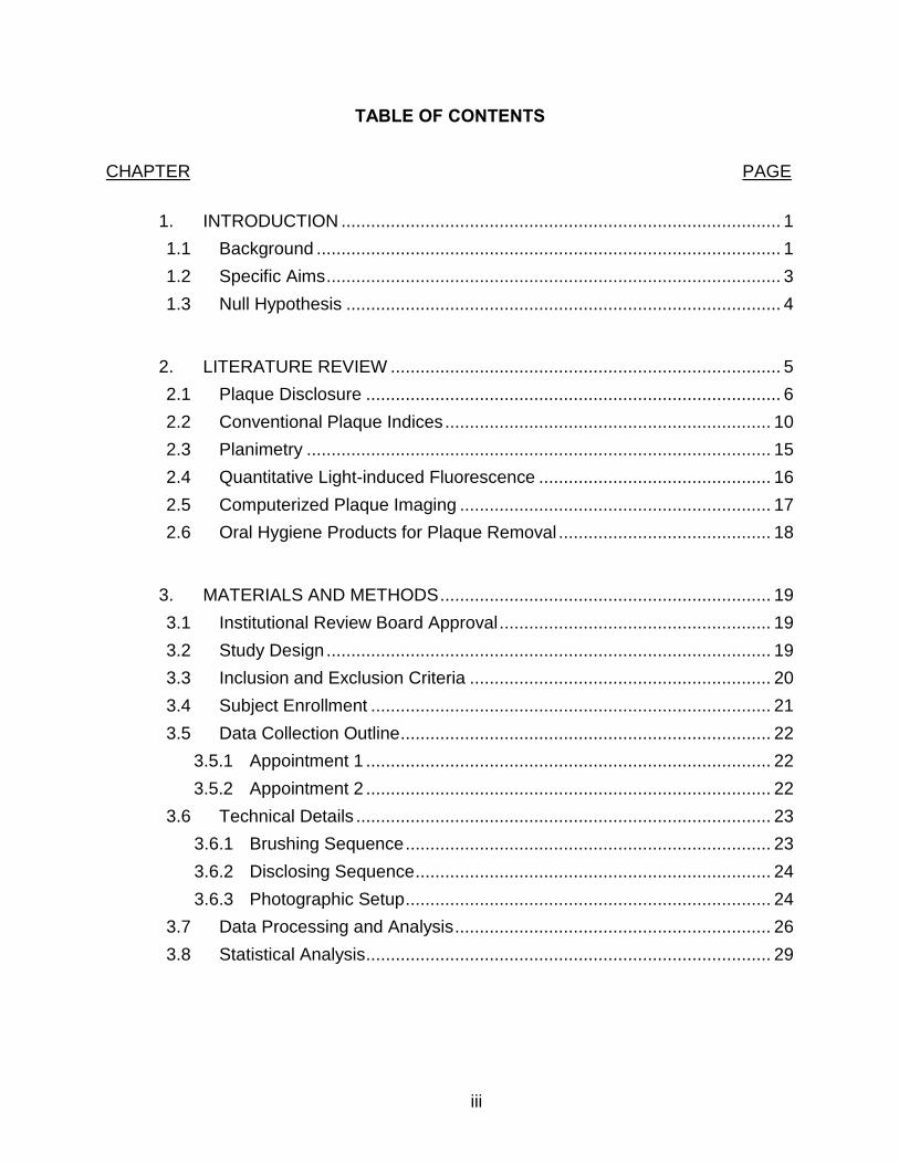
TABLE OF CONTENTS
CHAPTER PAGE
1. INTRODUCTION ......................................................................................... 1
1.1 Background .............................................................................................. 1
1.2 Specific Aims ............................................................................................ 3
1.3 Null Hypothesis ........................................................................................ 4
2. LITERATURE REVIEW ............................................................................... 5
2.1 Plaque Disclosure .................................................................................... 6
2.2 Conventional Plaque Indices .................................................................. 10
2.3 Planimetry .............................................................................................. 15
2.4 Quantitative Light-induced Fluorescence ............................................... 16
2.5 Computerized Plaque Imaging ............................................................... 17
2.6 Oral Hygiene Products for Plaque Removal ........................................... 18
3. MATERIALS AND METHODS ................................................................... 19
3.1 Institutional Review Board Approval ....................................................... 19
3.2 Study Design .......................................................................................... 19
3.3 Inclusion and Exclusion Criteria ............................................................. 20
3.4 Subject Enrollment ................................................................................. 21
3.5 Data Collection Outline ........................................................................... 22
3.5.1 Appointment 1 .................................................................................. 22
3.5.2 Appointment 2 .................................................................................. 22
3.6 Technical Details .................................................................................... 23
3.6.1 Brushing Sequence .......................................................................... 23
3.6.2 Disclosing Sequence ........................................................................ 24
3.6.3 Photographic Setup .......................................................................... 24
3.7 Data Processing and Analysis ................................................................ 26
3.8 Statistical Analysis .................................................................................. 29

iv
TABLE OF CONTENTS (continued)
CHAPTER PAGE
4. RESULTS .................................................................................................. 30
4.1 Photographic Analysis ............................................................................ 30
4.2 Initial Comparison of Groups .................................................................. 34
4.3 Change in Plaque Coverage Within Groups ........................................... 34
4.4 Comparison of Toothpastes ................................................................... 36
5. DISCUSSION ............................................................................................ 38
5.1 Interpretation of the Results ................................................................... 38
5.2 Subject Selection .................................................................................... 39
5.3 Test Toothpaste ..................................................................................... 39
5.4 Brushing Instructions .............................................................................. 40
5.5 Photographic Setup ................................................................................ 41
5.6 Rinse Components ................................................................................. 42
5.7 Photographic Processing ....................................................................... 43
5.8 Digital Plaque Imaging Software ............................................................ 43
5.9 Limitations of the Study and Future Research ....................................... 46
6. CONCLUSIONS ........................................................................................ 48
CITED LITERATURE ......................................................................................... 49
APPENDICES .................................................................................................... 55
APPENDIX A ............................................................................................. 56
APPENDIX B ............................................................................................. 59
APPENDIX C ............................................................................................. 60
VITA.. ................................................................................................................. 61

v
LIST OF TABLES
TABLE PAGE
I. STUDY CROSSOVER DESIGN .................................................................... 20
II. RESULTS OF PIXEL ANALYSIS BY DPIA SOFTWARE .............................. 32
III. COMPARISON OF PLAQUE COVERAGE (%) BETWEEN GROUPS AT APPOINTMENT 1 ......................................................................................... 34
IV. COMPARISON OF PLAQUE COVERAGE (%) OF THE CONTROL
GROUP BETWEEN APPOINTMENTS ........................................................ 35
V. COMPARISON OF PLAQUE COVERAGE (%) OF THE EXPERIMENTAL GROUP BETWEEN APPOINTMENTS........................................................ 36 VI. COMPARISON OF PLAQUE COVERAGE (%) BETWEEN GROUPS AT APPOINTMENT 2 ......................................................................................... 37

vi
LIST OF FIGURES
FIGURE PAGE
1. Sample photo of Plaque-A-Way disclosing plaque on teeth. ................... 24
2. Photographic setup. ................................................................................. 25
3. Photo processing and upload sequence.. ................................................ 27
4. Pixel class definition process.. ................................................................. 28
5. Comparison of photos and corresponding classification output between the present study and the work of Sagel et al…………………………….. 31
6. Comparison of differently-weighted outputs.. ........................................... 45

vii
LIST OF ABBREVIATIONS
3D Three-Dimensional BBI Bonded Bracket Index DMPI Distal Mesial Plaque Index DPIA Digital Plaque Imaging Analysis
FDA Food and Drug Administration FD and C Food, Drug, and Cosmetic
LED Light Emitting Diode NPI Navy Plaque Index OHI-S Simplified Oral Hygiene Index OPRS Office of the Protection of Research Subjects OTC Over-the-Counter PHP Patient Hygiene Performance PPI Plaque Percent Index QLF Quantitative Light-induced Fluorescence
RGB Red, Green, Blue SD Standard Deviation UV Ultraviolet UIC University of Illinois at Chicago

viii
SUMMARY
Inadequate oral hygiene practices in the general population lead to gingivitis,
periodontitis, and tooth decay. There are numerous oral care products on the market
which aim to assist the general public to improve plaque removal efficacy at home, such
as disclosing tablets, mouth rinses, and toothpastes containing antimicrobials. Plaque-
A-WayTM (TJA Health, Joliet, IL) is a newly developed dentifrice which incorporates a
disclosing agent into the formulation. The purpose of this study was to compare the
plaque removal efficacy of Plaque-A-WayTM to that of a placebo toothpaste.
There are several methods for measuring plaque in the oral cavity. These include
conventional plaque indices, which measure the presence, absence, or amount of
plaque in designated tooth locations; planimetry, which maps the outline of plaque and
calculates the percentage of coverage; quantitative light-induced fluorescence (QLF),
which illuminates the oral cavity with ultraviolet light that results in red auto-fluorescence
of plaque deposits; and digital plaque imaging analysis (DPIA), which discloses plaque
with fluorescein causing deposits to glow yellow-green when exposed to ultraviolet light
followed by a computerized photo analysis.
A total of 35 subjects completed this study. After a period of refraining from the
completion of oral hygiene, subjects were asked to brush their teeth with either the test
toothpaste (Plaque-A-WayTM) or a placebo toothpaste at two separate appointments. No
special brushing instructions were given to the subjects. A rinse sequence was
completed using fluorescein to disclose any remaining plaque after brushing. An

ix
SUMMARY (continued)
intraoral photo was captured and analyzed for percentage plaque coverage using
custom made DPIA software.
The changes of plaque percentages between appointments for the control and
test groups were calculated and there were no statistically significant differences found
between the two. This suggests that the use of Plaque-A-WayTM did not result in a
significant amount of plaque removal compared to the placebo with the testing protocol
used. This may have been due to several limitations to this study including: non-specific
brushing instructions, small sample size, inconsistent lighting parameters, and
investigator subjectivity during photo processing. Despite inconclusive results obtained
from the present study, the test toothpaste (Plaque-A-WayTM) demonstrates potential as
a valuable over-the counter (OTC) oral hygiene aid for the general public.

1
1. INTRODUCTION
1.1 Background
Ineffective dental plaque removal has been shown to cause demineralization,
caries, gingivitis, and periodontitis (Loe and Silness, 1963; Ower, 2003; Ferreira and
Mendes, 2005). These conditions result in physical and cosmetic damage to both soft
and hard tissues in the form of bleeding and swollen gums, white spot lesions, enamel
discoloration, the need for restorations, and potentially tooth loss. Prevalence of tooth
decay and periodontal disease is high despite many patients’ claims of following
recommended homecare guidelines for removing plaque. The discrepancy in what
patients report versus disease actually found is likely due to a variety of factors beyond
intentional misrepresentation by patients. For example, poor oral hygiene skill, restricted
dexterity, and a lack of dental knowledge, motivation, and ability to accurately evaluate
one’s oral status all have negative impacts on plaque removal.
In order to address the problem of ineffective dental plaque removal, there is a
need to elevate the level of homecare among patients in the general population.
Increasing education and technique instruction is one way to achieve that goal.
Improving homecare products themselves may be another effective way to address the
problem. This is especially important in populations of minorities, patients having low
socioeconomic status, and elderly patients, each of which traditionally have decreased
access to professional oral care (Kim et al., 2012). The link between higher
socioeconomic status and increased oral health knowledge, positive dental attitudes
and behaviors was echoed by Schou and Wight (1994).

2
There are a variety of methods to study the impacts of oral hygiene on the
disease process. Clinical plaque analysis has been performed for decades using
several manual indices developed by Ramfjord, Quigley and Hein, Silness and Loe,
O’Leary, and Elliott, as well as others (Fischman, 1986). However, reviews of such
manual indices, including that of Pretty, et al. (2005), found that “traditional plaque
indices are problematic due to their integral nature and their failure to detect small, but
potentially clinically relevant changes in plaque area.” These procedures are also time
consuming, subjective, and invasive to patients.
Alternatively, the digital plaque imaging analysis (DPIA) method introduced by
Sagel et al. (2000) makes use of clinical photography and computer software to
increase speed of data collection, operator consistency, reproducibility of results, the
ability to store data for later use and analysis, and most importantly patient comfort. The
disclosing agent used in DPIA, which is fluorescein (FD and C No. 8), has been well-
documented for intraoral plaque disclosure (Lang et al., 1972; Cohen et al., 1972). To
conduct DPIA, long wave UV light, similar to commercially available black lights and
dental curing lights, and commonly used in medical, scientific, and law enforcement
applications, is used to excite the fluorescein-incorporated plaque on intraoral structures
with enough photographic color separation to be analyzed quantitatively pixel by pixel
(Sagel et al., 2000). The efficacy, safety, and reliability of DPIA have been tested
thoroughly, and it has become a standard plaque analysis procedure at
Procter & Gamble (White et al., 2006; Klukowska et al., 2011).

3
Despite the availability of existing oral care products to aid patients with removing
plaque, such as disclosing tablets or mouth rinses, the inadequacy of patients’ oral
hygiene technique suggests the need for additional products that are simple, easily
accessible, and minimally invasive. Visualization of the location of plaque has the
potential to increase patients’ awareness and encourage them to be more thorough
when performing homecare (Block et al., 1972). Plaque-A-WayTM, the test product in
this study, incorporates a disclosing agent directly into the toothpaste. The dye in
Plaque-A-WayTM is an organic food colorant derived from the plant Annato (Bixa
orellana) and is registered with the FDA. The dye adheres to plaque and stains it green,
providing users with a visual indication of the location of plaque on their teeth to improve
brushing efficacy.
1.2 Specific Aims
The purpose of this pilot clinical trial was to compare subjects’ plaque removal
efficacy with the test toothpaste (Plaque-A-WayTM) versus the placebo toothpaste by
using digital plaque imaging analysis. We were to determine whether the presence of a
visual indicating dye in the toothpaste would cause a difference in the way subjects
brush their teeth even if the subjects were not told specifically to brush off the green
dye. Ultimately the goal would be to increase patient awareness of existing plaque
deposits and, therefore, improve the level of plaque removal during homecare. In
addition, a simple and objective method of plaque analysis that can be readily used in a
smaller scale clinical setting is presented.

4
1.3 Null Hypothesis
There is no mean difference in plaque reduction between an indicating dye-
containing toothpaste and a traditional non dye-containing toothpaste when subjects are
not given specific instructions to brush off the stained plaque.

5
2. LITERATURE REVIEW
The complex biofilm present in dental plaque has long been recognized as the
cause of caries and periodontitis (Loe and Silness, 1963).
The American Dental Association (Council on Dental Therapeutics, 1985) defines
plaque as “a highly variable entity resulting from the colonization and growth of micro-
organisms on the surfaces of the teeth and oral soft tissues and consisting of a number
of microbial species and strains embedded in an extracellular matrix.” The bacterial and
salivary components of plaque attach to the tooth surface in layers, starting with pioneer
species and progressing to a more diverse array of filamentous and anaerobic bacteria
(Marsh and Martin 1992). Compared to food deposits and other oral debris, plaque has
a specific adherent architecture and cannot simply be rinsed away (Block et al., 1972).
Therefore, mechanical removal of plaque has been the cornerstone of oral hygiene
practices for centuries. Studies aiming to further the understanding of periodontal
disease should evaluate subjects’ oral hygiene status and technique, and performing a
plaque index is an important part of that process (Silness and Loe, 1964).
The soft biofilm of plaque becomes calcified over time, making at-home removal
increasingly difficult for patients. Therefore, the general aim of oral hygiene protocols is
to direct patients to remove as much of the plaque as possible from the teeth on a daily
basis. The successful use of a toothbrush or dental floss to reduce soft deposits
depends on the awareness, skill, and motivation of the individual patient (Rustogi et al.,
1992). A person must be able to identify plaque deposits and visualize potential problem
areas where plaque tends to accumulate. Common sites include interproximal contact
regions, carious lesions, irregular gingival contours, occlusal fissures, poorly contoured

6
restorations, and the area around the junction of the teeth with removable or fixed
appliances, including orthodontic bands and brackets (Pretty et al., 2005). Effective
visual feedback can have a significant positive effect on patients’ oral hygiene technique
(Godin, 1976).
2.1 Plaque Disclosure
Even before the plaque-associated etiology of periodontal disease was
discovered, Skinner (1914) recognized the role of plaque disclosure in improving oral
health status. His approach was to stress the importance of prevention versus
restoration. By giving patients a visual tool to improve their oral hygiene, he also gave
them some responsibility and control over any subsequent outcomes. Later work by
Arnim (1963) recognized that plaque accumulation occurred too quickly to be the sole
responsibility of the dental practitioner. This realization led, in part, to a more
widespread use of disclosing agents as part of an oral care routine.
Because plaque deposits may be difficult for patients (and sometimes even
practitioners) to identify clearly, a disclosing agent can be used to stain the soft material,
which includes bacteria-related products and pellicle (Fischman, 1986; Marsh and
Martin 1992). The use of disclosing agents chairside and at home has now become a
common teaching tool to accompany oral hygiene instructions. Baab and Weinstein
(1983) suggested that “patients can be taught accurately to recognize and score plaque
in their own mouths using a self-instructional format.” Indeed self-evaluation is very
important for long-term oral hygiene improvement and the prevention of gingivitis and
periodontitis (Lindhe et al., 1984; Baab and Weinstein, 1986).
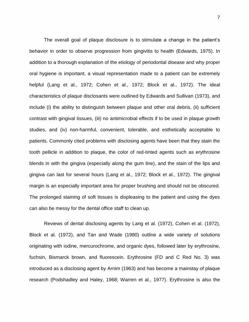
7
The overall goal of plaque disclosure is to stimulate a change in the patient’s
behavior in order to observe progression from gingivitis to health (Edwards, 1975). In
addition to a thorough explanation of the etiology of periodontal disease and why proper
oral hygiene is important, a visual representation made to a patient can be extremely
helpful (Lang et al., 1972; Cohen et al., 1972; Block et al., 1972). The ideal
characteristics of plaque disclosants were outlined by Edwards and Sullivan (1973), and
include (i) the ability to distinguish between plaque and other oral debris, (ii) sufficient
contrast with gingival tissues, (iii) no antimicrobial effects if to be used in plaque growth
studies, and (iv) non-harmful, convenient, tolerable, and esthetically acceptable to
patients. Commonly cited problems with disclosing agents have been that they stain the
tooth pellicle in addition to plaque, the color of red-tinted agents such as erythrosine
blends in with the gingiva (especially along the gum line), and the stain of the lips and
gingiva can last for several hours (Lang et al., 1972; Block et al., 1972). The gingival
margin is an especially important area for proper brushing and should not be obscured.
The prolonged staining of soft tissues is displeasing to the patient and using the dyes
can also be messy for the dental office staff to clean up.
Reviews of dental disclosing agents by Lang et al. (1972), Cohen et al. (1972),
Block et al. (1972), and Tan and Wade (1980) outline a wide variety of solutions
originating with iodine, mercurochrome, and organic dyes, followed later by erythrosine,
fuchsin, Bismarck brown, and fluorescein. Erythrosine (FD and C Red No. 3) was
introduced as a disclosing agent by Arnim (1963) and has become a mainstay of plaque
research (Podshadley and Haley, 1968; Warren et al., 1977). Erythrosine is also the

8
main ingredient in the popular GUM Red-Cote® disclosing tablets and liquid commonly
used in clinical trials and for patient education. Similarly to erythrosine, fuchsin stains
plaque a strong red-magenta color. However, both erythrosine and fuchsin have been
plagued by claims of potential carcinogenicity (Lang et al., 1972; Fischman, 1986).
A two-tone dye was developed by Block et al. (1972), which combines
erythrosine and fast green (FD and C Green No. 3) to create a bluish disclosant. This
combination is unique in that thinner, newer plaque coverage is stained red, while
thicker, older bacterial colonies are stained blue. Block’s study claims that the blue color
is more distinguishable to patients and there are no issues concerning persistent
staining. The study also proposed that this method would be helpful in studying the
bacterial composition of different regions and stages of plaque accumulation.
Kieser and Wade (1976) compared the use of food colorings to traditional
disclosants and suggested that the food colorings would be more cost-effective, have
better taste properties, and possibly have less concerns with carcinogenicity. Kieser and
Wade found the food colorings to have similar ability to stain plaque as the other agents
mentioned above. They also determined that colors in the blue range were more
effective than other colors because of the increased contrast between disclosed plaque
and the gingiva.
The use of fluorescein (FD and C Yellow No. 8) as a disclosant was first
introduced by Brilliant in 1971 (Cohen et al., 1972). Fluorescein is similar in chemical
structure to erythrosine. However, due to its pale yellow color under visible light, it has a
lower propensity for unpleasant staining of the oral cavity. When exposed to UV light,

9
the fluorescein-stained plaque glows bright yellow-green against darker plaque-free
tooth and gingival structures. The Plak-Lite® system (Brilliant Enterprises, Inc.,
Philadelphia, PA) using fluorescein as a disclosing agent was evaluated by Lang et al.
(1972). The illumination was in the range of 420-560nm which targets the peak
absorption spectrum of fluorescein. The study evaluated subjects via the Silness and
Loe plaque index method (1964), and compared disclosure with fluorescein and the
Plak-Lite® system to a more traditional disclosure with erythrosine. In addition to less
residual visible staining, fluorescein was also reported to have a more pleasing taste
than erythrosine and was found to be a more specific disclosant because it does not
adhere to the pellicle like erythrosine. Unlike erythrosine, fluorescein also has the added
benefit of not being antibacterial, which would make it more acceptable for use in long-
term plaque growth studies. Another study comparing plaque disclosure using
fluorescein and erythrosine by Cohen et al. (1972) also proposed that disclosure with
fluorescein was more successful and possibly more preferred by subjects due to better
taste, more vibrant visual disclosure, and less objectionable staining of the oral cavity.
Silva et al. (2004) compared DentPlaque (Axis Biotec, Brazil), a product which
incorporates a disclosing agent directly into toothpaste (although not currently available
in the United States), to traditional disclosing tablets. The study concluded that while the
tablets produced better plaque disclosure than the toothpaste, subjects preferred the
taste of the toothpaste over the disclosing tablets and were, therefore, more motivated
to use the product. The direct dispensing of a disclosing agent from a toothbrush has
been proposed in United States Patent No. 6371674 (Lerner, 2002), but such a product

10
does not currently appear to be on the market. Finally, Miranda et al. (2014) compared
the use of certain pre-brushing mouth rinses containing disclosing agents, but such
rinses showed no difference in plaque removal versus a placebo containing just water.
2.2 Conventional Plaque Indices
An increased focus on the identification and quantification of dental plaque both
for research purposes and patient education began in the 1950s (Podshadley and
Haley, 1968). The need arose to consistently identify plaque in order to study the
components of its biofilm as well as its removal efficacy by oral care products, such as
toothbrushes, toothpastes, and mouth rinses. As a result, numerous conventional
plaque indices have been developed and tested over the years, as outlined in reviews
by Mandel (1974) and Fischman (1986). Recommended requirements of an index
outlined by Davies (1968) included (i) ease of use with a large population while
minimizing time and cost, (ii) clear and reproducible criteria, (iii) suitability for statistical
analysis, and (iv) consistent sensitivity across the designated scale with distinction of
disease progression. In general, plaque indices attempt to quantify plaque deposits
either by defining and scoring coverage zones, or measuring the actual thickness or
volume of the soft debris itself (Fischman, 1986). Ideally, the information gained through
plaque analysis can be rapidly translated into education and treatment options for a
patient (Silberman et al., 1998).
One of the first references to a plaque index in the literature was by Ramfjord
(1959). Modified by Shick and Ash (1961) and eventually used as an adjunct with the
Periodontal Index (Ramfjord, 1967), Ramfjord’s index utilizes a scoring system that

11
accounts for presence of plaque along the gingival margins and interproximal surfaces
of six selected teeth stained with Bismarck brown solution. The scores range from 0
(absence of plaque) to 3 (plaque covering more than two-thirds of the selected site).
The site scores are then averaged to derive the overall score. Using a subset of
representative teeth allows for quick scoring by an examiner for research purposes, but
it may not be the best method for patient education.
Another plaque index commonly used for research purposes was created by
Quigley and Hein (1962) and modified by Turesky at el. (1970). Subjects were
instructed not to brush their teeth for three days prior to evaluation to increase the
presence of biofilm. Plaque disclosure was accomplished with fuchsin and photographs
were taken to create a permanent record of the data. The tooth surface subdivisions are
slightly different from Ramfjord’s index, and there is a longer scale from 0 (no plaque) to
5 (plaque covering two-thirds or more of the crown). Again, the total score is the mean
of the site scores and only selected teeth are scored. Scoring sites on the labial, buccal,
and lingual surfaces is valuable for anti-plaque studies which evaluate the efficacy of
various oral hygiene aids such as toothbrushes, dental floss and topical anti-plaque
agents and dentifrices (Fischman, 1986).
The simplified oral hygiene index (OHI-S) developed by Greene and Vermillion
(1964) evaluates all soft debris composed of salivary proteins and food as well as
bacterial components. The scoring ranges from 0 (no debris or stain) to 3 (soft debris
covering more than two-thirds of the tooth surface). The presence of debris is detected
by running an explorer along the non-disclosed surfaces of six teeth (four posterior and

12
two anterior). Fischman (1986) determined that this index gives inadequate weight to
deposits along the gingival margin and was deemed less suitable for study groups with
lower plaque levels. However, the developers of the index claim that advantages
include less discretionary decision-making by the examiner and reduced completion
time (Greene and Vermillion, 1964). Additionally, this index has been useful for
assessing oral hygiene education programs (Greene, 1967).
Similar to the OHI-S previously described, a plaque index method developed by
Silness and Loe (1964) and later updated by Loe (1967) can be performed without
disclosure because it measures soft deposits collected by running a probe over the
tooth surfaces. The scoring ranges from 0 (no plaque present visually or on the probe)
to 3 (heavy accumulation of visible plaque along the gingival margin and into the
interdental area). This index is more difficult to use in larger scale trials because the
plaque deposits are disturbed during data collection and therefore unable to be
evaluated repeatedly by additional examiners (Fischman, 1986; Pretty et al., 2005).
One of the first indices to take all teeth into account was originated by O’Leary
(1967a; O'Leary et al., 1972). His method was prompted by the need for a plaque index
that could be applied more easily by practitioners chairside, specifically in the military,
as compared to a research setting. O’Leary also intended to encourage “the patient to
visualize his own progress in learning plaque control” which subsequently increases
patient motivation (O'Leary et al., 1972). Each tooth is scored from 0 (no plaque) to 3
(plaque covering more than one-half of the crown), and only the highest scoring tooth in
each segment is fed into the overall mean score. This index has been criticized for

13
giving too little weight to the gingival margin zone (Fischman, 1986). However, Baab
and Weinstein (1983) found this method helpful because of its simplicity and because,
in their opinion, it does adequately highlight plaque at the gingival margin as well as
interproximally.
The patient hygiene performance (PHP) method developed by Podshadley and
Haley (1968) and modified by Martens and Meskin (1972) discloses plaque with
erythrosine tablets and evaluates six teeth. A score is given to each of the five
subdivisions per tooth and then the mean overall score is calculated. Only two scores
per subdivision are possible, 0 (no debris present) or 1 (debris definitely present). The
PHP was found to be more sensitive interproximally than the OHI-S when evaluating
oral hygiene aids (Anaise, 1977) and is useful for patient education (Silberman et al.,
1998).
The Navy Plaque Index (NPI) was developed by Elliot et al. (1972) and modified
by Rustogi et al. (1992). Plaque disclosure is carried out using fuchsin. Instead of a
score range, however, the presence of plaque in an individual zone is given a score of 1
and then all the zones are aggregated to give a final score. There are more designated
zones adjacent to the gingival margin compared to the rest of the tooth surface.
Therefore, unlike several of the previously described indices, the NPI more adequately
considers the importance of plaque along the gingival margin by giving a higher weight
to deposits located there (Fischman, 1986).
The distal mesial plaque index (DMPI) originated by Cancro in 1983 and outlined
by Fischman (1986) uses more detailed subdivisions and is more time consuming than

14
other indices. However, it is highly useful for clinical trials related to oral care product
testing due to the attention paid to the gingival margin and interproximal areas. Scores
range from 0 (absence of plaque) to 3 (plaque covering the entire area).
Numerous additional modifications to the concept of conventional plaque indices
have evolved over the last several decades including the hygiene analysis index (Love
et al., 1975), global plaque index (Benson et al., 1993), plaque assessment scoring
system (Butler et al., 1996), index of oral cleanliness (Bearn et al., 1996), and the
University of Mississippi oral hygiene index (Silberman et al., 1998). However, despite
these repeated attempts at redefining evaluation zones to improve plaque evaluation,
no consensus has been reached in the literature on the best one to use.
There are fewer examples in the literature of plaque indices devised specifically
for orthodontic patients. The bonded-bracket index (BBI) developed by Ciancio et al.
(1984) is a system that specifically assigns subdivisions of plaque assessment to the
teeth, gingiva and orthodontic brackets. Scores range from 0 (no plaque on the bracket
or tooth surfaces) to 5 (plaque on tooth, bracket and extension to gingiva). A recent
systematic review by Al-Anezi and Harradine (2012) determined that the most common
plaque index used for evaluating orthodontic subjects is that of Silness and Loe, but that
newer techniques such as planimetry and DPIA would increase validity and
reproducibility over traditional indices (although the latter are more technically complex).
The conventional plaque indices described above have been considered
complicated, time-consuming, cumbersome and tedious (Butler et al., 1996; Silberman
et al., 1998). Other limitations of such plaque indices involve inherent difficulties in
15
appropriately weighting certain critical areas of the tooth surface, such as the gingival
margin. Some indices fail to show a clear change in score even though the amount of
plaque has been significantly reduced. Indices are also somewhat subjective and
require specific training and strict calibration of all examiners (O'Leary, 1967b; Greene,
1967; Bentley and Disney, 1995; Shaloub and Addy, 2000). This makes it more difficult
to conduct larger scale and multicenter trials consistently because often intra-examiner
consistency is better than inter-examiner agreement (Shaloub and Addy, 2000).
Conversely, methods of analyzing plaque that increase precision, objectivity, and
reliability would potentially allow a reduction in time and number of study participants in
clinical trials (Pretty et al., 2005). In addition, many of the plaque indices appear more
suited for epidemiological studies and not necessarily practical for clinical trials or oral
hygiene evaluation in a private practice setting (Warren et al., 1977; Butler et al., 1996;
Silberman et al., 1998). Because different indices measure subdivisions that are
constructed in different ways, it can be very difficult to accurately compare them to each
other to determine which is most valid or useful (Poulsen et al., 1979).
2.3 Planimetry
Planimetric analysis involves the calculation of a Plaque Percent Index (PPI)
(Lang et al., 1972), which is accomplished by disclosing the plaque, taking a photo,
tracing and determining the area covered by plaque and then dividing by the total tooth
area. Planimetric analyses differ from traditional plaque indices because the areas of
plaque deposits are mapped and quantified, either manually or with the aid of a
computer, making the use of interval rather than ordinal scale data analysis possible

16
(Pretty et al., 2005). One key advantage of planimetry over traditional plaque indices is
that in planimetry, a photographic record is captured and saved, such that the same
sample may be used for repeated or future analysis (Addy et al., 1999). This is more
favorable than conventional plaque indices where the disclosed plaque sample may be
disturbed or degraded during data collection, and where such sample is only available
while the subject is present. Planimetry is still time-consuming. However, it has potential
as a highly efficient and reliable method with increased objectivity, reliability, precision,
and sensitivity when compared to traditional plaque indices (Soder et al., 1993; Pretty et
al., 2005). A study by Renton-Harper et al. (1999) compared previously calculated
plaque coverage area percentages to one of the most commonly used conventional
plaque indices and confirmed that planimetric areas can also be reliably converted to
other indices for research purposes.
2.4 Quantitative Light-induced Fluorescence
Quantitative light-induced fluorescence (QLF) is another method that has
applications both in research and patient education. Initially used to identify carious
lesions and tooth decalcification, QLF with blue light at 408nm has since been shown to
effectively highlight plaque deposits by causing them to appear red-orange against the
yellow-green tooth background (Amaechi and Higham, 2002; Raggio et al., 2010). The
obligate anaerobic bacteria contained in mature plaque are responsible for the red auto-
fluorescence. QLF has been determined to be a reliable method for identifying plaque
deposits targeted for removal by practitioners, and it may potentially be used for patient
education (Pretty et al., 2005). Advantages include ease of intraoral use, reduced

17
distortion and reflection, and advances are being made to increase positional
reproducibility.
2.5 Computerized Plaque Imaging
Based on the method developed by Sagel et al. (2000), digital plaque imaging
analysis (DPIA) makes use of fluorescein to disclose plaque deposits. When exposed to
long wave UV light, fluorescein is a bright yellow-green, which contrasts with the
surrounding tooth structure which appears darker and blue in color. An intraoral photo of
the properly illuminated subject is captured and fed into a computer program that
classifies each pixel in the image as plaque, tooth, gingiva, etc., using a least squared
distance algorithm. Similar to planimetry, a plaque coverage percentage is calculated
automatically and then aggregate sample data may be further analyzed. Computer
analysis removes much of the subjectivity that has hindered conventional plaque
indices. Drawbacks include insufficient application to posterior and lingual surfaces, and
patient positioning reproducibility can be an issue as well (Matthijs et al., 2001; Mohan
et al., 2012).
The DPIA method is now used frequently by researchers at Procter & Gamble to
test oral care products such as toothpaste and mouth rinses, and a version using
illumination with a white light only (non-UV) has been shown to be effective as well
(Bellamy et al., 2008). Klukowska et al. (2011) has successfully adapted the DPIA
method for studying orthodontic patients which is more complicated due to the presence
of fixed appliances on the teeth.

18
Other contemporary plaque imaging techniques were created by Smith et al.
(2001) and Carter et al. (2004) in an attempt to reduce inter-operator error. Their
procedures followed a similar progression as Sagel’s original DPIA method (i.e., plaque
disclosure, digital photo capture, software manipulation and computerized pixel
analysis). The main differences among them are that Smith used erythrosine and Carter
used methylene blue as the disclosants instead of fluorescein, and both Smith and
Carter incorporated white lighting versus the UV illumination used by Sagel.
In any event, these digital plaque assessment methods all attempt to better
quantify a percentage of total plaque coverage as compared to using tooth subdivisions
to assign ordinal scores as in traditional manual plaque indices. Quantifying a
percentage of total plaque coverage is important because with the conventional index
scoring methods, the same score may be obtained with different total amounts of
plaque present. In addition, teeth of different sizes may be assigned different scores
despite having the same amount of total plaque present (Carter et al., 2004).
2.6 Oral Hygiene Products for Plaque Removal
Commonly available over-the-counter (OTC) oral hygiene aids used for
mechanical plaque removal include toothbrushes, dental floss, disclosing tablets, rubber
tips, interdental picks and proxabrushes (Warren and Chater, 1996; Schiff et al., 2006).
Examples of chemical agents used to reduce dental plaque include toothpastes, mouth
rinses, and topical gels or foams which often contain antimicrobials such as fluoride,
oxygenating agents, anti-attachment agents, and non-ionic agents like triclosan (Gaffar
et al., 1997).

19
3. MATERIALS AND METHODS
3.1 Institutional Review Board Approval
This study was approved by the University of Illinois at Chicago (UIC) Institutional
Review Board, Office of the Protection of Research Subjects (OPRS), on February 14,
2013, IRB Protocol #2013-0113 (Appendix A).
3.2 Study Design
This was a pilot study to compare the effects on dental plaque removal of a test
toothpaste versus a placebo. From here on, the test toothpaste is defined as Plaque-A-
WayTM and contains a green disclosing dye. The placebo toothpaste has the same
basic formulation as the test toothpaste. However, it does not contain the green
disclosing dye and is white in color. The participants were divided into two groups, a
control (Group A) and an experimental group (Group B). A crossover design shown in
Table I was used where the control group brushed with the placebo at both Appointment
1 and Appointment 2, and the experimental group brushed with the placebo toothpaste
at Appointment 1 and then the test toothpaste at Appointment 2. The subjects were
asked to perform toothbrushing in their customary way. When applying the test
toothpaste No instruction was given to brush off the green dye on their teeth surfaces.
Statistics were then used to compare the two study groups to each other at both
Appointment 1 and Appointment 2, and also compare the individual groups between
appointments.

20
TABLE I
STUDY CROSSOVER DESIGN
Group Appointment 1
Toothpaste Appointment 2
Toothpaste
Control (Group A)
Placebo Placebo
Experimental (Group B)
Placebo Test (Plaque-A-WayTM)
3.3 Inclusion and Exclusion Criteria
Inclusion criteria were English-speaking adults 18 years of age or older; in good
general health (via self-assessment); with all 12 anterior teeth present (canine to canine
in both dental arches); and able to commit to two 30 minute visits. Exclusion criteria
were pregnant or nursing subjects; dental students and clinical faculty or staff;
antibiotics taken within two weeks of the data collection appointments; symptoms of dry-
mouth or significant food allergies; dental restorations or fixed appliances of any kind in
the anterior region (canine to canine in both dental arches); visible caries or staining
present in the anterior region; and new restorations or prophylaxis anywhere in the oral
21
cavity within 30 days prior to the first appointment or planned within the duration of
study participation.
3.4 Subject Enrollment
Subjects were recruited from around the campus through flyers and an
advertisement posted in the UIC online event calendar. The inclusion and exclusion
criteria were assessed of interested participants in person or via phone to confirm
general eligibility. Eligible participants were then scheduled for the first data collection
appointment and given pre-appointment instructions via phone or email. Subjects were
instructed to refrain from brushing, flossing, or using other oral hygiene aids and
chewing gum the evening prior and morning of their scheduled data collection
appointment.
A key code list was created with randomized order subject identification numbers
divided into the two study groups, control and experimental. Qualifying subjects were
assigned to one of the two groups by adding the subject's name to the next random
subject number on the key code list during study enrollment. A total of 37 subjects were
enrolled in the study and assigned subject numbers. Two subjects (Subjects 19 and 31)
were dropped due to failure to be present at the first appointment. Thirty-five subjects
completed both appointments and were included in the data analysis. The control group
consisted of 18 participants and the experimental group consisted of 17. The sample
consisted of 24 females and 11 males with ages ranging from 18 to 61 years.

22
3.5 Data Collection Outline
3.5.1 Appointment 1
Subjects reported to the Orthodontics Department Clinic, Room 131, where they
were provided with a subject information sheet and informed consent form, and any
questions were answered. A signed copy of the informed consent form was retained for
documentation purposes. Inclusion and exclusion criteria and completion of pre-
appointment instructions were verified verbally and visually. At the first appointment all
subjects in both study groups were asked to brush their teeth using the placebo
toothpaste for one minute. Brushing took place at the hallway brushing station in front of
the mirror. The subjects were then escorted to an administrative office and oriented to
the image capture steps, cheek retraction, and head positioning with a brief
demonstration and practice. Next they were asked to complete the disclosing sequence
with fluorescein and phosphate buffer. The subjects were then positioned in the chin
rest with cheeks retracted and teeth slightly apart, the room lights were turned off, and a
frontal intraoral image of the upper and lower teeth was captured.
3.5.2 Appointment 2
At the second appointment, the brushing, disclosing, and photographic
procedures above were repeated. The only difference was that the control group was
instructed to brush with the placebo toothpaste again and the test group brushed with
the test toothpaste (Plaque-A-WayTM) instead of the placebo. Immediately following the
intraoral photograph, there was a study debrief session where the subjects were
informed of the special dye in the test toothpaste, its purpose in the study, and whether

23
they used it or not. They were given the opportunity to withdraw their photographs if
they wished. They were also permitted to brush their teeth again if any of the green
toothpaste or fluorescein remained.
3.6 Technical Details
3.6.1 Brushing Sequence
Subjects were provided with a manual toothbrush and a 1 mL single-use syringe
of the assigned toothpaste. The specific brushing instructions were: “Brush your teeth in
the mirror for one minute.” The process was timed with a stopwatch, but there was
otherwise no intervention by the investigator.
The test toothpaste, Plaque-A-WayTM, is an FDA registered product which
contains a yellow-orange vegetable dye made from Annato (Bixa orellana) seed extract,
plus FD and C Blue No. 1, giving the toothpaste a green color that adheres to intraoral
plaque deposits. A sample intraoral photo of Plaque-A-WayTM in use is shown in
Figure 1. The Drug Facts are contained in Appendix B. The placebo toothpaste is the
same base product, but it does not contain the green dye components and is white.

24
Figure 1. Sample photo of Plaque-A-WayTM disclosing plaque on teeth.
3.6.2 Disclosing Sequence
The disclosing procedure was taken from Procter & Gamble’s DPIA experiments
(Klukowska et al., 2011). The phosphate buffer was composed of 3.62 g monosodium
phosphate and 0.349 g disodium phosphate in 2 L of water, pH 5.5. The disclosing
solution consisted of 1240-ppm fluorescein (FD and C yellow No. 8) in phosphate
buffer. Subjects were instructed to rinse for 10 seconds with 25 mL of phosphate buffer,
rinse for 1 minute with 5.0 mL of 1240-ppm fluorescein in phosphate buffer, and then
rinse 3 times for 10 seconds with 25 mL of phosphate buffer. Subjects expectorated the
solution after each rinse.
3.6.3 Photographic Setup
The photographic setup was modified from Sagel et al. (2000). The camera used
was a Canon EOS Rebel T3 (Canon, Melville, NY). A Tamron 90mm macro lens

25
(Tamron, Commack, NY) was attached to the camera and a Digi-Slave 3200 LED UV
ring flash (SR Electronics, Dallas, TX) was then added to the end of the lens.
The location was an administrative office with the windows blacked out. The
photographic setup is shown in Figure 2. Two tripods were placed opposite each other
on a desk and the positions marked with tape for consistency. The camera assembly
was attached to one tripod. A chin rest for patient positioning was attached to the other.
The distance from the end of the ring flash to the anterior edge of the chin cup was
measured to be 15 inches.
Figure 2. Photographic setup.

26
3.7 Data Processing and Analysis
Upon completion of data collection, the subjects’ photos were stored under a
filename string including only their de-identified subject number, study group,
appointment number, and age for sorting purposes. All other personal data used for
screening and contact purposes was destroyed.
Photos were assessed by a computer analysis method modified from Sagel et al.
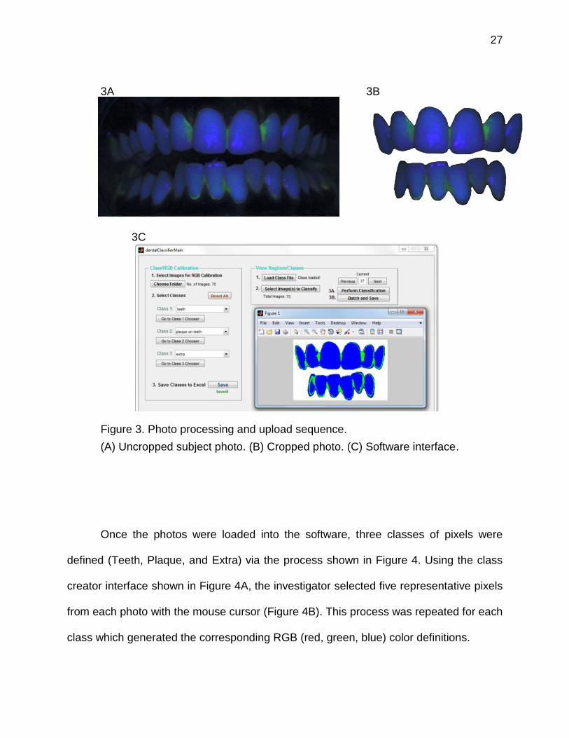
(2000). The photo processing and upload sequence is shown in Figure 3. Using
Photoshop (Adobe, San Jose, CA), the 12 anterior teeth in each intraoral digital photo
(Figure 3A) were cropped around the gingival margin and incisal edges using the
freeform pen tool and eraser tool to define the area of analysis (Figure 3B).
Data analysis software was developed by David Franz at the UIC Research
Resources Center. All cropped digital photos were uploaded into the software program
interface shown in Figure 3C.
27
3A 3B
3C
Figure 3. Photo processing and upload sequence.
(A) Uncropped subject photo. (B) Cropped photo. (C) Software interface.
Once the photos were loaded into the software, three classes of pixels were
defined (Teeth, Plaque, and Extra) via the process shown in Figure 4. Using the class
creator interface shown in Figure 4A, the investigator selected five representative pixels
from each photo with the mouse cursor (Figure 4B). This process was repeated for each
class which generated the corresponding RGB (red, green, blue) color definitions.

28
4A
4B
Figure 4. Pixel class definition process.
(A) Class creator interface. (B) Close up of five plaque pixel selections (black dots)
Next, the resulting pixel class definitions were applied by the software to
automatically analyze the batch of digital photos, assigning every pixel in each photo to
one of the three classes based on the RGB least squared distance color space
algorithm presented in Sagel et al. (2000). The data output consisted of both a visual
representation for each photo a numerical exportation to Excel (Microsoft, Redmond,

29
WA). The pixels designated as Extra were subtracted from the total number of pixels to
isolate only the area inside the gingival margin. The ratio of (Plaque pixels)/(Plaque +
Teeth pixels) x 100% was calculated to give an overall subject plaque percentage.
3.8 Statistical Analysis
Student paired t-tests were performed to test the mean paired differences within
each group and independent t-tests were used to test the mean differences between
groups. Statistical significance was set at p<0.05. SPSS version 22.0 (Chicago, IL) was
used for data analysis.

30
4. RESULTS
4.1 Photographic Analysis
There were 70 digital photographs used for analysis, 35 from Appointment 1 and
35 from Appointment 2. The photos were downloaded from the camera onto a desktop
computer and each saved with a unique de-identified filename that included subject
number, study group, appointment number, and age. The image of the anterior teeth in
all of the photos were then cropped using the computer program Photoshop (Adobe,
San Jose, CA). The cropped photos were loaded into the custom DPIA software, class
definitions were created, and then pixel composition analyzed. The complete Excel data
output summary from the present experiment is shown in Table II. A sample photo from
the present study and the corresponding visual representation of the classification
output were compared to the work of Sagel et al. (2000) and displayed in Figure 5.
Copyright permission to use the reproduced images is contained in Appendix C.

31
5A 5B
5C 5D
Figure 5. Comparison of photos and corresponding classification output between the present study and the work of Sagel et al.
(A) Sample clinical photo from the present study. (B) Corresponding classification output of the photo shown in (A) processed via the
software in this study. (C) Sample clinical photo from Sagel et al. (D) Corresponding classification output of the photo shown in (C) processed by the
software used by Sagel et al.

32
TABLE II
RESULTS OF PIXEL ANALYSIS BY DPIA SOFTWARE
Group Subj Age Sex Appt
Extra (#
Pixels)
Teeth (#
Pixels)
Plaque (#
Pixels)
Total (#
Pixels) Plaque
(%) Appt
Extra (#
Pixels)
Teeth (#
Pixels)
Plaque (#
Pixels)
Total (#
Pixels) Plaque
(%)
A 1 22 F 1 15827 688005 193143 896975 21.92 2 4688 819088 132994 956770 13.97
A 3 24 M 1 24485 938705 390474 1353664 29.38 2 2368 1083018 177273 1262659 14.07
A 6 51 F 1 4952 732141 427063 1164156 36.84 2 3008 820781 325660 1149449 28.41
A 7 20 F 1 6883 1002102 305858 1314843 23.38 2 3042 651284 820342 1474668 55.74
A 8 22 F 1 29473 681517 255310 966300 27.25 2 3687 592883 336147 932717 36.18
A 10 21 F 1 121 868229 205852 1074202 19.17 2 880 896515 99278 996673 9.97
A 11 24 M 1 2263 794567 203165 999995 20.36 2 10326 624716 368973 1004015 37.13
A 15 52 F 1 2447 955451 151962 1109860 13.72 2 38 962417 230502 1192957 19.32
A 16 18 F 1 125 674121 251208 925454 27.15 2 1170 823021 51480 875671 5.89
A 17 59 F 1 777 977659 170672 1149108 14.86 2 211 699861 383066 1083138 35.37
A 22 27 F 1 2212 756086 120439 878737 13.74 2 1550 758272 110884 870706 12.76
A 23 61 F 1 50438 894300 99273 1044011 9.99 2 9626 422041 526871 958538 55.52
A 26 28 F 1 46 864139 261858 1126043 23.26 2 332 969685 100202 1070219 9.37
A 30 24 F 1 6952 889678 212564 1109194 19.28 2 198 966917 144357 1111472 12.99
A 32 48 F 1 62 568965 369893 938920 39.40 2 965 765521 124146 890632 13.95
A 34 43 F 1 6706 759997 161250 927953 17.50 2 1102 751908 129459 882469 14.69
A 35 20 M 1 70355 986117 468298 1524770 32.20 2 8457 1244280 245862 1498599 16.50
A 36 42 F 1 48803 812127 333642 1194572 29.12 2 12563 870666 298673 1181902 25.54
Plaque % = (Plaque pixels)/(Plaque pixels + Teeth pixels) x 100%
33
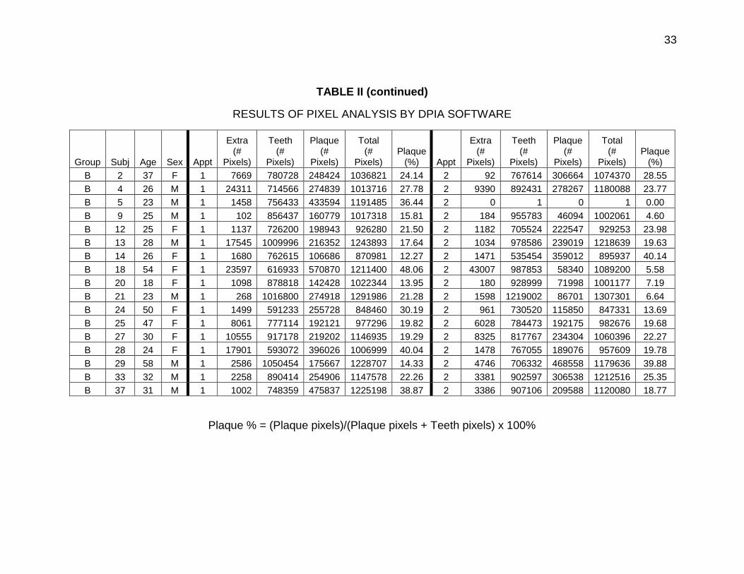
TABLE II (continued)
RESULTS OF PIXEL ANALYSIS BY DPIA SOFTWARE
Group Subj Age Sex Appt
Extra (#
Pixels)
Teeth (#
Pixels)
Plaque (#
Pixels)
Total (#
Pixels) Plaque
(%) Appt
Extra (#
Pixels)
Teeth (#
Pixels)
Plaque (#
Pixels)
Total (#
Pixels) Plaque
(%)
B 2 37 F 1 7669 780728 248424 1036821 24.14 2 92 767614 306664 1074370 28.55
B 4 26 M 1 24311 714566 274839 1013716 27.78 2 9390 892431 278267 1180088 23.77
B 5 23 M 1 1458 756433 433594 1191485 36.44 2 0 1 0 1 0.00
B 9 25 M 1 102 856437 160779 1017318 15.81 2 184 955783 46094 1002061 4.60
B 12 25 F 1 1137 726200 198943 926280 21.50 2 1182 705524 222547 929253 23.98
B 13 28 M 1 17545 1009996 216352 1243893 17.64 2 1034 978586 239019 1218639 19.63
B 14 26 F 1 1680 762615 106686 870981 12.27 2 1471 535454 359012 895937 40.14
B 18 54 F 1 23597 616933 570870 1211400 48.06 2 43007 987853 58340 1089200 5.58
B 20 18 F 1 1098 878818 142428 1022344 13.95 2 180 928999 71998 1001177 7.19
B 21 23 M 1 268 1016800 274918 1291986 21.28 2 1598 1219002 86701 1307301 6.64
B 24 50 F 1 1499 591233 255728 848460 30.19 2 961 730520 115850 847331 13.69
B 25 47 F 1 8061 777114 192121 977296 19.82 2 6028 784473 192175 982676 19.68
B 27 30 F 1 10555 917178 219202 1146935 19.29 2 8325 817767 234304 1060396 22.27
B 28 24 F 1 17901 593072 396026 1006999 40.04 2 1478 767055 189076 957609 19.78
B 29 58 M 1 2586 1050454 175667 1228707 14.33 2 4746 706332 468558 1179636 39.88
B 33 32 M 1 2258 890414 254906 1147578 22.26 2 3381 902597 306538 1212516 25.35
B 37 31 M 1 1002 748359 475837 1225198 38.87 2 3386 907106 209588 1120080 18.77
Plaque % = (Plaque pixels)/(Plaque pixels + Teeth pixels) x 100%

34
4.2 Initial Comparison of Groups
The percentage of plaque coverage remaining between the groups at
Appointment 1 after both brushed with the placebo toothpaste was analyzed with an
independent t-test and is displayed in Table III. The control group had a mean of
23.25% plaque coverage and the experimental group had a mean of 24.92%. No
significant difference was found between the two groups at Appointment 1 (p>0.05);
therefore, the groups were determined to be similar.
TABLE III
COMPARISON OF PLAQUE COVERAGE (%) BETWEEN GROUPS AT APPOINTMENT 1
Group N Mean ± SD Mean Diff
95% CI p-value
Lower Upper
Control 18 23.25 ± 8.16 -1.67 -8.10 4.76 0.601
Experimental 17 24.92 ± 10.45
4.3 Change in Plaque Coverage Within Groups
Paired samples t-tests were used to analyze the mean plaque percentages within
each group. The change in plaque coverage between Appointment 1 and Appointment
2 for the control is shown in Table IV. When the subjects brushed with the placebo
toothpaste initially at Appointment 1, the mean percentage of remaining plaque was

35
23.25%. For the same participants, after brushing with the placebo toothpaste at
Appointment 2, the mean plaque percentage was 23.19%. There was no significant
difference (p>0.05) for the control group between the two appointments.
TABLE IV
COMPARISON OF PLAQUE COVERAGE (%) OF THE CONTROL GROUP BETWEEN APPOINTMENTS
Appointment N Mean ± SD Mean Diff
95% CI p-value
Lower Upper
1 18 23.25 ± 8.16 -0.064 -9.40 9.27 0.989
2 18 23.19 ± 15.17
The difference in plaque coverage after brushing of the experimental group
between Appointment 1 (subjects brushed with the placebo) and Appointment 2
(subjects brushed with the test toothpaste) is shown in Table V. The mean plaque
coverage percentage using the placebo toothpaste was 24.92% whereas that using the
test toothpaste was 18.79%. The mean plaque percentage difference was -6.12.
However, the difference was not statistically significant (p>0.05).

36
TABLE V
COMPARISON OF PLAQUE COVERAGE (%) OF THE EXPERIMENTAL GROUP BETWEEN APPOINTMENTS
Appointment N Mean ± SD Mean Diff
95% CI p-value
Lower Upper
1 17 24.92 ± 10.45 -6.12 -15.63 3.37 0.190
2 17 18.79 ± 11.57
4.4 Comparison of Toothpastes
Using an independent t-test, the percentage plaque coverage after brushing at
Appointment 2 between the control group using the placebo toothpaste and the
experimental group using the test toothpaste were compared and the results are shown
in Table VI. The mean percentages of plaque coverage between the control and the
experimental groups were 23.19% and 18.79% respectively. While there was less
percent plaque associated with the test toothpaste, the difference was not statistically
significant (p>0.05).

37
TABLE VI
COMPARISON OF PLAQUE COVERAGE (%) BETWEEN GROUPS AT APPOINTMENT 2
Group N Mean ± SD Mean Diff
95% CI p-value
Lower Upper
Control 18 23.19 ± 15.17 4.39 -4.93 13.71 0.344
Experimental 17 18.79 ± 11.57

38
5. DISCUSSION
5.1 Interpretation of the Results
The study groups could be considered reasonably similar with regards to plaque
coverage after Appointment 1 where both groups brushed with the placebo toothpaste
because there was no significant difference shown in the mean plaque percentages.
Both the control and the experimental groups showed an overall reduction in mean
plaque percentage following Appointment 2 (-0.064 and -6.12 respectively). The total
difference in remaining plaque between the control and the experimental group after
brushing at Appointment 2 was 4.39%. However, neither plaque reduction comparison
was statistically significant.
The overall reduction in plaque percentages in both groups may be attributed to
several factors, including subjects’ awareness of being observed and photographed and
familiarity with the study procedures when they repeated the steps at Appointment 2. A
study using Plak-Lite® by Friedman et al. (1974) highlights the fact that simply
participating in a brushing study calls subjects’ attention to the presence of plaque, and
can have an impact on the results by altering their awareness or inspiring a more
meticulous or frequent brushing technique. The greater overall reduction in plaque
percentage of the experimental group vs. the control may also indicate that the
presence of the green dye in the test toothpaste did help those subjects identify areas of
more plaque accumulation than the subjects that brushed with the white placebo
toothpaste the second time. However, the statistical analysis cannot guarantee that it
was due to the test effect and not chance in this study.

39
5.2 Subject Selection
Subjects were selected to represent the general population as best as possible in
a comparable manner as studies at Procter & Gamble (Sagel et al., 2000; Klukowska et
al., 2011). Major exclusions were to protect minors, pregnant or nursing women, or
those subjects not in good health. Dental students and clinical faculty were excluded
due to their presumed heightened awareness of oral hygiene. Based on the
photographic method involved, a full complement of anterior teeth with no caries,
restorations, or major stains was required. The criteria regarding no dental procedures
within a month, antibiotics within two weeks and lack of dry mouth symptoms were to
prevent significant impact on intraoral plaque during the study.
5.3 Test Toothpaste
The purpose of the green dye that is present in the test toothpaste (Plaque-A-
WayTM) is to adhere to plaque deposits on the teeth allowing better visualization and
therefore encouraging users to improve their brushing efficacy. In the past, plaque
disclosure by an indicating dye had only been incorporated into chewable tablets and a
mouth rinses in the United States (Cohen et al., 1972; Miranda et al., 2014). Both
require an extra step in the oral hygiene routine which takes more time for the user, and
they are often messy, leaving the mouth bright pink or purple for some time after use.
The green dye in Plaque-A-WayTM is persistent enough to highlight areas of plaque
accumulation, but it is easily brushed away and mostly unnoticeable by the end of each
brushing session. Because the dye is incorporated into the toothpaste itself, the proper
brushing technique and plaque removal lessons learned by users can be accomplished
40
efficiently every time they brush their teeth. In April 2014, the market name of the test
toothpaste was changed to Plaque HD®.
5.4 Brushing Instructions
The intention of the instructions: “Brush your teeth in the mirror for one minute,”
was to avoid calling specific attention to the presence of the green dye in the test
toothpaste. Initially, we did not want to introduce a psychological reason for subjects to
change the way they brushed their teeth between appointments in one group and not
the other (Friedman et al., 1974). It was expected that the subjects in the experimental
group may notice the change in color of the toothpaste (green vs. white) the second
time they brushed, but no questions about it were answered until the debrief session
following the completion of the two appointments. In retrospect, the plaque-indicating
dye in the test toothpaste is the primary mechanism by how it functions. The visual
indication of plaque deposits is what allows users to improve their plaque removal
efficacy. Therefore, the presence of the dye need not be avoided in the brushing
instructions and should be mentioned either before or during brushing.
Additionally, despite instructing subjects to look in the mirror while brushing, it
was noticed after the study started that many subjects were not actually looking in the
mirror. Possibly that was not part of their normal brushing routine. For consistency, we
decided not intervene or redirect the subjects at the time. However, it can be assumed
that if the subjects in the experimental group were not looking in the mirror while
brushing, the dye in test toothpaste may not have been as effective as if they had been
watching.

41
5.5 Photographic Setup
The photographic setup was modified from that outlined in Sagel et al. (2000) in
order to simplify the necessary equipment, reduce costs, and be more easily replicated
in a small-scale clinical setting. We chose a readily available digital camera and lens to
simulate what many practitioners would already use for clinical photos. Similar to a non-
UV model that many clinicians may be familiar with, a ring flash was chosen instead of
the complex flash assembly shown in Sagel’s article for its ease of setup by attaching
directly to the lens. Batteries were used to supply the ring flash LEDs and after
analyzing the photos, the inconsistency in flash intensity was noticed. This was likely
due to declining battery power over time. It would have been better to use a wall outlet-
based power supply to keep the flash level constant throughout the duration of the
study. In addition, although convenient, the ring flash had a tendency to produce a
purple halo in the most reflective areas of the teeth and gingiva. This was partially
compensated for during pixel analysis by including purple pixels as the Teeth category
definition. However, it did introduce a source of inaccuracy. This complication would
have been mitigated by using two separate flashes at a 45 degree angle as in the
original DPIA method to reduce direct reflection of the flash into the camera.
The subject positioning in the chin rest was difficult to replicate at each
appointment. The distance from the rest assembly to the camera was kept constant, but
specific head orientation was not exactly the same between subjects or at each visit.
The subjects’ original photos from Appointment 1 were consulted during positioning for

42
the second photo at Appointment 2, but using a forehead rest may have provided
additional stability.
Because of the UV lighting aspect, manual camera focus had to be employed.
This introduced focus inconsistencies that were not apparent until photo processing.
One of the photos was not focused well enough for pixel analysis to be performed by
the DPIA software (Table II, Subject 5, Appointment 2). Focal depth was also a minor
problem since some subjects’ arches were more anteriorly tapered than others, which
made it difficult to keep the line of all anterior teeth from canine to canine in focus at
once. It would be helpful to find a method of using auto focus to improve accuracy and
consistency.
5.6 Rinse Components
The use of the sodium phosphate buffer solution was important to regulate
subjects’ intraoral pH during rinsing. The adsorption of fluorescein into the plaque
material is pH dependent (Lang et al., 1972). Keeping the pH close to 5.5 was also
critical to avoid initiation of demineralization of the enamel that may occur at a lower pH,
potentially causing harm to the subjects’ tooth enamel.
Fluorescein’s glowing quality when excited by UV light provides improved
contrast between tooth structure, plaque, and gingiva versus visible light dyes.
However, pooling of the fluorescein solution tends to occur and collection in between
the tooth contacts and along the gingival sulcus can falsely indicate the presence of
plaque when there is none. This is a drawback to the method we used, and it was not

43
possible to eliminate this issue in the study procedures and photo analysis. The study
by Lang et al. (1972) using fluorescein recommended having subjects rinse with water
for 30 seconds following the plaque disclosure step to reduce pooling. That is a
potential area for improvement in future studies as long as the presence of disclosed
plaque is not excessively diminished.
5.7 Photographic Processing
We found that fluorescein-stained deposits on gingiva and other areas of the oral
cavity are difficult to distinguish from those on the teeth during photo analysis. Because
the area of interest in this study was plaque on tooth structure, including immediately
adjacent to the gingival margin, we decided to crop the photos in order to mask the
teeth and remove some of the more ambiguous parts of the photos similar to the
method outlined in Klukowska et al. (2011). Areas including the bulk of the gingiva, lips,
cheeks, tongue and retractors were eliminated. This was a fairly cumbersome and
subjective process using the freeform pen and eraser tools and likely introduced some
error. More experience and training with Photoshop or a more efficient tool to crop the
photos would increase the integrity of the data.
5.8 Digital Plaque Imaging Software
Once the cropped photos were uploaded into the DPIA software, three classes
were created to categorize each pixel: Teeth, Plaque, and Extra. The Extra category
was used because the cropping was not an exact process and a small area outside the
gingival margin and incisal edges of the teeth was included to be sure all tooth structure

44
and plaque covered tooth structure was available for data analysis. However, the Extra
pixels were not to be used in the plaque percentage calculation, so they were
subtracted from the total pixels in the cropped photos during Excel output by the DPIA
software (Table II).
In order for the DPIA software to automatically classify every pixel in each photo,
the three classes had to first be defined by assigning RGB color profiles. This was a
visual process completed by the investigator, which involved cycling through each
digital photo and selecting five pixels from each of the three classes in each photo
through the DPIA software interface. The pixel selection by the investigator was
subjective. Meaning a Teeth pixel appears blue, a Plaque pixel (stained by fluorescein)
appears green, and an Extra pixel around the edges appears black. For this study, the
most representative pixels of each class were selected despite there being a noticeable
gradient of colors to choose from. This introduced error because the class definition
exercise could be completed with a variety of RGB color selections, which would each
give a somewhat different end result for the plaque percentage calculations. For
example, when choosing pixels to represent the Plaque category, if the investigator
includes more ambiguous blue-green pixels towards the transition edge of a plaque
deposit, the software analysis may result in more pixels being designated as Plaque
(plaque-weighted output) compared to a class definition where those same blue-green
pixels are chosen to represent the Teeth class instead (teeth-weighted output). To
illustrate this aspect of potential subjectivity, the same photo, deliberately re-processed

45
with plaque-weighted or a teeth-weighted class definition, was compared to the
investigator’s initial best-representation output (Figure 6).
6A 6B 6C
Figure 6. Comparison of differently-weighted outputs.
(A) Plaque-weighted. (B) Best-representation. (C) Teeth-weighted.
In this study, we were comparing changes in plaque percentage between groups
and appointments. Therefore, any investigator biases in pixel class definitions would be
applied to all photos equally, which we believe reduced the negative impact on the
outcomes. Additionally, the visual outputs were compared to the original photos after
DPIA analysis to make sure that the processed classification output appeared as close
to real-life as possible. It would have been helpful to be able to identify a visually valid
RGB color profile for each class and then have another input box in the software where
those same RGB values could be entered again for consistency in pixel analysis.

46
5.9 Limitations of the Study and Future Research
There were several limitations to the present study. First, the sample size was
very small which made the ability to show a statistical difference difficult. It is
recommended that larger scale clinical studies be conducted to better analyze the
clinical potential of the test toothpaste and increase the power of the study. Because
the brushing instructions did not highlight the presence of the dye, subjects may not
have clearly made the educational connection between the presence of the dye and any
remaining plaque that they should brush away. In future studies, the brushing
instructions should be modified to reflect the packaging instructions of the test
toothpaste. In addition, more intervention should be made by the investigator to direct
subjects’ attention to the mirror for the entire duration of the brushing sequence.
Regarding the photographic setup and analysis, a power cord should be used for
the ring flash in order to maintain lighting consistency. Or possibly investigators could
switch to the more powerful and less glare-prone wired flash setup used by Procter &
Gamble. More testing should also be done prior to data collection to optimize the
photographic environment to ensure the best quality photos are obtained. The digital
photo analysis software developed for this study was an excellent entry-level tool.
However, time should be spent to increase the software sophistication allowing for more
consistent pixel analysis and specific RGB value input for increased repeatability.
It would also be interesting to conduct a future study on orthodontic subjects in
order to see if using an indicating dye-containing toothpaste can help patients reduce
plaque accumulation around fixed appliances, which is a major problem during

47
orthodontic treatment. Future studies could also compare the test toothpaste’s efficacy
and patients’ palatability and affinity to that of disclosing tablets or mouth rinses.
Regardless of the inconclusive results of this study, the test toothpaste has valuable
potential as an educational oral hygiene adjunct for dental patients of all ages.

48
6. CONCLUSIONS
The null hypothesis was retained. There was no statistically significant difference in
mean plaque reduction between an indicating dye-containing toothpaste and a
traditional non dye-containing toothpaste when subjects are not given specific
instructions to brush off the stained plaque.
Digital plaque imaging analysis (DPIA) is a valuable method for analyzing subject
plaque accumulation and brushing efficacy. However, more work needs to be done to
improve the adapted version of DPIA employed in this study.
Despite the lack of statistically significant differences between the test toothpaste and
the placebo, the test toothpaste does represent a promising educational oral hygiene
aid.

49
CITED LITERATURE
Addy, M., Renton-Harper, P., and Newcombe, R.: Plaque regrowth studies: discriminatory power of plaque index compared to plaque area. J. Clin. Periodontol., 26:110-112, 1999.
Al-Anezi, S.A. and Harradine, N.W.: Quantifying plaque during orthodontic treatment: Angle Orthod., 82:748-753, 2012.
Amaechi, B.T. and Higham, S.M.: Quantitative light-induced fluorescence: a potential tool for general dental assessment. J. Biomed. Opt., 7:7-13, 2002.
American Dental Association Council on Dental Therapeutics: Guidelines for acceptance of chemotherapeutic products for the control of supragingival dental plaque and gingivitis. Chicago, 1985.
Anaise, J.Z.: A comparison of two oral hygiene indexes for measuring the effectiveness of dental floss in plaque removal. J. Public Health Dent., 37:62-67, 1977.
Arnim, S.S.: The use of disclosing agents for measuring tooth cleanliness. J. Periodontol., 34:227-245, 1963.
Baab, D.A. and Weinstein, P.: Longitudinal evaluation of a self-inspection plaque index in periodontal recall patients. J. Clin. Periodontol., 13:313-318, 1986.
Baab, D.A. and Weinstein, P.: Oral Hygiene Instruction using a self-inspection plaque index. Community Dent. Oral Epidemiol., 11:174-179, 1983.
Bearn, D.R., Aird, J.C., Jenkins, W.M., and Kinane, D.F.: Index of oral cleanliness (I.O.C.). A new oral hygiene index for use in clinical audit. Br. J. Orthod., 23:145-151, 1996.
Bellamy, P.G., Jhaj, R., Mussett, A.J., Barker, M.L., Klukowska, M., and White, D.J.: Comparison of a stabilized stannous fluoride/sodium hexametaphosphate dentifrice and a zinc citrate dentifrice on plaque formation measured by digital plaque imaging (DPIA) with white light illumination. J. Clin. Dent., 19:48-54, 2008.
Benson, B.J., Henyon, G., and Grossman, E.: Clinical plaque removal efficacy of three toothbrushes. J. Clin. Dent., 4:21-25, 1993.
Bentley, C.D. and Disney, J.A.: A comparison of partial and full mouth scoring of plaque and gingivitis in oral hygiene studies. J. Clin. Periodontol., 22:131-135, 1995.
Block, P.L., Lobene, R.R., and Derdivanis, J.P.: A two-tone dye test for dental plaque. J. Periodontol., 43:423-426, 1972.

50
Butler, B.L., Morejon, O., and Low, S.B.: An accurate, time-efficient method to assess plaque accumulation. J. Am. Dent. Assoc., 127:1763-1766, 1996.
Carter, K., Landini, G., and Walmsley, A.D.: Automated quantification of dental plaque accumulation using digital imaging. J. Dent., 32:623-628, 2004.
Ciancio, S.G., Cunat, J., Mather, M., and Harvey, D.J.: A comparison of plaque accumulation in bonded vs. banded teeth. J. Dent. Res., 64:359, 1985.
Cohen, D.W., Stoller, N.H., Chace, R., Jr., and Laster, L.: A comparison of bacterial plaque disclosants in periodontal disease. J. Periodontol., 43:333-338, 1972.
Davies, G.N.: The different requirements of periodontal indices for prevalence studies and clinical trials. Int. Dent. J.,18:560, 1968.
Edwards, R.C.: Bleeding index: a new indicator in personal plaque control. J. Am. Soc. Prev. Dent., 5:20,2, 35-37, 1975.
Edwards, R.C. and Sullivan, W.W.: An evaluation of plaque disclosants: clinical significance. U.S. Navy. Med., 62:28-30, 1973.
Elliott, J.R., Bowers, G.M., Clemmer, B.A., and Rovelstad, G.H.: Evaluation of an oral physiotherapy center in the reduction of bacterial plaque and periodontal disease. J. Periodontol., 43:221-224, 1972.
Ferreira, M.A. and Mendes, N.S.: Factors associated with active white enamel lesions. Int. J. Paediatr. Dent., 15:327-334, 2005.
Fischman, S.L.: Current status of indices of plaque. J. Clin. Periodontol., 13:371,4, 379-380, 1986.
Friedman, L.A., Evans, R.I., Paver, R.C., Bridges, J.T., and Burdine, J.T.: Bacterial plaque disclosure survey. J. Periodontol., 45:439-443, 1974.
Gaffar, A., Afflitto, J., Nabi, N.: Chemical agents for the control of plaque and plaque microflora: an overview. Eur. J. Oral. Sci., 105:502-507, 1997.
Godin, M.C.: The effect of visual feedback and self-scaling on plaque control behavior. J. Periodontol., 47:34-37, 1976.
Greene, J.C.: The oral hygiene index--development and uses. J. Periodontol., 38:Suppl:625-637, 1967.

51
Greene, J.C. and Vermillion, J.R.: The simplified oral hygiene index. J. Am. Dent. Assoc., 68:7-13, 1964.
Kieser, J.B. and Wade, A.B.: Use of food colourants as plaque disclosing agents. J. Clin. Periodontol., 3:200-207, 1976.
Kim, J.K., Baker, L.A., Seirawan, H., and Crimmins, E.M.: Prevalence of oral health problems in U.S. adults, NHANES 1999-2004: exploring differences by age, education, and race/ethnicity. Spec. Care Dentist., 32:234-241, 2012.
Klukowska, M., Bader, A., Erbe, C., Bellamy, P., White, D.J., Anastasia, M.K., and Wehrbein, H.: Plaque levels of patients with fixed orthodontic appliances measured by digital plaque image analysis. Am. J. Orthod. Dentofacial Orthop., 139:e463-470, 2011.
Lang, N.P., Ostergaard, E., and Loe, H.: A fluorescent plaque disclosing agent. J. Periodontal. Res., 7:59-67, 1972.
Lerner, S.: Plaque Disclosing Agent Dispensing Toothbrush. U.S. 6,371,674, Appl. 09/705,791, 6 Nov 2000; 6pp, 16 Apr 2002.
Lindhe, J., Westfelt, E., Nyman, S., Socransky, S.S., and Haffajee, A.D.: Long-term effect of surgical/non-surgical treatment of periodontal disease. J. Clin. Periodontol., 11:448-458, 1984.
Loe, H.: The gingival index, the plaque index and the retention index systems. J. Periodontol., 38:Suppl:610-616, 1967.
Loe, H. and Silness, J.: Periodontal disease in pregnancy. I. Prevalence and severity. Acta Odontol. Scand., 21:533-551, 1963.
Love, W.D., Ramirez, J.M., and Fultz, R.P.: Comparative toothbrush efficiency utilizing a new oral hygiene index. J. Public Health Dent., 35:231-236, 1975.
Mandel, I.D.: Indices for measurement of soft accumulations in clinical studies of oral hygiene and periodontal disease. J. Periodontal Res. Suppl., 14:106-110, 1974.
Marsh, P.D. and Martin, M.V.: Oral Microbiology. 3rd ed., London, Chapman and Hall, 1992.
Martens, L.V. and Meskin, L.H.: An innovative technique for assessing oral hygiene. J. Dent. Child., 39:12-14, 1972.

52
Matthijs, S., Sabzevar, M.M., and Adriaens, P.A.: Intra-examiner reproducibility of 4 dental plaque indices. J. Clin. Periodontol., 28:250-254, 2001.
Miranda Rda, S., Marques, R.A., Dummel, C., Soares, F.Z., Oliveira, M.D., and Rocha Rde, O.: The influence of prebrushing mouthwashes on plaque removal in children. Pediatr. Dent., 36:211-215, 2014.
Mohan, N., Mahesh, M.R., Varghese, V.I., Pretty, I.A., Taylor, A.M., and Ellwood, R.P.: Evaluation of the sensitivity of a digital plaque imaging system on different tooth surfaces. J. Clin. Dent., 23:11-16, 2012.
O'Leary, T.J.: The periodontal screening examination. J. Periodontol., 38:Suppl:617-624, 1967a.
O'Leary, T.J.: A study of periodontal examination systems. Parodontol. Acad. Rev., 1:7-12, 1967b.
O'Leary, T.J., Drake, R.B., and Naylor, J.E.: The plaque control record. J. Periodontol., 43:38, 1972.
Ower, P.: The role of self-administered plaque control in the management of periodontal diseases: I. A review of the evidence. Dent Update, 30:60-68, 2003.
Podshadley, A.G. and Haley, J.V.: A method for evaluating oral hygiene performance. Public Health Rep., 83:259-264, 1968.
Poulsen, S., Holm-Pedersen, P., and Kelstrup, J.: Comparison of different measurements of development of plaque and gingivitis in man. Scand. J. Dent. Res., 87:178-183, 1979.
Pretty, I.A., Edgar, W.M., Smith, P.W., and Higham, S.M.: Quantification of dental plaque in the research environment. J. Dent., 33:193-207, 2005.
Quigley, G.A. and Hein, J.W.: Comparative cleansing efficiency of manual and power brushing. J. Am. Dent. Assoc., 65:26-29, 1962.
Raggio, D.P., Braga, M.M., Rodrigues, J.A., Freitas, P.M., Imparato, J.C., and Mendes, F.M.: Reliability and discriminatory power of methods for dental plaque quantification. J. Appl. Oral Sci., 18:186-193, 2010.
Ramfjord, S.P.: Indices for prevalence and incidence of periodontal disease. J. Periodontol., 30:51-59, 1959.

53
Ramfjord, S.P.: The periodontal disease index (PDI). J. Periodontol., 38:Suppl:602-610, 1967.
Renton-Harper, P., Claydon, N., Warren, P., Newcombe, R.G., and Addy, M.: Conversion of plaque-area measurements to plaque index scores. An assessment of variation and discriminatory power. J. Clin. Periodontol., 26:429-433, 1999.
Rustogi, K.N., Curtis, J.P., Volpe, A.R., Kemp, J.H., McCool, J.J., and Korn, L.R.: Refinement of the modified Navy plaque index to increase plaque scoring efficiency in gumline and interproximal tooth areas. J. Clin. Dent., 3:C9-12, 1992.
Sagel, P.A., Lapujade, P.G., Miller, J.M., and Sunberg, R.J.: Objective quantification of plaque using digital image analysis. Monogr. Oral Sci., 17:130-143, 2000.
Schiff, T., Proskin, H.M., Zhang, Y.P., Petrone, M., and DeVizio, W.: A clinical investigation of the efficacy of three different treatment regimens for the control of plaque and gingivitis. J. Clin. Dent., 17:138-144, 2006.
Schou, L. and Wight, C.: Does dental health education affect inequalities in dental health? Community Dent. Health, 11:97-100, 1994.
Shaloub, A. and Addy, M.: Evaluation of accuracy and variability of scoring-area-based plaque indices. A laboratory model. J. Clin. Periodontol., 27:16-21, 2000.
Shick, R.A. and Ash, M.M.: Evaluation of the vertical method of toothbrushing. J. Periodontol., 32:346-353, 1961.
Silberman, S.L., Le Jeune, R.C., Serio, F.G., Devidas, M., Davidson, L., and Vernon, K.: A method for determining patient oral care skills: the University of Mississippi oral hygiene index. J. Periodontol., 69:1176-1180, 1998.
Silness, J. and Loe, H.: Periodontal disease in pregnancy. II. Correlation between oral hygiene and periodontal condition. Acta Odontol. Scand., 22:121-135, 1964.
Silva, D.D., Goncalo Cda, S., Sousa Mda, L., and Wada, R.S.: Aggregation of plaque disclosing agent in a dentifrice. J. Appl. Oral Sci., 12:154-158, 2004.
Skinner, F.H.: The prevention of pyorrhea and dental caries by oral prophylaxis. D. Cosmos, 56:299, 1914.
Smith, R.N., Brook, A.H., and Elcock, C.: The quantification of dental plaque using an image analysis system: reliability and validation. J. Clin. Periodontol., 28:1158-1162, 2001.

54
Soder, P.O., Jin, L.J., and Soder, B.: Computerized planimetric method for clinical plaque measurement. Scand. J. Dent. Res., 101:21-25, 1993.
Tan, A.E. and Wade, A.B.: The role of visual feedback by a disclosing agent in plaque control. J. Clin. Periodontol., 7:140-148, 1980.
Turesky, S., Gilmore, N.D., and Glickman, I.: Reduced plaque formation by the chloromethyl analogue of victamine C. J. Periodontol., 41:41-43, 1970.
Warren, P.R., Chater, B.V.: An overview of established interdental cleaning methods. J. Clin. Dent., 7:65-69, 1996.
Warren, W.C., Rustogi, K.N., Hansen, K.R., Burke, C., and Volpe, A.R.: A plaque assessment index for measuring tooth brush efficiency. J. N. J. Dent. Assoc., 48:38-45, 1977.
White, D.J., Kozak, K.M., Gibb, R., Dunavent, J., Klukowska, M., and Sagel, P.A.: A 24-hour dental plaque prevention study with a stannous fluoride dentifrice containing hexametaphosphate. J. Contemp. Dent. Pract., 7:1-11, 2006.

55
APPENDICES

56
APPENDIX A

57
APPENDIX A (continued)

58
APPENDIX A (continued)

59
APPENDIX B

60
APPENDIX C
From: "Rights and Permissions" <[email protected]>
Subject: AW: Requested Karger Material
Date: Wed, September 10, 2014 1:30 am
To: "[email protected]" <[email protected]>
Cc: "Meier, Silvia" <[email protected]>,"Thommen, Moritz"
Dear Dr. Stevens
Thank you for your email. As to your request, I am pleased to inform you that
permission is granted herewith to use figure 6 and 7 from the book
Faller RV (ed): Assessment of Oral Health. Monogr Oral Sci. Basel, Karger,
2000, vol 17, pp 130-143 (DOI:10.1159/000061638)
to be reproduced in your Master thesis. This permission covers the right to
archive the Master thesis including the above-mentioned figures in the
university's repository.
Please note that this is a non-exclusive permission, hence any further use,
edition, translation or distribution, either in print or electronically,
requires written permission again as this permission is valid for the above
mentioned purpose only. This permission applies only to copyrighted content
that S. Karger AG owns, and not to copyrighted content from other sources. If
any material in our work appears with credit to another source, you must also
obtain permission from the original source cited in our work. All content
reproduced from copyrighted material owned by S. Karger AG remains the sole
and exclusive property of S. Karger AG. The right to grant permission to a
third party is reserved solely by S. Karger AG.
Thank you for your understanding and cooperation.
Hopefully, I have been of assistance to you with the above.
Best regards,
David Schaub
David Schaub
Legal Advisor
S. Karger AG
+41 61 306 12 55
[email protected]<mailto:[email protected]>
S. Karger AG, Medical and Scientific Publishers, Allschwilerstrasse 10, 4009
Basel,
Switzerland
t +41 61 306 11 11, f +41 61 306 12 34,
www.karger.com<http://www.karger.com/>
[Grafik Signatur]<http://www.karger.com/services/rightspermissions>
61
VITA
NAME: EDUCATION:
Katharine Emilia Stevens B.S., Chemistry, University of Wisconsin-Madison, Madison, Wisconsin, 2000 D.D.S., University of Illinois at Chicago, Chicago, Illinois, 2012 M.S., Oral Sciences, University of Illinois at Chicago, Chicago, Illinois, 2015 Certificate, Orthodontics, University of Illinois at Chicago, Chicago, Illinois, 2015
HONORS:
Lina B. Tharp Award for Leadership from Omicron Kappa Upsilon, 2012 Eleanor Bushee Senior Woman Dental Student Award, 2012 Daniel Seldin Award for Excellence in Oral Surgery, 2012 American Institute of Orthodontic Research Award, 2012 American Academy of Oral Medicine Certificate of Merit, 2012 Academy of Dental Materials Award for Excellence in Scholarship, 2012 Navy Commendation Medal, 2005 Navy Achievement Medal, 2002 LTJG Norris Memorial Sword for Academic Excellence, 2000
PROFESSIONAL MEMBERSHIP: EXPERIENCE:
American Association of Orthodontists American Dental Association Chicago Dental Society Illinois Society of Orthodontists United States Navy, Lieutenant, 2000-2005 Boston Public Schools, Budget Coordinator, 2005-2008 Illinois Army National Guard, Captain, 2009-Present