Embed Size (px)
Citation preview

386 J Nippon Med Sch 2021; 88 (5)
―Original―
Effectiveness and Long-term Outcomes of Nerve-Sparing Radical Hysterectomy
for Cervical Cancer
Akihito Yamamoto, Seiryu Kamoi, Mariko Ikeda,
Takashi Yamada, Koichi Yoneyama and Toshiyuki Takeshita
Department of Obstetrics and Gynecology, Nippon Medical School, Tokyo, Japan
Background: Radical hysterectomy (RH) is a type of radical surgery for cervical cancer. Urinary dys-
function due to RH worsens postoperative quality of life of patients with cervical cancer. Nerve-sparing
RH (NSRH) technique has been used as an effective means to conserve urinary function. However, few
reports have examine long-term outcomes after NSRH. This study describes the details and long-term
outcomes of our nerve-sparing technique.
Methods: Sixty-one patients underwent radical hysterectomy in a 5-year period during which nerve-
sparing technique was introduced; of these, 31 patients underwent NSRH and 30 underwent conven-
tional RH. We retrospectively examined their medical records and compared postoperative urinary
function and treatment outcomes between these two groups.
Results: The median time required for urinary residual volume to fall to �50 mL after removal of the
urinary catheter was 6 days (range, 2-20 days) in the NSRH group and 13.5 days (range, 3-46 days) in
the RH group. The results were significantly better in the NSRH group (p < 0.05). The mean follow-up
period was 2456.3 days (range, 48-4,213 days). Analysis of curability revealed no significant difference
between the two groups in local recurrence or long-term survival rates. The 5-year survival rate was
0.861 in the NSRH group and 0.782 in the RH group; the 10-year survival rate was 0.861 in the NSRH
group and 0.679 in the RH group.
Conclusions: NSRH significantly improved postoperative urinary function without worsening local re-
currence rates or long-term outcomes. (J Nippon Med Sch 2021; 88: 386―397)
Key words: uterine cervical neoplasms, radical hysterectomy, urination disorders, prognosis, organ-
sparing treatments
Introduction
Radical hysterectomy (RH), a surgical treatment for cervi-
cal cancer, includes removal of the uterus, as well as the
parametrium and upper vagina, and also includes bilat-
eral pelvic lymphadenectomy. This surgical approach,
which can cure cervical cancer, was first described by
Wertheim more than 100 years ago, subsequently modi-
fied by Okabayashi in 1921, and repopularized by Meigs
in the 1950s and by Piver in the 1970s1―4. In Japan,
Okabayashi-style RH has been used as optimal therapy
for International Federation of Gynecology and Obstet-
rics (FIGO) stage Ib-IIb cervical cancer. Although RH has
good therapeutic efficacy, it may result in damage to the
pelvic autonomic nervous system that causes bladder
dysfunction as a long-term postoperative complication5―8.
The incidence of postoperative bladder dysfunction has
been reported to be 70%-85%9. The pelvic splanchnic
nerve is the pathway for neural control of the rectum,
bladder, and sexual function. The hypogastric nerve is a
sympathetic nerve fiber involved in relaxation of the
bladder detrusor and contraction of the urethral sphinc-
ter. To maintain postoperative bladder function, these
neural networks should be preserved, to the extent possi-
ble, without sacrificing the benefits of surgery.
Correspondence to Akihito Yamamoto, Department of Obstetrics and Gynecology, Nippon Medical School, 1―1―5 Sendagi,
Bunkyo-ku, Tokyo 113―8602, Japan
E-mail: [email protected]
https://doi.org/10.1272/jnms.JNMS.2021_88-503
Journal Website (https://www.nms.ac.jp/sh/jnms/)

Nerve-Sparing Radical Hysterectomy
J Nippon Med Sch 2021; 88 (5) 387
Cervical cancer is one of the most important cancers
affecting women. Age of onset is lower than that of other
cancers, and maintaining postoperative quality of life is a
critical issue. Achieving maximum therapeutic effect with
minimal invasiveness in the surgical treatment of inva-
sive cervical cancer has always been a challenge for gy-
necological oncologists. In 1961, Kobayashi proposed a
surgical procedure to preserve the pelvic plexus and its
bladder branch10. Nerve-sparing radical hysterectomy
(NSRH) was later improved and is now widely accepted
as a procedure that can maintain postoperative urinary
function11―22. However, the anatomical structures of the
pelvic autonomic nerves have not been completely de-
scribed, and it is extremely difficult to clearly visualize
these structures in all patients. Therefore, to completely
preserve postoperative urinary function, surgeons should
familiarize themselves with the nerve fiber tracts, which
cannot be clearly seen, and spare them to the greatest ex-
tent possible.
We have improved these surgical procedures and es-
tablished nerve-sparing techniques. The most important
surgical procedure we perform is to completely dissect
and preserve nerve fibers around the pelvic plexus, from
the paracolpium outward, as described below. In addi-
tion, although results after nerve-sparing surgery are fre-
quently reported in the short-term, reports of outcomes
after more than 5 years are rare. In this study, we report
long-term outcomes of nerve-sparing surgery.
Materials and Methods
Patient Selection
The patient enrollment period was 5 years―from
March 2007 through February 2012―which included the
time before and after modification of the surgical proce-
dure. The period until February 2020 was established as
the prognostic study period, and data were extracted
from medical records. This patient enrollment period was
the operative transition period; most patients underwent
RH during the first half of the study, and most under-
went NSRH during the second half. All consecutive pa-
tients who received a diagnosis of cervical cancer during
this period and underwent RH at our hospital were in-
cluded in this study. The exclusion criteria included pre-
operative voiding dysfunction, previous pelvic radiation
therapy, previous pelvic reconstruction surgery, and his-
tory of cerebrospinal disease. Before surgery, all patients
underwent a detailed medical review, physical examina-
tion, serum biochemical examination, analysis of tumor
markers, and chest radiography, abdominal and pelvic
computed tomography, and pelvic magnetic resonance
imaging studies. Tumor tissue resected during surgery
was sent for histopathological examination, and the stage
of tumor progression was confirmed by microscopy in all
cases. All surgeries were performed by gynecological on-
cologists.
This study was approved by the ethics committee at
our institution (No. 30-01-1068). All patients received a
written explanation of and provided consent for the sur-
gery performed. This study was conducted retrospec-
tively by examining the patients’ medical records. The
data analyzed were age, body mass index, histopa-
thological type, staging, operation time, intraoperative
blood loss, operation-related complications, postoperative
urination, number of days to establish urinary function,
presence or absence of local recurrence, disease-free sur-
vival, and overall survival. Local recurrence was defined
as any recurrence in the lesser pelvis, including the va-
gina and pelvic lymph nodes.
Surgical Techniques
RH
Even when using conventional methods, some consid-
eration is given to avoiding damage to the autonomic
nerve, but this is not sufficient. Nerve injury due to am-
putation of the cardinal ligament is considered the lead-
ing cause of urination disorders. The nerve-sparing
method that we have conventionally performed involves
one additional step in which cutting of the cardinal liga-
ment was moved to the uterine side―by cutting the
blood vessel part of the cardinal ligament and then lift-
ing the end to the uterine side.
NSRH
The key points of the nerve-sparing procedure we de-
veloped and performed are described below.
1. The ureter and hypogastric nerve are detached from
the dorsal pelvic peritoneum, marked with tape, and
kept outside the body. The pararectal space is developed
by advancing the detachment to the dorsal side.
2. Detachment of the hypogastric nerve is advanced
caudally to the uterosacral ligament. Beyond this liga-
ment, the hypogastric nerve is joined to the pelvic
plexus. To avoid damage to the pelvic plexus, we do not
advance the dissection beyond it.
3. The uterine artery is cut from the internal iliac artery
bifurcation and isolated toward the uterus. Sufficient iso-
lation beyond the intersection with the ureter is then per-
formed.
4. The anterior layer of the vesicouterine ligament is

A. Yamamoto, et al
388 J Nippon Med Sch 2021; 88 (5)
Fig. 1 The point at which resection of the anterior layer of the vesicouterine liga-
ment was completed.
The left ureter was moved completely and laterally.
excised and the ureter is moved outward. It is excised
from the entrance of the ureter tunnel, and instead of be-
ing cut all at once, it is divided into small parts of ap-
proximately several millimeters each, as if the roof of the
tunnel is broken down gradually. Because blood vessels
are abundant in the vesicouterine ligament, severe bleed-
ing interferes with subsequent detailed operations; thus,
these resections are performed carefully. The anterior
layer of the vesicouterine ligament is unfolded, and the
ureter moved outward up to the site where the vesi-
coureteral junction is completely exposed (Fig. 1). When
the ureter is sufficiently removed, the ureteral insertion
angle into the bladder is observed horizontally. Sufficient
separation between the back of the bladder and anterior
vaginal wall is necessary for subsequent development of
the paravaginal space. If the bladder detaches in a shal-
low manner, the paravaginal tissue including the auto-
nomic nervous bladder branch is not visible during sub-
sequent procedures.
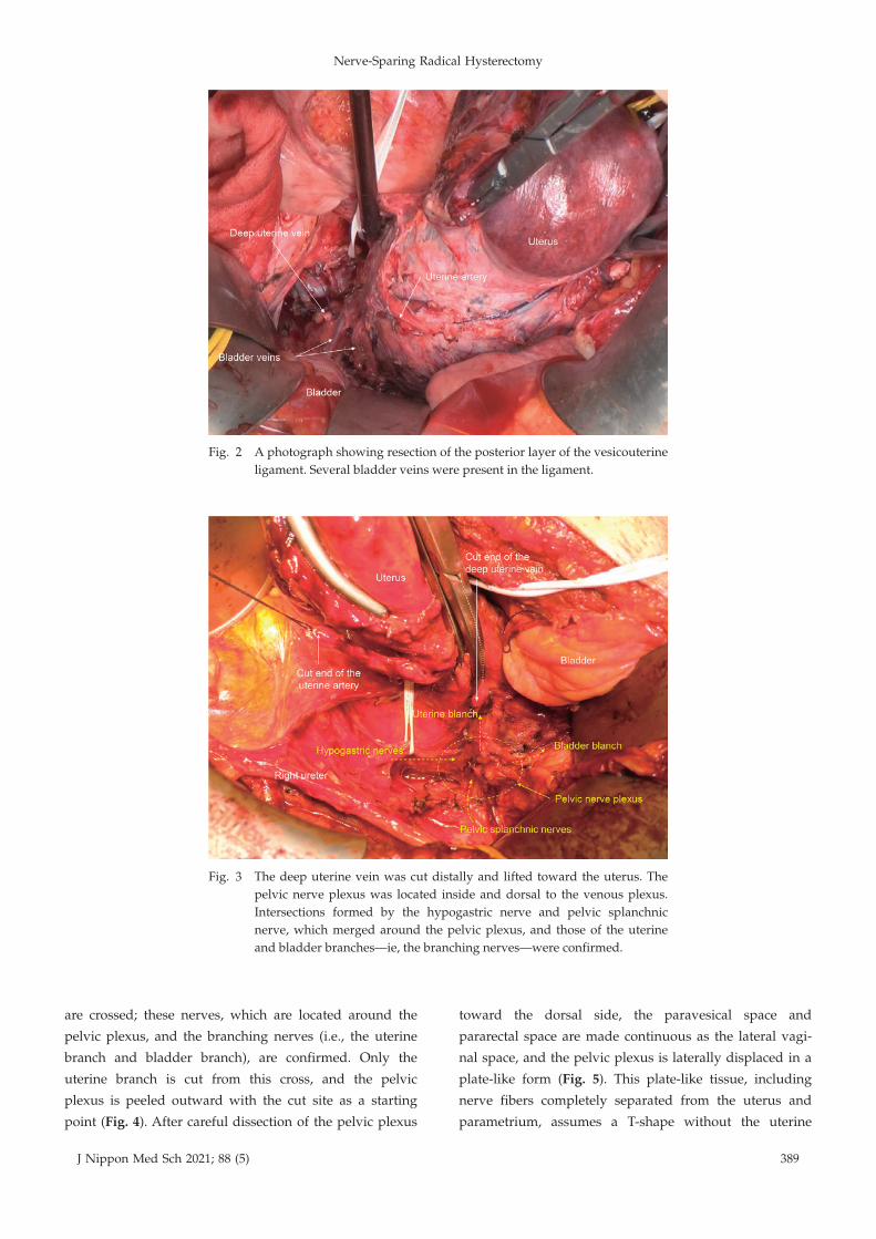
5. The posterior layer of the vesicouterine ligament is
carefully separated and ligated in small portions. During
this procedure, a venous plexus is observed between the
bladder and the cervix. In our experience, two or three
bladder veins are usually found, which are cut from the
deep uterine veins (Fig. 2). The posterior layer of the
vesicouterine ligament and paracolpium, including the
autonomic bladder branch, is continuous and borderless.
Therefore, damage to the bladder branch must be pre-
vented. The point of processing is, first, to exfoliate the
tissue from the position on the uterine side from the
middle part of the ligament and, second, to finish exfo-
liation at the depth that the bladder veins are cut and
separated from the deep uterine vein. A wide incision in
the caudal and dorsal layers of the posterior vesicouter-
ine ligament would damage the bladder branch of the
pelvic plexus.
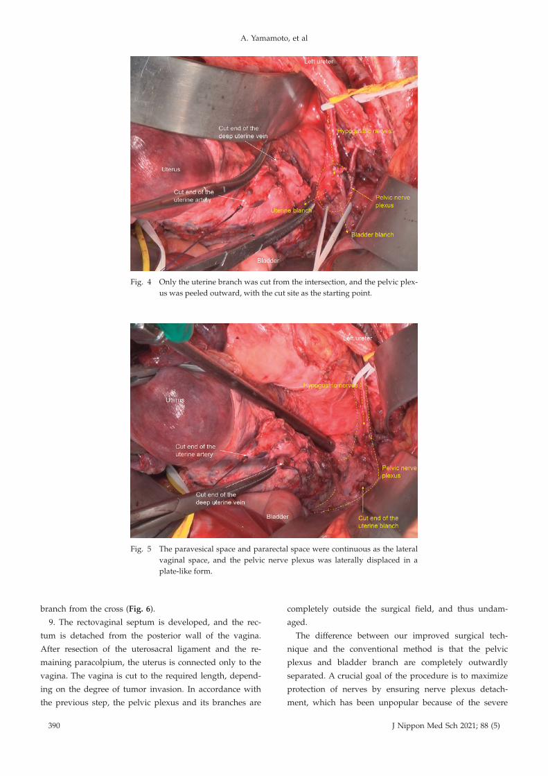
6. A portion of the cardinal ligament vessel, which is
well-exposed after lymphadenectomy, is cut distally and
lifted toward the uterus. Because the bladder veins that
meet from the anterior have already been cut, the deep
uterine vein can be easily lifted to a shallow position.
The pelvic nerve plexus is located inside and dorsal to
the venous plexus (Fig. 3).
7. The nerve plexus is formed where the autonomic
nerves exit S2-S4 and the hypogastric nerve exits the cra-
nial side and is joined at the site of the cardinal ligament.
Branches of the nerve are then advanced toward the
uterus and bladder, from the plexus. A procedure is per-
formed to protect this bladder branch passing through
the lateral portion of the paracolpium that was exposed
in the previous step. The outer portion of the paracol-
pium is detached from the vagina toward the paravesical
space, taking care not to damage the vascular plexus in-
side the paracolpium. This is marked and protected by
using tape and moving it outward.
8. The hypogastric nerve and pelvic splanchnic nerve
Nerve-Sparing Radical Hysterectomy
J Nippon Med Sch 2021; 88 (5) 389
Fig. 2 A photograph showing resection of the posterior layer of the vesicouterine
ligament. Several bladder veins were present in the ligament.
Fig. 3 The deep uterine vein was cut distally and lifted toward the uterus. The
pelvic nerve plexus was located inside and dorsal to the venous plexus.
Intersections formed by the hypogastric nerve and pelvic splanchnic
nerve, which merged around the pelvic plexus, and those of the uterine
and bladder branches—ie, the branching nerves—were confirmed.
are crossed; these nerves, which are located around the
pelvic plexus, and the branching nerves (i.e., the uterine
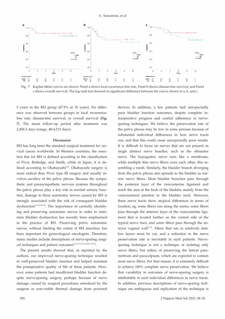
branch and bladder branch), are confirmed. Only the
uterine branch is cut from this cross, and the pelvic
plexus is peeled outward with the cut site as a starting
point (Fig. 4). After careful dissection of the pelvic plexus
toward the dorsal side, the paravesical space and
pararectal space are made continuous as the lateral vagi-
nal space, and the pelvic plexus is laterally displaced in a
plate-like form (Fig. 5). This plate-like tissue, including
nerve fibers completely separated from the uterus and
parametrium, assumes a T-shape without the uterine
A. Yamamoto, et al
390 J Nippon Med Sch 2021; 88 (5)
Fig. 4 Only the uterine branch was cut from the intersection, and the pelvic plex-
us was peeled outward, with the cut site as the starting point.
Fig. 5 The paravesical space and pararectal space were continuous as the lateral
vaginal space, and the pelvic nerve plexus was laterally displaced in a
plate-like form.
branch from the cross (Fig. 6).
9. The rectovaginal septum is developed, and the rec-
tum is detached from the posterior wall of the vagina.
After resection of the uterosacral ligament and the re-
maining paracolpium, the uterus is connected only to the
vagina. The vagina is cut to the required length, depend-
ing on the degree of tumor invasion. In accordance with
the previous step, the pelvic plexus and its branches are
completely outside the surgical field, and thus undam-
aged.
The difference between our improved surgical tech-
nique and the conventional method is that the pelvic
plexus and bladder branch are completely outwardly
separated. A crucial goal of the procedure is to maximize
protection of nerves by ensuring nerve plexus detach-
ment, which has been unpopular because of the severe

Nerve-Sparing Radical Hysterectomy
J Nippon Med Sch 2021; 88 (5) 391
Fig. 6 The plate-like tissue, including nerve fibers that were completely separat-
ed from the uterus and parametrium, assumed a T-shape without the uter-
ine branch from the intersection of fibers.
bleeding caused by damage to the venous plexus. There-
fore, the procedure, as it relates to hysterectomy, was
modified. The procedure that was historically performed
from the cranial side to the caudal side (in the order of
cardinal ligament, uterosacral ligament, vesicouterine
ligament, paracolpium, vagina) was modified and is now
performed from the ventral side to the dorsal side (in the
order of vesicouterine ligament, cardinal ligament, nerve
plexus dissection, uterosacral ligament, paracolpium, va-
gina). This was done because completing excision of the
posterior layer of the vesicouterine ligament and substan-
tially raising the cardinal ligament to the uterine corpus
are essential steps in the outward detachment of the pel-
vic plexus and its bladder branch.
Residual Urine Measurement
An indwelling bladder catheter was placed at the time of
surgery and removed 7 days later. After removal of the
catheter, patients were free to urinate, and urination vol-
ume and residual urine volume were recorded at six pre-
specified times each day. On-time measurement of resid-
ual urine volume was performed at 3:00, 7:00, 11:00,
15:00, 19:00, and 23:00. The residual urine volume was
accurately measured by a nurse immediately after urina-
tion, by inserting a catheter into the bladder cavity of the
patient. When residual urine volume was 50 mL or less
at the same time of day for 2 consecutive days, measure-
ment of residual urine was stopped only at that time.
When all measurement times were cleared, urination
ability was considered completely established. In addi-
tion to measurement of residual urine volume, micturi-
tion desire was also recorded. If no improvement in uri-
nary function was observed even after more than 3
weeks after the start of residual urine measurement, the
patient was given instruction on the method of self-
catheterization and withdrawn from residual urine meas-
urement. If urinary function improved over time, even
after more than 3 weeks, measurement of residual urine
was continued until urination was established.
Follow-up
All patients were staged according to the FIGO and TNM
classification systems. Patients at high risk for recurrence
received additional postoperative adjuvant therapy. After
treatment, patients underwent medical checkups for the
number of months equal to the number of years passed.
After more than 6 years, all patients underwent regular
medical examinations every 6 months for at least 10
years after treatment was completed. At the regular
medical examinations, patients underwent an internal ex-
amination, transvaginal ultrasonography, and serum bio-
chemistry tests, including detection of tumor markers, to
assess recurrence. In addition, patients underwent annual
computed tomography scans of the chest, abdomen, and
pelvis to evaluate the presence or absence of recurrent le-
sions, including those at distant sites. All patient clinical

A. Yamamoto, et al
392 J Nippon Med Sch 2021; 88 (5)
data were stored in an in-hospital electronic medical re-
cords system.
Statistical Analysis
The results are expressed as means ± SD, or medians, as
appropriate. The normality of the distribution was exam-
ined for all continuous variables. The t-test was used for
comparisons of age and body mass index between the
two groups, when the data were normally distributed.
For comparisons between the two groups with respect to
operative time, amount of blood loss, and number of
days until establishment of urination, the difference be-
tween medians was evaluated with the Mann-Whitney U
test when the data were not normally distributed.
Fisher’s exact test was used to compare nominal vari-
ables between the two groups. The log-rank test was
used to compare outcomes between the two groups. All
tests were two-sided, and p < 0.05 was considered statis-
tically significant. All statistical analyses were performed
with EZR (Saitama Medical Center, Jichi Medical Univer-
sity, Saitama, Japan), a graphical user interface for R (The
R Foundation for Statistical Computing, Vienna, Austria).
More precisely, it is a modified version of R Commander
that is designed to add statistical functions frequently
used in biostatistics23.
Results
During the 5-year period from March 2007 to February
2012, 61 patients with cervical cancer underwent RH. The
patients were divided into two groups according to the
type of treatment received: 31 patients were in the NSRH
group and 30 were in the RH group. No significant dif-
ferences in the measured variables, including staging and
histology, were observed between the two groups (Table
1). In the NSRH group, one patient had diabetes, two
had depression, one had panic disorder, and one had
Hashimoto’s disease. In the RH group, two patients had
diabetes, one had schizophrenia, and four had hyperten-
sion. All patients in both groups received appropriate
treatment for complications, which were controlled. In
the NSRH group, five patients underwent unilateral
nerve-sparing surgery after preoperative evaluation of tu-
mor invasion and intraoperative findings. Postoperative
histopathological examination revealed tumor invasion of
the cardinal ligaments in seven patients in the NSRH
group and in six patients in the RH group; however, the
resected margins were negative in all cases. Pelvic lymph
node metastases were diagnosed pathologically in six pa-
tients in the NSRH group and in nine patients in the RH
group. On the basis of the stage of postoperative patho-
logical diagnosis, 23 patients in the NSRH group and 21
patients in the RH group received postoperative adjuvant
therapy. In the NSRH group, 19 patients received chemo-
therapy and four patients received concurrent chemora-
diotherapy. In the RH group, 15 patients received chemo-
therapy, four patients received radiotherapy, and two pa-
tients received concurrent chemoradiotherapy.
To evaluate surgical invasiveness, we compared opera-
tive time, intraoperative blood loss, and intraoperative
complications between groups. In the NSRH group, me-
dian operative time was 390.0 minutes (range, 253-580
minutes) and median blood loss was 1,212.0 g (range,
500-3,195 g). In the RH group, median operative time
was 361.5 minutes (range, 255-555 minutes) and median
blood loss was 1,562.5 g (range, 500-4,780 g). No signifi-
cant difference was observed between the two groups
(Table 2), and no intraoperative complications occurred
in either group.
To evaluate urinary function, postoperative micturition
desire and the number of days required to establish uri-
nation without a catheter were compared between the
two groups. A self-reported subjective survey showed
that 80.6% (25/31) of patients in the NSRH group were
aware of urination after removal of the indwelling blad-
der catheter, while 46.7% (14/30) of patients in the RH
group were aware. The median time required to establish
urination after removal of the indwelling bladder cathe-
ter was 6 days (range, 2-20 days) in the NSRH group and
13.5 days (range, 3-46 days) in the RH group. The results
were significantly better in the NSRH group (p < 0.05).
No patient in the NSRH group required self-
catheterization at discharge, whereas five patients in the
RH group required self-catheterization (Table 2). The
prevalence of dysuria was reported to be higher in pa-
tients with diabetes or mental illnesses such as depres-
sion24―27. However, in this study, postoperative urinary
function did not differ significantly between patients
with and without these conditions.
The results for the five patients who underwent a uni-
lateral nerve-sparing procedure in the NSRH group were
analyzed. The median time required to establish urina-
tion was 7 days (range, 5-15 days) in those patients. This
value was slightly worse than that of patients who un-
derwent the bilateral nerve-sparing procedure but was
significantly better than that of patients in the RH group.
This result is consistent with the findings of a previous
study, which reported that the contractile function of the
detrusor muscle of the bladder was maintained if unilat-

Nerve-Sparing Radical Hysterectomy
J Nippon Med Sch 2021; 88 (5) 393
Table 1 Patient characteristics
NSRH (n = 31) RH (n = 30) P value
Age (years) 50.8 ± 10.3 49.7 ± 11.3 0.682
BMI (kg/m2) 22.6 ± 3.0 23.1 ± 4.2 0.619
FIGO stage (%) 0.742
IA 1 (3.2) 0 (0.0)
IA2 2 (6.5) 0 (0.0)
IB1 14 (45.2) 15 (50.0)
IB2 6 (19.4) 4 (13.3)
IIA 1 (3.2) 1 (3.3)
IIB 7 (22.6) 9 (30.0)
IIIB 0 (0.0) 1 (3.3)
TNM stage
pT (%) 0.895
1a 1 (3.2) 0 (0.0)
1a1 1 (3.2) 1 (3.3)
1a2 2 (6.5) 0 (0.0)
1b 1 (3.2) 0 (0.0)
1b1 12 (38.7) 14 (46.7)
1b2 4 (12.9) 4 (13.3)
2a 2 (6.5) 4 (13.3)
2b 7 (22.6) 6 (20.0)
3a 1 (3.2) 1 (3.3)
pN (%) 6 (19.4) 9 (30.0) 0.384
pM (%) 0 (0) 0 (0) N/A
Postoperative treatment (%) 23 (76.7) 21 (70.0) 0.771
Histology (%) 0.097
Squamous cell carcinoma 19 (61.3) 25 (83.3)
Adenosquamous carcinoma 1 (3.2) 0 (0.0)
Mucinous carcinoma 7 (22.6) 4 (13.3)
Endometrioid carcinoma 3 (9.7) 0 (0.0)
Serous carcinoma 1 (3.2) 0 (0.0)
Small-cell carcinoma 0 (0.0) 1 (3.3)
Values are number (%) or mean ± SD
BMI, body mass index; TNM stage, the TNM classification of malignant tu-
mors established by the Union for International Cancer Control; pT, pN, pM,
TNM classification determined by histopathologic examination of a surgical
specimen
Table 2 Postoperative urination and surgical invasiveness
NSRH (n = 31) RH (n = 30) P value
Operative time (min) 390.0 [253.0, 580.0] 361.5 [255.0, 555.0] 0.155
Blood loss (g) 1,212.0 [500.0, 3,195.0] 1,562.5 [500.0, 4,780.0] 0.074
Micturition desire (%) 25 (80.6) 14 (46.7) 0.008*
Time to establish urination (days) 6.0 [2.0, 20.0] 13.5 [3.0, 46.0] 0.002*
Self-catheterization (%) 0 (0.0) 5 (16.7) 0.024*
Values are number (%) or median [range]
*p<0.05
eral nerve preservation was successful22.
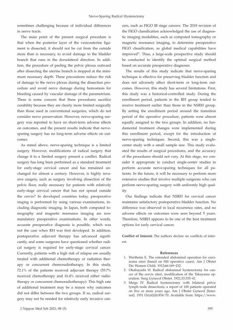
To assess curability, we compared the local recurrence-
free rate, disease-free survival, and overall survival be-
tween the two groups. The local recurrence-free rate was
87.1% (27/31) for patients in the NSRH group and 83.3%
(25/30) for the RH group. The disease-free survival rate
in the NSRH group was 70.0% at 5 years (70.0% at 10
years); in the RH group, it was 68.3% at 5 years (63.1% at
10 years). The overall survival rate was 86.1% in the
NSRH group at 5 years (86.1% at 10 years) and 78.2% at
A. Yamamoto, et al
394 J Nippon Med Sch 2021; 88 (5)
Fig. 7 Kaplan-Meier curves are shown. Panel a shows local recurrence-free rate, Panel b shows disease-free survival, and Panel
c shows overall survival. The log-rank test showed no significant difference between the curves shown in a, b, and c.
5 years in the RH group (67.9% at 10 years). No differ-
ence was observed between groups in local recurrence-
free rate, disease-free survival, or overall survival (Fig.
7). The mean follow-up period after treatment was
2,456.3 days (range, 48-4,213 days).
Discussion
RH has long been the standard surgical treatment for cer-
vical cancer worldwide. In Western countries, the resec-
tion line for RH is defined according to the classification
of Piver, Rutledge, and Smith, while in Japan, it is de-
fined according to Okabayashi4,28. Okabayashi surgery is
more radical than Piver type III surgery and usually in-
volves sacrifice of the pelvic plexus. Because the sympa-
thetic and parasympathetic nervous systems throughout
the pelvic plexus play a key role in normal urinary func-
tion, damage to these autonomic nerves caused by RH is
strongly associated with the risk of consequent bladder
dysfunction8,19―21,29―32. The importance of carefully identify-
ing and preserving autonomic nerves in order to mini-
mize bladder dysfunction has recently been emphasized
in the practice of RH. Preserving pelvic autonomic
nerves, without limiting the extent of RH resection, has
been important for gynecological oncologists. Therefore,
many studies include descriptions of nerve-sparing surgi-
cal techniques and patient outcomes5,6,12,13,15,17,18,20,21,29―38.
The present results showed that, as reported by the
authors, our improved nerve-sparing technique resulted
in well-preserved bladder function and helped maintain
the postoperative quality of life of these patients. How-
ever, some patients had insufficient bladder function de-
spite nerve-sparing surgery, perhaps because of nerve
damage caused by surgical procedures unnoticed by the
surgeon or non-visible thermal damage from powered
devices. In addition, a few patients had unexpectedly
poor bladder function outcomes, despite complete in-
traoperative progress and careful adherence to nerve-
sparing techniques. We believe the preservation rate of
the pelvic plexus may be low in some persons because of
substantial individual differences in how nerve tracts
run, and that this could cause unexpectedly poor results.
It is difficult to focus on nerves that are not present as
single distinct nerve bundles, such as the obturator
nerve. The hypogastric nerve runs like a membrane,
while multiple thin nerve fibers cross each other, this re-
sembling a mesh. Similarly, the bladder branch diverging
from the pelvic plexus also spreads to the bladder as nar-
row nerve fibers. Most bladder branches pass through
the posterior layer of the vesicouterine ligament and
reach the area at the back of the bladder, mainly from the
vesicoureteral junction to the bladder neck. However,
these nerve tracts show atypical differences in terms of
location, eg, some fibers run along the ureter, some fibers
pass through the anterior layer of the vesicouterine liga-
ment that is located further on the ventral side of the
typical nerve tract, and some fibers pass through the an-
terior vaginal wall39―42. Fibers that run in relatively shal-
low layers must be cut, and a reduction in the nerve
preservation rate is inevitable in such patients. Nerve-
sparing technique is not a technique of isolating only
nerve fibers, but rather, of preserving the lateral para-
metrium and paracolpium, which are expected to contain
most nerve fibers. For that reason, it is extremely difficult
to achieve 100% complete nerve preservation. We believe
that variability in outcomes of nerve-sparing surgery is
attributable to such individual differences in nerve tracts.
In addition, previous descriptions of nerve-sparing tech-
nique are ambiguous and replication of the technique is
Nerve-Sparing Radical Hysterectomy
J Nippon Med Sch 2021; 88 (5) 395
sometimes challenging because of individual differences
in nerve tracts.
The main point of the present surgical procedure is
that when the posterior layer of the vesicouterine liga-
ment is dissected, it should not be cut from the outside
more than is necessary, to avoid damage to the bladder
branch that runs in the dorsolateral direction. In addi-
tion, the procedure of peeling the pelvic plexus outward
after dissecting the uterine branch is stopped at the mini-
mum necessary depth. These precautions reduce the risk
of damage to the nerve plexus during the dissection pro-
cedure and avoid nerve damage during hemostasis for
bleeding caused by vascular damage of the parametrium.
There is some concern that these procedures sacrifice
curability because they are clearly more limited surgically
than those used in conventional surgeries, which do not
consider nerve preservation. However, nerve-sparing sur-
gery was reported to have no short-term adverse effects
on outcomes, and the present results indicate that nerve-
sparing surgery has no long-term adverse effects on out-
comes.
As stated above, nerve-sparing technique is a limited
surgery. However, modifications of radical surgery that
change it to a limited surgery present a conflict. Radical
surgery has long been performed as a standard treatment
for early-stage cervical cancer and has remained un-
changed for almost a century. However, is highly inva-
sive surgery, such as surgery involving dissection of the
pelvic floor, really necessary for patients with relatively
early-stage cervical cancer that has not spread outside
the cervix? In developed countries today, preoperative
staging is performed by using various examinations, in-
cluding diagnostic imaging. In Japan, both computed to-
mography and magnetic resonance imaging are now
mandatory preoperative examinations. In other words,
accurate preoperative diagnosis is possible, which was
not the case when RH was first developed. In addition,
postoperative adjuvant therapy has advanced signifi-
cantly, and some surgeons have questioned whether radi-
cal surgery is required for early-stage cervical cancer.
Currently, patients with a high risk of relapse are usually
treated with additional chemotherapy or radiation ther-
apy or concurrent chemoradiotherapy. In this study,
72.1% of the patients received adjuvant therapy (55.7%
received chemotherapy and 16.4% received either radio-
therapy or concurrent chemoradiotherapy). This high rate
of additional treatment may be a reason why outcomes
did not differ between the two groups. If so, radical sur-
gery may not be needed for relatively early invasive can-
cers, such as FIGO IB stage cancers. The 2018 revision of
the FIGO classification acknowledged the use of diagnos-
tic imaging modalities, such as computed tomography or
magnetic resonance imaging, to determine preoperative
FIGO classification, as global medical capabilities have
improved43. Thus, a large-scale prospective study should
be conducted to identify the optimal surgical method
based on accurate preoperative diagnoses.
The results of this study indicate that nerve-sparing
technique is effective for preserving bladder function and
does not adversely affect short-term or long-term out-
comes. However, this study has several limitations. First,
this study was a historical-controlled study. During the
enrollment period, patients in the RH group tended to
receive treatment earlier than those in the NSRH group.
By setting the enrollment period around the transition
period of the operative procedure, patients were almost
equally assigned to the two groups. In addition, no fun-
damental treatment changes were implemented during
this enrollment period, except for the introduction of
nerve-sparing techniques. Second, this was a single-
center study with a small sample size. This study evalu-
ated the results of surgical procedures, and the accuracy
of the procedures should not vary. At this stage, we con-
sider it appropriate to conduct single-center studies to
perform accurate nerve-sparing techniques for all pa-
tients. In the future, it will be necessary to perform more
extensive studies that involve multiple surgeons who can
perform nerve-sparing surgery with uniformly high qual-
ity.
Our findings indicate that NSRH for cervical cancer
maintains satisfactory postoperative bladder function. No
difference was observed in local recurrence rates, and no
adverse effects on outcomes were seen beyond 5 years.
Therefore, NSRH appears to be one of the best treatment
options for early cervical cancer.
Conflict of Interest: The authors declare no conflicts of inter-
est.
References1.Wertheim E. The extended abdominal operation for carci-
noma uteri (based on 500 operative cases). Am J Obstet
Dis Women Child. 1912;66:169―232.
2.Okabayashi H. Radical abdominal hysterectomy for can-
cer of the cervix uteri, modification of the Takayama op-
eration. Surg Gynecol Obstet. 1921;33:335―41.
3.Meigs JV. Radical hysterectomy with bilateral pelvic
lymph node dissections; a report of 100 patients operated
on five or more years ago. Am J Obstet Gynecol [Inter-
net]. 1951 Oct;62(4):854―70. Available from: https://www.

A. Yamamoto, et al
396 J Nippon Med Sch 2021; 88 (5)
ncbi.nlm.nih.gov/pubmed/14885271
4.Piver MS, Rutledge F, Smith JP. Five classes of extended
hysterectomy for women with cervical cancer. Obstet Gy-
necol [Internet]. 1974 Aug;44(2):265―72. Available from: ht
tps://www.ncbi.nlm.nih.gov/pubmed/4417035
5.Ercoli A, Delmas V, Gadonneix P, et al. Classical and
nerve-sparing radical hysterectomy: an evaluation of the
risk of injury to the autonomous pelvic nerves. Surg Ra-
diol Anat. 2003 Jul-Aug;25(3-4):200―6.
6.Maas CP, Trimbos JB, DeRuiter MC, van de Velde CJ,
Kenter GG. Nerve sparing radical hysterectomy: latest de-
velopments and historical perspective. Crit Rev Oncol
Hematol [Internet]. 2003 Dec;48(3):271―9. Available from:
https://www.ncbi.nlm.nih.gov/pubmed/14693339
7.Zullo MA, Manci N, Angioli R, Muzii L, Panici PB. Vesi-
cal dysfunctions after radical hysterectomy for cervical
cancer: a critical review. Crit Rev Oncol Hematol [Inter-
net]. 2003 Dec;48(3):287―93. Available from: https://www.
ncbi.nlm.nih.gov/pubmed/14693341
8.Landoni F, Maneo A, Cormio G, et al. Class II versus
class III radical hysterectomy in stage IB-IIA cervical can-
cer: a prospective randomized study. Gynecol Oncol [In-
ternet]. 2001 Jan;80(1):3―12. Available from: https://www.
ncbi.nlm.nih.gov/pubmed/11136561
9.Kindermann G, Debus-Thiede G. Postoperative urological
complications after radical surgery for cervical cancer.
Baillieres Clin Obstet Gynaecol [Internet]. 1988 Dec;2(4):
933―41. Available from: https://www.ncbi.nlm.nih.gov/p
ubmed/3229061
10.Kobayashi T. [Abdominal radical hysterectomy with pel-
vic lymphadenectomy for cancer of cervix]. Shikyukeigan
Shujutsu [Cervical cancer surgery]. Tokyo: Nanzando;
1961. p. 178―87. Japanese.
11.Höckel M, Konerding MA, Heussel CP. Liposuction-
assisted nerve-sparing extended radical hysterectomy: on-
cologic rationale, surgical anatomy, and feasibility study.
Am J Obstet Gynecol [Internet]. 1998 May;178(5):971―6.
Available from: https://www.ncbi.nlm.nih.gov/pubmed/
9609569
12.Possover M, Stober S, Plaul K, Schneider A. Identification
and preservation of the motoric innervation of the blad-
der in radical hysterectomy type III. Gynecol Oncol. 2000
Nov;79(2):154―7.
13.Trimbos JB, Maas CP, Deruiter MC, Peters AA, Kenter
GG. A nerve-sparing radical hysterectomy: guidelines and
feasibility in Western patients. Int J Gynecol Cancer [In-
ternet]. 2001 May-Jun;11(3):180―6. Available from: http
s://www.ncbi.nlm.nih.gov/pubmed/11437922
14.Querleu D, Narducci F, Poulard V, et al. Modified radical
vaginal hysterectomy with or without laparoscopic nerve-
sparing dissection: a comparative study. Gynecol Oncol
[Internet]. 2002 Apr;85(1):154―8. Available from: https://
www.ncbi.nlm.nih.gov/pubmed/11925136
15.Raspagliesi F, Ditto A, Fontanelli R, et al. Nerve-sparing
radical hysterectomy: a surgical technique for preserving
the autonomic hypogastric nerve. Gynecol Oncol [Inter-
net]. 2004 May;93(2):307―14. Available from: http://www.
sciencedirect.com/science/article/pii/S0090825804000733
16.Papp Z, Csapó Z, Hupuczi P, Mayer A. Nerve-sparing
radical hysterectomy for stage IA2-IIB cervical cancer: 5-
year survival of 501 consecutive cases. Eur J Gynaecol
Oncol [Internet]. 2006;27(6):553―60. Available from: http
s://www.ncbi.nlm.nih.gov/pubmed/17290582
17.Sakamoto S, Takizawa K. An improved radical hysterec-
tomy with fewer urological complications and with no
loss of therapeutic results for invasive cervical cancer.
Baillieres Clin Obstet Gynaecol [Internet]. 1988 Dec;2(4):
953―62. Available from: http://www.sciencedirect.com/sc
ience/article/pii/S0950355298800229
18.Yabuki Y, Asamoto A, Hoshiba T, Nishimoto H,
Nishikawa Y, Nakajima T. Radical hysterectomy: An
anatomic evaluation of parametrial dissection. Gynecol
Oncol [Internet]. 2000;77(1):155―63. Available from: htt
p://www.sciencedirect.com/science/article/pii/S0090825
899957232
19.Kuwabara Y, Suzuki M, Hashimoto M, Furugen Y,
Yoshida K, Mitsuhashi N. New method to prevent blad-
der dysfunction after radical hysterectomy for uterine cer-
vical cancer. J Obstet Gynaecol Res [Internet]. 2000 Feb;26
(1):1―8. Available from: https://doi.org/10.1111/j.1447-075
6.2000.tb01192.x
20.Fujii S, Takakura K, Matsumura N, et al. Anatomic identi-
fication and functional outcomes of the nerve sparing
Okabayashi radical hysterectomy. Gynecol Oncol [Inter-
net]. 2007 Oct;107(1):4―13. Available from: http://www.sci
encedirect.com/science/article/pii/S009082580700652X
21.Sakuragi N, Todo Y, Kudo M, Yamamoto R, Sato T. A sys-
tematic nerve-sparing radical hysterectomy technique in
invasive cervical cancer for preserving postsurgical blad-
der function. Int J Gynecol Cancer [Internet]. 2005 Mar-
Apr;15(2):389―97. Available from: https://www.ncbi.nlm.
nih.gov/pubmed/15823132
22.Katahira A, Niikura H, Kaiho Y, et al. Intraoperative elec-
trical stimulation of the pelvic splanchnic nerves during
nerve-sparing radical hysterectomy. Gynecol Oncol. 2005
Sep;98(3):462―6.
23.Kanda Y. Investigation of the freely available easy-to-use
software ‘EZR’ for medical statistics. Bone Marrow Trans-
plant. 2013 Mar;48(3):452―8.
24.Yamaguchi C, Sakakibara R, Uchiyama T, et al. Overac-
tive bladder in diabetes: a peripheral or central mecha-
nism? Neurourol Urodyn [Internet]. 2007;26(6):807―13.
Available from: https://www.ncbi.nlm.nih.gov/pubmed/
17357115
25.Daneshgari F, Liu G, Birder L, Hanna-Mitchell AT,
Chacko S. Diabetic bladder dysfunction: current transla-
tional knowledge. J Urol [Internet]. 2009 Dec;182(6
Suppl):S18―26. Available from: https://www.ncbi.nlm.nih.
gov/pubmed/19846137
26.Kirschner-Hermanns R, Daneshgari F, Vahabi B, Birder L,
Oelke M, Chacko S. Does diabetes mellitus-induced blad-
der remodeling affect lower urinary tract function? ICI-RS
2011. Neurourol Urodyn [Internet]. 2012 Mar;31(3):359―64.
Available from: https://www.ncbi.nlm.nih.gov/pubmed/
22415965
27.Sakakibara R, Ito T, Yamamoto T, et al. Depression, anxi-
ety and the bladder. Low Urin Tract Symptoms [Internet].
2013 Sep;5(3):109―20. Available from: https://www.ncbi.nl
m.nih.gov/pubmed/26663445
28.Okabayashi H. Radical abdominal hysterectomy for can-
cer of the cervix uteri, modification of the Takayama op-
eration. Surg Gynecol Obstet. 1921;33:335―41.
29.Yabuki Y, Asamoto A, Hoshiba T, Nishimoto H, Satou N.
A new proposal for radical hysterectomy. Gynecol Oncol
[Internet]. 1996 Sep;62(3):370―8. Available from: http://w
ww.sciencedirect.com/science/article/pii/S0090825896902
516
30.Kato T, Murakami G, Yabuki Y. A new perspective on
nerve-sparing radical hysterectomy: nerve topography
and over-preservation of the cardinal ligament. Jpn J Clin
Oncol [Internet]. 2003 Nov;33(11):589―91. Available from:
https://www.ncbi.nlm.nih.gov/pubmed/14711985

Nerve-Sparing Radical Hysterectomy
J Nippon Med Sch 2021; 88 (5) 397
31.Mantzaris G, Rodolakis A, Vlachos G, et al. Magnifying
lenses assisted nerve-sparing radical hysterectomy and
prevention of nerve plexus trauma. Int J Gynecol Cancer
[Internet]. 2008 Jul-Aug;18(4):868―75. Available from: http
s://www.ncbi.nlm.nih.gov/pubmed/17892457
32.Possover M, Quakernack J, Chiantera V. The LANN tech-
nique to reduce postoperative functional morbidity in la-
paroscopic radical pelvic surgery. J Am Coll Surg [Inter-
net]. 2005 Dec;201(6):913―7. Available from: https://www.
ncbi.nlm.nih.gov/pubmed/16310695
33.Fujii S. Original film of the Okabayashi’s radical hysterec-
tomy by Okabayashi himself in 1932, and two films of
the precise anatomy necessary for nerve-sparing Oka-
bayashi’s radical hysterectomy clarified by Shingo Fujii.
Int J Gynecol Cancer [Internet]. 2008 Mar-Apr;18(2):383―5.
Available from: https://www.ncbi.nlm.nih.gov/pubmed/
17587316
34.Maas K, Moriya Y, Kenter G, Trimbos B, van de Velde C.
A plea for preservation of the pelvic autonomic nerves.
Lancet [Internet]. 1999 Aug;35(9180):772―3. Available
from: https://www.ncbi.nlm.nih.gov/pubmed/10475214
35.Kato K, Suzuka K, Osaki T, Tanaka N. Unilateral or bilat-
eral nerve-sparing radical hysterectomy: a surgical tech-
nique to preserve the pelvic autonomic nerves while in-
creasing radicality. Int J Gynecol Cancer. 2007 Sep-Oct;17
(5):1172―8.
36.Höckel M, Horn LC, Hentschel B, Höckel S, Naumann G.
Total mesometrial resection: high resolution nerve-sparing
radical hysterectomy based on developmentally defined
surgical anatomy. Int J Gynecol Cancer [Internet]. 2003
Nov-Dec;13(6):791―803. Available from: https://www.ncb
i.nlm.nih.gov/pubmed/14675316
37.Raspagliesi F, Ditto A, Kusamura S, et al. Nerve-sparing
radical hysterectomy: a pilot study. Tumori [Internet].
2003 Sep-Oct;89(5):497―501. Available from: https://www.
ncbi.nlm.nih.gov/pubmed/14870771
38.Fujii S. Anatomic identification of nerve-sparing radical
hysterectomy: A step-by-step procedure. Gynecologic On-
cology [Internet]. 2008 Nov;111(2, Supplement):S33―41.
Available from: http://www.sciencedirect.com/science/ar
ticle/pii/S0090825808005398
39.Spradling K, Khoyilar C, Abedi G, et al. Redefining the
autonomic nerve distribution of the bladder using 3-
dimensional image reconstruction. J Urol [Internet]. 2015
Dec;194(6):1661―7. Available from: https://www.ncbi.nlm.
nih.gov/pubmed/26003207
40.Kraima AC, Derks M, Smit NN, Van De Velde CJ, Kenter
GG, DeRuiter MC. Careful dissection of the distal ureter
is highly important in nerve-sparing radical pelvic sur-
gery: A 3D reconstruction and immunohistochemical
characterization of the vesical plexus. Int J Gynecol Can-
cer [Internet]. 2016 Jun;26(5):959―66. Available from: http
s://www.ncbi.nlm.nih.gov/pubmed/27101584
41.Purves JT, Spruill L, Rovner E, et al. A three dimensional
nerve map of human bladder trigone. Neurourol Urodyn.
2017 Apr;36(4):1015―9.
42.Ripperda CM, Jackson LA, Phelan JN, Carrick KS, Corton
MM. Anatomic relationships of the pelvic autonomic
nervous system in female cadavers: clinical applications
to pelvic surgery. Am J Obstet Gynecol [Internet]. 2017
Apr;216(4):388.e1-.e7. Available from: https://www.ncbi.n
lm.nih.gov/pubmed/27956200
43.Bhatla N, Berek JS, Cuello Fredes M, et al. Revised FIGO
staging for carcinoma of the cervix uteri. Int J Gynaecol
Obstet [Internet]. 2019 Apr;145(1):129―35. Available from:
https://www.ncbi.nlm.nih.gov/pubmed/30656645
(Received,
(Accepted,
(J-STAGE Advance Publication,
April
July
August
29, 2020)
2, 2020)
1, 2020)
Journal of Nippon Medical School has adopted the Creative Com-mons Attribution-NonCommercial-NoDerivatives 4.0 InternationalLicense (https://creativecommons.org/licenses/by-nc-nd/4.0/) forthis article. The Medical Association of Nippon Medical School re-mains the copyright holder of all articles. Anyone may download,reuse, copy, reprint, or distribute articles for non-profit purposesunder this license, on condition that the authors of the articles areproperly credited.


![Nerve-sparing radical hysterectomy: time for a new standard of … · 2015-04-13 · et al. [16] reported on an RCT comparing conventional RH and NSRH, which included 92 cervical](https://img.pdfslide.us/doc/110x75/5f3b181bf2a48f65051316bf/nerve-sparing-radical-hysterectomy-time-for-a-new-standard-of-2015-04-13-et-al.jpg)

![Efficacy and outcomes of facial nerve–sparing treatment ... · the patient at significant risk of morbidity and mortality. Damage to the facial nerve (cranial nerve [CN] VII) is](https://img.pdfslide.us/doc/110x75/5edf22ebad6a402d666a7cb2/efficacy-and-outcomes-of-facial-nerveasparing-treatment-the-patient-at-significant.jpg)