Embed Size (px)
Citation preview

Ultrasonics Sonochemistry 17 (2010) 628–632
Contents lists available at ScienceDirect
Ultrasonics Sonochemistry
journal homepage: www.elsevier .com/locate /u l tsonch
Effect of ultrasound parameters for unilamellar liposome preparation
Raquel Silva, Helena Ferreira, Collin Little, Artur Cavaco-Paulo *
Dept. Textile Eng., University of Minho, 4800-058 Guimarães, Portugal
a r t i c l e i n f o
Article history:Received 7 July 2009Received in revised form 21 September2009Accepted 20 October 2009Available online 24 October 2009
Keywords:LiposomeSonicationReactor optimizationPhoton-correlation spectroscopyZeta-potential measurements
1350-4177/$ - see front matter � 2009 Elsevier B.V. Adoi:10.1016/j.ultsonch.2009.10.010
* Corresponding author. Tel.: +351 253 510271; faxE-mail address: [email protected] (A. Cavaco-P
a b s t r a c t
In this study, it was investigated the effects of ultrasound, namely power input, distance from ultrasoundtip to base of reactor and treatment time, in the formation of liposomes. Results indicate a dependence oncavitation events that are a function of power input, and consequently dependent on the position of theprobe within the reaction vessel and the wave behaviour. Short treatment times are required to achievenanosized vesicles in anti-nodal (k/4; 19 mm) reactor geometries. In this wave point the cavitation phe-nomenon is more pronounced when compared with the nodal point (k/2; 38 mm). Therefore, the consid-eration of the above parameters is vital if dependable and repeatable results are to be achieved.
� 2009 Elsevier B.V. All rights reserved.
1. Introduction path-length had a marked effect on rates of temperature rise and
Liposome applications can been found in many different areasas biochemistry, molecular biology, food technology, pharmaceuti-cal and medical. Each application requires vesicles with differentcharacteristics, which will be dependent for example, on the mate-rial to be encapsulated, as well as on the different release proper-ties [1–3]. Different methodologies are described in the literatureto produce multi-lamellar vesicles (MLVs), large unilamellar vesi-cles (LUVs) and small unilamellar vesicles (SUVs) [1,2,4,5]. Theuse of ultrasound methods to produce LUVs and SUVs are widelyreported in the literature [5–7]. However, the productions of thesephospholipid vesicles are poorly reproducible, since that ultra-sound experimental set-up is poorly described.
Ultrasound phenomena in liquid media enhance mass trans-ports of their constituents in a non-homogeneous fashion allowingthe fast formation of vesicles [8,9]. Several authors had pointed thefact that the most claimed ultrasound characteristics are in directdependence of power input and duration of sonication effects[10–12]. However, the control of these two parameters still leavesthe possibility of variation sound field intensity arising from therelationship between the frequency of ultrasound, position ofprobe tip from the base of the vessel and the phase of the soundwave upon reflection at the base. It is well known that ultrasoundmechanical waves generate cavitation in liquids with the forma-tion of local hot spots and free radicals [13,14]. Previous work,done by Little et al., has shown that variations of the ultrasound
ll rights reserved.
: +351 253 510293.aulo).
radical production within the bulk solution [15]. Therefore, thepower input, the duration of treatment and the position of theultrasound source within the solution, will have an outcome onthe ultrasound conditions imposed on the solution. This will affectthe levels of hydroxyl radicals (�OH) generated in solution. In fact,the chemical effects of the cavitation bubble collapse, namely �OHradical formation, are rarely considered in detriment of the extentof the tensile stresses imposed by ultrasound, which are usually re-ported [15].
The present work intends to show the need of control experi-mental set of operating parameters to engineer the characteristicsof phospholipids vesicles. For that it was performed an extensivereactor characterization process. The methodology that was usedexplored the effects of the three parameters referred, namelypower input, sonication time and depth (measure from the baseof the vessel), in the production of hydroxyl radicals and conse-quently in the formation of vesicles. These conditions were relatedwith lipid vesicle size, the polydispersity index (PDl) and surfacecharge (before and after sonication).
2. Materials and methods
2.1. Materials
2.1.1. ReagentsSodium phosphate dibasic, monosodium phosphate, tere-
phthalic acid, sodium hydroxide, chloroform and 1,2-dipalmitoyl-sn-glycerol-3-phosphocholine (DPPC) were purchased from SigmaChemicals and used as supplied, without further purification.
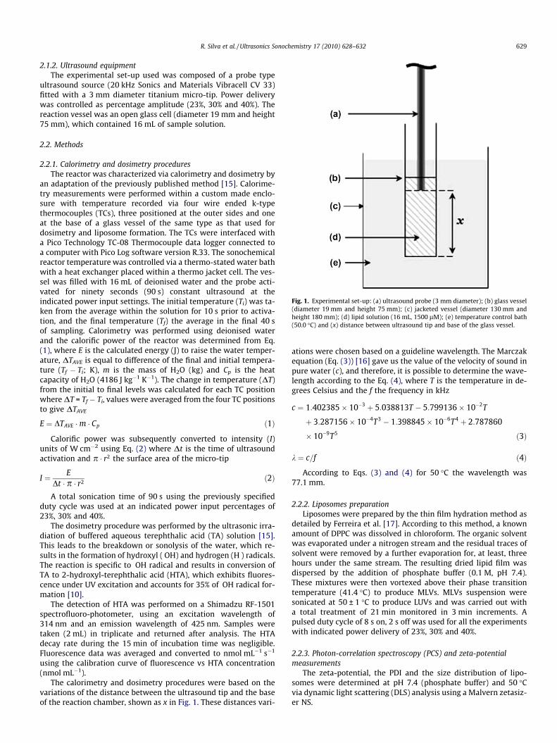
Fig. 1. Experimental set-up: (a) ultrasound probe (3 mm diameter); (b) glass vessel(diameter 19 mm and height 75 mm); (c) jacketed vessel (diameter 130 mm andheight 180 mm); (d) lipid solution (16 mL, 1500 lM); (e) temperature control bath(50.0 �C) and (x) distance between ultrasound tip and base of the glass vessel.
R. Silva et al. / Ultrasonics Sonochemistry 17 (2010) 628–632 629
2.1.2. Ultrasound equipmentThe experimental set-up used was composed of a probe type
ultrasound source (20 kHz Sonics and Materials Vibracell CV 33)fitted with a 3 mm diameter titanium micro-tip. Power deliverywas controlled as percentage amplitude (23%, 30% and 40%). Thereaction vessel was an open glass cell (diameter 19 mm and height75 mm), which contained 16 mL of sample solution.
2.2. Methods
2.2.1. Calorimetry and dosimetry proceduresThe reactor was characterized via calorimetry and dosimetry by
an adaptation of the previously published method [15]. Calorime-try measurements were performed within a custom made enclo-sure with temperature recorded via four wire ended k-typethermocouples (TCs), three positioned at the outer sides and oneat the base of a glass vessel of the same type as that used fordosimetry and liposome formation. The TCs were interfaced witha Pico Technology TC-08 Thermocouple data logger connected toa computer with Pico Log software version R.33. The sonochemicalreactor temperature was controlled via a thermo-stated water bathwith a heat exchanger placed within a thermo jacket cell. The ves-sel was filled with 16 mL of deionised water and the probe acti-vated for ninety seconds (90 s) constant ultrasound at theindicated power input settings. The initial temperature (Ti) was ta-ken from the average within the solution for 10 s prior to activa-tion, and the final temperature (Tf) the average in the final 40 sof sampling. Calorimetry was performed using deionised waterand the calorific power of the reactor was determined from Eq.(1), where E is the calculated energy (J) to raise the water temper-ature, DTAVE is equal to difference of the final and initial tempera-ture (Tf � Ti; K), m is the mass of H2O (kg) and Cp is the heatcapacity of H2O (4186 J kg�1 K�1). The change in temperature (DT)from the initial to final levels was calculated for each TC positionwhere DT = Tf � Ti, values were averaged from the four TC positionsto give DTAVE
E ¼ DTAVE �m � Cp ð1Þ
Calorific power was subsequently converted to intensity (I)units of W cm�2 using Eq. (2) where Dt is the time of ultrasoundactivation and p � r2 the surface area of the micro-tip
I ¼ EDt � p � r2 ð2Þ
A total sonication time of 90 s using the previously specifiedduty cycle was used at an indicated power input percentages of23%, 30% and 40%.
The dosimetry procedure was performed by the ultrasonic irra-diation of buffered aqueous terephthalic acid (TA) solution [15].This leads to the breakdown or sonolysis of the water, which re-sults in the formation of hydroxyl (�OH) and hydrogen (H�) radicals.The reaction is specific to �OH radical and results in conversion ofTA to 2-hydroxyl-terephthalic acid (HTA), which exhibits fluores-cence under UV excitation and accounts for 35% of �OH radical for-mation [10].
The detection of HTA was performed on a Shimadzu RF-1501spectrofluoro-photometer, using an excitation wavelength of314 nm and an emission wavelength of 425 nm. Samples weretaken (2 mL) in triplicate and returned after analysis. The HTAdecay rate during the 15 min of incubation time was negligible.Fluorescence data was averaged and converted to nmol mL�1 s�1
using the calibration curve of fluorescence vs HTA concentration(nmol mL�1).
The calorimetry and dosimetry procedures were based on thevariations of the distance between the ultrasound tip and the baseof the reaction chamber, shown as x in Fig. 1. These distances vari-
ations were chosen based on a guideline wavelength. The Marczakequation (Eq. (3)) [16] gave us the value of the velocity of sound inpure water (c), and therefore, it is possible to determine the wave-length according to the Eq. (4), where T is the temperature in de-grees Celsius and the f the frequency in kHz
c ¼ 1:402385� 10�3 þ 5:038813T � 5:799136� 10�2T
þ 3:287156� 10�4T3 � 1:398845� 10�6T4 þ 2:787860
� 10�9T5 ð3Þ
k ¼ c=f ð4Þ
According to Eqs. (3) and (4) for 50 �C the wavelength was77.1 mm.
2.2.2. Liposomes preparationLiposomes were prepared by the thin film hydration method as
detailed by Ferreira et al. [17]. According to this method, a knownamount of DPPC was dissolved in chloroform. The organic solventwas evaporated under a nitrogen stream and the residual traces ofsolvent were removed by a further evaporation for, at least, threehours under the same stream. The resulting dried lipid film wasdispersed by the addition of phosphate buffer (0.1 M, pH 7.4).These mixtures were then vortexed above their phase transitiontemperature (41.4 �C) to produce MLVs. MLVs suspension weresonicated at 50 ± 1 �C to produce LUVs and was carried out witha total treatment of 21 min monitored in 3 min increments. Apulsed duty cycle of 8 s on, 2 s off was used for all the experimentswith indicated power delivery of 23%, 30% and 40%.
2.2.3. Photon-correlation spectroscopy (PCS) and zeta-potentialmeasurements
The zeta-potential, the PDI and the size distribution of lipo-somes were determined at pH 7.4 (phosphate buffer) and 50 �Cvia dynamic light scattering (DLS) analysis using a Malvern zetasiz-er NS.

20 24 28 32 36 400
2
4
6
OH
.fo
rmat
ion
(nm
ol.m
L-1.s
ec-1
)
Power Input (%)
19 (mm)38 (mm)
Fig. 3. Variation of rate of �OH radical formation (nmol mL�1 s�1) with power input(%), at different depths (19 and 38 mm).
3 6 9 12 15 18 210
100
200
300
400
500
600
700 Z-average (23%)Z-average (30%)Z-average (40%)PDI (23%)PDI (30%)PDI (40%)
Z-av
erag
e (n
m)
0.0
0.2
0.4
0.6
0.8
1.0
1.2
PDI
(a) 19 mm
630 R. Silva et al. / Ultrasonics Sonochemistry 17 (2010) 628–632
3. Results and discussion
The reactor was characterized via calorimetry and dosimetryprocedures, as described in Section 2.2.1, prior to testing the lipo-some behaviour to the sonication. These two procedures weremade at 19 mm (anti-nodal point; k/4) and 38 mm (nodal point;k/2) of depth, which were calculated based on an estimated wave-length of 77.1 mm. The nodal point is known as a point where thewave has the minimal amplitude. The opposite of a nodal point isan anti-nodal point, where the amplitude of the wave is maximum.
The calorimetry results are presented in Fig. 2. In this figure, it ispossible to observe that the energy deposition for these two depths(19 and 38 mm) increase with the power input. The highest valueof input energy (about of 50 W cm�2) was obtained at 19 mm ofdepth and 40% of power input. Although, when it was used the23% of power input at two different depths (19 and 38 mm), itwas obtained the same input energy inside of the reactor (about15 W cm�2). After the calorimetry method it was performed theTA dosimeter, which is extensively used as an �OH radical indicator[11,12]. The extent of the conversion of TA to HTA obtained in thedosimetry procedure is shown in Fig. 3. The behaviour at the38 mm (k/2) position showed an almost linear production of hy-droxyl radicals. Conversely, the 19 mm (k/4) position displayed asignificant increase in hydroxyl radical production at 40% powerinput. The possible reason for this is that whilst cavitation bubbleimplosion is regarded as necessary for �OH radical formation, and isalso a contributor of heat, other factors are present which contrib-ute heat energy to the solution when conditions do not favor �OHradical production. These include cavitation bubble implosion ofinsufficient energy to form radicals, fluid friction within the bulksolution from the mixing effect, and friction between the bulk solu-tion with the stationary boundary layer adjacent to the side of thevessel. These friction forces could continue to provide heat energyeven when the differential in acoustic pressure is not enough tosustain effective cavitation [15]. After this characterization, itwas possible to identify the minima and maxima hydroxyl radicalactivity points as occurring at 38 mm (nodal point) and 19 mm(anti-nodal point) positions, respectively.
The characterization described above is essential, once that theultrasound can promote the hydrolysis and the oxidation of phos-pholipids, via the free radicals produced in the cavitation bubblescollapse. Additionally, high temperatures accelerate phosphocho-line hydrolysis. Thus, during the sonication procedure, tempera-ture should be controlled otherwise oxidation and hydrolysisreactions are favored [18,19]. However, according to Rabinovich-
24 28 32 36 40
20
30
40
50
Inpu
t Ene
rgy
W.c
m-2
Power Input (%)
19 (mm)38 (mm)
Fig. 2. Variation of measured input energy (W cm�2) with power input (%), atdifferent depths (19 and 38 mm).
Guilatt et al. [20] a temperature of 50 �C over 24 h induces only1.6% of phosphocholine hydrolysis. In this context, in order to min-imize the hydrolysis, the temperature was controlled during all theexperiment using a thermo-stated bath and the sonication was car-ried out in time intervals of 3 min. Therefore, in our working con-ditions this hydrolysis should be negligible.
Time (min)
Z-average (23%)Z-average (30%)Z-average (40%)PDl (23%)PDl (30%)PDl (40%)
(b) 38 mm
0
100
200
300
400
500
600
700
Z-av
erag
e (n
m)
3 6 9 12 15 18 21Time (min)
0.0
0.2
0.4
0.6
0.8
1.0
1.2
PDI
Fig. 4. Effect of sonication on DPPC liposomes (1500 lM) sizes using 19 mm (a) and38 mm (b) of depth, applying different amplitudes (23%, 30%, 40%) after 3, 6, 9, 12,15, 18 and 21 min, at 50 �C and pH 7.4.

0
10
20
30
40
1 10 100 1000 10000
Volu
me
(%)
Size (d.nm)
Size Distribution by Volume
0
10
20
30
40
1 10 100 1000 10000
Volu
me
(%)
Size (d.nm)
Size Distribution by Volume
a
b
Fig. 5. Size distribution of liposomes (1500 lM) using 19 mm (a) and 38 mm (b) of depth, after 21 min of sonication, with 40% of amplitude, at 50 �C and pH 7.4.
0
10
20
30
40
1 10 100 1000 10000
Volu
me
(%)
Size (d.nm)
Size Distribution by Volume
0
10
20
30
1 10 100 1000 10000
Volu
me
(%)
Size (d.nm)
Size Distribution by Volume
a
b
Fig. 6. Size distribution of liposomes (1500 lM) using 19 mm (a) and 38 mm (b) of depth, after 21 min of sonication, with 23% of amplitude, at 50 �C and pH 7.4.
R. Silva et al. / Ultrasonics Sonochemistry 17 (2010) 628–632 631

632 R. Silva et al. / Ultrasonics Sonochemistry 17 (2010) 628–632
Afterwards, the ultrasonic treatment of the liposomes was car-ried out at amplitudes of 23%, 30% and 40%, using the depths of 38and 19 mm, measured from the base of the vessel. The variation ofthe vesicles size with sonication time, at different fixed sonicationpowers and different depths, was analyzed by DLS.
Size distribution is a crucial parameter for the characterizationof liposomes and can be weighted by number, surface area, volumeor any other property of the particle being measured. These differ-ent measurements are dependable of the liposomes applications.Liposomal delivery of an encapsulated hydrophilic drug, for exam-ple, is best described by a volume-weighted histogram to deter-mine the liposome size at which most of the drug is carried.Delivery of a membrane-bound molecule may be better describedby a surface-area weighted histogram [7]. In this work, the size ofliposomes was measured in terms of volume, so these liposomescan be used as vehicle for controlled release [21]. Fig. 4 showsthe vesicle size, after increasing sonication times and using con-stant sonication amplitudes: 23%, 30% and 40%, for the system con-taining 1500 lM of DPPC. This figure shows a decrease of theparticle size with the increase of the sonication time, until a pla-teau size was obtained after 21 min of sonication. These resultsalso show that the vesicle sizes decreased when sonication ampli-tude increased.
In addition, using different sonication times and differentdepths, a difference in the sizes were observed. Considering a con-stant sonication time and amplitude, a higher value of liposomesizes were observed when the treatment was made at 38 mm.On the other hand, at the anti-nodal point (19 mm) the cavitationphenomenon is more pronounced promoting a higher hydroxylradical formation. These phenomena could affect the compositionof samples and the formation of vesicles with lower sizes. How-ever, in the nodal point (38 mm) the size obtained was not so dif-ferent, after 21 min of treatment, with the advantage to decreasethe possibility of phospholipid oxidation since at this depth the�OH radical production is much lower. Fig. 4 also shows that usinghigher amplitude it was possible to obtain a decrease in PDl. In fact,the size and PDl decreased as the higher power exerts greater shearforces within the solution. The greater extent of streaming fromthe ultrasound source promotes higher mixing of the solutionand consequently more homogeneity. Therefore, at 19 mm andafter 21 min of treatment a drop in the physical size and PDl wasobserved. However, at 38 mm the difference of size was not so sig-nificant and the possibility of occurring oxidative reactions is low-er. This is very important when it is used polyunsaturatedphospholipids, because they are easily oxidised. In Figs. 5 and 6it can be observed two different sizes of population. Current theo-ries postulate that sonication, as other methods of liposome forma-tion, randomly fragment MLVs into what are termed LUVs [1,22].These disc-like fragments are thought to fold up into thermody-namically stable liposomes [23]. Alternatively, tiny unstable lipo-somes, formed during sonication, may fuse together to formslightly larger, stable liposomes [24,25].
The determinations of zeta-potential were made before andafter sonication. After the measurements it was verified that thepotential surface of liposomes did not change significantly by theuse of ultrasound (�3.90 and �3.24 mV, before and after ultra-sound, respectively).
4. Conclusions
The results of this work show the importance of reactor charac-terization to attain the control of liposome sizes using an ultrasonicprobe system. It was considered that the three principal factors ofultrasound which could influence the ranges of size and zeta-po-
tential of liposomes are: depth, power input and extent of treat-ment. Indeed, these factors that could influence the cavitationphenomenon have an impact on the rate and structure of the ves-icles formed. At 19 mm of depth, 40% of amplitude and 21 min oftreatment, carried out in time intervals of 3 min, it is possible toobtain a more homogeneous population of nanosized vesicles than38 mm. These two positions are of importance when using ultra-sound for the breakup of multi-lamellar liposome layer stuff, inparticular the position at which constructive interference (anti-no-dal point; 19 mm) occurs as this maximises cavitation events andassociated phenomena.
These findings seem to indicate the usefulness of the ultrasoundmethod to obtain unilamellar liposomes, particularly when theparameters are controlled.
References
[1] D.D. Lasic, Liposomes-from Physics to Applications, Elsevier, New York, 1998.[2] S. Vemuri, C.T. Rhodes, Preparation and characterization of liposomes as
therapeutic delivery systems: a review, Pharm. Acta Helv. 1994 (1995) 95–111.[3] K.A. Edwards, A.J. Baeumner, Analysis of liposomes, Talanta 68 (2006) 1432–
1441.[4] A. Gómez-Hens, J.M. Fernández-Romero, The role of liposomes in analytical
processes, Trends Anal. Chem. 24 (2005) 9–19.[5] R.R.C. New, Liposomes – A Practical Approach, Oxford University Press, New
York, 1990.[6] M. Kiyoshi, S. Yuklo, H. Masao, Formation and mechanical stability of
phospholipid vesicles, Membrane 17 (1992) 257–262.[7] J. Pereira-Lachataignerais, R. Pons, P. Panizza, L. Courbin, J. Rouch, O. López,
Study and formation of vesicles systems with low polydispersity index byultrasound method, Chem. Phys. Lipid 140 (2006) 88–97.
[8] C. Basto, T. Tzanov, A. Cavaco-Paulo, Combined ultrasound-laccase assistedbleaching of cotton, Ultrason. Sonochem. 14 (2007) 350–354.
[9] A. Kumar, P.R. Gogate, A.B. Pandit, H. Delmas, A.M. Wilhelm, Gas–Liquid masstransfer studies in sonochemical reactors, Ind. Eng. Chem. Res. 43 (2004)1812–1819.
[10] T.J. Mason, D. Peters, Practical Sonochemistry: Uses and Applications ofUltrasound, Horwood Publishing, Chischester, West Sussex, UK, 2002.
[11] T.J. Mason, J.P. Lorimer, D.M. Bates, Y. Zhao, Dosimetry in sonochemistry: theuse of aqueous terephthalate ion as a fluorescence monitor, Ultrason.Sonochem. 1 (1994) 91–95.
[12] X. Fang, G. Mark, C. von Sonntag, OH radical formation by ultrasound inaqueous solutions. Part I: the chemistry underlying the terephthalatedosimeter, Ultrason. Sonochem. 3 (1996) 57–63.
[13] J. Rae, M. Ashokkumar, O. Eulaerts, C. von Sonntag, J. Reisse, F. Grieser,Estimation of ultrasound induced cavitation bubble temperatures in aqueoussolutions, Ultrason. Sonochem. 12 (2005) 325–329.
[14] M. Ashokkumar, J. Lee, S. Kentish, F. Grieser, Bubbles in an acoustic field: anoverview, Ultrason. Sonochem. 14 (2007) 470–475.
[15] C. Little, M. El-Sharif, M.J. Hepher, The effect of solution level on calorific anddosimetric results in a 70 kHz tower type sonochemical reactor, Ultrason.Sonochem. 14 (2007) 375–379.
[16] W. Marczak, Water as a standard in the measurements of speed of sound inliquids, J. Acoust. Soc. Am. 102 (1997) 2776–2779.
[17] H. Ferreira, M. Lúcio, J. Lima, C. Matos, S. Reis, Interaction of clonixin with EPCliposomes used as membrane model, J. Pharm. Sci. 94 (2005) 1277–1287.
[18] R. Almog, R. Forward, C. Samsonoff, Stability of sonicated aqueous suspensionsof phospholipids under air, Chem. Phys. Lipid 60 (1991) 93–99.
[19] C. Genot, B. Métro, M. Viau, B. Bouchet, Characterization and stability duringstorage of liposomes made of muscle phospholipids, Lebensm.-Wiss. Technol.32 (1999) 167–174.
[20] L. Rabinovich-Guilatt, C. Dubernet, K. Gaudin, G. Lambert, P. Couvreur, P.Chaminade, Phospholipid hydrolysis in a pharmaceutical emulsion assessed byphysicochemical parameters and new analytical method, Eur. J. Pharm.Biopharm. 61 (2005) 69–76.
[21] R. Silva, C. Little, H. Ferreira, A. Cavaco-Paulo, Incorporation of peptides inphospholipid aggregates using ultrasound, Ultrason. Sonochem. 15 (2008)1026–1032.
[22] B.A. Korgel, J.H. van Zanten, H.G. Monbouquette, Vesicle size distributionsmeasured by flow field-flow fractionation coupled with multiangle lightscattering, Biophys. J. 74 (1998) 3264–3272.
[23] P. Fromherz, D. Ruppel, Lipid vesicle formation – the transition from opendisks to closed shells, FEBS Lett. 179 (1985) 155–159.
[24] J. Lasch, V. Weissig, M. Brandl, Preparation of liposomes, in: Liposomes: APractical Approach, Oxford University Press, 2003, pp. 3–27.
[25] M. Brandl, D. Bachmann, M. Dreschler, K. Bauer, Liposome preparation usinghigh-pressure homogenizers, in: Liposome Technology: Liposome Preparationand Related Techniques, CRC Press, 1993, pp. 49–65.