Embed Size (px)
Citation preview
October 21, 2019
Effect of topical melatonin
application on dental implant
osseointegration and marginal bone
level (Clinical and Radiographic
Evaluation)
Republic of Iraq Ministry of Higher Education And Scientific Research University of Baghdad College of Dentistry
Title Effect of topical melatonin application on dental
implant osseointegration and marginal bone level
(Clinical and Radiographic Evaluation)
Proposal thesis
Submitted to the Department of Periodontics, College of
Dentistry/University of Baghdad in Partial Fulfillment of the
Requirements for Master's Degree in Periodontics.
By Zaid Mohannad Yasser
B.D.S.
Supervised by: Prof. Dr. Saif S.Saliem
B.D.S., M.Sc. (Periodontics)
2019-2020
Introduction:- Melatonin (N-acetyl-5-methoxy-tryptamine) is an indoleamine synthesized and
secreted by the pineal gland and other organs, such as the retina, bone marrow,
and intestines in a circadian pattern. Extrapineal sites contribute poorly, or only
upon specific stimuli to circulating melatonin(Hardeland et al., 2006). Melatonin
influences numerous physiological actions that may be mediated by the binding of
the indoleamine to membrane receptors in all tissues(Girgert et al., 2009).
Because of its lipophilic properties, melatonin passes through cell membranes to gain
access to subcellular organelles (Hevia et al., 2008), being capable to bind to some
cytosolic proteins like kinase-C (Macías et al., 2003). Currently, melatonin is not
considered a hormone in the classical sense of the term, because it is synthesized
in several organs and does not exert effects on a specific target (Tan et al., 2003),
but it is rather a powerful cell protector against molecular damage. For the
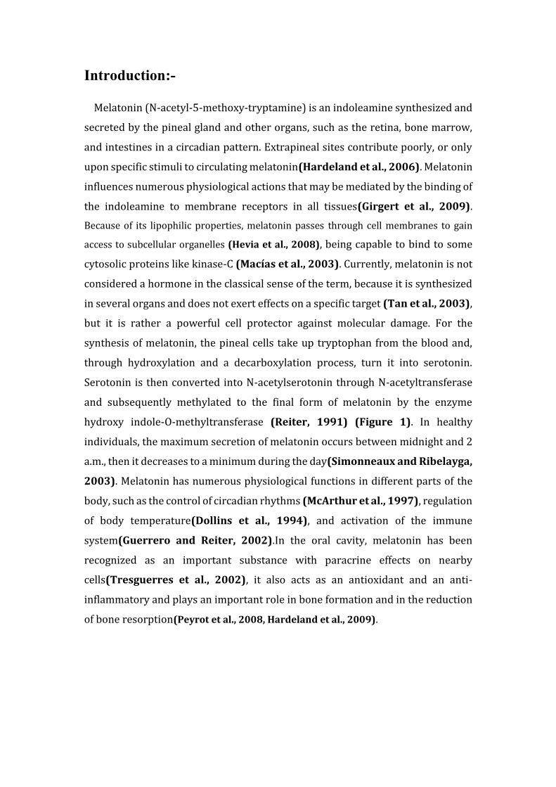
synthesis of melatonin, the pineal cells take up tryptophan from the blood and,
through hydroxylation and a decarboxylation process, turn it into serotonin.
Serotonin is then converted into N-acetylserotonin through N-acetyltransferase
and subsequently methylated to the final form of melatonin by the enzyme
hydroxy indole-O-methyltransferase (Reiter, 1991) (Figure 1). In healthy
individuals, the maximum secretion of melatonin occurs between midnight and 2
a.m., then it decreases to a minimum during the day(Simonneaux and Ribelayga,
2003). Melatonin has numerous physiological functions in different parts of the
body, such as the control of circadian rhythms (McArthur et al., 1997), regulation
of body temperature(Dollins et al., 1994), and activation of the immune
system(Guerrero and Reiter, 2002).In the oral cavity, melatonin has been
recognized as an important substance with paracrine effects on nearby
cells(Tresguerres et al., 2002), it also acts as an antioxidant and an anti-
inflammatory and plays an important role in bone formation and in the reduction
of bone resorption(Peyrot et al., 2008, Hardeland et al., 2009).
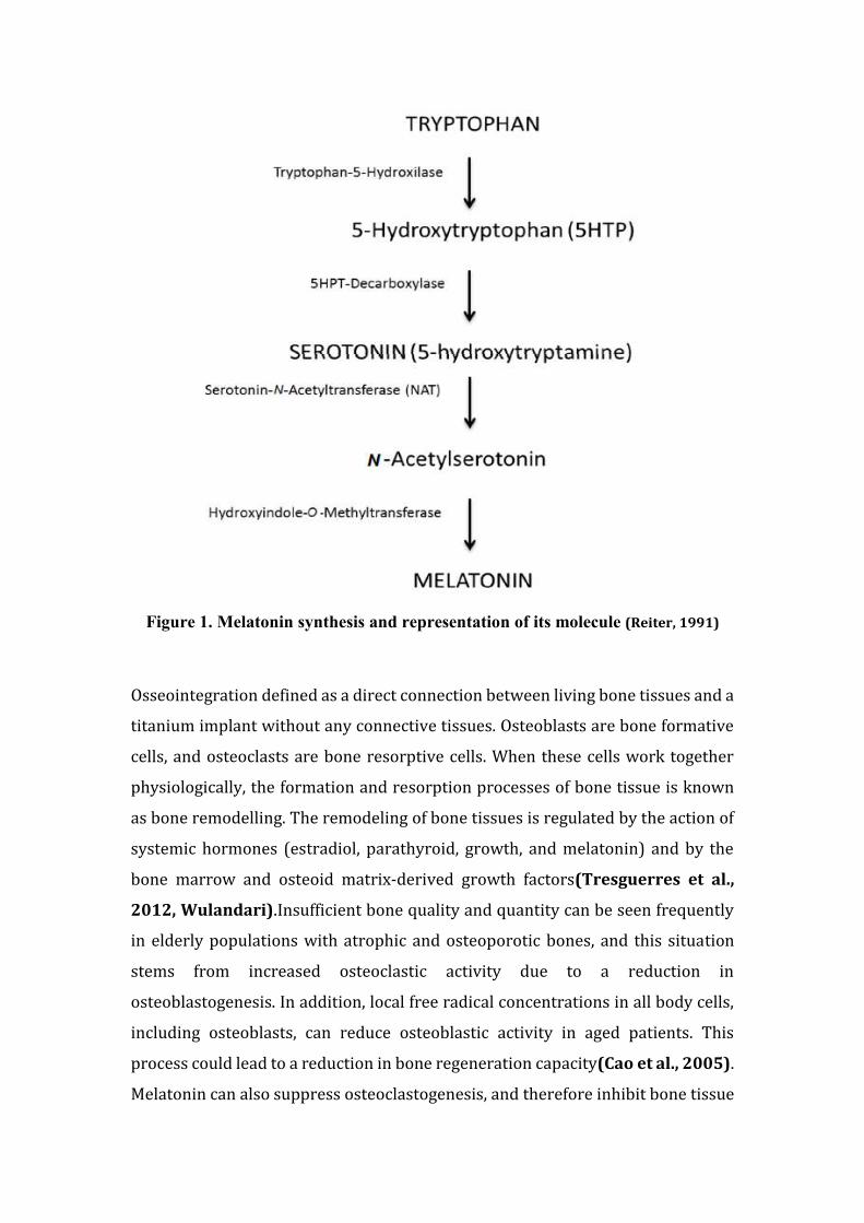
Figure 1. Melatonin synthesis and representation of its molecule (Reiter, 1991)
Osseointegration defined as a direct connection between living bone tissues and a
titanium implant without any connective tissues. Osteoblasts are bone formative
cells, and osteoclasts are bone resorptive cells. When these cells work together
physiologically, the formation and resorption processes of bone tissue is known
as bone remodelling. The remodeling of bone tissues is regulated by the action of
systemic hormones (estradiol, parathyroid, growth, and melatonin) and by the
bone marrow and osteoid matrix-derived growth factors(Tresguerres et al.,
2012, Wulandari).Insufficient bone quality and quantity can be seen frequently
in elderly populations with atrophic and osteoporotic bones, and this situation
stems from increased osteoclastic activity due to a reduction in
osteoblastogenesis. In addition, local free radical concentrations in all body cells,
including osteoblasts, can reduce osteoblastic activity in aged patients. This
process could lead to a reduction in bone regeneration capacity(Cao et al., 2005).
Melatonin can also suppress osteoclastogenesis, and therefore inhibit bone tissue
resorption(Cutando et al., 2008, Witt‐Enderby et al., 2006). In addition to this,
in vitro research has reported that melatonin can increase osteoblast proliferation
and differentiation(López-Martínez et al., 2012, Muñoz et al., 2012). So was
proposed that the local application of melatonin during surgical implant
placement procedures would be an effective treatment technique for dental
implant osseointegration(Dundar et al., 2016). Worthy osseointegration is a
prerequisite for dental implants. Optimal osseointegration depends on the
formation of new bone around implants, which may be stimulated by the
application of biomimetic agents(Ramazanoglu et al., 2011). Considering the
bone metabolism, melatonin acts directly on the osteoclast, a multinucleated cell,
which resorbs the extracellular matrix through various mechanisms, including the
production of free radicals(Roth et al., 1999). Moreover, in pre-osteoblast
cultures from rats, melatonin, in a dose-dependent manner, promoted the
development of bone sialoprotein and other protein bone markers, including
alkaline phosphatase, osteopontin, and osteocalcin, and speeds up their period of
differentiation into osteoblasts from the normal rate, which is 21 days, to 12 days,
this reaction is mediated by the membrane receptors for the indole(Roth et al.,
1999). Also, melatonin, may interfere with the function of the osteoclast and
thereby inhibit bone resorption(Cardinali et al., 2003). (Cutando et al., 2008)
conducted an experimental study using melatonin with dental implants in dogs.
Two weeks after implant insertion, melatonin significantly increased all
parameters of osteointegration. It has been observed that melatonin, increases
bone mass by suppressing resorption through down-regulation of the RANKL-
mediated osteoclast formation and activation(Cutando et al., 2007). These data
point towards an osteogenic effect of melatonin that may be of clinical importance,
as it could be used as a therapeutic agent in situations in which bone formation
would be advantageous, such as in the treatment of fractures or of osteoporosis
(Cardinali et al., 2003).
Toxicology of Melatonin:-
The physiological functions of the pineal hormone melatonin are extremely
diverse. The functions include direct and indirect modulations of anti-oxidative
defense, blood pressure, body temperature, cortisol rhythm, reproduction and
immune function (Claustrat and Leston, 2015). Correspondingly, exogenous
melatonin has been investigated as a treatment for a number of medical and
surgical diseases, demonstrating encouraging results (Andersen et al., 2014,
Gitto et al., 2011). In the USA, melatonin is available as an over-the-counter non-
prescription drug. In most European countries, however, melatonin remains a
prescription drug (Circadin) and has only been approved as a treatment for
primary insomnia in people over 55 years of age. However, a recent Norwegian
register study documented a 3- to 5-times increase in off-label use among children
and adolescents in the time period 2004 to 2012 (Hartz et al., 2015). Melatonin
is generally considered safe (Yousaf et al., 2010), but the increasing clinical use
with potentially increasing doses necessitates further investigations of the risks
of both mild and serious adverse effects. In experimental animal studies,
exogenously administered melatonin has been given in doses up to 800 mg/kg
without any acute toxic effects (Barchas et al., 1967). A number of studies in
preterm infants have investigated the anti-oxidative/anti-inflammatory and
clinical effects of exogenous melatonin in different conditions, such as pain during
tracheal intubation (10 mg/kg x 10 doses, randomized)(Gitto et al., 2012),
asphyxia (10 mg x 8 doses, randomized) (Fulia et al., 2001), respiratory distress
(10 mg/kg x 10 doses, randomized)(Gitto et al., 2004b). Despite repeated high
doses of intravenous melatonin, no signs of adverse effects were observed (Gitto
et al., 2004c, Gitto et al., 2004a). Oral melatonin in doses of 0.5–10 mg was
administered daily from 10 days to 12 weeks to improve sleep quality (Wright et
al., 2011, Cortesi et al., 2012). The studies performed detailed registration of
possible adverse effects, demonstrating a number of mild adverse effects, such as
agitation, dizziness, headache, nausea, and sleepiness (Wasdell et al., 2008, Jain
et al., 2015). Another study was to assess the toxicology of melatonin (10 mg),
administered for 28 days to 40 volunteers randomly assigned to groups receiving
either melatonin (N_30) or placebo (N_10) in a double-blind fashion, according to
the parameters analyzed, there is no toxicological effect that might compromise
the use of melatonin at a dose of 10 mg for the period of time utilized in this
study(Seabra et al., 2000). A randomized, double-blind, placebo-controlled study
including 54 female patients undergoing breast cancer surgery were given 6 mg
of oral melatonin or placebo to improve depressive symptoms and anxiety during
a three month investigational period (Hansen et al., 2014). Finally, a randomized,
double-blind, placebo-controlled study was conducted in elderly patients
suffering from Alzheimer’s disease who were administered 2 mg of melatonin or
placebo for a 24-week period (Wade et al., 2014). A substantial number of both
animal and human studies document that short-term use of melatonin is safe, even
in extreme doses. No studies indicate that exogenous melatonin possesses any
serious adverse effects. Also, randomized clinical studies indicate that long-term
administration only induces mild adverse effects comparable to placebo
treatment. (Andersen et al., 2016).
Periotest M device:-
Periotest device was developed to measure the damping characteristics of natural
teeth and has been used to evaluate implant (Cehreli et al., 2009). It was
developed by Schulte in 1983 to quantify TM (Shulte et al., 1983), and then
utilized by Teerlinck et al. (1991), to measure implant stability and to overcome
destructive methods in measuring the implant stability like histologic analysis,
tensional test, push-out/pull-out test and removal torque analysis. The
(Classic/Wired) Periotest device has been the subject of several studies with
generally favorable results. These results showed that the Periotest device
generally demonstrated a high degree of repeatability and reliability. A wireless
version of the Periotest (Periotest “M”) has been introduced to the profession.
According to information provided by the electronic page of the Periotest M
/wireless device manufacturer (Medizintechnik Gulden, Modautal, Germany),
Periotest M is simpler to perform an objective evaluation of an implant's stability
compared to classic Periotest. In Periotest M wireless design, the user can benefit
from maximum freedom of movement. The device can be used for taking
measurements on a wide variety of implants, without the need for any special
accessories, such as a smart peg (Pang et al., 2014). The Periotest has an
important advantage against the others like the Osstell device: it can be applied
directly to the implant superstructure (Geckili et al., 2009).
The limitations of Periotest are the inability of the instrument to measure the
mesiodistal mobility, the possible effect of position and angle of the rods on the
measured value (Satwalekar et al., 2015). The readings of PTV are from (−8 to
+50) according to information provided by the device manufacturer
(Medizintechnik Gulden, Modautal, Germany) (Pang et al., 2014), (Table 1).
These readings are displayed digitally on a monitor as (PTVs), (Aparicio et al.,
2006).
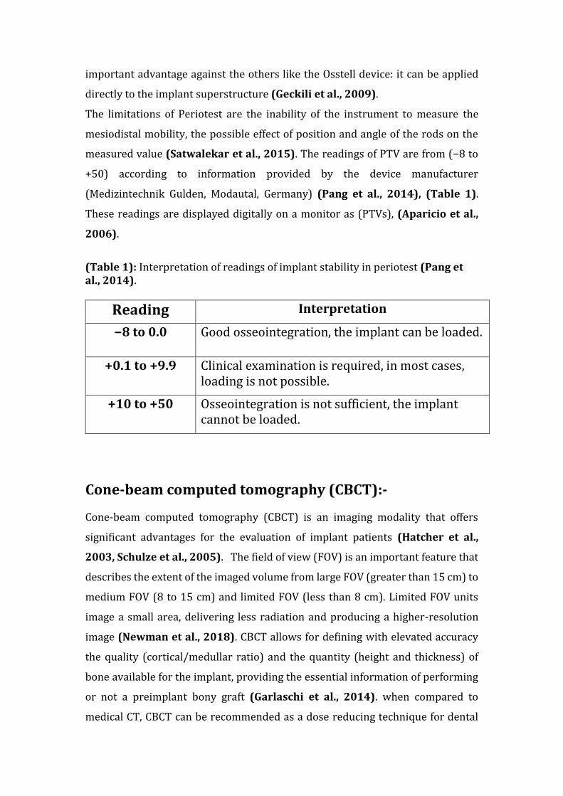
(Table 1): Interpretation of readings of implant stability in periotest (Pang et al., 2014).
Reading Interpretation
−8 to 0.0 Good osseointegration, the implant can be loaded.
+0.1 to +9.9 Clinical examination is required, in most cases, loading is not possible.
+10 to +50 Osseointegration is not sufficient, the implant cannot be loaded.
Cone-beam computed tomography (CBCT):-
Cone-beam computed tomography (CBCT) is an imaging modality that offers
significant advantages for the evaluation of implant patients (Hatcher et al.,
2003, Schulze et al., 2005). The field of view (FOV) is an important feature that
describes the extent of the imaged volume from large FOV (greater than 15 cm) to
medium FOV (8 to 15 cm) and limited FOV (less than 8 cm). Limited FOV units
image a small area, delivering less radiation and producing a higher-resolution
image (Newman et al., 2018). CBCT allows for defining with elevated accuracy
the quality (cortical/medullar ratio) and the quantity (height and thickness) of
bone available for the implant, providing the essential information of performing
or not a preimplant bony graft (Garlaschi et al., 2014). when compared to
medical CT, CBCT can be recommended as a dose reducing technique for dental
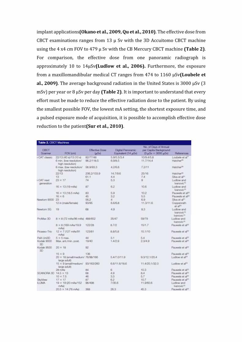
implant applications(Okano et al., 2009, Qu et al., 2010). The effective dose from
CBCT examinations ranges from 13 µ Sv with the 3D Accuitomo CBCT machine
using the 4 x4 cm FOV to 479 µ Sv with the CB Mercury CBCT machine (Table 2).
For comparison, the effective dose from one panoramic radiograph is
approximately 10 to 14µSv(Ludlow et al., 2006). Furthermore, the exposure
from a maxillomandibular medical CT ranges from 474 to 1160 µSv(Loubele et
al., 2009). The average background radiation in the United States is 3000 µSv (3
mSv) per year or 8 µSv per day (Table 2). It is important to understand that every
effort must be made to reduce the effective radiation dose to the patient. By using
the smallest possible FOV, the lowest mA setting, the shortest exposure time, and
a pulsed exposure mode of acquisition, it is possible to accomplish effective dose
reduction to the patient(Sur et al., 2010).
Aim and objectives:-
Aim: - To evaluate the osseointegration following the application of melatonin
around dental implants.
Objectives: -
1- Measuring primary and secondary stability of the dental implant by Periotest
M device.
2- Measuring marginal bone level around the dental implant in baseline and after
6 months follow up using a cone-beam computed tomography (CBCT).
Research Hypothesis:-
The topical application of melatonin powder in the osteotomy site has an effect
on osseointegration around the dental implant and minimize marginal bone loss.
Null hypothesis:-
The topical application of melatonin powder in the osteotomy site has no effect
on osseointegration around the dental implants and in minimizing marginal
bone loss.
Methodology:-
Study design: - a split-mouth clinical trial.
Setting & Subjects:
The study will be conducted in a split-mouth design on patients male with
subjects the ,)molar area st1 lower jaw (canine to upper orthe teeth in missing
will be selected depending on bone density and from those attending to the
department of periodontics.
Inclusion criteria:
1. Patients male have good oral hygiene.
2. Patients were periodontally healthy.
stto 1 canine( lower jaw upper or Patients had at least two missing teeth in the. 3
molar area) indicated for the dental implant.
Exclusion criteria:
1-Patients with any systemic diseases that influence bone healing such as
osteoporosis and diabetes mellitus.
2-Fully edentulous.
3-Patients who had parafunctional habits.
4-Smokers.
5-Patients who were not able to follow the treatment protocol.
Sample Size:
Twenty single-piece endosseous implants use for patients with at least two
.)area smolar stlower jaw (canine to 1 upper orthe teeth in missing
Materials:-
-from (Sigma) methoxytryptamine-5-Acetyl-N( Melatonin powder-1
Aldrich/Germany).
2-Periotest M device (Medizintechnik Gulden, Germany).
3-Dentium implant surgical kit system and DI fixtures (Dentium Co., Korea).
Procedure:
Twenty single-piece endosseous implants (Dentium Co, Korea) will use to restore
missing teeth in the upper or lower jaw (canine to 1st molars area) from both sides.
The study will be split-mouth technique, each patient serves as his own control
(served into 2 groups), the study side (topical application of melatonin in the
implant side), and the control side (no melatonin in the other implant side of the
same patients). According to the study reported by (Cutando et al., 2008) the
estimated dose of melatonin required to enhance osseointegration of dental
implant and minimize the marginal bone resorption is 1.2 mg of melatonin powder
for each implant. CBCT for all patients before implant placement to determine
bone density, dimension, and anatomical landmarks. Prior to surgery, all patients
will instruct to use chlorhexidine 0.12% mouthwash as antiseptic. After local
anesthesia, a mucoperiosteal flap will reflect. The manufacturer’s instructions
should be followed for the preparation of the implant osteotomy site. For the study
side, 1.2 mg of melatonin powder will place in the osteotomy site before the
insertion of the implant. For the control side, no melatonin powder will be used
and implants will be inserted directly in the prepared implant site. After dental
implant installation, the gingival former will insert into the body of the fixture,
then the Periotest M device will use to measure the primary stability of the DI
fixture. After that, the gingival former will be removed and cover screw place and
tightened into the fixture. Sutures will place after flap replacement. Patients
should be instructed to use antibiotics amoxicillin 500mg and metronidazole
500mg 3 times/day for 5 days after surgical procedure, soft diet and proper oral
hygiene measures. Also, CBCT immediately after implant placement to record
baseline bone level, CBCT after 6 months follow up to measure marginal bone level
around implant comparing with baseline measurement, and measure the
secondary stability using periotest M device.
Stopping rules
The occurrence of an unexpected include implant failure...
Budget and funding
This study is self-funded.
Justification for ethical approval:-
According to the college of the dentistry/university of Baghdad following the
Declaration of Helsinki / Tokyo on medical protocol and ethics.
Dissemination
Postgraduate Thesis.
References
ANDERSEN, L., WERNER, M., ROSENBERG, J. & GöGENUR, I. 2014. A systematic review of peri‐operative melatonin. Anaesthesia, 69, 1163-1171
ANDERSEN, L. P. H., GöGENUR, I., ROSENBERG, J. & REITER, R. J. 2016. The safety of
melatonin in humans. Clinical drug investigation, 36, 169-175.
BARCHAS, J., DACOSTA, F. & SPECTOR, S. 1967. Acute pharmacology of melatonin. Nature, 214, 919. CAO, J. J., WRONSKI, T. J., IWANIEC, U.,
PHLEGER, L., KURIMOTO, P., BOUDIGNON, B. & HALLORAN, B. P. 2005. Aging increases stromal/osteoblastic cell‐induced osteoclastogenesis and alters the osteoclast precursor pool in the mouse. Journal of Bone and Mineral Research, 20, 1659-1668
CARDINALI, D. P., LADIZESKY, M. G., BOGGIO, V., CUTRERA, R. A. & MAUTALEN, C. 2003. Melatonin effects on bone: experimental facts and clinical perspectives. Journal of pineal research, 34, 81-87.
CLAUSTRAT, B. & LESTON, J. 2015. Melatonin: Physiological effects in humans. Neurochirurgie, 61, 77-84
CORTESI, F., GIANNOTTI, F., SEBASTIANI, T., PANUNZI, S & .VALENTE, D. 2012.
Controlled‐release melatonin, singly and combined with cognitive behavioural therapy, for persistent insomnia in children with autism spectrum disorders: a randomized placebo‐controlled trial. Journal of sleep research, 21, 700-709.
CUTANDO, A., GóMEZ‐MORENO, G., ARANA, C., ACUñA‐CASTROVIEJO, D. & REITER, R. J. 2007. Melatonin: potential functions in the oral cavity. Journal of periodontology, 78, 1094-1102.
CUTANDO, A., GóMEZ‐MORENO, G., ARANA, C., MUñOZ, F., LOPEZ‐PEñA, M.,
STEPHENSON ,J. & REITER, R. J. 2008. Melatonin stimulates osteointegration of dental implants. Journal of Pineal Research, 45, 174-179.
DOLLINS, A. B., ZHDANOVA, I. V., WURTMAN, R. J., LYNCH, H. J. & DENG, M. H. 1994. Effect of inducing nocturnal serum melatonin concentrations in daytime on sleep, mood, body temperature, and performance. Proceedings of the National Academy of Sciences, 91, 1824-1828.
DUNDAR, S., YAMAN, F., SAYBAK, A., OZUPEK, M. F., TOY, V. E., GUL, M. & OZERCAN, İ. H.
2016. Evaluation of effects of topical melatonin application on osseointegration of dental implant: an experimental study. Journal of Oral Implantology, 42, 386-389.
EL-GAMMAL, M. Y., SALEM, A. S., ANEES, M. M. & TAWFIK, M. A. 2016. Clinical and radiographic evaluation of immediate loaded dental implants with local application of melatonin: a preliminary randomized controlled clinical trial. Journal of Oral Implantology, 42, 119-125.
FULIA, F., GITTO, E., CUZZOCREA, S., REITER, R. J., DUGO, L., GITTO, P., BARBERI, S., CORDARO, S. & BARBERI, I. 2001. Increased levels of malondialdehyde and
nitrite/nitrate in the blood of asphyxiated newborns: reduction by melatonin. Journal of pineal research, 31, 343-349.
GIRGERT, R., HANF, V., EMONS, G. & GRUNDKER, C. 2009. Membrane‐bound melatonin receptor MT1 down‐regulates estrogen responsive genes in breast cancer cells. Journal of pineal research, 47, 23-31.
GITTO, E., AVERSA, S., REITER, R. J., BARBERI, I. & PELLEGRINO, S. 2011. Update on the use of melatonin in pediatrics. Journal of pineal research, 50, 21-28.
GITTO, E., AVERSA, S., SALPIETRO, C. D., BARBERI, I., ARRIGO, T., TRIMARCHI, G., REITER, R. J. & PELLEGRINO, S. 2012. Pain in neonatal intensive care: role of
melatonin as an analgesic antioxidant. Journal of pineal research, 52, 291-295.
GITTO, E., REITER, R. J., AMODIO, A., ROMEO, C., CUZZOCREA, E., SABATINO, G., BUONOCORE, G., CORDARO, V., TRIMARCHI, G. & BARBERI, I. 2004a. Early indicators of chronic lung disease in preterm infants with respiratory distress syndrome and their inhibition by melatonin. Journal of pineal research, 36, 250-255.
GITTO, E., REITER, R. J., CORDARO, S. P., LA ROSA, M., CHIURAZZI, P., TRIMARCHI, G., GITTO, P., CALABRó, M. P. & BARBERI, I. 2004b. Oxidative and inflammatory parameters in respiratory distress syndrome of preterm newborns: beneficial effects of melatonin. American journal of perinatology, 21, 209-216.
GITTO, E., ROMEO, C., REITER, R. J., IMPELLIZZERI, P., PESCE, S., BASILE, M., ANTONUCCIO, P., TRIMARCHI, G., GENTILE, C. & BARBERI, I. 2004c. Melatonin reduces oxidative stress in surgical neonates. Journal of pediatric surgery, 39, 184-189.
GUERRERO, J. M. & REITER, R. J. 2002. Melatonin-immune system relationships. Current topics in medicinal chemistry, 2, 167-179
HANSEN, M. V., ANDERSEN, L. T ,.MADSEN, M. T., HAGEMAN, I., RASMUSSEN, L. S.,
BOKMAND, S., ROSENBERG, J. & GöGENUR, I. 2014. Effect of melatonin on depressive symptoms and anxiety in patients undergoing breast cancer surgery:
a randomized, double-blind, placebo-controlled trial. Breast cancer research and treatment, 145, 683-695.
HARDELAND, R., PANDI-PERUMAL, S. & CARDINALI, D. P. 2006. Melatonin. The international journal of biochemistry & cell biology, 38, 313-316.
HARTZ, I., HANDAL, M., TVERDAL, A. & SKURTVEIT, S. 2015. Paediatric Off‐Label Use of Melatonin–A Register Linkage Study between the Norwegian Prescription Database and Patient Register. Basic & clinical pharmacology & toxicology, 117, 267-273.
JAIN, S. V., HORN, P. S., SIMAKAJORNBOON, N., BEEBE, D. W., HOLLAND, K., BYARS ,A.
W. & GLAUSER, T. A. 2015. Melatonin improves sleep in children with epilepsy: a randomized, double-blind, crossover study. Sleep medicine, 16, 637-644.
LóPEZ-MARTíNEZ, F., OLIVARES PONCE, P. N., GUERRA RODRíGUEZ, M. & MARTíNEZ PEDRAZA, R. 2012. Melatonin: bone metabolism in oral cavity. International journal of dentistry, 2012.
LOUBELE, M., BOGAERTS, R., VAN DIJCK, E., PAUWELS, R., VANHEUSDEN, S., SUETENS, P., MARCHAL, G., SANDERINK, G. & JACOBS, R. 2009. Comparison between
effective radiation dose of CBCT and MSCT scanners for dentomaxillofacial applications. European journal of radiology, 71, 461-468.
LUDLOW, J. B., DAVIES-LUDLOW, L., BROOKS, S. & HOWERTON, W. 2006. Dosimetry of 3
CBCT devices for oral and maxillofacial radiology: CB Mercuray, NewTom 3G and i-CAT. Dentomaxillofacial Radiology, 35, 219-226.
MCARTHUR, A. J., HUNT, A. E. & GILLETTE, M. U. 1997. Melatonin action and signal transduction in the rat suprachiasmatic circadian clock: activation of protein kinase C at dusk and dawn. Endocrinology, 138, 627-634.
MUñOZ, F., LóPEZ‐PEñA, M., MIñO, N., GóMEZ‐MORENO, G., GUARDIA, J. & CUTANDO, A. 2012. Topical application of melatonin and growth hormone accelerates bone healing around dental implants in dogs. Clinical implant dentistry and related research, 14, 226-235.
RAMAZANOGLU, M., LUTZ, R., ERGUN, C., VON WILMOWSKY, C., NKENKE, E. & SCHLEGEL, K. A. 2011. The effect of combined delivery of recombinant human bone morphogenetic protein‐2 and recombinant human vascular endothelial
growth factor 165 from biomimetic calcium‐phosphate‐coated implants on osseointegration. Clinical oral implants research, 22, 1433-1439.
REITER, R. J. 1991. Pineal melatonin: cell biology of its synthesis and of its physiological
interactions. Endocrine reviews, 12, 151-180.
ROTH, J. A., KIM, B.-G., LIN, W.-L. & CHO, M.-I. 1999. Melatonin promotes osteoblast differentiation and bone formation. Journal of Biological Chemistry, 274, 22041-22047.
SEABRA, M. D. L. V., BIGNOTTO, M., PINTO JR, L. R. & TUFIK, S. 2000. Randomized, double‐blind clinical trial, controlled with placebo, of the toxicology of chronic melatonin treatment. Journal of pineal research, 29, 193-200.
SIMONNEAUX, V. & RIBELAYGA, C. 2003. Generation of the melatonin endocrine
message in mammals: a review of the complex regulation of melatonin synthesis by norepinephrine, peptides, and other pineal transmitters. Pharmacological reviews, 55, 325-395.
SUR, J., SEKI, K., KOIZUMI, H., NAKAJIMA, K. & OKANO, T. 2010. Effects of tube current on cone-beam computerized tomography image quality for presurgical implant planning in vitro. Oral Surgery, Oral Medicine, Oral Pathology, Oral Radiology, and Endodontology, 110, e29-e33.
TAN, D. X., MANCHESTER, L. C., HARDELAND, R., LOPEZ‐BURILLO, S., MAYO, J. C., SAINZ,
R. M . &REITER, R. J. 2003. Melatonin: a hormone, a tissue factor, an autocoid, a paracoid, and an antioxidant vitamin. Journal of pineal research, 34, 75-78.
TRESGUERRES, I. F., CLEMENTE, C., BLANCO, L., KHRAISAT, A., TAMIMI, F. & TRESGUERRES, J. A. 2012. Effects of local melatonin application on implant osseointegration. Clinical implant dentistry and related research, 14, 395-399.
TRESGUERRES, I. F., CLEMENTE, C., DONADO, M., GóMEZ‐PELLICO, L., BLANCO, L., ALOBERA, M. A. & TRESGUERRES, J. A. 2002. Local administration of growth
hormone enhances periimplant bone reaction in an osteoporotic rabbit model: an histologic, histomorphometric and densitometric study. Clinical oral implants research, 13, 631-636
WADE, A. G., FARMER, M., HARARI, G., FUND, N., LAUDON ,M., NIR, T., FRYDMAN-MAROM, A. & ZISAPEL, N. 2014. Add-on prolonged-release melatonin for cognitive function and sleep in mild to moderate Alzheimer’s disease: a 6-month, randomized, placebo-controlled, multicenter trial. Clinical interventions in aging,
9, 947.
WASDELL, M. B., JAN, J. E., BOMBEN, M. M., FREEMAN, R. D., RIETVELD, W. J., TAI, J., HAMILTON, D. & WEISS, M. D. 2008. A randomized, placebo‐controlled trial of controlled release melatonin treatment of delayed sleep phase syndrome and impaired sleep maintenance in children with neurodevelopmental disabilities. Journal of pineal research, 44, 57-64.
WITT‐ENDERBY, P. A., RADIO, N. M., DOCTOR, J. S. & DAVIS, V. L. 2006. Therapeutic treatments potentially mediated by melatonin receptors: potential clinical uses in the prevention of osteoporosis, cancer and as an adjuvant therapy. Journal of pineal research, 41, 297-305.
WRIGHT, B., SIMS, D., SMART, S., ALWAZEER, A., ALDERSON-DAY, B., ALLGAR, V.,
WHITTON, C., TOMLINSON, H., BENNETT, S. & JARDINE, J .2011 .Melatonin versus placebo in children with autism spectrum conditions and severe sleep problems not amenable to behaviour management strategies: a randomised controlled crossover trial. Journal of autism and developmental disorders, 41, 175-184.
WULANDARI, A. Jumlah Sel Osteoklas yang Mengekspresikan Interleukin 1 Beta (IL-1Β) Akibat Induksi Gaya Mekanik Ortodonti Dengan Pemberian Natrium Fluorida (NaF) Secara Topikal.
YOUSAF, F., SEET, E., VENKATRAGHAVAN, L., ABRISHAMI, A. & CHUNG, F. 2010. Efficacy
and Safety of Melatonin as an Anxiolytic and Analgesic in the Perioperative PeriodA Qualitative Systematic Review of Randomized Trials. Anesthesiology: The Journal of the American Society of Anesthesiologists, 113, 968-976.
Peyrot, F. and Ducrocq, C., 2008. Potential role of tryptophan derivatives in stress
responses characterized by the generation of reactive oxygen and nitrogen
species. Journal of pineal research, 45(3), pp.235-246.
Hardeland, R., Tan, D.X. and Reiter, R.J., 2009. Kynuramines, metabolites of melatonin
and other indoles: the resurrection of an almost forgotten class of biogenic
.126-47(2), pp.109 Journal of pineal research, amines.
analysis of methods used -Cehreli, M.C., Karasoy, D., Akca, K. and Eckert, S.E., 2009. Meta
).6(24 International Journal of Oral & Maxillofacial Implants, to assess implant stability.
SHULTE, W., D’HOEDT, B., LUKAS, D., MUHIBRADT, L., SCHOLZ, F. & BRETSCHI, J. 1983. Periotest-neues messverfahren der function des paradontiums. Zahnarzti Mitt, 73, 1229-40.
PANG, K. M., LEE, J. W. & LEE, J. Y. 2014. Clinical outcomes of magnesium-incorporated oxidised implants: a randomised double-blind clinical trial. Clin Oral Implants Res, 25, 616-21. GECKILI, O., BILHAN, H. & BILGIN, T. 2009. A 24-week prospective study comparing the stability of titanium dioxide grit-blasted dental implants with and without fluoride treatment. Int J Oral Maxillofac Implants, 24, 684-8. SATWALEKAR, P., NALLA, S., REDDY, R. & CHOWDARY, S. G. 2015. Clinical evaluation of osseointegration using resonance frequency analysis. J Indian Prosthodont Soc, 15(3), 192-9. PANG, K. M., LEE, J. W. & LEE, J. Y. 2014. Clinical outcomes of magnesium-incorporated oxidised implants: a randomised double-blind clinical trial. Clin Oral Implants Res, 25, 616-21. APARICIO, C., LANG, N. P. & RANGERT, B. 2006. Validity and clinical significance of biomechanical testing of implant/bone interface. Clin. Oral Imp Res, 17(2), 2-7. Hatcher, D.C., Dial, C. and Mayorga, C., 2003. Cone beam CT for pre-surgical assessment of implant sites. CDA, 31(11), pp.825-834. Schulze, D., Heiland, M., Blake, F., Rother, U. and Schmelzle, R., 2005. Evaluation of quality of reformatted images from two cone-beam computed tomographic systems. Journal of Cranio-Maxillofacial Surgery, 33(1), pp.19-23. Newman, M.G., Takei, H., Klokkevold, P.R. and Carranza, F.A., 2018. Newman and Carranza's Clinical Periodontology E-Book. Elsevier Health Sciences. Garlaschi, G., 2014. Cone Beam CT and 3D Imaging: A Practical Guide. Springer. Okano, T., Harata, Y., Sugihara, Y., Sakaino, R., Tsuchida, R., Iwai, K., Seki, K. and Araki, K., 2009. Absorbed and effective doses from cone beam volumetric imaging for implant planning. Dentomaxillofacial Radiology, 38(2), pp.79-85. Qu, X.M., Li, G., Ludlow, J.B., Zhang, Z.Y. and Ma, X.C., 2010. Effective radiation dose of ProMax 3D cone-beam computerized tomography scanner with different dental protocols. Oral Surgery, Oral Medicine, Oral Pathology, Oral Radiology, and Endodontology, 110(6), pp.770-776.


![Feasibility of melatonin for treatment (MEL-T) of …...Perioperative melatonin & delirium • >20 years; elective Sx with planned post-op ICU stay >48h [plasma] melatonin 08:00 before](https://img.pdfslide.us/doc/110x75/5f1f61cce84d081c1e42da29/feasibility-of-melatonin-for-treatment-mel-t-of-perioperative-melatonin-.jpg)