Embed Size (px)
Citation preview

95
Vol. XV, No. 1, Jan. 2007Mansoura J. Forensic Med. Clin. Toxicol.
El-Meligy et al...
EFFECT OF NICOTINE ADMINISTRATION AND ITS WITHDRAWAL ON THE REPRODUCTIVE ORGANS, FERTILITY,
AND PREGNANCY OUTCOME IN FEMALE RATS.
BY
Manal M. S. El-Meligy; Randa H. Abdel Hady*; Amal R. Abdel Samaei and Heba M. Saad Eldien**
Departments of Anatomy, Forensic Medicine and Clinical Toxicology* and Histology**
Faculty of Medicine, Assiut University
ABSTRACT
There is clear support for an association between smoking and decreased female fertility. Substantial
harmful effects of cigarette smoke on fecundity and reproduction have become apparent but not generally
appreciated. Therefore the present work studied the effect of nicotine on some reproductive organs and
the morphological abnormalities of offsprings of nicotine treated mothers. The animals were divided into
two main groups: Group A (non pregnant rats) and group B (pregnant rats).Group A was subdivided into
three subgroups: (1) Saline treated control group, (2) Nicotine treated group, that was injected daily with
nicotine in a dose of 0.4mg/100. gm body weight which is equivalent to the amount of nicotine passing to
the blood of the heavy smoker subcutaneously for 3 weeks, (3) Rehabilitation group that was injected with
nicotine in the same previous dose and duration then left for 2 months after stoppage of injection. Group
B was subdivided into two subgroups: (1) Control group which was left to get pregnant and injected with
0.9% saline subcutaneously daily from the day 6-20 of pregnancy. (2) Nicotine treated group that was in-
jected with nicotine in the same dose used in group (A) from the day 6-20 of pregnancy. Non pregnant an-
imals of group A 1, 2 were sacrificed at the end of 3 weeks, whereas those of groups A 3 were sacrificed 2
months later. Uterine and ovarian specimens were fixed in 10% formaline solution for histopathological
examination. Pregnant rats of group B (1),(2) were sacrificed on the 20th day of gestation, and fetuses
were extracted, examined by naked eye and some of them underwent an Alizarin red stain for skeletal ex-
amination. Placenta was fixed in 10% formaline solution for histopathological examination. As regard
the histological changes in ovary, nicotine caused retardation in the follicular growth, a decrease in
healthy follicles and increase in the atretic and cystic follicles. Apoptotic granulosa cells were clearly ob-
served. Fatty degenerative changes in corpora lutea could be observed. In rehabilitation group, there
was an improvement in the follicular growth as well as the degenerative changes in corpora lutea but did
not reach the control level .In uterus, nicotine caused reduction in the endometrial and myometrial thick-
ness and marked reduction in the endometrial glands. In rehabilitation group, endometrium and myome-

El-Meligy et al...96
Vol. XV, No. 1, Jan. 2007Mansoura J. Forensic Med. Clin. Toxicol.
1996) menstrual abnormalities and earlyonset of menopause (Midgette and Baron,1990). Interestingly, women who smokehave a decreased risk of breast cancer(Vessey et al., 1983) and endometrial can-cer (Cramer et al., 1986). Because thesephenomena are all estrogen dependent, ithas been suggested that smoking has anti-estrogenic effects (Baron et al., 1990). Infact, female smokers had significantly low-er levels of estriol, estradiol, and estroneduring the luteal phase of menstrual cy-cles and tended to have lower levels ofthese estrogens during the follicular phasecompared to nonsmokers (MacMahon etal., 1982). Treatment of rats with nicotinewas associated with a decrease in estro-gen-dependent parameters including ute-rine weight, diameter and thickness of themyometrium and endometrium (Patil, etal., 1999). Also, nicotine and M-nicotine (anicotine metabolite ) can induce a sort ofluteal insufficiency by inhibiting progeste-rone release, probably through modula-
INTRODUCTlON
Cigarette smoke is a complex mixtureof toxic chemicals including nicotine, car-bon monoxide, and several recognizedcarcinogens and mutagens (Stedman,1968). These toxicants are absorbedthrough the pulmonary vasculature andtransported via the bloodstream causingcytotoxicity, genotoxicity, and tumorige-nicity throughout the body (Stillman,.etal.,, 1986.and. Clair,et al.,1994). Nicotine ismetabolized primarily by the liver, and toa lesser extent, the lung and kidney ,withthe primary metabolite being cotinine(Kyerematen et al., 1990 a&b). In additionto the deleterious effects on cardiovascularand pulmonary physiology, cigarettesmoking affects the reproductive systemand imposes a number of unique risksspecific to women, as smoking have beenfound to be associated with infertility , ec-topic pregnancy (Saraiya et al., 1998),spontaneous abortion (Kendrick et al.,
trium looked more or less similar to control apart from reduced endometrial glands in some animals.
Placental degenerative changes were observed and reflected on the newborns in the form of abnormal
reduction in body size and weight, wrinkled skin, misshaped head, severe kyphosis of body, reduced neck
region, micromelia (short limbs), spina bifida and meningoencephalocele. Also skeletal examination of
Alizarin red stained fetuses revealed retarded ossification of axial and appendicular skeleton. nicotine
adversely influences follicular growth by an increase in apoptotic cell death as well as necrosis. Nicotine
induced cell apoptosis and necrosis may represent the mechanisms underlying the well established link
between smoking and fertility disorders. Also placental degenerative changes may be considered at least
in part, the underlying etiology of the observed fetal anomalies.
Key words : Nicotine, Uterus, Ovary. Placenta, Pregnancy outcome.
97
Vol. XV, No. 1, Jan. 2007Mansoura J. Forensic Med. Clin. Toxicol.
El-Meligy et al...
of nicotine on ovarian, uterine and pla-cental structure. The second focuseson the fetal complications associatedwith maternal smoking during preg-nancy. So the present study was car-ried out to declare the possible mecha-nisms by which nicotine administrationcan interfere with female fertility andaffect pregnancy outcome, and whetherits withdrawal has an improving effector not.
MATERIAL AND METHODS
A total number of 40 adult femalealbino Wistar rats aged 3 months oldwere used in this study. The rats wereobtained from animal house, AssiutUniversity. They were housed in stain-less steel cages under 12 h light /darkcycle at 25oC and allowed water and food(laboratory chow) add libitum. The ex-periments reported here were approvedby Faculty of Medicine Assiut Universi-ty ethics committee. The animals weredivided into two main groups: (A &B),group A consists of 24 (non pregnantrats) and group B consists of 16 (pregnantrats).
Group A (non pregnant rats) weresubdivided into three subgroups, 8 ratseach:
1- Control, which were injected with 1ml 0.9% saline subcutaneously dailyfor 3 weeks.
tion of the prostaglandin (PG) system(Miceli et al., 2005).
Clearly, additional investigations areneeded to clarify the effects of nicotine onfollicular steroidogenesis and to demon-strate the nicotine induced histopathologi-cal changes in the ovary and uterus.
New ham et al., (1990) ,Went et al.,(1990), Ashfaq et al., (2003), Vaglenova etal., (2004), Ronco et al., (2006) and Jauni-aux and Burton, (2007) reported that ac-tive and passive maternal smoking hasbeen found to cause a damaging effect inevery trimester of human pregnancy. Cig-arette smoke contains scores of toxinswhich exert a direct effect on the placentaland fetal cell proliferation and differentia-tion and can explain the increased riskof miscarriage, fetal growth restriction,stillbirth, preterm birth, placental abrup-tion, increased perinatal morbidity, andchildhood cognitive and behavioral defi-cits .All these changes were associatedwith extensive loss of trophoblasts byapoptosis.
Although many adverse effects ofcigarette smoking on reproduction havebecome clear, the findings of previousstudies have largely been contradicto-ry.
Aim of the work: is divided into twomain parts: The first focuses on the effects
El-Meligy et al...98
Vol. XV, No. 1, Jan. 2007Mansoura J. Forensic Med. Clin. Toxicol.
Pregnant rats of group B (1, 2) weresacrificed on the 20th day of gestation,after being anaesthetized by ether earlyin the morning to prevent the mothersfrom devouring any damaging off-spring. Before opening either uterinehorn the number of living and deadfetuses, as indicated by their movementfollowing a gentle pressure, was record-ed (Wilson, 1964). The weight, length andgeneral morphology of fetuses were ex-amined. The placentae were kept in 10%formaline solution for histopathologicalexamination using Hx & E stain forgeneral histological examination, Mas-son's trichrome for collagen fibres andperiodic acid schief for polysacharide(Drury and Wallington, 1980). Two thirdsof the fetuses were fixed in Bouin’s solu-tion for external and visceral examination.Transverse sections were made in off-springs of control and nicotine treatedmothers to investigate the presence of anyfetal abnormalities that could be apparentby naked eye.
The other third of fetuses was eviscerat-ed and preserved in 95% alcohol for skele-tal examination using Alizarin red stain(Wilson, 1964).
Statistical analysis was done for fetalweight and crown crumb length for bothcontrol and nicotine treated groups usingstatistical package for social sciences ver-sion II (SSPS).
2- Nicotine treated group, that were in-jected daily with nicotine in a dose of0.4mg/100 gm body weight subcuta-neously for 3 weeks which is equiva-lent to the amount of nicotine pass-ing to the blood of the heavy smoker(Aydos et al., 2001). Nicotine (liquid)was purchased from Fluka BioChemica (Switzerland).
3- Rehabilitation group that was inject-ed with nicotine in a dose of 0.4mg/100 gm body weight subcutaneouslydaily for 3 weeks (Aydos et al., 2001),then left for 2 months after stoppageof injection.
Group B (pregnant rats) were subdi-vided into two subgroups, 8 rats each:
(1) Control group: Rats were left to getpregnant and injected with 0.9% sa-line subcutaneously daily from theday 6-20 of pregnancy.
(2) Nicotine treated group rats were in-jected with nicotine in the samedose used in group (A) from theday 6-20 of pregnancy.
Non pregnant animals of group A (1, 2)were sacrificed at the end of 3 weeks, whe-reas those of groups A (3) were sacrificed2 months later. Uterine and ovarian(in thediestrus phase) specimens were fixed in10% formaline solution for histopathologi-cal examination using H & E stain.

99
Vol. XV, No. 1, Jan. 2007Mansoura J. Forensic Med. Clin. Toxicol.
El-Meligy et al...
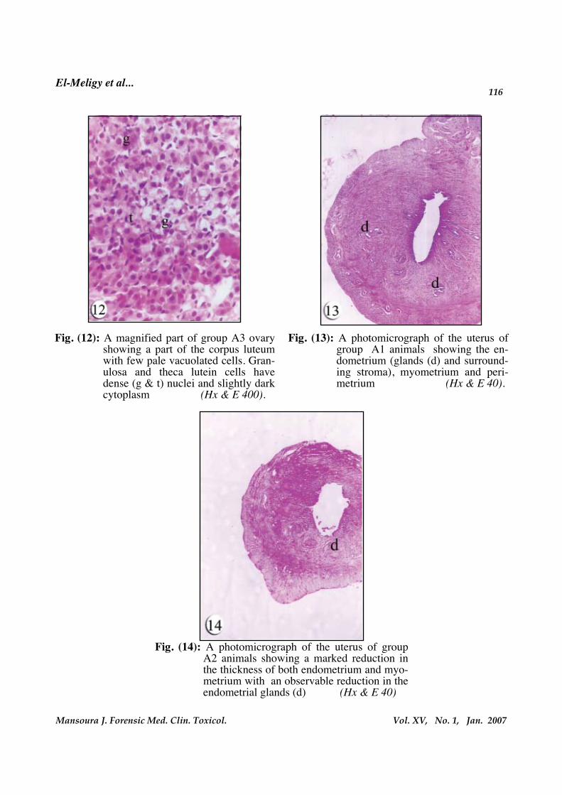
ca lutein cells appear with dense nucleiand slightly dark cytoplasm, while oth-ers appear with pale vacuolated cyto-plasm (Fig. 12).
Examination of the uterus of the controlanimals reveals the normal histologicalstructure of the endometrium (glands andsurrounding stroma), myometrium andperimetrium (Fig. 13). Examination of theuterus of the nicotine treated animalsshows a marked reduction in the thicknessof both endometrium and myometriumwith an observable reduction in the endo-metrial glands (Fig. 14). In the rehabilitat-ed animals, endometrium and myometri-um appear more or less similar to those ofcontrol, however fewer glands are ob-served, compared to the control, in someparts of the endometrium (Fig. 15).
Group (B): Examination of the placenta of the con-
trol animals (in the basal plate) reveals thematernal part decidual cells and multiplematernal blood spaces that are separatedby fetal trophoblast cells (fig.16). Multiplebranching chorionic villi have blood ves-sels inside their cores with intact endothe-lial linning (Figs. 17). Examination of thecontrol placenta by Masson's trichromedemonstrates collagen fibres in moderateamount in the core of villi and in the sub-trophoblastic membranes (Fig. 18). Posi-tive periodic acid schief reactions are ob-served in the basement membranes of the
RESULTS
3.1. Histological results:Group (A):Examination of the ovary of the con-
trol animals reveals that the cortex isoccupied by follicles in various stagesof development and multiple corpora lu-tea (Fig. 1). Examination of the ovary ofthe nicotine treated animals reveals multi-ple cystic and degenerated follicles in vari-ous stages of degeneration. Collapsed fol-licles are observed on the surface of theovary (Figs. 2 & 3). Multiple apoptoticcells are present among the follicular cells,also degenerated ova are observed insome growing follicles (Figs. 4 & 5). Multi-ple degenerated granulosa cells with vacu-olated cytoplasm and dense nuclei arepresent (Figs. 6 & 7).
Irregularity in the capsule of some cor-pora lutea is associated with a large groupof pale vacuolated cells immediately be-neath the capsule, some of these cells ap-pear anucleated (Figs. 8 & 9).
As regards the rehabilitated animals,there are somewhat increase in the healthyfollicles and corpora lutea and reductionin the degenerated follicles and corporalutea but do not reach the control level(Fig. 10). Mild degenerative changes areobserved in ovum, corona radiata andgranulosa cells of some animals (Fig. 11).In some corpora lutea, granulosa and the-
El-Meligy et al...100
Vol. XV, No. 1, Jan. 2007Mansoura J. Forensic Med. Clin. Toxicol.
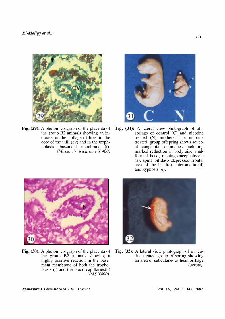
the trophoblasts and blood capillaries, sothey appeare thicker than those of controlplacenta (Fig. 30).
3.2. Fertility and pregnancy outcomeresults:
This study reveals decrease in femalerat fertility, however statistically it do notreach the level of significance. The preg-nancy outcome of nicotine treated mothersdeclares a marked reduction in compari-son to control mothers, the difference isfound to be highly significant (Table 1).
3.3. Gross morphological results of the
offsprings :- As regard fetal body weight, it is
found that fetuses of nicotine treatedmothers are lighter than those of con-trol mothers. Statistically, the differ-ence in fetal body weight betweenthese two groups of offsprings is re-corded to be highly significant (Ta-ble 2).
- As regard fetal crown rumb length, itis found that fetuses of nicotine treat-ed mothers are shorter than those ofcontrol mothers. Statistically, this dif-ference is reported to be highly signif-icant (Table 2).
3.4. Naked eye examination of off-springs:
The newborn offsprings of nicotine
trophoblasts and those of blood capillaries(Fig. 19).
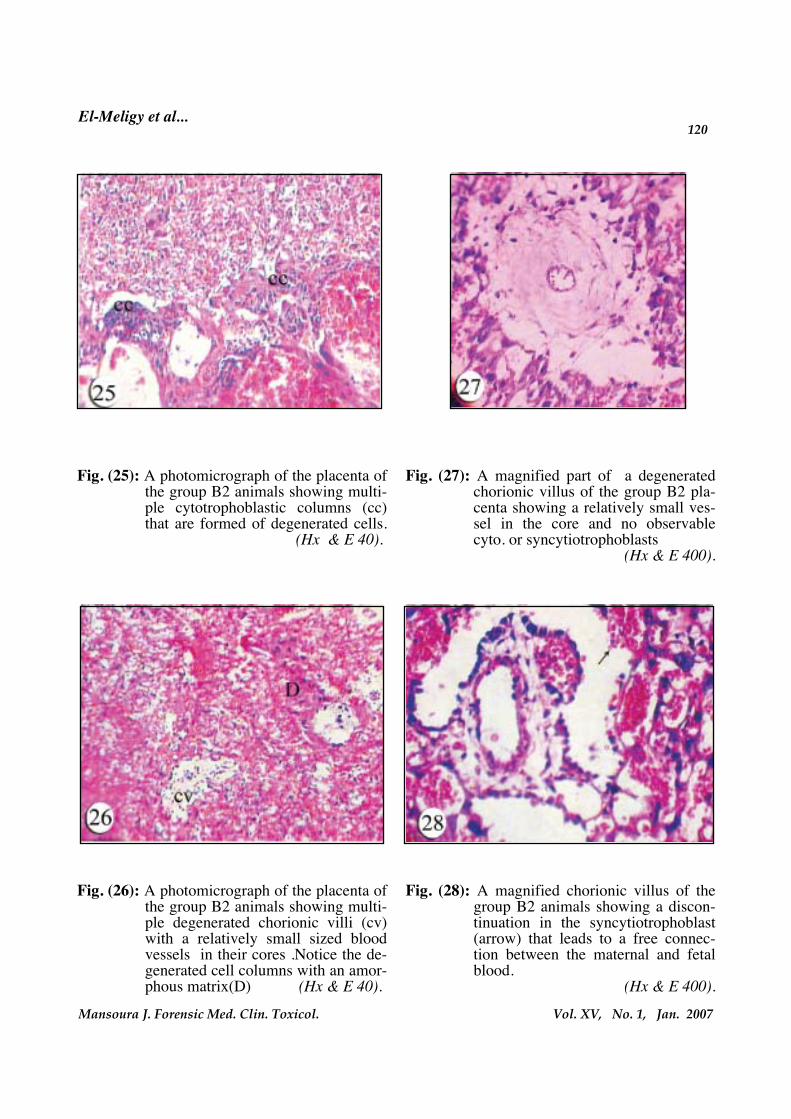
Examination of the placenta of the treat-ed animals reveals separation of the decid-ual cells from the basal plate and alsofrom trophoblastic cell columns. Necroticas well as apoptotic changes are observedin the decidual cells and multiple degener-ated cell columns are present (Fig. 20).Wide maternal blood spaces that are sur-rounded by degenerated decidual cells arealso observed (Fig. 21). Apoptotic signsare seen in the decidual cells in the form offragmentation of the nucleus with separa-tion of the nuclear fragments (Fig. 22).Margination of the nuclear chromatin isassociated with cytoplasmic blebbing (Fig.23), and the nucleus is fragmented intomultiple dense rounded globules (Fig. 24).There are multiple cytotrophoblast col-umns that are formed of degenerated cells(Figs. 25). Multiple degenerated chorionicvilli with a relatively small sized bloodvessel in their cores as well as degeneratedcell columns with an amorphous matrixare observed (Figs. 26 & 27). Discontinua-tion in the syncytiotrophoblast that leadsto free connection between the maternaland fetal blood is present in some animals(Fig. 28). Masson's trichrome demon-strates an increase in the collagen fibres inthe core of the villi and in the trophoblas-tic basement membrane (Fig. 29). Highlypositive periodic acid schief reaction isseen in the basement membranes of both


103
Vol. XV, No. 1, Jan. 2007Mansoura J. Forensic Med. Clin. Toxicol.
El-Meligy et al...
One reason for these conflicting resultsmay be the variation in the nicotine con-centration in the different used prepara-tions. Another important factor to consid-er when comparing the results of past andpresent studies is the level of ATP insidethe tissue which is an important determi-nant of the form of cell death, since the re-duction of ATP inside the tissue is in favorof necrosis. These suggestions were inagreement with the work of Sugano andIto (2000) who found that nicotine changesthe form of H2O2-induced cell death fromapoptosis to necrosis in U937 cells by re-ducing the level of intracellular ATP.
The observed pale vacuolated cyto-plasm either in the follicular cells or in theluteal cells of the corpus luteum may indi-cate a large amount of accumulated lipidsthat dissolved during the preparation ofslides for haematoxyline and Eosin stain-ing that may reflect an impairment in thesteroidogenesis. These findings are in ac-cordance with those of the work of severalauthors who found an increase in theovarian cholesterol level in nicotine treat-ed rats that indicates the inhibition of thesteroidogenesis (Patil et al., 1999). Othersreported that the antiestrogenic effects ofnicotine may be exerted indirectly by act-ing at the level of the theca interna to de-crease androgen biosynthesis (Sanders etal., 2002) or at the level of granulosa cellaromatase that is responsible for the con-version of androstenedione to estradiol
since they found that benzopyrene, cad-mium, or cotinine may have a role inthe reduction of blood circulation after ex-posure to smoke, which can lead to a de-creased quantity of mature oocytes. Theyadded that, nicotine may be responsiblefor the impairment in the vascular endo-thelial growth factor A which is consid-ered to be one of the substances ableto increase ovarian blood circulation, andtherefore improves oocyte maturation.Several authors reported necrosis as aform of death in different tissues afternicotine exposure. In human coronary ar-tery endothelial cells, Hakki et al., (2002)found that nicotine significantly inhibitsapoptosis by decreased expression levelof active caspases. Also, Suzuki et al.,(2003) reported an inhibitory effect ofsmoking in the cardiac apoptosis. Similarobservations were reported in the chickciliary ganglion neurons and in the spinalcord neurons, the later neuron-protectiveeffects were exerted by the increased ex-pression of nerve growth factor (Pugh andMargiotta, 2000 and Garrido et al., 2003).However Pugh and Margiotta (2000)and Garrido et al. (2003) noticed apop-tosis, and \ or necrosis in pancreatic can-cer cells and in pulmonary tissue after nic-otine exposure. Lastely, Bordel, et al.(2006) reported a dose dependent inhibi-tion in the follicular growth and an in-creased apoptotic cell death specially witha high dose of nicotine in Syrian goldenhamsters.
El-Meligy et al...104
Vol. XV, No. 1, Jan. 2007Mansoura J. Forensic Med. Clin. Toxicol.
tions are in accordance with those of thework of Bao et al. (2002) who investigatedthe effects of cigarette smoke related hy-drocarbons (benzo (a) pyrene) on uterineCYP1A1/2 and 1B1 (enzymes involved inestrogen metabolism), and they suggestedthat CYP1A2 and CYP1B1 are not inducedby(benzo (a) pyrene) in the endometrialcells. Also, it has been found that smokingis associated with decreased incidence ofuterine fibroids, endometriosis and ute-rine cancer, which may reflect inhibitoryeffects of smoke constituents on uterinecell proliferation and extracellular matrixinteractions. In addition, increased miscar-riage rate observed among mothers whosmoke might be related to direct adverseeffects of nicotine, cadmium and polyaro-matic hydrocarbons on trophoblast inva-sion and proliferation (Shiverick and Sala-fia, 1999).
The observed increased amount of col-lagen in chorionic villi and increasedthickness of subtrophoblastic as well asfetal capillary basement membranes inthe present work reflect the degenerativechanges that may result from nicotine it-self and / or its metabolites and lead toa noticeable retardation in the fetalgrowth and low birth weight. Nicotineand carbon monoxide both induce de-generative changes that affect the func-tional component of an organ by reducingits nutritive and excretory functionswhich may be the cause of low birth
(Barbieri et al., 1986). However, otherssuggested that nicotine free base aug-ments estradiol secretion and inhibits pro-gesterone secretion by human granulosacells in a dose-dependent manner (Bódiset al., 1997). This increase in the estradiolsecretion may have a suppressive effect onthe progesterone produced by the follicu-lar cells. These suggestions were support-ed by the work of Fortune and Hanse,(1979) who noticed that high intrafollicu-lar concentrations of estradiol may inhibitfollicular progesterone production in vivobefore the LH surge .So, nicotine and M-nicotine (a metabolite of nicotine) can in-duce a sort of luteal insufficiency by inhib-iting progesterone release, probablythrough modulation of the Prostaglandinsystem (Miceli et al., 2005).
The reduced thickness of the endome-trium and myometrium and decrease ofnumber of the endometrial glands ob-served in the present study may resultfrom the impaired hormonal support (es-trogen and progesterone), since estro-gen is responsible for the proliferationof the uterine muscles and progeste-rone is responsible mainly for the de-velopment of the uterine glands and gly-cogen deposition, however the directtoxicity of nicotine or its related metabo-lites cannot be excluded. The later factormay explain the degenerative and atroph-ic changes observed specially in the ute-rine epithelium and glands. These sugges-

105
Vol. XV, No. 1, Jan. 2007Mansoura J. Forensic Med. Clin. Toxicol.
El-Meligy et al...
oblasts and the blood capillaries as wellas degeneration of the villous capillaryendothelial cells, and reduced diameter ofthe fetal capillaries in the present workmay have a role in the impairment of thefeto-maternal exchange of gases and nu-trients and so, fetal hypoxaemia .Thesesuggestions are in accordance with thoseof Larsen et al. (2002) who found that sig-nificant increase of the trophoblast volumein the mothers who smoked cigarettes wasassociated with a significant reduction inthe lengths of villous capillaries .Also,thevolume density of the fetal vessels in theterminal villi of the smoker's placenta wasshown to be decreased and so, the ex-change area of the smoker's placenta (VanDer Velde et al., 1983 and 1985). Similarly,Bush et al., (2000) reported that, the in-creased haematocrits suggests that fetusesof smoking mothers suffer hypoxic stress.However, others reported that some of thefetal changes cannot be explained on thisbasis and that these may possibly be dueeither to cadmium toxicity or to accumula-tion of polycyclic aromatic hydrocarbonsin the placenta and/or in the fetal tissues(Van Der Veen and fox, 1982). Zenzes etal. (1997) supported the later opinion andadded the immunodetection of cotinine, amajor metabolite of nicotine, and benzo (a)pyrene, a potent mutagen and carcinogen,in the embryos as well as granulosa-luteincells of women in IVF exposed actively orpassively to cigarette smoke. In agreementwith these results Jauniaux and Burton,
weight babies in smokers (Ashfaq et al.,2003).
The observed degenerative and necroticchanges in the trophoblast cells in thepresent study may be responsible for thepremature aging changes in the placentathat may affect the intrauterine fetalgrowth. These suggestions are in agree-ment with those of Ashfaq et al. (2003)who found that the extensive loss oftrophoblasts by apoptosis and syncytialbuds may lead to hormonal imbalance,premature labour and abnormal outcomeof pregnancy in smokers.
The observed increase in cell columnsmay be explained as a compensatory hy-perplasia in the trophoblast cells in a trialto replace the lost trophoblasts, however,the predominant degenerated cells inthese columns in the placentae of smokingmothers, may result from the toxaemic ef-fect of nicotine that interferes with theconnection of the chorionic villi with theuterine wall that may lead to impairmentof the placental barrier and so, the trans-ports between the mother and the fetus.Zdravkovic et al., (2006) supported thesesuggestions and mentioned that nicotineimpaires the development of cell columnsby acting through the l-selectin adhesionsystem.
The observed irregular thickening ofthe basement membrane of both the troph-
El-Meligy et al...106
Vol. XV, No. 1, Jan. 2007Mansoura J. Forensic Med. Clin. Toxicol.
syncytial pinocytotic activity that maylead to impairment of placental barrier,and these morphological changes are like-ly to compromise, rather than assist, trans-placental oxygen transfer.
As regard the effect of nicotine with-drawal on the ovarian and uterine tissue,the results of the present work declaredthat stoppage of nicotine intake leads toimprovement of some degenerativechanges as the tissue became more or lesssimilar to that of the control. This comes inaccordance with the observation of Galalet al. (2005) in the testicular tissue after ex-posure to nicotineThey suggested that anyreduction in the sperm count is potentiallyreversible. However, Riesenfeld and Oli-va, (1988) disagreed with the current find-ings and reported that the toxic effects ofnicotine are irreversible and persisting forthe entire life as does infertility. Anyway,it is believed that complete recovery mayoccur if the period of rehabilitation be-comes more prolonged or is accompaniedwith administration of an adjuvant antiox-idant therapy(ElAdlali, 2000).
As regard the effect of nicotine on fe-male fertility and pregnancy outcome, thiswork revealed a serious influence of nico-tine on women's reproductive ability. Thismay be due to the adverse effect of nico-tine on several of the crucial steps withinthe reproductive process required forachieving a pregnancy or its maintenance,
(2007) reported that, in the placenta, smok-ing is associated from early in pregnancy,with a thickening of the trophoblasticbasement membrane, an increase in colla-gen content of the villous mesenchymeand a decrease in vascularisation.Theseanatomical changes are associated withchanges in placental enzymatic and syn-thetic functions. In particular,nicotine de-presses active amino acid uptake by hu-man placental villi and trophoblastinvasion and cadmium decreases the ex-pression and activity of 11 beta- dehy-drogenase type 2 which is causally linkedto FGR. Maternal smoking also dysregu-lates trophoblast expression of moleculesthat govern cellular responses to oxygentension. The later findings raised the pos-sibility that, the perinatal morbidity asso-ciated with cigarette smoking duringpregnancy is probably more related to theischemic and/or toxic effects of severalcompounds in tobacco smoke, partly onplacental function and also directly on thefetus.
The observed perforation in the syncy-tium in the present study and so connec-tion between fetal and maternal blood asa result of syncytial degenerative andatrophic changes may expose the fetus totoxic substances in the maternal blood.However, Demir et al. (1994) mentionedthat focal syncytial necrosis, is associatedwith degenerated cytoplasmic organelles,abnormalities of the microvilli, decreased
107
Vol. XV, No. 1, Jan. 2007Mansoura J. Forensic Med. Clin. Toxicol.
El-Meligy et al...
cluded that environmental tobacco smokeexposure in pregnant women adverselyaffects pregnancy by increasing fetal mor-tality and preterm delivery at higher expo-sure levels and showing fetal growth re-tardation across all levels of exposure. Inaddition, the results of the study ofSheung et al. (2006) demonstrated that in-halation exposure of pregnant mice to alow dose of mainstream cigarette smoke(MCS) shortened gestation and alteredhormone secretory patterns, which are im-portant for maintaining pregnancy.
Considering the fetal growth, thepresent study declared a significant reduc-tion of the fetal growth parameters in theoffsprings of nicotine treated mothers.This could be attributed to the interferenceof nicotine with the fetal nutrition throughits serious effect on the placenta. Thiscomes in accordance with the suggestionof Kulal et al. (2001) who found that chil-dren of mothers who were moderate toheavy smokers during pregnancy, hadlower birth weight, shorter body lengthand smaller head circumference thanthose of non-smokers. They stated thatnicotine can adversely affect uterine andplacental blood flow by causing constric-tion of the blood vessels.
Also, Van derVeen and Fox,(1982);Shivericka and Salafia, (1999) stated thatretardation of fetal growth could be attrib-uted to ultrastructural changes of the pla-
as this study declared the deleterious ef-fect of nicotine on the female reproductiveorgans exemplified in the ovary and uter-us. Also, its detrimental effect on the pla-centa cannot be ignored. This result agreeswith those of New ham., et al. (1990),Went et al. (1990), Economides and Braith-waite (1994), Cnattingius and Nordstrom.,1996, Nelson et al. (1999), and Gruslin etal. (2001) who stated that maternal smok-ing is associated with spontaneous abor-tion, reduced fetal growth, prematurity,and increased perinatal mortality andmorbidity. Also, these results agree withthe latter observation of Zdravkovic et al.(2005) who suggested that tobacco constit-uents exert direct effects on cytotropho-blast proliferation and differentiation andhelp to explain the mechanisms by whichsmoking negatively affects human preg-nancy outcome.
Similar observations were reported byRostand et al. (1999) in human, they foundthat the maternal smoking was detrimen-tal to the very early embryo, and even ifsmoking was stopped, the effects wouldpersist at least for some days and therewas no immediate catch-up growth. Also,Pauly et al. (2004) suggested persistentgeneral dependant changes in the behav-ior of newborn after oral nicotine admin-istration to pregnant mice .However ,thesepoints are in need for further study.
Moreover, Kharrazi et al. (2004) con-
El-Meligy et al...108
Vol. XV, No. 1, Jan. 2007Mansoura J. Forensic Med. Clin. Toxicol.
biparietal diameter observed among in-fants with prenatal exposure to tobaccosmoke.
This work revealed a variety of congen-ital malformations including wrinkledskin, malformed head (scaphocephaly), se-vere kyphosis, shortened neck region, mi-cromelia, spina bifida and meningoence-phalocele. This reflects the severedetrimental effect of nicotine on the devel-oping emberyo. These results agree withthose of Saad et al. (1995) who concludedthat nicotine had a serious effect on gener-al growth and development as well as onpalatogenesis of mice Also, Rostand et al.(1990) found a large number of craniofa-cial characteristics in the infants of heavysmokers. A great link between cigarettesmoke and teratogenicity in rats werefound by Reckzeh et al. (1975) and Nelson.et al. (1999). They found also a dose-dependent reduction in fetal body weight,sizes, lengths and bitemporal diameters.Gartner et al. (1997) suggested that nico-tine interferes with both palatal and me-senchymal components in utero affectinggreatly the tongue development. Further-more, the studies of Vaglenova et al.(2004) demonstrated that prenatal nicotineexposure produced significant long-termdevelopmental and behavioral teratogeniceffects.
In particular, smoking interferesstrongly with the fetal brain and pan-
centa that are believed to contribute to re-duced placental nutrient and oxygentransfer. These placental changes areconsistent with the association betweenmaternal smoking and fetal growth re-striction. Ronco et al. (2006) showed thatmetallothionein -2 isoform was specifical-ly induced in smokers’ placentas. Thiscould be involved in placental cadmiumand zinc retention that could contribute toreduce that transference of zinc to the fe-tus, which in turn, may be associated withdetrimental effects on fetal growth, finallyleading to the reduced birth weights ob-served in neonates born from smokingmothers.
These results come in accordance withthose of Jauniaux and Burton (2007) whostated that in the fetus, smoking is asso-ciated with a reduction of weight, fat massand most anthropometric parameters andas in the placenta with alterations in pro-tein metabolism and enzyme activity.These alterations are the results of a directtoxic effect on the fetal cells or an indirecteffect through damage to, and/or func-tional disturbances of the placenta.
In addition, Kalinka et al. (2005) foundthat tobacco smoke exposure is a signifi-cant factor inducing increased resistanceof umbilical blood flow as measured in 20-24 weeks’ gestation. They stated that thiscould be one of the main mechanismsleading to decrease birth weight and fetal

109
Vol. XV, No. 1, Jan. 2007Mansoura J. Forensic Med. Clin. Toxicol.
El-Meligy et al...
smoking may be associated with particu-lar vascular patterns of damage to the de-veloping brain that can predispose to a hy-dranencephalic malformation. Moreover,Dempsey and Benowitz, (2001), studies in-dicated that nicotine adversely affects thedeveloping fetal central nervous systemand nicotine effects on the brain may beinvolved in the pathophysiology of sud-den infant death syndrome (SIDS).
Also, Qiao et al. (2005) reported thatnicotine elicited oxidative damage to de-veloping neural cells both in vitro and invivo, a mechanism that explains some ofthe neurodevelopmental endpoints thatare common to that agent.
During a critical developmental period,nicotine exposure produces stimulation ofcholinergic receptors in target cells duringwhich provides signals that influence cellreplication and differentiation. According-ly, promotes cholinergic activity thatevokes neurodevelopmental damage. Nic-otine might induce mitotic arrest in braincells possessing high concentrations of nic-otinic cholinergic receptors (Slotkin et al.,1999).
Furthermore, it was indicated that ma-ternal exposure to nicotine might inducesignificant neurobehavioral deficits, a de-crease in the surviving neurons and an in-creased expression of glial fibrillary acidicprotein in cerebellum and hippocampus of
creas biological parameters and induceschromosomal instability, which is associat-ed with an increase in the risk of cancer,especially childhood malignancies (Jauni-aux and Burton , 2007).
As regard the mechanisms of nicotinetoxicity, increasing evidence points to rolefor oxidative stress in toxicity by nicotineentailing major body organs, including thelung cardiovascular system, central ner-vous system, liver, kidney, testis, ovary,pancreas and esophagus (Kovacic andCooksey, 2005). On the other hand, the re-sults of the work of Seller and Bnait,(1995) indicated that tobacco smoke wasnot a potent teratogen in the mouse, butthat it might have minor effects in thoseindividuals genetically predisposed to anabnormality. Also, the results of the workof Daeninck et al. (1991) revealed that fol-lowing exposure to nicotine during earlygestation, dysmorphogenesis was not ob-served.
Regarding the effect of nicotine on thecentral nervous system, the present workrevealed the major deleterious effect ofnicotine on that system development rep-resented by spina bifida and meningoen-cephalocele. This supports the suggestionof great relationship between maternalsmoking and fetal congenital central ner-vous system malformations. This comes inaccordance with the suggestion of To andTang, (1999) who concluded that maternal
El-Meligy et al...110
Vol. XV, No. 1, Jan. 2007Mansoura J. Forensic Med. Clin. Toxicol.
that maternal passive smoking duringpregnancy produces a widespread ossifi-cation abnormalities in all exposed groupsregardless of the dose. Such a retardationwas not limited to a particular bone. Thatresults supported epidemiologic data ondevelopmental toxicity of passive smok-ing and further provided evidence on anunfavorable osteopathic effect of sidestream exposure of mother on fetal devel-opment. Furthermore, Carmines et al.(2003)reported that with in utero exposureto cigarette smoke, skeletal examinationsrevealed delayed ossification (supraoccipi-tal and sternebrea) in fetuses.
On the other hand, and on the contraryto the above mentioned research results, amultivariate analysis as regard smokingand the occurrence of congenital malfor-mations and spontaneous abortions wasdone by Hemminki et al. (1983). Theyfound that the possibility for thesmoker’s child to be born with central ner-vous system defects, oral cleft, or muscu-loskeletal malformations revealed statisti-cally non significant differences. Thatcontroversy in the results of the differentresearches on nicotine teratogenic effectsmay be due to the differences in the dosesand routes of administration of nicotine.Also, it may be due to variation in the ge-netic predisposition and sensitivity to nic-otine.
Lastly, it is our recommendations for
the offspring on postnatal days 30 and 60in addition to chromosomal instability inthe brain. So , it is suggested that nicotineexposure may produce long-term neuro-pathological alterations in the offspring(Mattson et al., 2002; Abdel-Rahman et al.,2005, Huizink and Mulder, 2005, Jauniauxand Burton, 2007 and Paz et al., 2007).
Regarding the effect of nicotine on thefetal skeletal system, the present workshowed a partial ossification of ribs andan impaired ossification of vertebral col-umn and metatarsal bones. This may bedue to toxic effect of nicotine on the devel-opment of bone ossification centres. Thiscomes in line with the statement of Sellerand Bnait, (1995) who found reduction inthe number of skeletal ossification centres,and reported that there was a develop-mental delay. In C7BL mice, one rib abnor-mality occurred, in the curly tail mutantmice, there was a modest increase in thefrequency of open spina bifida and exen-cephaly.
The delayed ossification in both axialand appendicular skeletons noticed in thiswork coincides with the results of thework of Paulson et al. (1994) who conclud-ed that high dose result in significantgrowth retardation and decreased ossifica-tion.
This study results agree with the resultsof Nelson. et al. (1999) who concluded
111
Vol. XV, No. 1, Jan. 2007Mansoura J. Forensic Med. Clin. Toxicol.
El-Meligy et al...
the reproductive health. Perhaps, this willpersuade individuals to stop and stimu-late further research into this health prob-lem.
Acknowledgment :The authors gratefully acknowledge
critical help by Dr .Gamal Hasan Abdel-Sabour,Department of Zoology,Faculty ofScience Assiut University.
preventive measures to be undertakenand to encourage females in the reproduc-tive age and pregnant women to stopsmoking and avoid passive exposure to to-bacco smoke from the beginning of preg-nancy.
In the end, it is our hope that thepresent study results super-light upon theadverse effects that smoking imparts on

El-Meligy et al...112
Vol. XV, No. 1, Jan. 2007Mansoura J. Forensic Med. Clin. Toxicol.
Table (1): Effects of nicotine on pregnancy outcome.
Paired samples test
Group Mean
+ S. D.
t Sig.
(2-tailed)
Correlation
(r)
Pair
(1)
Number of pregnant
control animals - number
of pregnant nicotine
treated animals
0.3750 ±±±±
0.51752.049 0.080
Pair
(2)
Number of fetuses of
control animals –
number of fetuses of
nicotine treated animals
4.2500 ±±±±
3.1053**3.871 0.006 - 1.000
Pair
(3)
Number of living fetuses
of control animals of
living fetuses of nicotine
treated animals
5.3750 ±±±±
2.1998**6.911 0.000 - 0.980
P<0.05
Table (2): Influence of nicotine on fetal growth .
Group Fetal weight (gm)
(X ±±±± SD)
crown rumb length
(cm)
Control (8) 1.24 ±±±± 3.3 E-02 2.28 ±±±± 0.11
Treatment (nicotine) (8) 0.25** ±±±± 5.23 E-02 1.16** ±±±± 0.10
P<0.05

113
Vol. XV, No. 1, Jan. 2007Mansoura J. Forensic Med. Clin. Toxicol.
El-Meligy et al...
Fig. (1) : A photomicrograph of the ovary of a control animal, (group A/1)showingthe cortex occupied by follicles (f) in various stages of development andmultiple corpora lutea (CL). (Hx & E 40).
Fig. (2) : A photomicrograph of the ovary of anicotine treated animal (group A2 )showing multiple cystic folliles (cs)and degenerated follicles (de) in vari-ous stages of degeneration. Noticetwo collapsed follicles (co) on thesurface of the ovary. (Hx & E 40).
Fig. (3) : A magnified part of group A2 ovaryshowing a large cystic follicle withlocalized thickening at one sidewhich is formed of multiple dark de-generated granulosa cells.
(Hx &E 200).
El-Meligy et al...114
Vol. XV, No. 1, Jan. 2007Mansoura J. Forensic Med. Clin. Toxicol.
Fig. (4) : A magnified part of group A2 ovaryshowing a part of cystic follicle withdetached apoptotic cells (arrow) to-wards the lumen. Notice the two ad-jacent follicles with degenerated ova(R) and surrounded by degeneratedfollicular cells. (Hx &E 200).
Fig. (5): A magnified part of group A2 ovaryshowing a part of the follicular wallwith multiple apoptotic cells (≠) be-tween the surrounding dark cells.
(Hx & E 1000).
Fig. (6): A magnified part of group A2 ovaryshowing an atretic growing folliclewith no observable ovum. Multiplevacuoles (v) of degenerated granulo-sa cells are present between the sur-rounding dark cells. (Hx &E 400).
Fig. (7): A magnified part of the previous pic-ture showing condensed nuclei thatare surrounded by vacuolated cyto-plasm (K).
(Hx & E 1000)

115
Vol. XV, No. 1, Jan. 2007Mansoura J. Forensic Med. Clin. Toxicol.
El-Meligy et al...
Fig. (8): A photomicrograph of the group A2ovary showing multiple corpora luteawith marked irregularity in their cap-sules (two arrows) (Hx & E 40).
Fig. (9): A magnified part of the group A2ovary showing a part of corpus lu-teum with a large group of pale vacu-olated cells immediately beneath thecapsules, some of these cells appearanucleated (N). (Hx & E 400).
Fig. (10): A photomicrograph of the rehabilita-tion group ovary (groupA3) showingmultiple more or less healthy folliclesand corpora lutea (CL) (Hx & E 40).
Fig. (11): A magnified part of group A3 ovaryshowing a well rounded secondaryfollicle with a degenerated ovum. Co-rona radiata (arrow) and granulosacells (G) look dense in comparison tothose of control (Hx & E 200).

El-Meligy et al...116
Vol. XV, No. 1, Jan. 2007Mansoura J. Forensic Med. Clin. Toxicol.
Fig. (12): A magnified part of group A3 ovaryshowing a part of the corpus luteumwith few pale vacuolated cells. Gran-ulosa and theca lutein cells havedense (g & t) nuclei and slightly darkcytoplasm (Hx & E 400).
Fig. (13): A photomicrograph of the uterus ofgroup A1 animals showing the en-dometrium (glands (d) and surround-ing stroma), myometrium and peri-metrium (Hx & E 40).
Fig. (14): A photomicrograph of the uterus of groupA2 animals showing a marked reduction inthe thickness of both endometrium and myo-metrium with an observable reduction in theendometrial glands (d) (Hx & E 40)
117
Vol. XV, No. 1, Jan. 2007Mansoura J. Forensic Med. Clin. Toxicol.
El-Meligy et al...
Fig. (15): A photomicrograph of the uterus of group A3 animals showing that the endo-metrium and myometrium are more or less similar to those of control, how-ever, the glands (d) appear fewer in some parts of the endometrium. (Hx & E 40).
Fig. (16): A photomicrograph of the basal plateof the control placenta (groupB1)which constitutes the maternal partshowing decidual cells(dc), multiplematernal blood spaces(ms) separatedby fetal trophoblast cells.
(Hx & E 40).
Fig. (17): A photomicrograph of the placenta ofthe group B1 animals showing multi-ple floating branching chorionic villi.
(Hx & E 100)

El-Meligy et al...118
Vol. XV, No. 1, Jan. 2007Mansoura J. Forensic Med. Clin. Toxicol.
Fig. (18): A photomicrograph of the placenta ofthe group B1 animals showing thecollagen fibres in the core of the villiand in the trophoblastic basementmembrane (t).
(Masson 's trichrome X400)
Fig. (19): A photomicrograph of the placenta ofthe group B1 animals showing apositive reaction in the basementmembranes of the trophoblasts(t) andthose of the blood capillaries (b).
(PAS X400)
Fig. (20): A photomicrograph of the placenta of the nicotine treated animals(group B2) showing separation of the decidual cells from the basalplate and also from the trophoblastic cell columns. Necrotic as wellas apoptotic changes are observed in the decidual cells (dc). Noticemultiple degenerated cell columns (cc) (Hx & E 40).

119
Vol. XV, No. 1, Jan. 2007Mansoura J. Forensic Med. Clin. Toxicol.
El-Meligy et al...
Fig. (21): A photomicrograph of the placenta ofgroup B2 animals showing maternalblood spaces that are surrounded bydegenerated decidual cells(dc).
(Hx & E 40).
Fig. (22): A magnified view of decidual cells ofthe group B2 animals showing frag-mented nuclei (n).
(Hx & E 1000).
Fig. (23): A magnified view of decidual cells ofthe group B2 animals showing margi-nation of the nuclear chromatin (n)associated with cytoplasmic blebbing
(Hx & E 1000).
Fig. (24): A magnified view of decidual cells ofthe group B2 animals showing frag-mented nuclei into multiple denserounded globules (n).
(Hx & E 1000).

El-Meligy et al...120
Vol. XV, No. 1, Jan. 2007Mansoura J. Forensic Med. Clin. Toxicol.
Fig. (25): A photomicrograph of the placenta ofthe group B2 animals showing multi-ple cytotrophoblastic columns (cc)that are formed of degenerated cells.
(Hx & E 40).
Fig. (26): A photomicrograph of the placenta ofthe group B2 animals showing multi-ple degenerated chorionic villi (cv)with a relatively small sized bloodvessels in their cores .Notice the de-generated cell columns with an amor-phous matrix(D) (Hx & E 40).
Fig. (27): A magnified part of a degeneratedchorionic villus of the group B2 pla-centa showing a relatively small ves-sel in the core and no observablecyto. or syncytiotrophoblasts
(Hx & E 400).
Fig. (28): A magnified chorionic villus of thegroup B2 animals showing a discon-tinuation in the syncytiotrophoblast(arrow) that leads to a free connec-tion between the maternal and fetalblood.
(Hx & E 400).
121
Vol. XV, No. 1, Jan. 2007Mansoura J. Forensic Med. Clin. Toxicol.
El-Meligy et al...
Fig. (29): A photomicrograph of the placenta ofthe group B2 animals showing an in-crease in the collagen fibres in thecore of the villi (cv) and in the troph-oblastic basement membrane (t).
(Masson 's trichrome X 400)
Fig. (30): A photomicrograph of the placenta ofthe group B2 animals showing ahighly positive reaction in the base-ment membrane of both the tropho-blasts (t) and the blood capillaries(b)
(PAS X400).
Fig. (31): A lateral view photograph of off-springs of control (C) and nicotinetreated (N) mothers. The nicotinetreated group offspring shows sever-al congenital anomalies includingmarked reduction in body size, mal-formed head, meningoencephalocele(a), spina bifida(b),depressed frontalarea of the head(c), micromelia (d)and kyphosis (e).
Fig. (32): A lateral view photograph of a nico-tine treated group offspring showingan area of subcutaneous heamorrhage
(arrow).
El-Meligy et al...122
Vol. XV, No. 1, Jan. 2007Mansoura J. Forensic Med. Clin. Toxicol.
Fig (33): A photograph of a transverse sectionin the abdominal region of a nicotinetreated group offspring showing anabnormal spinal cord; spina bifidawith some degrees of rachischisis(failure of fusion of several vertebralarches) and myeloschisis (failure ofclosure of neural folds) (1). (Note) :Li = liver, and In =intestine.
Fig. (34): A ventral view photograph of a con-trol rat group offspring stained withAlizarin red showing complete ossifi-cation of the metacarpal (1), metatar-sal (2), ribs (3), ilium (4), ischium(5), pubis (6),and vertebral column(7) bone centres.
Fig. (35): A lateral view photograph of a nico-tine treated group offspring stainedwith Alizarin red showing partial os-sification of ribs (1).
Fig. (36): A lateral view photograph of a nico-tine treated group offspring stainedwith Alizarin red showing an im-paired ossification of vertebral col-umn (1), and metatarsal bones (2).

123
Vol. XV, No. 1, Jan. 2007Mansoura J. Forensic Med. Clin. Toxicol.
El-Meligy et al...
ette smoking in women”. Am. J. Obstet.Gynecol., 162 : 502-514.
Bo-dis, J.; Hanf, V.; Torok, A.; Tinne-berg, H. R.; Borsay, P. and Szabo, I.(1997) : “Influence of nicotine on progeste-rone and estradiol production of culturedhuman granulosa cells. Early pregnancy:biology and medicine” the official Journalof the Society for the Investigation of EarlyPregnancy, 3 : 34-37.
Bordel, R.; Laschke, M. W.; Manger,M. D. and Vollmar, B. (2006) : “Nicotinedoes not affect Vascularisation but inhibitsgrowth of freely transplanted ovarian fol-licles by inducing granulosa cell apopto-sis”. Human Reproduction, 21 : 610-617.
Bush, P. G.; Mayhew, T. M.; Abramo-vich, D. R.; Aggett, P. J.; Burke, M. D. andPage, K. R. (2000) : “A quantitative studyon the effects of maternal smoking on pla-cental morphology and cadmium concen-tration”. Placenta, 21 : 247-56.
Carmines, E. L.; Gaworski, C. L.; Fagi,A. S. and Roiendran, N. (2003) : “In uteroexposure to 1R4F reference cigarettesmoke: evaluation of developmental toxic-ity”. Toxicol. Scis., 75 : 134-147 .
Clair, D. K.; Jordan, J. A.; Wan, X.S. and Gairola, C. G. (1994) : “Protectiverole of manganese super-oxide dismu-tase against cigarette smoke-induced
REFERENCES
Abdel-Rahman, A.; Dechkovskaia, A.M.; Sutton, J. M.; Chen, W. C.; Guan, X.;Khan, W. A. and Abou-Donia M. B.(2005) : “Maternal exposure of rats to nico-tine via infusion during gestation produc-es neurobehavioral deficits and elevatedexpression of glial fibrillary acidic proteinin the cerebellum and CA1 subfield in theoffspring at puberty”, Toxiology, 209 :245-61.
Ashfaq, M.; Janjua, M. Z. and Nawaz,M.. (2003) : “Effects of maternal smokingon placental morphology”. J. Ayub MedColl Abbottabad, 15 : 12-15.
Aydos, K.; Guven, M. C.; Can, B. andErgun, A., (2001) : “Nicotine toxicity to theultrastructure of the rat testis”. BJU Int,88 : 622-626.
Bao, H.; Vepakomma, M. and Sarkar,A M. (2002) : “Benzo (a) pyrene exposureinduces CYP1A1 activity and expressionin human endometrial cells”. J Steroid Bi-ochem Mol Bioly, 81 : 37-45.
Barbieri, R. L.; McShane, P. M. andRyan, K.J. (1986) : “Constituents of cigar-ette smoke inhibit human granulosa cellaromatase”. Fertil. and Steril., 46 : 232-236.
Baron, J. A.; LaVecchia, C. and Levi, F.,(1990) : “The antiestrogenic effect of cigar-
El-Meligy et al...124
Vol. XV, No. 1, Jan. 2007Mansoura J. Forensic Med. Clin. Toxicol.
Drug Saf., 24 : 277-322.
Drury, R. and Wallington, E. A. (1980) :"Carleton's Histological Techniques." 5thed. Oxford University Press, 235-241.
Economides, D. and Braithwaite J.(1994) : “Smoking, pregnancy and the fe-tus”. TR Soc Health, 114 : 198-201.
ELAdlali, (2000) : “The role of seleniumin resolution of reproductive toxicity ofmethoxyethanol and nicotine of male albi-no rat”. MD. Thesis, Faculty of science,Mansura University.
Fawcett, D. W. (1994) : A Text Book ofHistology, 11th ed. W.B. Saunders Co Phil-adelphia., 769-799.
Fortune, J. E. and Hansel, W. (1979) :“The effects of 17-estradiol on progeste-rone secretion by bovine theca and gran-ulosa cells”. Endocrinology, 104 : 1834-1838.
Galal, A. T.; Sayed, S. A.; El-Meligy,M. M. and Abd-El-Samea, A. R. (2005) :“Stereological and histological studies onthe effect of nicotine administration andits withdrawal on the seminiferous tu-bules of the albino rat testis”. Med. J. Cai-ro. Univ., 73:69-105.
Galitovsky, V.; Chowdhury, P. and.Zharov, P. V. (2004) : “Photothermal de-
cytotoxicity”. J. Toxicol. Environ., Health,43 : 239-249.
Cnattingius, S. and Nordstrom M. L.(1996) : “Maternal smoking and feta- in-fant mortality biological pathways andpublic health significance”. Acts paediats,85 : 1400-1402.
Cramer, D. W.; Wilson, E. and Still-man, R. J. (1986) : “The relation of endo-metriosis to menstrual characteristics,smoking and exercise”. JAMA, 255 : 1904-1908.
Daeninck, P. J.; Messiah, N. and Per-saud T. V. (1991) : “Intrauterine develop-ment in the rat. following continuous ex-posure to nicotine from gestational day 6through 12". Anat Anz ., 172 : 257-261.
Demir, R.; Demir, A. Y. and Yinanc, M.(1994) : “Structural changes in placentalbarrier of smoking mother. A quantitativeand ultrastructural study”. Pathol ResPract., 190 : 656-667.
Demiralay, R.; Gürsanb, N. and Er-demb, H. (2006) : “The effects of erdos-teine, N-acetylcysteine, and vitamin E onnicotine-induced apoptosis of pulmonarycells”. Toxicology, 219 : 197-207.
Dempsey, D. A. and Benowitz N. L(2001) : “Risks and benefits of nicotine toaid smoking cessation in pregnancy”.

125
Vol. XV, No. 1, Jan. 2007Mansoura J. Forensic Med. Clin. Toxicol.
El-Meligy et al...
Helen, K.; Krishnakumar, P. L.; Vijay-ammal, K. T. and Augusti, A., (2003) : “Acomparative study of antioxidants S-allylcysteine sulfoxide and vitamin E. on thedamages induced by nicotine in rats”.Pharmacology, 67 : 113-117.
Hellermann, G. R.; Nagy, S B.; Kong,X.; Richard, F.; Lockey, R. F. and Moha-patra, S. S. (2002) : “Mechanism of cigar-ette smoke condensate-induced acute in-flammatory response in human bronchialepithelial cells”. Respir. Res., 3 : 22.
Hemminki, K.; Mutatnen, P. andSaloniemi, I. (1983) : “Smoking and theoccurrence of congenital malformationsand spontaneous abortion: multivariateanalyics”. Am. J. Obstet. Gynecol., 145 : 61-66.
Huizink A. C. and Mulder E. J. (2006) :“Maternal smoking, drinking or cannabisuse during pregnancy and neurobehavior-al and cognitive functioning in human off-spring”. Neurosci and Biolehavl. Rev., 30 :24-41.
Jauniaux, E. and Burton, G. J., (2007) :“Morphological and biological effects ofmaternal exposure to tobacco smoke onthe feto-placental unit”. Early Human De-velopment, 83 : 699-706.
Jay, M.; Kojima, S. and Gillespie, M.N. (1986) : “Nicotine potentiates superox-
tection of nicotine-induced apoptotic ef-fects in pancreatic cancer cells”. Life Sci-ences. 75(22), 15, 2677-2687.
Garrido, R.; King-Pospisil, K.; Son, K.W.; Hennig, B. and Toborek, M. (2003) :“Nicotine upregulates nerve growth factorexpression and prevents apoptosis of cul-tured spinal cord neurons”. Neurosci.Res., 47 : 349-355.
Gartner, L P.; Saad, A. Y. and Hiatt, T.L. (1997) : “Effects of nicotine on tonguedevelopment in the CD-1 mouse”. Eur J.Morphol., 35 : 337 - 43.
Gocze, M. P. and Freeman, D. A.(2000) : “Cytotoxic effects of cigarettesmoke alkaloids inhibit the progeste-rone production and cell growth of cul-tured MA-10 Leydig tumor cells”. Eur. J.Obstet & Gynecol. and Reprod. Biolo., 93 :77-83.
Gruslin, A.; qiu, Q. andTsang, B. K.(2001) : “Influence of maternal smoking ontrophoblast apoptosis throughout devel-opment: possible involvement of xiap reg-ulation”. Biology of Reproduction, 65 :1164-1169.
Hakki, A.; Friedman, H. and Pross, S.(2002) : “Nicotine modulation of apoptosisin human coronary artery endothelialcells”. Int Immunopharmacol, 2 : 1403-1409.
El-Meligy et al...126
Vol. XV, No. 1, Jan. 2007Mansoura J. Forensic Med. Clin. Toxicol.
ner, G.; Martin, L. F. and Vessell, E. S.(1990b) : “Metabolism of nicotine by hepa-tocytes”. Biochem. Pharmacol., 40 : 1747-1756.
Kyerematen, G. A.; Morgan, M. L.;Chattopadhyay, B.; DeBethizy, J. D. andVesell, E. S. (1990a) : “Disposition of nico-tine and eight metabolites that are longerlived than cotinine”. Clin. Pharmacol.Ther. 48 : 641-651.
Larsen, L. G.; Clausen, H. V. andJønsson, L., (2002) : “Stereologic examina-tion of placentas from mothers who smokeduring pregnancy”. Am J. Obstet Gyne-col., 186 : 531-537.
MacMahon, B.; Trichopoulos, D.; Cole,P. and Brown, J. (1982) : “Cigarette smokeand urinary estrogens”. N. Engl. J. Med.,307 : 1062-1065.
Mattson, S. N.; Calarco, K. E.; Cham-bers, C. D. and Jones, K. L. (2002) : “Inter-action of maternal smoking and otherfactors in pregnancy exposures: analyticconsiderations”. Neurotoxicol. Teratol.,24 : 359-3 67.
Miceli, F.; Minici, F.; Tropea, A.; Cati-
no, S.; Orlando, M.; Lamanna, G.; Sagnel-la, F.; Tiberi, F.; Bompiani, A.; Mancuso,S.; Lanzone, A. and Apa, A., (2005) : “Ef-fects of nicotine on human luteal cells invitro”: A possible role on reproductive
ide anion generation by human neutroph-ils”. Toxicol. App. Pharmacol, 86 : 484-487.
Kalinka, J.; Hanke, W. and Sobala, W.,(2005) : “Impact of prenatal tobacco smokeexposure, as measured by midgestationserum cotinine levels, on fetal biometryand umbilical flow velocity waveforms”.Am. J. Perinatol., 22 : 41-47.
Kendrick, J. S.; Tierney, E. F. and Law-son, H. W. (1996) : “Previous cesarean de-livery and the risk of ectopic pregnancy.Obstet. Gynecol., 87 : 297-301.
Kharrazi, M.; Delorenze, G. N.; Kauf-man, F. L.; Eskenazi, B.; Bernert, J. T.;Graham, S.; Pearl, M. and Pirkle, J.(2004) : “Environmental tobacco smokeand pregnancy outcome”. Epidemiology,15 : 660-670.
Kovacic, P. K. and Cooksey A. (2005) :“Iminium metabolite mechanism for nico-tine toxicity and addiction : oxidativestress and electron transfer”. Med. Hy-potheses., 64 : 104-111.
Kulal, L.; Hruba, D. and Tyrlik, M.(2001) : “European Longitudinal Study ofthe Pregnancy and childhood : smokingand damages- of reproduction : evidenceof ELSPAC”. Cent. Eur. J. Public Health,9 : 59-63.
Kyerematen, G. A.; Morgan, M.; War-
127
Vol. XV, No. 1, Jan. 2007Mansoura J. Forensic Med. Clin. Toxicol.
El-Meligy et al...
Biol., 14:16-25.
Pauly, J. R.; Sparks, T. A.; Hauser, K. E.and Pauly, T. H. (2004) : “In utero nicotineexposure causes persistent, gender - de-pendant changes in locomotor activity andsensitivity to nicotine in C57 B 1/6 mice”.Int. J. Dev. Neurosci., 22 : 329 - 337.
Paz, R.; Barsness, B.; Martenson, T.;Tanner, D. and Allan, A. M. (2007) : “Be-havioral teratogenicity induced by non-forced maternal nicotine consumption”.Neuropsychopharmacol. 32 : 693-699.
Pugh, C. P. and Margiotta, F. J. (2000) :“Nicotinic acetylcholine receptor agonistspromote survival and reduce apoptosis ofchick ciliary ganglion neurons”. Molecularand Cellular Neuroscience, 15 : 113-122.
Qiao, D.; Seidler, F. J. and Slotkin, T.A. (2005) : “Oxidative mechanisms con-tributing to the developmental neurotoxic-ity of nicotine”. Toxicol. Appl. Pharmacol.,1 : 17-26.
Reckzeh, G.; Dontewill, W. andLeuschner, F. (1975) : “Testing of cigarettesmoke inhalation for teratogenicity inrats”. Toxicology, 4 : 289 -295.
Riesenfeld, A. and Oliva. H. (1988) :“The effect of nicotine on the fertility, cy-tology and life span of rats”. Acta Ana-tomica (Basel), 131 : 171-176.
outcome for smoking women. Biol. of Re-prod., 72 : 628-632.
Midgette, A. S. and Baron, J. A. (1990) :“Cigarette smoking and the risk of naturalmenopause”. Epidemiology, 1 : 474-479.
Motejlek, K.; Palluch, F.; Neulen, J.and Grümmer, R. (2006) : “Smoking im-pairs angiogenesis during maturation ofhuman oocytes”. Fertil and steril. 86 : 186-191.
Nelson, E.; Jodscheit, K. and Guo, Y.(1999) : “Maternal passive smoking duringpregnancy and fetal developmental toxici-ty. Part 1 : gross morphological effects”.Hum. Exp. Toxicol., 18 : 252-256.
New ham, J. P.; Patterson, L.; James, I.and Reid, S. E. (1990) : “Effects of mater-nal cigarette smoking on ultrasonic meas-urements of fetal growth and on Dopplerflow velocity waveforms”. Early Hum.Dev., 24:23-36.
Patil, S.; Patil, S.; Bhaktaraj, B. and Pa-til, S. B. (1999) : “Effect of graded doses ofnicotine on ovarian and uterine activitiesin albino rats”. Indian J. Exp. Biol., 37 :184-186.
Paulson, R. B.; Shanfeld, J.; Mullet,D.; Cole, J. and Paulson, J. O. (1994) :“Prenatal smokeless tobacco effects onthe rat fetus”. J. Craniofac. Genet. Dev.
El-Meligy et al...128
Vol. XV, No. 1, Jan. 2007Mansoura J. Forensic Med. Clin. Toxicol.
Seller, M. J. and Bnait, K. S. (1995) :“Effects of tobacco smoke inhalation onthe developing mouse embryo and fetus”.Reprod. Toxicol., 9 : 449 -459.
Sheung, P. N.; Bernard, G.; Steinetz,Salamia G., Lasano, and Judith, T.; Zelik-off (2006) : “Hormonal changes accompa-nying cigarette smoke - induced pretermbirths in a mouse model”. Exp. Biol. Med.,231 : 1403-1409.
Shivericka, K. T. and Salafia, C.(1999) : “Cigarette smoking and pregnan-cy I : ovarian, uterine and placental ef-fects”. Placenta, 20 : 265-272.
Slotkin, T. A.; Epps, T. A.; Steger, M.L.; Sawyer, K. J. and Seidler, F. J. (1999) :“Cholinergic receptors in heart and brain-stem of rats exposed to nicotine during de-velopment: implications for hypoxia toler-ance and perinatal mortality”. Brain ResDev., 12 : 1-12.
Stedman, R. L. (1968) : “The chemicalcomposition of tobacco and tobaccosmoke”. Chem. Rev., 68 : 153.
Stillman, J. R.; Rosenberg, M. J. andSachs, P. B. (1986) : “Smoking and repro-duction”. Fertil. Steril., 46 : 545-566.
Sugano, N. and Ito, K. (2000) : “Nico-tine switches the form of H2O2-inducedcell death from apoptosis to necrosis in
Riyadh Daily Newspaper, (2004) :Wednesday 31; 13067.
Ronco, A. M.; Garrido, F.; Llanons, M.N. (2006) : “Smoking specifically inducesmetallothionein - 2 isoform in human pla-centa at term”. Toxicology, 223 : 46-53.
Rostand, A.; Kaminski, M.; Lelong,N.; Dehaene, P.; Delestret, I.; Klein -Bertrand, C.; Querleu, D. and Crepin,G. (1990) : “Alcohol use in Pregnancy,Craniofacial features, and fetal growth”. J.Epdemiol. Community Health, 44 : 302 -306.
Saad, A. Y.; Gartner, L. P. and Hiatt, J.L. (1995) : “Teratogenic effects of nicotineon Palate formation in mice”. Biol. Struct.Morphol., 91 : 31-35.
Sanders, R. S.; Cuneo, P. S. and Tur-zillo, M. A. (2002) : “Effects of nicotineand cotinine on bovine theca interna andgranulosa cells”. Reproductive Toxicolo-gy, 16 : 795-800.
Saraiya, M.; Berg, C. and Kendrick, J.S. (1998) : “Cigarette smoking as a risk fac-tor for ectopic pregnancy”. Gynecology,178 : 493-498.
Schardein, J. L. (2000) : Tobacco Smok-ing In : Chemically Induced Birth Defects3rd ed., Marcel Dekker, New York, P.P.716-722.

129
Vol. XV, No. 1, Jan. 2007Mansoura J. Forensic Med. Clin. Toxicol.
El-Meligy et al...
the placentae of smoking mothers: a quan-titative study”. Placenta, 4 : 231-40.
Vessey, M.; Baron, J. and Poll, R.(1983) : “Oral contraceptives and breastcancer, final report of an epidemiologicalstudy”. Br. J. Cancer, 47: 455.
Went, S. W.; Goldenberg, K. L.; Cutter,G. C.; Hoffman, H. J.; liver, S. P.; Davis,R. O. and Dullard, M. B. (1990) : “Smok-ing, maternal age, fetal growth and gesta-tional age at delivery”. Am. J. Obstet.Gynecol., 162 : 53-58.
Wilson, J. G. (1964) : Teratology Princi-ples and Techniques. 10th ed. Chicago Illi-nois, the University of Chicago Press, P.P.134-137.
Yildiz, D.; Ercal, N. and Armstrong,W. D. (1998) : “Nicotine enantiomers andoxidative stress”. Toxicology, 130 : 155-165.
Zdravkovic, T.; Genbacev, O.; Mc Mas-ter, M. T. and Fisher, S. J. (2005) : “Theadverse effects of maternal smoking onthe human placenta : a review”. Placenta,26 : S81-86.
Zdravkovica, T.; Genbaceva, O.; Pra-kobphola, A.; Cvetkovicd, M.; Schanza,A.; McMastera, M. and Susan J. Fish-era, B. C. (2006) : “Nicotine downregu-lates the l-selectin system that medi-
U937 cells”. Immunology Letters, 72 : 163-166.
Suzuki, J.; Bayna, E.; Molle, D. E. andWilbur, Y. W. and Lew, F. A. (2003) :“Nicotine inhibits cardiac apoptosis in-duced by lipopolysaccharide in rats”. JAm College of Cardiology, 41 : 482-488.
To, W. W. and Tang, M. H. (1999) :“The association between maternal smok-ing and fetal hydranencephaly”. J. Obstet.Gynecol. Res., 25 : 39 - 42.
Vaglenova, J.; Birru, S.; Pandiella, N.M. and Breese, C. R. (2004) : “An assess-ment of the long - term developmentaland behavioral teratogenicity of prenatalnicotine exposure”. Be. haw. Brain Res., 2 :159-70.
Van Der Veen, F. and Fox, H. (1982) :“The effects of cigarette smoking on thehuman placenta: a light and electron mi-croscopic study”. Placenta, 3 : 243-56.
Van Der Velde, W. J.; Peereboom-Stegeman, J. H.; Treffers, P. E. and James,J. (1985) : “Basal lamina thickening in theplacentae of smoking mothers”. Placenta,6 : 329-40.
Van Der Velde, W. J.; Copius Peere-boom-Stegeman J. H.; Treffers, P. E. andJames, J. (1983) : “Structural changes in
El-Meligy et al...130
Vol. XV, No. 1, Jan. 2007Mansoura J. Forensic Med. Clin. Toxicol.
Update, 6 : 122-131.
Zenzes, M. T.; Puy, L. A. and Bie-lecki, R. (1997) : “Immunodetection ofbenzo [a]pyrene in spare embryos ofwomen in IVF-ET exposed to cigarettesmoke”. Fertil. and Steril., 68 : S155-S156.
ates cytotrophoblast emigration from cellcolumns and attachment to the uterinewall”. Reprod Toxicol., 22 : 69-76.
Zenzes, T. M. (2000) : “Smoking and re-production: gene damage to human gam-etes and embryos”. Human Reproduction
131
Vol. XV, No. 1, Jan. 2007Mansoura J. Forensic Med. Clin. Toxicol.
El-Meligy et al...
W�uB)«Ë WOK�UM��« ¡UC�_« vK� t�UDI�≈ r� 5�uJOM�« ¡UD�≈ dO�Q�WG�U�« Ê«–d'« ÀU�≈ v
Y���« v� Êu�d�A*«
*ÈœUN�«b� 5�� Áb�— Æœ v U� œu‡L� ‰UM Æœ
**s�b�« bF� bL� t� Æœ lOL��«b� V�«— q √ Æœ
** vÇu�u��N�« ¨ *v�dA�« VD�« ¨`�dA��« ÂU�√
◊uO�√ W‡‡‡F U� ≠ V‡‡D�« W‡OK�
Ác� XL��≈ p�c� ÆbF� q UJ�U� UN�O{u� r�� r?� WöF�« Ác� dO�H� sJ�Ë ÎU uL� ÀU�ù« v� W�uB)« hI�Ë 5�b?��« 5� WIO�Ë Wö� b�u�U?N?�?'UF? X9 U?N? _ b?O�«u*« v� W?OKJA�« «d?O?G?��« v�≈ W?�U?{ùU� WO?K�UM��« ¡U?C?�_« iF� vK� ÎU?C�√ t?�UDI�«Ë 5�uJOM�« d?O�Q?�� W?�«—b�«
Æ5�uJOM�U�
U�u?L�? Àö� v�≈ W�uL?:« Ác� XL�?Ë q «u(« d?O� Ê«d�?H�« qLA�Ë √ W?�uL?� ∫ 5�O?�U�√ 5�?�uL�? v�≈ U�«uO?(« XL�? bË∫ WO�d�
ÆjI� v�K ‰uK�0 UNMI� -Ë WD�UC�« W�uL:« ≠±
ÆlO�U�√ W�ö� …b* r�'« Ê“Ë s r��Ør� —¥ W�d�� 5�uJOM�U� bK'« X% UNMI� - v��« W�uL:« ≠≤
ÆÃö� ÊËb� s�dN! …b* X�d� r� 5�I�U��« …b*«Ë W�d'« fHM� 5�uJOM�U� UNMI� - v��« W�uL:« ≠≥
Æ5�O�d� 5��uL� v�≈ UNLO�I� -Ë q «u(« Ê«d�H�« qLA�Ë » W�uL�
ÆqL(« s s�dAF�« ÂuO�« v�≈ ”œU��« ÂuO�« s jI� `K*« ‰uK�0 XMI�Ë WD�UC�« W�uL:« ±
ÆqL(« s s�dAF�« ÂuO�« v�≈ ”œU��« ÂuO�« s r�'« Ê“Ë s r��Ør� —¥ W�d�� 5�uJOM�U� XMI� v��« W�uL:« ≤
v� …œu�u*« öB�u?(« u/ v� d�Q� v�≈ Èœ√ 5�uJOM�« Ê√ b�ËË √ W�uL?:« v� r�d�«Ë 5CO�*« s UMO� c�√ Y�U?#�« Ÿu��ù« bM�ËÆ¡«dHB�« ÂU��_« v� p�UN� Àb� UL� ¨UNM WLOK��« v� ÿu�K hI�Ë WJ�UN�*« pK� v� …œU�“Ë i�U�*«
W?OK�«b�« W?I?�D�« pL?� v� hI� k�uK� r�dK?� W�?�M�U� ¨W?D�UC�« W?�u?L?:« v�≈ qB� r� tMJ?�Ë ÿu�K? s�% Àb?� 5�uJ?OM�« ŸUDI�≈ l ËW?�uL?:« v�≈ ÎU?C�√ qB� r� tMJ�Ë 5�uJ?OM�« ŸUDI�≈ l s�?% Àb� b?Ë W?OK�«b�« W?I�D?�« v� …œu�u*« œb?G�« p�c?�Ë r�d�« —«b?� s vD�u�«Ë
ÆWD�UC�«
v� ÁuA� ¨bK'« v� b?O�U& dN$ U?L� ¨ÎUO�?�� r�(« v� …dOG?+ dN$ v��« bO�«u*« vK� d�√ U2 `{«Ë p�U?N� Àb� bI� W?LOALK� W?��M�U�5�O�M�« v� o�H� ÎU�u�B ÈdIH�« œuLF�« v� o! ÀËb?� v�≈ W�U{ùU� ¨·«d,_«Ë oMF�« WIDM v� ‘ULJ�≈ ¨dNE�« v� ”uI� ¨WL�L'« ÂUE�

El-Meligy et al...132
Vol. XV, No. 1, Jan. 2007Mansoura J. Forensic Med. Clin. Toxicol.
ÆÈdIH�« œuLF�« rEF� v� d�Q�Ë v<«Ë vzU���«
vK� v?�K��« Ád??O�Q� d??�?H?� U2 Z d??�*« U�ö??)« u? …œU?�“ o�d, s� W??O??C?O??�*« ö??B�u??(« u/ qODF?� vK� qL??F� 5�uJO?M�« Ê√ Z�M�??�«Ë Æ5�uJOMK� rN�UN √ W{dF*« bO�«u*« v� WOIK)« U�uA�K� W���*« q «uF�« sL{ ÊuJ� U0— WLOA*« v� ÿu�K*« p�UN��« ÎUC�√ ÆW�uB)«