Embed Size (px)
Citation preview
Effect of Knee Flexion Angle on AchillesTendon Force and Ankle JointPlantarflexion Moment DuringPassive Dorsiflexion
Karl F. Orishimo, MS, Gideon Burstein, MD, Michael J. Mullaney, DPT,Ian J. Kremenic, MEng, Marcus Nesse, MS, Malachy P. McHugh, PhD,and Steven J. Lee, MD
Early mobilization exercises are advocated following Achilles tendon (AT) repair, but forces on the repairduring passive range of motion are unknown. The extent to which these forces change with flexion of theknee is also not known. Estimated AT forces were measured using 3 models: cadaveric, uninjuredsubjects, and in both legs of subjects 6 weeks following unilateral AT repair. For cadaveric testing,estimated AT force was recorded using a force transducer while cycling the ankle from 10° plantarflexionto maximum dorsiflexion at 3 different knee flexion angles (0°, 45°, and 90°). For in vivo testing, subjectswere seated in an isokinetic dynamometer, and their ankles passively cycled from plantarflexion todorsiflexion with the knee extended and flexed 50°. Passive plantarflexion moment recorded by thedynamometer was converted to AT force by estimating the AT moment arm. In the cadaveric model, kneeflexion reduced estimated AT forces during dorsiflexion by more than 40% (P � .036). In vivo testingshowed that estimated AT force was reduced in knee flexion in healthy subjects (P � .001) and in theuninvolved leg AT repair subjects (P � .021), but not in the AT repaired leg (P � .387). Normal AT showeda marked reduction in estimated AT force with knee flexion which was not present in repaired AT. Thiscould be because of elongation of the repair, causing more slack in the tendon that would need to betaken up before force transmission occurs. ACFAS Level of Clinical Evidence: 4. (The Journal of Foot& Ankle Surgery 47(1):34–39, 2008)
Key Words: Achilles tendon, repair, tension, passive dorsiflexion
The goal of early passive mobilization following Achillestendon (AT) repair is to promote tendon healing and toavoid motion loss. Biomechanical studies have shown thatmobilization during healing improves tensile strength andtendon vascularity (1–3). Clinically, patients managed withearly mobilization after Achilles tendon repair had shorterrehabilitation time and shorter return-to-sport time versuspatients who were immobilized (4, 5).
Previous research has shown that the Achilles tendonundergoes significant changes in length during passive dor-siflexion of the ankle (6). Herbert et al (7) demonstrated thatmost of the strain imposed by passive dorsiflexion wasabsorbed by the tendon. Examination of their data on mus-cle tendon unit and medial gastrocnemious fascicle length
Nicholas Institute of Sports Medicine and Athletic Trauma, New York, NY.Address correspondence to: Karl F. Orishimo, MS, Nicholas Institute of
Sports Medicine and Athletic Trauma, 130 E. 77th Street, 10th Floor, NewYork, NY 10021. E-mail: [email protected]
Copyright © 2008 by the American College of Foot and Ankle Surgeons
1067-2516/08/4701-0007$34.00/0doi:10.1053/j.jfas.2007.10.00834 THE JOURNAL OF FOOT & ANKLE SURGERY
changes showed that the muscle tendon unit elongated byapproximately 6 mm from 10° to 20° of dorsiflexion. Thiselongation translates into a considerable amount of forcebeing transmitted through the Achilles tendon. However,the magnitude of this force is not known.
Commonly used Achilles tendon repair techniques havebeen shown to fail at forces ranging from 45 to 250 N (4,8–11). The strongest reported techniques are the “triplebundle technique” failing at 453 N (12) and a 4-strandKrakow repair with epitendinous augmentation failing at323 N (13). However, these augmented repair techniquesare not widely used. Although early mobilization exercisesare advocated following repair, the magnitude of the forcetransmitted through the Achilles tendon during passiverange of motion (ROM) must not exceed the failure strengthof the repair.
Because the gastrocnemius is a bi-articulate muscle (ie,crosses both the ankle and the knee) it may be possible toreduce Achilles tendon forces during passive dorsiflexionexercises to a safe range by simply flexing the knee. This
would give the patient the benefits of early mobilization and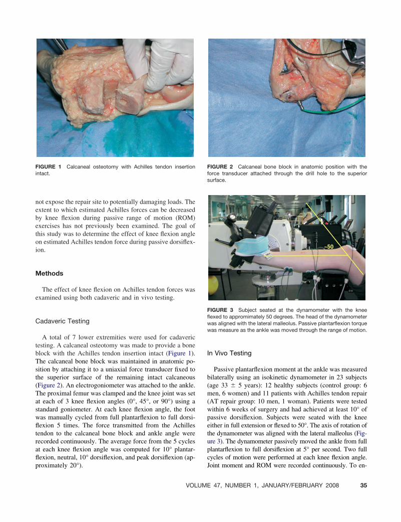
not expose the repair site to potentially damaging loads. Theextent to which estimated Achilles forces can be decreasedby knee flexion during passive range of motion (ROM)exercises has not previously been examined. The goal ofthis study was to determine the effect of knee flexion angleon estimated Achilles tendon force during passive dorsiflex-ion.
Methods
The effect of knee flexion on Achilles tendon forces wasexamined using both cadaveric and in vivo testing.
Cadaveric Testing
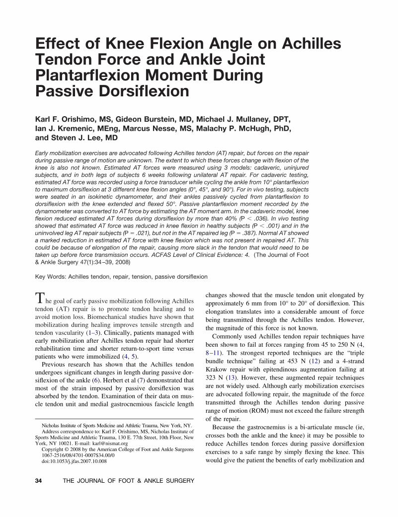
A total of 7 lower extremities were used for cadaverictesting. A calcaneal osteotomy was made to provide a boneblock with the Achilles tendon insertion intact (Figure 1).The calcaneal bone block was maintained in anatomic po-sition by attaching it to a uniaxial force transducer fixed tothe superior surface of the remaining intact calcaneous(Figure 2). An electrogoniometer was attached to the ankle.The proximal femur was clamped and the knee joint was setat each of 3 knee flexion angles (0°, 45°, or 90°) using astandard goniometer. At each knee flexion angle, the footwas manually cycled from full plantarflexion to full dorsi-flexion 5 times. The force transmitted from the Achillestendon to the calcaneal bone block and ankle angle wererecorded continuously. The average force from the 5 cyclesat each knee flexion angle was computed for 10° plantar-flexion, neutral, 10° dorsiflexion, and peak dorsiflexion (ap-
FIGURE 1 Calcaneal osteotomy with Achilles tendon insertionintact.
proximately 20°).
VOLUME
In Vivo Testing
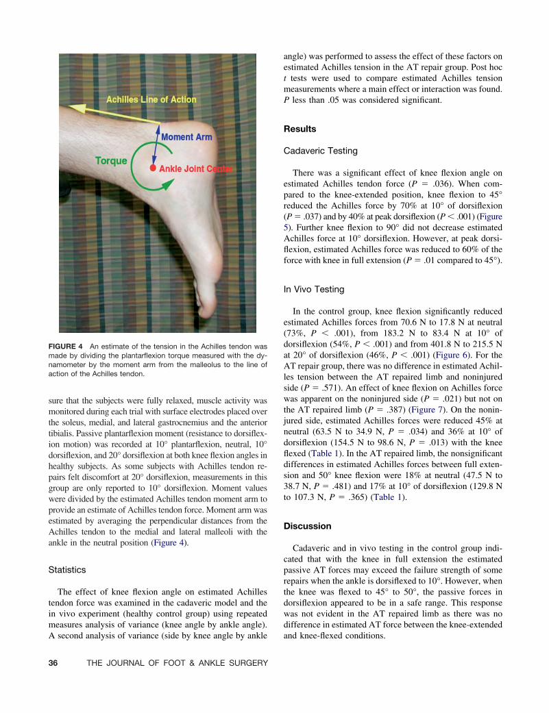
Passive plantarflexion moment at the ankle was measuredbilaterally using an isokinetic dynamometer in 23 subjects(age 33 � 5 years): 12 healthy subjects (control group: 6men, 6 women) and 11 patients with Achilles tendon repair(AT repair group: 10 men, 1 woman). Patients were testedwithin 6 weeks of surgery and had achieved at least 10° ofpassive dorsiflexion. Subjects were seated with the kneeeither in full extension or flexed to 50°. The axis of rotation ofthe dynamometer was aligned with the lateral malleolus (Fig-ure 3). The dynamometer passively moved the ankle from fullplantarflexion to full dorsiflexion at 5° per second. Two fullcycles of motion were performed at each knee flexion angle.
FIGURE 2 Calcaneal bone block in anatomic position with theforce transducer attached through the drill hole to the superiorsurface.
FIGURE 3 Subject seated at the dynamometer with the kneeflexed to appromimately 50 degrees. The head of the dynamometerwas aligned with the lateral malleolus. Passive plantarflexion torquewas measure as the ankle was moved through the range of motion.
Joint moment and ROM were recorded continuously. To en-
47, NUMBER 1, JANUARY/FEBRUARY 2008 35
sure that the subjects were fully relaxed, muscle activity wasmonitored during each trial with surface electrodes placed overthe soleus, medial, and lateral gastrocnemius and the anteriortibialis. Passive plantarflexion moment (resistance to dorsiflex-ion motion) was recorded at 10° plantarflexion, neutral, 10°dorsiflexion, and 20° dorsiflexion at both knee flexion angles inhealthy subjects. As some subjects with Achilles tendon re-pairs felt discomfort at 20° dorsiflexion, measurements in thisgroup are only reported to 10° dorsiflexion. Moment valueswere divided by the estimated Achilles tendon moment arm toprovide an estimate of Achilles tendon force. Moment arm wasestimated by averaging the perpendicular distances from theAchilles tendon to the medial and lateral malleoli with theankle in the neutral position (Figure 4).
Statistics
The effect of knee flexion angle on estimated Achillestendon force was examined in the cadaveric model and thein vivo experiment (healthy control group) using repeatedmeasures analysis of variance (knee angle by ankle angle).
FIGURE 4 An estimate of the tension in the Achilles tendon wasmade by dividing the plantarflexion torque measured with the dy-namometer by the moment arm from the malleolus to the line ofaction of the Achilles tendon.
A second analysis of variance (side by knee angle by ankle
36 THE JOURNAL OF FOOT & ANKLE SURGERY
angle) was performed to assess the effect of these factors onestimated Achilles tension in the AT repair group. Post hoct tests were used to compare estimated Achilles tensionmeasurements where a main effect or interaction was found.P less than .05 was considered significant.
Results
Cadaveric Testing
There was a significant effect of knee flexion angle onestimated Achilles tendon force (P � .036). When com-pared to the knee-extended position, knee flexion to 45°reduced the Achilles force by 70% at 10° of dorsiflexion(P � .037) and by 40% at peak dorsiflexion (P � .001) (Figure5). Further knee flexion to 90° did not decrease estimatedAchilles force at 10° dorsiflexion. However, at peak dorsi-flexion, estimated Achilles force was reduced to 60% of theforce with knee in full extension (P � .01 compared to 45°).
In Vivo Testing
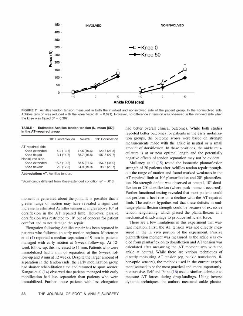
In the control group, knee flexion significantly reducedestimated Achilles forces from 70.6 N to 17.8 N at neutral(73%, P � .001), from 183.2 N to 83.4 N at 10° ofdorsiflexion (54%, P � .001) and from 401.8 N to 215.5 Nat 20° of dorsiflexion (46%, P � .001) (Figure 6). For theAT repair group, there was no difference in estimated Achil-les tension between the AT repaired limb and noninjuredside (P � .571). An effect of knee flexion on Achilles forcewas apparent on the noninjured side (P � .021) but not onthe AT repaired limb (P � .387) (Figure 7). On the nonin-jured side, estimated Achilles forces were reduced 45% atneutral (63.5 N to 34.9 N, P � .034) and 36% at 10° ofdorsiflexion (154.5 N to 98.6 N, P � .013) with the kneeflexed (Table 1). In the AT repaired limb, the nonsignificantdifferences in estimated Achilles forces between full exten-sion and 50° knee flexion were 18% at neutral (47.5 N to38.7 N, P � .481) and 17% at 10° of dorsiflexion (129.8 Nto 107.3 N, P � .365) (Table 1).
Discussion
Cadaveric and in vivo testing in the control group indi-cated that with the knee in full extension the estimatedpassive AT forces may exceed the failure strength of somerepairs when the ankle is dorsiflexed to 10°. However, whenthe knee was flexed to 45° to 50°, the passive forces indorsiflexion appeared to be in a safe range. This responsewas not evident in the AT repaired limb as there was nodifference in estimated AT force between the knee-extended
and knee-flexed conditions.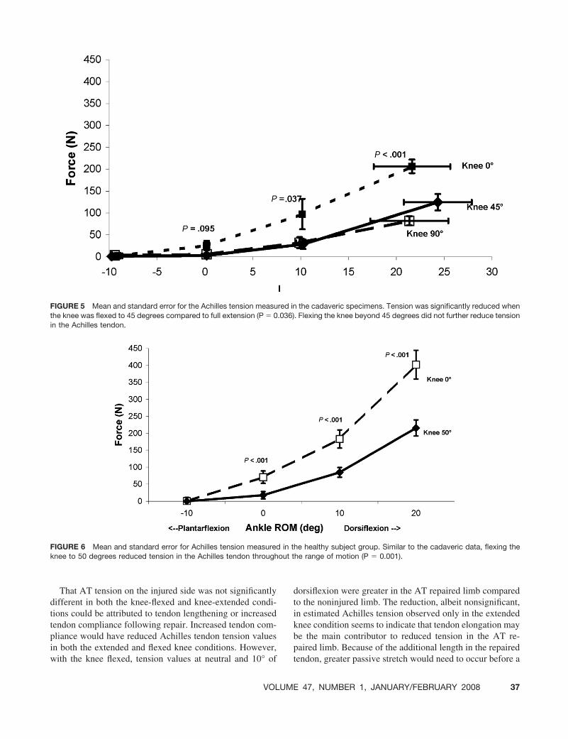
out t
That AT tension on the injured side was not significantlydifferent in both the knee-flexed and knee-extended condi-tions could be attributed to tendon lengthening or increasedtendon compliance following repair. Increased tendon com-pliance would have reduced Achilles tendon tension valuesin both the extended and flexed knee conditions. However,
FIGURE 5 Mean and standard error for the Achilles tension measuthe knee was flexed to 45 degrees compared to full extension (P � 0in the Achilles tendon.
FIGURE 6 Mean and standard error for Achilles tension measuredknee to 50 degrees reduced tension in the Achilles tendon through
with the knee flexed, tension values at neutral and 10° of
VOLUME
dorsiflexion were greater in the AT repaired limb comparedto the noninjured limb. The reduction, albeit nonsignificant,in estimated Achilles tension observed only in the extendedknee condition seems to indicate that tendon elongation maybe the main contributor to reduced tension in the AT re-paired limb. Because of the additional length in the repaired
the cadaveric specimens. Tension was significantly reduced when. Flexing the knee beyond 45 degrees did not further reduce tension
he healthy subject group. Similar to the cadaveric data, flexing thehe range of motion (P � 0.001).
red in.036)
in t
tendon, greater passive stretch would need to occur before a
47, NUMBER 1, JANUARY/FEBRUARY 2008 37
moment is generated about the joint. It is possible that agreater range of motion may have revealed a significantincrease in estimated Achilles tension at angles above 10° ofdorsiflexion in the AT repaired limb. However, passivedorsiflexion was restricted to 10° out of concern for patientcomfort and to not damage the repair.
Elongation following Achilles repair has been reported inpatients who followed an early motion regimen. Mortensenet al (4) reported a median separation of 9 mm in patientsmanaged with early motion at 6-week follow-up. At 12-week follow-up, this increased to 11 mm. Patients who wereimmobilized had 5 mm of separation at the 6-week fol-low-up and 9 mm at 12 weeks. Despite the larger amount ofseparation in the tendon ends, the early mobilization grouphad shorter rehabilitation time and returned to sport sooner.Kangas et al (14) observed that patients managed with earlymobilization had less separation than patients who were
FIGURE 7 Achilles tendon tension measured in both the involveAchilles tension was reduced with the knee flexed (P � 0.021). Hothe knee was flexed (P � 0.387).
TABLE 1 Estimated Achilles tendon tension (N, mean [SD])in the AT-repaired group
10° Plantarflexion Neutral 10° Dorsiflexion
AT-repaired sideKnee extended 4.2 (13.8) 47.5 (16.6) 129.8 (21.3)Knee flexed �3.1 (14.7) 38.7 (16.8) 107.3 (27.7)
Noninjured sideKnee extended 15.5 (19.3) 63.5 (21.6) 154.5 (31.0)Knee flexed* �2.3 (17.3) 34.9 (19.9) 98.6 (29.7)
Abbreviation: AT, Achilles tendon.
*Significantly different from Knee-extended condition (P � .013).
immobilized. Further, those patients with less elongation
38 THE JOURNAL OF FOOT & ANKLE SURGERY
had better overall clinical outcomes. While both studiesreported better outcomes for patients in the early mobiliza-tion groups, the outcome scores were based on strengthmeasurements made with the ankle in neutral or a smallamount of dorsiflexion. In these positions, the ankle mus-culature is at or near optimal length and the potentiallynegative effects of tendon separation may not be evident.
Mullaney et al (15) tested the isometric plantarflexionstrength of 20 patients after Achilles tendon repair through-out the range of motion and found marked weakness in theAT-repaired limb at 10° plantarflexion and 20° plantarflex-ion. No strength deficit was observed at neutral, 10° dorsi-flexion or 20° dorsiflexion (where peak moment occurred).Further functional testing revealed that most patients couldnot perform a heel rise on a decline with the AT-repairedlimb. The authors hypothesized that these deficits in end-range plantarflexion strength could be because of excessivetendon lengthening, which placed the plantarflexors at amechanical disadvantage to produce sufficient force.
There are a few limitations in this experiment that war-rant mention. First, the AT tension was not directly mea-sured in the in vivo portion of the experiment. Passiveplantarflexion moment was measured as the ankle was cy-cled from plantarflexion to dorsiflexion and AT tension wascalculated after measuring the AT moment arm with theankle at neutral. While there are various techniques ofdirectly measuring AT tension (eg, buckle transducers, fi-ber-optic sensors), the methods used in the current experi-ment seemed to be the most practical and, more importantly,noninvasive. Self and Paine (16) used a similar technique tomeasure AT forces during drop-landings. Using inverse
d noninvolved side of the patient group. In the noninvolved side,r, no difference in tension was observed in the involved side when
d anweve
dynamic techniques, the authors measured ankle plantar-
flexion moments during landing. Then, using an estimationof AT moment arm at each ankle angle, they approximatedAT force.
A second limitation of this experiment was that the ATmoment arm was assumed to be constant over the ROM.Rugg et al (17) measured AT moment arm as a function ofankle angle using magnetic resonance images (MRI) andfound only a 10% change in AT moment arm over the ROMused in the in vivo portion of this study (10° plantarflexionto 20° dorsiflexion). While small changes in the AT momentarm may have led to a small amount of error in the esti-mated AT forces, any uncertainty in the measurements willaffect the data from each subject in a similar manner. Themain concern of this experiment was to measure the changein AT forces when the knee was flexed.
Finally, the position of the force transducer in the cadav-eric experiment may not have been exactly in line with theAchilles tendon force. Depending on the relationship be-tween the transducer and the Achilles tendon line-of-pull,the actual tension in the Achilles tendon may have beenover- or underestimated. While there was uncertainty in theforces measured using this technique, the main goal of theexperiment was to examine the change in Achilles tensionas the knee is flexed. Achilles tension estimates were com-pared at each knee-flexion angle within each specimen.Therefore, the uncertainty due to transducer placement wascontrolled for in each specimen.
In conclusion, in the normal intact Achilles tendon, forcesduring passive dorsiflexion were substantially reduced byflexing the knee to 45° to 50°. However, in repaired Achillestendons this effect of knee flexion was not apparent. Thismay be further evidence of tendon elongation and increasedcompliance following surgery. While the benefits of earlymobilization regimens have been well documented, theseprotocols may contribute to elongation of the repair bystressing the healing tendon. Therefore, to reduce thesepotential effects and to protect the repair, it is recommendedthat early mobilization exercises be performed with the kneeflexed and to not exceed neutral ankle position in the rangeof motion. Future work in this area should prospectivelyexamine whether early mobilization exercises performed inthis manner have an effect on tendon elongation and/or
compliance.VOLUME
References
1. Enwemeka CS, Spielholz NI, Nelson AJ. The effect of early functionalactivities on experimentally tenotomized Achilles tendons in rats.Am J Phys Med Rehabil 67:264–269, 1988.
2. Pneumaticos SG, Noble PC, McGarvey WC. The effects of earlymobilization in the healing of Achilles tendon repair. Foot Ank Int21:551–557, 2000.
3. Mabit C, Bellaubre JM, Charissoux JL, Caix M. Study of the experi-mental biomechanics of tendon repair with immediate active mobili-zation. Surg Radiol Anat 8:29–35, 1986.
4. Mortensen HM, Skov O, Jensen PE. Early motion of the ankle afteroperative treatment of a rupture of the Achilles tendon: a prospective,randomized clinical and radiographic study. J Bone Joint Surg 81A:983–990, 1999.
5. Maffulli N, Tallon C, Wong J, Lim KP, Bleakney R. Early weight-bearing and ankle mobilization after open repair of acute midsubstancetears of the Achilles tendon. Am J Sports Med 31:692–700, 2003.
6. Alexander RM, Bennet-Clark HC. Storage of elastic strain energy inmuscle and other tissues. Nature 265:114–117, 1977.
7. Herbert RD, Moseley AM, Butler JE, Gandevia SC. Change in lengthof relaxed muscle fascicles and tendons with knee and ankle move-ment in humans. J Physiology 539:637–645, 2002.
8. Cretnik A, Zlajpah L, Smrkolj V, Kosanovic M. The strength ofpercutaneous methods of repair of the Achilles tendon: a biomechani-cal study. Med Sci Sports Exerc 32:16–20, 2000
9. Mandelbaum BR, Myerson MS, Forster R. Achilles tendon ruptures. Anew method of repair, early range of motion, and functional rehabil-itation. Am J Sports Med 23:392–395, 1995.
10. Mortensen NH, Saether J. Achilles tendon repair: a new method ofAchilles tendon repair tested on cadaverous materials. J Trauma 31:381–384, 1991.
11. Watson TW, Jurist KA, Yang KH, Shen KL. The strength of Achillestendon repair: an in vitro study of the biomechanical behavior inhuman cadaver tendons. Foot Ankle Int 16:191–195, 1995.
12. Jaakkola JI, Hutton WC, Beskin JL, Lee GP. Achilles tendon rupturerepair: biomechanical comparison of the triple bundle technique versusthe Krakow locking loop technique. Foot Ankle Int 21:14–17, 2000.
13. Lee SJ, Matarazzo MF, Nicholas SJ, McHugh MP, Kremenic IJ,Ben-Avi S. Epitendinous suture augmentation in Achilles tendon re-pair. Paper presented at: American Academy of Orthopaedic Surgeons70th Annual Meeting; February 5–9, 2003; New Orleans, LA.
14. Kangas J, Pajala A, Ohtonen P, Leppilahti J. Achilles tendon elonga-tion after rupture repair: a randomized comparison of 2 postoperativeregimens. Am J Sports Med 35:59–64, 2007.
15. Mullaney MJ, McHugh MP, Tyler TF, Nicholas SJ, Lee SJ. Weaknessin end-range plantar flexion after Achilles tendon repair. Am J SportsMed 34:1120–1125, 2006.
16. Self BP, Paine D. Ankle biomechanics during four landing techniques.Med Sci Sports Exerc 33:1338–1344, 2000.
17. Rugg SG, Gregor RJ, Mandelbaum BR, Chiu L. In vivo moment armcalculations at the ankle using magnetic resonance imaging (MRI).
J Biomech 23(5):495–501, 1990.47, NUMBER 1, JANUARY/FEBRUARY 2008 39