Embed Size (px)
Citation preview

Vol. 8(24), pp. 3107-3112, 27 June, 2013 DOI: 10.5897/AJAR12.2075
ISSN 1991-637X ©2013 Academic Journals
http://www.academicjournals.org/AJAR
African Journal of Agricultural
Research
Full Length Research Paper
Effect of ingestion of thermally oxidized sunflower oil on the fatty acid composition and histological alteration
of rat liver and adipose tissue in development
Sadoudi R.1*, Ammouche A.2 and Ali Ahmed D.1
1Département d’Agronomie, Faculté des Sciences Biologiques et Agronomiques, Université de Tizi-Ouzou, Algérie.
2Département de Technologie alimentaire et de Nutrition Humaine, Ecole nationale supérieure agronomique (ENSA, Ex.
INA). Hassen badi, El-Harrach (Alger), Algérie.
Accepted 14 June, 2013
The effect of ingestion of thermoxidized sunflower oil (TOS) on fatty acids composition and histological alteration was investigated in rats. The animals were divided into 2 groups, the first group fed on fresh sunflower oil (FSO) diet and a second group fed on TOS diet during 9 weeks. This oil was heated at 99 ± 2°C with incorporation of 9 L of oxygen/second during 52 h continuously. Changes that occurred in oil fatty acid composition were similar to those found in tissues (liver and adipose tissue) of rat, nourished with oxidized oil. Indeed, our results showed a change in the tissues profile of the fatty-acids; the rates of saturated and monounsaturated fatty-acids increased with the detriment of the linoleic acid and its higher derivative, arachidonic acid. Thus, the total unsaturation of the tissues lipids decreased. In addition, unlike linoleic acid, the palmitic acid is converted into C16:1 n-7. Probably, this was due to the activation of ∆9-desaturase and the reduction of the ∆6- and ∆5-desaturases by the generated products in thermoxidized sunflower oil. Microscopic examination of the test group showed a significant histological change, when compared with the control, such as increase in cell size. Key words: Thermoxidized sunflower oil, rat, fatty acid composition, histology, tissues.
INTRODUCTION Sunflower oil, derived from the seeds of Heliantalus annus linnaeus is the most widely used frying and cooking oil in Algeria. It is considered by some as desirable as olive oil. In our study, sunflower oil was selected due to its high use in food as it is a rich source of linoleic acid (essential fatty-acid). Furthermore, it is light in taste and appearance and has a high vitamin E content (Shahidi et al., 1992). Lastly, this oil contains more vitamin E than any other vegetable oil.
Although the high level of unsaturation of fatty acids (FA) contained in vegetable oils is appreciated by nutritionists, it causes severe technological problems,
because of their high susceptibility to oxidation. The fats oxidation degrades the organoleptic quality of food, reduces its nutritional value. Moreover, products of the oxidation processes can contribute in the aging of an organism and in the aetiology of cardiovascular diseases and cancer (Yoshikawa et al., 2000). Indeed, at high temperature in the presence of air, many chemical reactions are able to be observed in oil: hydrolysis, polymerization, oxidation and isomerization (Rossell, 2001). Thus, new and unstable compounds potentially toxic at low concentrations may be generated after destroy of the linoleic acid (C18:2 n-6) (Nawar, 1996;
*Corresponding author. E-mail: [email protected]. Tel : 213 93 27 79 73.
3108 Afr. J. Agric. Res. Min and Boff, 2001).
The safety of thermally-abused fats and oils that are destined for human and animal consumption has been the subject of research over many years. Feeding oxidized lipids to different species can engender wide variety of symptoms including appetite reduction, growth of depression, diarrhea, histological changes in tissues and even death (Izaki et al., 1984). Jimoh and Odutuga (2004) had studied the effect of ingestion of dietary oxidized groundnut oil on the histology of Wistar rat brain (cerebral cortex), liver, and kidney. These authors showed mild to severe distortion of the normal architecture in animals fed with replenished and not-replenished thermoxidized groundnut oil, respectively.
There is limited information on the effect of other dietary oxidized oils on the histology of other tissues. In this study, therefore, we have investigated histological changes in the liver and adipose tissue due to dietary thermoxidized sunflower oil (TOS) ingestion by growing Wistar rats. The objective of this project is double. After evaluating the deterioration level of sunflower oil submitted at thermoxidative treatment (in the absence of foodstuff), we studied the effect of consumption of this oil on tissues FA composition and histological alteration. MATERIALS AND METHODS
In our study, the oxidation of the sunflower oil was carried out according to the laboratory technical developed by Drozdowski and Szukalska (1987), and modified by Blanc-Gondardmary et al. (1989). Sunflower oil was selected due to its high use in food as it is a rich source of linoleic acid. Moreover, thanks to its high unsaturation degree, it can easily undergo rancidity (Shahidi et al., 1992). The sunflower oil was heated at 99 ± 2°C with incorporation of 9 liters of oxygen/second during 52 h continuously. The oil oxidation degree was monitored through several physicochemical analysis. Following parameters were studied: free-fatty acid (FFA) contents, peroxide value (PV), refractive index (RI), humidity, iodine value (IV), saponification value (SV) density (AOCS, 1989) and fatty acid (FA) composition (AOAC, 1999). Animals, experimental protocol and diets
Male Wistar rats (n = 10) were obtained at the age of 3 weeks from the Pasteur institute (Kouba, Algeria). They were housed at 24 ± 2°C with 60 ± 20% relative humidity, with a 12 h light-dark cycle, and fed with a commercial non purified diet for 4 weeks. After acclimation to the housing conditions, rats were allotted to two groups of 5 animals each. Rats received a diet which was supplemented with (5 /100 g) fresh sunflower oil (FSO) for (control group) or TOS for (test group).
Each group was fed for 9 consecutive weeks. All diets were prepared once per week and stored at +4°C until needed. The composition by weight as percent of total was: sucrose 21.94; corn starch 44.9; casein 20; DL-Methionine 0.16; agar-agar 2; vitamin mixture 2; salt mixture 4, and fat 5. Every morning, fresh portions sufficient for 1 day supply were fed to the animals. Each rat was fed 25 g/day (one rat per cage) of the experimental diet. Then, rats were maintained in cages with free access to water and the experimental diets until they were killed.
After 6 weeks of receiving their respective diets (9 week old
animals), the rats (food deprived for 24 h) were sacrificed and the liver and adipose tissue were rapidly dissected as previously described by Winter et al. (1994). It has established that, the liver detoxifies a large number of lipid oxidation products by a complex series of chemical reactions. Mitchell and Petersen (1989) showed that, rat hepatic microsomal aldehyde dehydrogenase (ALDH) may serve a biological role for detoxification of reactive aldehydes produced by lipid peroxidation of microsomal membranes. Hodson et al. (2008) suggested that, adipose tissue is a suitable biomarker of dietary fatty acid intake. Ideally, adipose tissue and dietary questionnaires should complement, rather than substitute for each other in epidemiologic studies.
Extraction of total lipids from the tissues was carried out following the method of Folch et al. (1957) modified by Pollet et al. (1978). The lipids were then methylated following the method of Morrisson and Smith (1964).
For histological study, permanent preparations using routine methods were made. The livers and adipose tissues were fixed in 10% buffered formalin (10 ml formalin in 100 ml normal saline). Sections were cut at 5 µm on a rotator microtome. They were flattened on warm water and mounted onto albumerised slides and dried overnight. The sections were dewaxed in xylene and hydrated through descending grades of ethanol to water. They were initially stained in Harris haematoxylin and differentiated in acid alcohol and thereafter stained with methylene blue. Then, they were dehydrated in 95% alcohol, stained in 10% alcohol eosin, dehydrated in absolute alcohol, cleaned in xylene and mounted in Canada balsam. The resulting slides are then viewed under the light microscope. Statistical analysis
Statistical comparison between the tissues’ fatty acids contents were performed using Student’s t-test, with P < 0.05 considered significantly different. RESULTS AND DISCUSSION From the results of various investigated parameters, we concluded that, the treatment applied in our study caused a high level of deterioration to this oil (Table 1). FFA content, AV, PV, density and moisture of the TOS increased significantly (p = 0.000); the IV and SV were found to decrease significantly (p = 0.000); lastly the RI remained constant (p = 0.475). All of these modifications conferred a thick texture and faded flavor to thermoxidized sunflower oil.
In addition, this applied treatment had an adverse effect on the quantitative FA composition of this oil. The most significant effects were on C18:2 n-6, C18:1 n-9 and palmitic acid (C16:0) contents. A decrease in linoleic acid (p = 0.000) and an increase in oleic acid (p = 0.006) were noted in TOS. It engendered an increase in the oleic acid to linoleic acid ratio (O/L), indicating a preferential use of C18:2 n-6 in oxidation reactions. Decrease of linoleic acid content is used as an indicator of lipid oxidation (Maniak et al., 2009). Our results corroborate those obtained by Morgado et al. (1999). Oxidation of the oils or fats destroys essential fatty acids and produces toxic compounds and oxidized polymers. Thus, oxidation is very important in terms of palatability, nutritional quality

Sadoudi et al. 3109
Table 1. Physico-chemical properties of fresh and TSO.
Characteristic Fresh oil Thermoxidized oil
Physical state at room temperature Fluid Thick texture, flavor faded
Peroxide value (meq/Kg) 5.83 ± 0.76 152.5 ± 5.33
Acid value (mg KOH/g) 0.182 ± 0.04 2.51 ± 0.11
Free Fatty Acids (%) 0.093 ± 0.02 1.25 ± 0.07
Iodine value (g I2/100g) 125.84 ± 1.59 80.51 ± 1.37
Saponification value (mg KOH/g) 192.60 ± 2.91 183.79 ± 1.32
Refraction index 1.461 ± 0.037 1.476 ± 0.023
Density at 20°C (g/ml) 0.910 ± 0.018 0.985 ± 0.010
Moisture (%) 0.100 ± 0.012 2.006 ± 0.221
C16 :0 content (%) 6.14 ± 0.451 9.48 ± 0.367
C18 :1 n-9 content (%) 33.500 ± 2.317 46.040 ± 3.939
C18 :2 n-6 content (%) 58.140 ± 2.475 40.590 ± 0.829
Values are mean±standard deviation of triplicate determinations. and toxicity of edible oils (Kamal-Eldin et al., 2003).
The effect of feeding TOS on the FA composition of rat tissues was also investigated. Significant decreases (p = 0.001 and p = 0.000) in total polyunsaturated FA (PUFA) of neutral lipids of liver and adipose tissue were observed between the two groups; however, total saturated FA (SFA) and monounsaturated FA (MUFA) increased even slightly (p > 0.05) (Table 2). Nevertheless, high changes were observed in the proportions of principally SFA and MUFA (palmitic and oleic acids) in both tissues. C16:0 was more abundant than C18:0. The content of C18:1 n-9 increased significantly (p = 0.000) in neutral lipids of liver, while in adipose tissue it remained constant (p = 0.205). Oleic acid is formed from stearic acid by the enzyme stearoyl Co-A desaturase, a major lipogenic enzyme. Reduction of PUFA, particularly linoleic acid and an increase in oleic acid led to an increase O/L ratio in both lipids tissues of rat that was nourished with thermoxidized sunflower oil. Lastly, a very significant increase (p < 0.05) in the proportion of palmitoleic acid (C16:1 n-7 not detected in oils incorporated to diet) was observed in neutral lipids of both tissues analysed in rats fed TSO compared with control group, as well as a decrease in PUFA like arachidonic acid, long chain products of linoleic acid. These PUFA are indispensables for physiological functions; they are metabolized to produce bioactive substances called eicosanoids (Russo, 2009).
The results are consistent with the concept that, heating PUFAs causes a greater degree of lipid peroxidation than heating SFA or MUFA. The tissue content in FA depends on the lipids proportion contained in the diets; it is likely that tissue FA compositions of rats fed on FSO would differ more substantially from those of rats fed on thermoxidized sunflower oil. Indeed, chronic consumption of TOS diet by rats has been observed to cause modification in tissues FA composition.
Comparatively to rats nourished with fresh sunflower oil, all tissues of rats feeding diet containing oxidized sunflower oil showed a significant increase in MUFA and a reduction in total PUFA contents, particularly linoleic acid. The lower tissues’ content in linoleic acid of the rat that was nourished with oxidized oil was caused by a diet containing a lower ratio of oxidized oil. One possible reason may explain the limited accumulation of linoleic and arachidonic acids in both tissues. Indeed, C18:2 n-6 was readily oxidized during thermoxidative treatment.
Thus, the total unsaturation of tissue-lipid fractions was significantly decreased in adipose tissue (p = 0.001) and liver (p = 0.000) of rats fed this TOS containing diet. Indeed, Wood et al. (2008) suggested that, linoleic acid derive entirely from the diet. It passes through the pig’s stomach unchanged and is then absorbed into the blood stream in the small intestine and incorporated from there into tissues.
Two clear trends emerge from the present study. It is apparent that, the TOS suppressed the formation of arachidonic acid in both tissues and increased the conversion of C16:0 to C16:1 n-7 (p = 0.038) in adipose tissue and C18:0 to C18:1 n-9 (p = 0.000) in lipids liver of rats fed thermoxidized sunflower oil. Components coming from oxidation of oil may be responsible for the effects. Indeed, reduction in the activity of several enzymes has been reported as one of the consequences of oxidized oil ingestion (Odutuga et al., 1997; Odutuga and Ologan, 1999).
Conversion of linoleic acid to arachidonic acid requires elongase and desaturase. Decrease in C20:4 n-6 content of both tissues may be due to the suppressive effect of the hazardous constituents of thermally oxidized sunflower oil on enzymatic activity. This was supported by experiments of many authors (Mesembe et al., 2004). Ruiz-Gutierrez and Muriana (1992) suggested that, the heated edible oils deteriorate the activity of the

3110 Afr. J. Agric. Res.
Table 2. Fatty acid composition, as mean% of total fatty acids of neutral lipids extracted from organs of rats fed fresh oil (FO) or (TOS).
Fatty acid Adipose tissues Liver
FO TOS p FO TOS p
C10 : 0 0.80 ± 0.17 ND - 0.25 ± 0.07 ND -
C12 : 0 0.16 ± 0.08 0.22 ± 0.14 0.4430 ND 0.20 ± 0.00 -
C14 : 0 1.74 ± 1.06 2.13 ± 1.29 0.6176 1.44 ± 0.13 2.18 ± 0.31 0.0002
C15 : 0 ND 0.11 ± 0.10 - ND 0.17 ± 0.00 -
C16 : 0 27.57 ± 1.93 30.11 ± 1.24 0.0355 25.05 ± 1.20 30.52 ± 0.87 0.0000
C16 : 1, ω9 0.82 ± 0.26 0.81 ± 0.25 0.9068 0.87 ± 0.10 0.89 ± 0.56 0.7707
C16 : 1, ω7 6.20 ± 0.84 7.75 ± 1.11 0.0389 4.70 ± 2.38 7.26 ± 0.10 0.0564
C18 : 0 6.43 ± 0.42 5.41 ± 1.20 0.1129 11.27 ± 0.97 5.68 ± 0.82 0.0000
C18 : 1, ω9 48.40 ± 2.12 46.72 ± 1.73 0.2057 40.91 ± 1.38 47.77 ± 0.60 0.0000
C18 : 1, ω7 2.83 ± 0.19 3.50 ± 0.96 0.1684 2.87 ± 0.64 3.16 ± 0.02 0.3636
C18 : 2, ω6 4.98 ± 1.33 2.27 ± 0.48 0.0026 7.44 ± 1.48 0.99 ± 0.00 0.0000
C18 : 3, ω3 ND ND - 0.26 ± 0.11 ND -
C20 : 0 0.10 ± 0.10 0.10 ± 0.01 0.8610 ND ND -
C20 : 1, ω9 0.16 ± 0.15 0.55 ± 0.19 0.0079 0.14 ± 0.07 0.99 ± 0.04 0.0000
C20 : 4, ω6 0.25 ± 0.05 ND - 4.09 ± 0.86 ND -
C20 : 3, ω3 ND ND - ND ND -
C20 : 5, ω3 ND ND - 0.19 ± 0.04 ND -
C22 : 0 ND ND - ND ND -
C22 : 4, ω6 ND ND - ND ND -
ΣAGS 36.20 ± 2.67 38.15 ± 3.08 0.5004 38.22 ± 0.66 38.83 ± 0.67 0.1380
ΣAGMI 58.41 ± 2.22 59.33 ± 3.28 0.6262 49.63 ± 2.44 60.08 ± 0.02 0.0000
ΣAGPI 5.41 ± 1.33 2.55 ± 0.48 0.0017 12.59 ± 2.26 1.09 ± 0.00 0.0000
Values are mean ± standard deviation of five determinations. desaturases in the hepatic microsomes. Products of oxidation and isomerization of the linoleic acid inhibit metabolism of essential FA by reducing the activities of ∆6-désaturase and ∆5-désaturase. However, the activity of ∆9-désaturase was promoted, other possible reasons may explain the limited accumulation of PUFA observed in our study. Otherwise, these products may be responsible for the severe alterations in hepatocytes and adipose cells of rats fed oxidized sunflower oil diet.
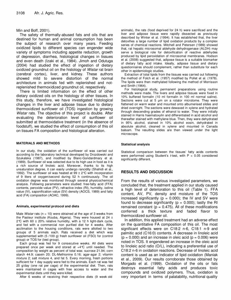
Indeed, for the histological examination, the results indicate that, structural abnormalities had occurred in the rats that were fed with TSO. The livers and adipose tissues from rats that fed on the TSO diet had different histological characteristics as those fed with the FSO diet, such as increase in cell size. Histological examination of thick sections of liver specimens showed that liver cells were swollen. The liver showed widening of the liver sinusoids (Figures 1 and 2). Hepatocytes showed large and spherical nuclei that were centrally located, with one or two prominent nucleolus (Figure 3). Adipose cells had nuclei that migrated to the cell periphery (Figure 4); the cell membrane was destroyed (Figure 5).
Consumption of peroxidized lipids has been shown to be injurious to health (Frankel, 1980; Kubow, 1992). There is a paucity of information on this aspect. Jimoh
and Odutuga (2004) showed disintegration of alveoli membrane and collapse of alveoli spaces in the lungs of rats fed oxidized groundnut oil. These authors also showed disorganization of the spatial arrangement of the heart cells which might lead to weakness of the cardiac muscle which may consequently lead to cardiac failure. This same authors (Jimoh and Odutuga, 2004) showed structural abnormalities in brain (cerebral cortex), liver, and kidney in animals fed with same thermoxidized groundnut oil. The liver showed mild to severe distortion of the normal architecture, as well as prominence and widening of the liver sinusoids. Conclusion The aimed of this study is to, first, determine the physical and chemical characteristics of TSO. Second, show the effects that come out from feeding diet containing this heated oil on the FA composition and histopathology of rat tissues (liver and adipose tissue). Results obtained showed a high loss in linoleic acid of oxidized oil and modification of FA composition of both tissues analyzed. The decrease in total PUFA observed in these tissues was in agreement with the observed increase in total SFA and MUFA.
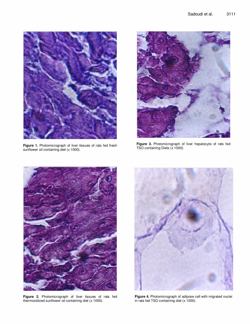
Figure 1. Photomicrograph of liver tissues of rats fed fresh
sunflower oil containing diet (x 1000).
Figure 2. Photomicrograph of liver tissues of rats fed thermoxidized sunflower oil containing diet (x 1000).
Sadoudi et al. 3111
Figure 3. Photomicrograph of liver hepatocyte of rats fed TSO containing Diets (x 1000).
Figure 4. Photomicrograph of adipose cell with migrated nuclei
in rats fed TSO containing diet (x 1000).

3112 Afr. J. Agric. Res.
Figure 5. Photomicrograph showed destroyed membrane
of adipose cell in rats fed TSO containing diet (× 1000).
Liver and adipose tissue histology were adversely affected by the inclusion of TSO. Hypertrophy of cells and cores have been observed in rats fed with TSO. If the results are applicable to man, they suggest that, there is reason for concern regarding adverse biochemical and histological consequences that comes out from a chronic consumption of thermally oxidized sunflower oil diets, what may be dangerous to health since may engender an inactivation of ∆6 and ∆5 desaturases and a liver damage. Therefore, eating a lot of fried foods is not a good idea. If a food is to be fried, one should use as few amount of oil as possible.
REFERENCES AOAC (1999). Official Methods of Analysis (16
th ed) International,
Gaithersburg. AOCS (1989). Official and Recommended Practices of the American oil
Chemists Society (5th
ed) Champaign, IL. pp. 48-62. Blanc-Gondardmary P, Revol A, Pacheco H (1989). Chronical ingestion
of oxidized oil in young rat. Effect on lipid composition and cytidyl transferase activity. Biomembranes et nutrition, colloque INSERM., Paris, Nutriments affectant la composition lipidique et les propriétés des membranes cellulaires. Inserm (eds), 195:481.
Drozdowski B, Szukalska EA (1987). Rapid instrumental method for the
evaluation of the stability of fats. J. Am. Oil. Chem. Soc. 64:1008-1011.
Folch J, Lees M, Stanley GHS (1957). A simple method for the isolation and purification of total lipids from animal tissues. J. Biol. Chem. 226:497-509.
Hodson L, Skeaff CM, Fielding BA (2008). Fatty acid composition of adipose tissue and blood in humans and its use as a biomarker of dietary intake. Prog. Lipid Res. 47:348-380.
Izaki Y, Yoshikawa S, Uchiyama M (1984). Effect of ingestion of thermally oxidized frying oil on peroxidative criteria in rats. Lipids 19:324-331.
Jimoh FO, Odutuga AA (2004). Histological changes of selected rat tissues following the ingestion of thermally oxidized groundnut oil. BIOKEMISTRI 16:1-10.
Kamal-Eldin A, Mäkinen M, Lampi A (2003). The challenging contribution of hydroperoxides to the lipid oxidation mechanism. In Kamal-Eldin A (eds) Lipid Oxidation Pathways, Champaign Illinois, AOCS Press. pp. 1-35.
Maniak B, Szmigielski M, Piekarski W, Markowsha A (2009). Physics-chemical changes of post-frying sunflower oil. Int. Agro-Phys. 23:243-248.
Mesembe OE, Ibanga I, Osim EE (2004). The effects of fresh and thermoxidized palm oil diets. Niger. J. Physiol. Sci. 19:86-91.
Min DB, Boff JF (2001). Lipid oxidation of edible oil. In C Akoh, DB Min (eds) Food lipids, New York, Marcel Dekker. pp. 335-363.
Mitchell DY, Peterson DR (1989). Oxidation of aldehydic products of lipid peroxidation by rat liver microsomal aldehyde dehydrogenase. Arch. Biochem. Biophys. 269:11-17.
Morgado N, Sanhueza J, Galleguillos A, Garrido A, Nieto S, Valenzuela A (1999). Effet de l’huile de poisson hydrogénée sur le profil des lipoprotéines plasmatiques et sur la composition en acides gras des différents tissus du rat. Annales de la nutrition et le métabolisme. 43:310-318.
Morrisson W, Smith L (1964). Preparation of fatty acid methyl esters and dimethylacetals from lipids with boron fluoride-methanol. J. Lipid. Res. 5:600-608.
Nawar WW (1996). Lipids OR. In Fennema (eds) Food chemistry (3rd
ed), New York, Marcel Dekker. pp. 225-319.
Odutuga AA, Obaleye JA, Ologan FO (1997). Thermoxidized soybean oil: Spectroscopic investigation and effects on selected rat tissues. Biokemistri 1:45-58.
Odutuga AA, Ologan FO (1999). Effect of thermally oxidized soyabean oil on alkaline and acid phosphatases in rat liver and serum. Biosci. Res. Commun. 11:281-285.
Pollet S, Ermidou S, LeSaux F, Monge M, Baumann N (1978). Microanalysis of brain lipids: Multiple two dimensional thin-layer chromatography. J. Lipi. Res. 19:916-921.
Rossell JB (2001). Frying. Improving quality. In JB Rossell (eds) Cambridge England, Woodhead Publishing Limited. P. 369.
Ruiz-Gutierrez V, Muriana FJG (1992). Effect of ingestion of thermally oxidized frying oil on desaturase activities and fluidity in rat–liver microsomes. J. Nutr. Biochim. 3:75-79.
Russo GL (2009). Dietary n-6 and n-3 polyunsaturated fatty acids: from biochemistry to clinical implications in cardiovascular prevention. Biochem. Pharmacol. 77:937-946.
Shahidi F, Janitha PK, Wanasundara PD (1992). Phenolic antioxidants. Crit. Rev. Food Sci. Nutr. 32:67-103.
Winter BL, Yeh SME, Yeh YY (1994). Linolenic acid provides a source of docosahexaenoic acid for artificially reared rat pups. J. Nutr. 124:1654-1659.
Wood JD, Enser M, Fisher AV, Nute GR, Sheard PR, Richardson RI (2008). Fat deposition, fatty acid composition and meat quality. A Rev. Meat Sci. 78:343-358.
Yoshikawa T, Toyokuni S, Yamamoto Y, Naito Y (2000). Free radicals in Chemistry Biology and Medicine, OICE International, Saint Lucin, London. P. 580.


![Thermally oxidized aluminum as catalyst-support...thermal oxidation in air was preferable to single-crystalline sapphire [6]. However, the surface analysis of air and/or thermally-oxidized](https://img.pdfslide.us/doc/110x75/60f87e34de46c858bb0a26ce/thermally-oxidized-aluminum-as-catalyst-support-thermal-oxidation-in-air-was.jpg)