Embed Size (px)
Citation preview
A Dissertation On
“EFFECT OF IAYT (INTEGRATED APPROACH OF YOGA THERAPY)
ON MOTOR NERVE CONDUCTIVITY IN SPASTIC
HEMIPLEGIC MEN”
Submitted by
Dr. R. VIDHYALAKSHMI, B.N.Y.S (Reg. No. 461412005)
Under the guidance of
Prof. Dr. S.T.VENKATESWARAN, N.D (OSM), M.Sc (Y.N), P.G.D.Y,
P.G.D.O.M, D.N.H.E, (MBA)
Submitted to
The Tamilnadu Dr.M.G.R.Medical University, Chennai
In partial fulfillment of the requirements for the award of degree of
DOCTOR OF MEDICINE
BRANCH – II: YOGA
POST GRADUATE DEPARTMENT OF YOGA
GOVERNMENT YOGA AND NATUROPATHY MEDICAL COLLEGE AND HOSPITAL,
CHENNAI – 600 106.
FEBRUARY 2018

GOVERNMENT YOGA AND NATUROPATHY MEDICAL COLLEGE
AND HOSPITAL, CHENNAI, TAMILNADU
CERTIFICATE BY THE GUIDE
This is to certify that “EFFECT OF IAYT (INTEGRATED APPROACH OF YOGA
THERAPY) ON MOTOR NERVE CONDUCTIVITY IN SPASTIC
HEMIPLEGIC MEN” is a bonafide work done by the Post graduate
Dr.R.VIDHYALAKSHMI , Department of Yoga, Government Yoga and
Naturopathy Medical College and Hospital, Chennai-600106, under my guidance and
supervision in partial fulfillment of regulations of The Tamilnadu Dr.M.G.R.Medical
University, Chennai for the award of degree of DOCTOR OF MEDICINE (M.D) –
Yoga, BRANCH – II during the academic period 2014 to 2018.
Place: Chennai. SIGNATURE OF THE GUIDE
Date: Dr.S.T.VENKATESWARAN,
N.D. (OSM), M.Sc (Y&N), P.G.D.Y, P.G.D.O.M,
D.N.H.E, (MBA)
Head of the Department - Department of Yoga,
Government Yoga and Naturopathy Medical College and Hospital,
Chennai – 106.
GOVERNMENT YOGA AND NATUROPATHY MEDICAL COLLEGE AND
HOSPITAL, CHENNAI, TAMILNADU
ENDORSEMENT BY THE HEAD OF THE DEPARTMENT
I certify that the dissertation entitled “EFFECT OF IAYT (INTEGRATED
APPROACH OF YOGA THERAPY) ON MOTOR NERVE CONDUCTIVITY IN
SPASTIC HEMIPLEGIC MEN” is the record of original research work carried out by
Dr. R. VIDHYALAKSHMI, Department of Yoga, Government Yoga and Naturopathy
Medical College and Hospital, Chennai – 600 106, submitted for the degree of
DOCTOR OF MEDICINE M.D – Branch – II (Yoga) under my guidance and
supervision, and that this work has not formed the basis for the award of any degree,
diploma, associate ship, fellowship or other titles in this University or any other
University or Institution of higher learning.
Place: Chennai. SIGNATURE OF THE H.O.D
Date: Dr.S.T.VENKATESWARAN,
N.D. (OSM), M.Sc(Y&N),P.G.D.Y, P.G.D.O.M,
D.N.H.E, (MBA),
Head of the Department - Department of Yoga,
Government Yoga and Naturopathy Medical College and Hospital,
Chennai – 106.

GOVERNMENT YOGA AND NATUROPATHY MEDICAL COLLEGE AND
HOSPITAL, CHENNAI, TAMILNADU
ENDORSEMENT BY THE PRINCIPAL
I certify that the dissertation entitled “EFFECT OF IAYT (INTEGRATED
APPROACH OF YOGA THERAPY) ON MOTOR NERVE CONDUCTIVITY IN
SPASTIC HEMIPLEGIC MEN” is the record of original research work carried out by
Dr. R. VIDHYALAKSHMI, Department of Yoga, Government Yoga and Naturopathy
Medical College and Hospital, Chennai – 600 106 submitted for the award of degree of
DOCTOR OF MEDICINE (M.D) Branch – II (Yoga) under my guidance and
supervision, and that this work has not formed the basis for the award of any degree,
diploma, associate ship, fellowship or other titles in this University or any other
University or Institution of higher learning.
Place: Chennai. SIGNATURE OF THE PRINCIPAL
Date: Dr. N.MANAVALAN,
N.D. (OSM), M. A (G.T), M.Sc (Y&N), M. Phil,
P.G.D.Y, P.G.D.H.M, P.G.D.H.H,
Government Yoga and Naturopathy Medical College and Hospital,
Chennai – 106.
GOVERNMENT YOGA AND NATUROPATHY MEDICAL COLLEGE AND
HOSPITAL, CHENNAI, TAMILNADU.
DECLARATION BY THE CANDIDATE
I, Dr. R. VIDHYALAKSHMI solemnly declare that this dissertation entitled
“EFFECT OF IAYT (INTEGRATED APPROACH OF YOGA THERAPY) ON
MOTOR NERVE CONDUCTIVITY IN SPASTIC HEMIPLEGIC MEN” is a bonafide
and genuine research work carried out by me at Government Yoga and Naturopathy
Medical College and Hospital, Chennai from June 2016 - May 2017 under the
guidance and supervision of Dr. S.T.VENKATESWARAN, N.D. (OSM), M.Sc
(Y&N), P.G.D.Y, P.G.D.O.M, D.N.H.E, (MBA), Head of the Department -
Department of Yoga. This dissertation is submitted to The Tamilnadu
Dr.M.G.R.Medical University, Chennai towards partial fulfillment of requirements for
the award of M.D. Degree (Branch – II –Yoga) in Yoga and Naturopathy.
Place: Chennai Signature of the candidate
Date: (Dr. R. Vidhyalakshmi)

INSTITUTIONAL ETHICAL COMMITTEE
GOVERNMENT YOGA AND NATUROPATHY MEDICAL
COLLEGE AND HOSPITAL, CHENNAI – 600 106.
CERTIFICATE OF APPROVAL
The Insti tutional Ethical Committee of Government Yoga &
Naturopathy Medical College and Hospital , Chennai reviewed and
discussed the appl ication for approval of “EFFECT OF IAYT (INTEGRATED
APPROACH OF YOGA THERAPY) ON MOTOR NERVE CONDUCTIVITY IN SPASTIC
HEMIPLEGIC MEN”, project work submit ted by Dr. R. Vidhyalakshmi,
2n d
year M.D.Yoga, Post graduate, Government Yoga and Naturopathy
Medical College and Hospita l , Chennai.
The proposal is APPROVED.
The Insti tutional Ethical Committee expects to be informed about the
progress of the study and adverse drug reactions during the course of
the study and any change in the protocol and patient information sheet/
informed consent and asks to be provided a copy o f the f inal report .

COPYRIGHT
DECLARATION BY THE CANDIDATE
I hereby declare that the Tamilnadu Dr.M.G.R.Medical
Universi ty, Chennai, Tamilnadu shal l have the r ights to preserve, use
and disseminate this Dissertation / Thesis in print or electronic format
for academic / research purpose.
Place: Chennai Signature of the candidate,
Date: (Dr. R.Vidhyalakshmi)
© The Tamilnadu Dr.M.G.R.Medical Universi ty , Chennai , Tamilnadu

ACKNOWLEDGEMENT
First and foremost I am so grateful to the LORD and also express my thanks
to YOGIS who gave me the strength and owing me with their blessings to complete
this dissertation work.
I sincerely thank the respected Prof. Dr. N. Manavalan, Principal,
Government Yoga & Naturopathy Medical College, Chennai - 106, who had been
helpful in completing my dissertation.
I wish to place wonderful thank to my guide Prof. Dr. S.T. Venkateswaran,
Head of the Yoga Department, Government Yoga & Naturopathy Medical College,
Chennai – 106 for his excellent guidance, inspiration and encouragement, right from
the time of choosing this topic to submitting this dissertation book with perfection and
for his necessary advice at every step of my dissertation work, valuable suggestions,
and good command during this study.
I also afford my deep sense of gratitude to Prof. Dr. Himeshwari., N.D(OSM)., Head of
the department (Acupuncture ),Government Yoga and Naturopathy Medical College
Hospital, Chennai-600106, for her valuable guidance and suggestions with constant
encouragement and moral support during the course of my study.
I would like to thank Dr. Mahesh MSc (Physiology), doing his PhD in Sri
Ramachandra Medical University for his support and constant guidance and time
throughout the completion of this dissertation and for helping me throughout the
statistical analysis and its interpretations needed for this study.
I thank my Husband Mr. Vivek Sankaran for giving me a helping hand in the making
of the manuscript of this study. I thank my kids for adjusting my absence.
I also thank all the non-teaching staff of the above mentioned colleges who have
endlessly helped me for the conduction of the yoga sessions and data extraction.
I express my thanks to my parents Mr. V. Ramani and Mrs. R. Rajeswari for always
being there and helping me with their moral support. My heartfelt gratitude also goes
out to my beloved brothers Dr. R. Sathish and Mr. R. Ganesh who always made their
supporting presence felt through the ups and downs of the conduction of this study.
My sincere thanks to all my Post-graduate and Undergraduate friends who have been
there at all phases of this study including the preparation of this dissertation. I also
acknowledge the support of all the subjects who participated in the study.
Last but not the least, with sincere gratitude; I thank all the patients who contributed so
much to this study, without them this study could not have been possible.

LIST OF ABBREVIATIONS USED
IAYT Integrated Approach of Yoga Therapy
MNC Motor Nerve Conduction
BMI Body Mass Index
CG Control Group
DBP Diastolic Blood Pressure
EG Experimental Group
HR Heart Rate
DoP Double Product
NSP Nadishodhana Pranayama
SBP Systolic Blood Pressure
NCS Nerve Conduction Study
RPP Rate Pressure Product
DRT Deep Relaxation Technique
PP Pulse Pressure
SNS Sympathetic Nervous System
PNS Parasympathetic Nervous System
LMN Lower Motor Neuron


I
ABSTRACT
Background: Hemiplegia, or paralysis of one side of the body, is caused by injury or
illness (for example, a stroke), and leads to other disabilities. People with hemiplegia are
limited physically in their daily activities. This limitation affects their social well-being
and thus can lead to depression. This study was planned to evaluate the effect of IAYT on
the changes in motor conductivity in spastic hemiplegic men.
Methods: A total of seventy-five subjects, mean aged (48.04±11.28) were randomly
assigned into two groups after satisfying the inclusion and exclusion criteria.
Experimental group (EG, n=40) and Control group (CG, n=35). Both groups
were assessed at baseline and after 56 days for MNC. During these 56 days the
experimental group practiced Sushma Vyayama, Nadishuddhi pranayama, AUM chanting
and DRT once daily and no intervention for control group. Finally experimental group
(n=33) and control group (n=31) completed the study.
II
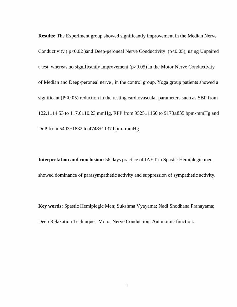
Results: The Experiment group showed significantly improvement in the Median Nerve
Conductivity ( p<0.02 )and Deep-peroneal Nerve Conductivity (p<0.05), using Unpaired
t-test, whereas no significantly improvement (p>0.05) in the Motor Nerve Conductivity
of Median and Deep-peroneal nerve , in the control group. Yoga group patients showed a
significant (P<0.05) reduction in the resting cardiovascular parameters such as SBP from
122.1±14.53 to 117.6±10.23 mmHg, RPP from 9525±1160 to 9178±835 bpm-mmHg and
DoP from 5403±1832 to 4748±1137 bpm- mmHg.
Interpretation and conclusion: 56 days practice of IAYT in Spastic Hemiplegic men
showed dominance of parasympathetic activity and suppression of sympathetic activity.
Key words: Spastic Hemiplegic Men; Sukshma Vyayama; Nadi Shodhana Pranayama;
Deep Relaxation Technique; Motor Nerve Conduction; Autonomic function.

III
CONTENTS

IV
TABLE OF CONTENTS
Sl. No.
INDEX
PAGE
NO.
1
INTRODUCTON
1
2
AIMS AND OBJECTIVES
5
3
REVIEW OF LITERATURE
7
4
MATERIALS AND METHODS
48
5
RESULTS
72
6
DISCUSSION
78
7
CONCLUSION
84
8
SUMMARY
86
9
LIMITATIONS
89
10
BIBLIOGRAPHY
90
11
ANNEXURES
103

V
LIST OF TABLES
TABLE
NO.
TOPIC
PAGE
NO.
1
Distribution of studies on Autonomic Variable & Yoga for
Wellbeing
32
2
Demographic details of the Subjects
50
3
Primary and Secondary Variables
58
4
Intervention Chart
68
5
Anthropometric variables between Yoga and Control group
73
6
Resting Cardio-vascular parameters before and after between
Yoga and Control group
74
7
Motor Nerve Conductivity before and after between the
Yoga and Control group
75
VI
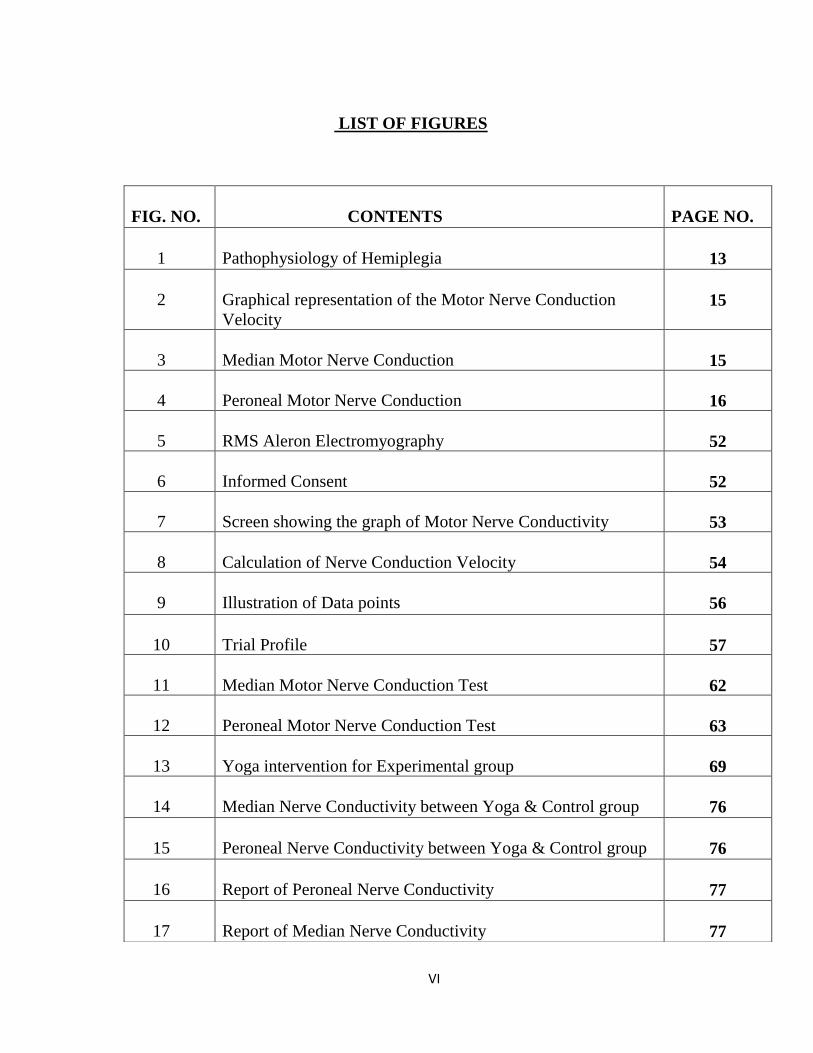
LIST OF FIGURES
FIG. NO.
CONTENTS
PAGE NO.
1
Pathophysiology of Hemiplegia
13
2
Graphical representation of the Motor Nerve Conduction
Velocity
15
3
Median Motor Nerve Conduction
15
4
Peroneal Motor Nerve Conduction
16
5
RMS Aleron Electromyography
52
6
Informed Consent
52
7
Screen showing the graph of Motor Nerve Conductivity
53
8
Calculation of Nerve Conduction Velocity
54
9
Illustration of Data points
56
10
Trial Profile
57
11
Median Motor Nerve Conduction Test
62
12
Peroneal Motor Nerve Conduction Test
63
13
Yoga intervention for Experimental group
69
14
Median Nerve Conductivity between Yoga & Control group
76
15
Peroneal Nerve Conductivity between Yoga & Control group
76
16
Report of Peroneal Nerve Conductivity
77
17
Report of Median Nerve Conductivity
77


1
INTRODUCTION
2
1.0 INTRODUCTION
Hemiplegia, or paralysis of one side of the body, is caused by injury or illness, and
leads to other disabilities. Hemilpegic have a limited daily physical activities which
affects their social well-being and leads to depression (Zenobia, C Y, Chan, 2012).
In 2007, WHO estimated that there are more than 600 million people with
disabilities worldwide (World Health Organization, 2007), and hemiplegia is one of
the more common disabling conditions and may be caused by neurological
problems like stroke and brain injury.
Pedretti . et.al., (2001) defined Hemiplegia as the paralysis of one side of the body.
Savinelli, R. et.al., (1978) reported that Hemiplegia is caused by injury to the
brain or due to some disease which leads to difficulties in locomotor functioning,
cardiopulmonary function, and sensory functioning.
Kong, K. H, & Yang, S. Y, (2006) found that these difficulties affect their
activities in daily living and thus have a negative impact on the quality of their life .
Lazaridou, A. et.al., (2013) explained that the experience of stroke can have a
negative impact on both psychological and physical health and on quality of life.
They suggested that Yoga practices were one of the effective therapies that have
been used for patients with a variety of ailments.
Yoga is the India's oldest tradition which harmonizes the mind, body and soul. It
has evolved over 5,000 years ago, which has moral and ethical precepts, mental
attitudes, and physical practices (Feuerstein G, 2000)

3
The word Yoga means ‘unity’ or oneness and is derived from the Sanskrit word
YUJ which means ‘to join’. Yoga unites the individual consciousness with the
universal consciousness to attain the supreme reality. It is the science of right living
and works on all aspects of the person: the physical, mental, vital, emotional, and
spiritual.
Swami Satyananda Saraswati said, “Yoga is not an ancient myth buried in oblivion
but the most valuable inheritance of the present. It is the essential need of today and
the culture of tomorrow” (Swami Satyananda saraswati, 2002, P-1)
Goyeche, J R M, (1979) suggests that Yoga and mindfulness can be regarded as a
main form of alternative medicine therapy.
Schmid, A A. et.al., (2014) observed that therapeutic yoga intervention may
improve multiple aspects of physical functioning after stroke and may be
complementary to traditional rehabilitation .
Immink, M A. et.al., (2014) concluded that the yoga intervention had a significant
improvement in quality of life associated with perceived motor function and
improvements in perceived recovery approached significance. They also found that
the memory significantly improved after yoga intervention, and those in the Yoga
intervention group exhibited decreased state and trait anxiety.
4
Schmid, A A. et.al., (2012) conducted a pilot study where they reported that the
Yoga group data demonstrated significant improvement in balance BBS (Berg
Balance Scale) and FoF (Fear of Falling). They concluded that yoga intervention
based on a group performance has potential in improving multiple post stroke
variables for people with chronic stroke.
These disabilities, make them to face difficulties in their daily lives, such as
depending on others for their daily needs. They may feel insecured, angry,
depressed, and guilty while facing the challenges. This is true with men, on whom
the masculinity is projected, including the attributes of power, control, strength,
independence, responsibility and dominance. This image may hinder men from
expressing their difficulties in dealing with their disabilities (Tager, D. et.al., 2006)
As counselling is found to reduce depressive moods and facilitate communication
(Courtenay, 2001)
A study conducted on the effect of Yoga and exercise in depression and anxiety in
people with post stroke disability revealed greater improvements in the mood of
post stroke (Chan, W. et.al., 2012)
All the above studies suggested to conduct a detailed study on the effect of Yoga on
Hemiplegia and as such there has been no study conducted yet on the effect of yoga
on motor nerve conductivity, which motivated us to conduct this study for the
betterment of hemiplegic patients.

5
AIM
&
OBJECTIVES

6
2.0 AIMS AND OBJECTIVES
2.1 Aim:
The aim of this study was to assess the effect of IAYT on changes in the Motor nerve
conductivity in Spastic Hemiplegic Men.
2.2 Objectives of the study:
The objectives of the present study are:
To record the Motor Nerve Conductivity in Median nerve and Peroneal nerve
before and after intervention
To record the Resting Cardio-respiratory parameters before and after
intervention
To compare the Motor nerve conductivity and Resting Cardio-respiratory
parameters before and after intervention

7
REVIEW
OF
LITERATURE

8
3.0 REVIEW OF LITERATURE
3.1 Hemiplegia
Hemiplegia is the condition where one side of the body is paralyzed or weakened due
to stroke. It occurs when the flow of blood to the brain is disturbed or obstructed, due
to which a part of the brain dies. Hemiplegia is caused by the damage to central
nervous system (brain and spinal cord): where the orders of movements are not
transmitted to the muscles. In addition to motor problems, memory or sensitivity may
also get affected.
Stroke is defined by the World Health Organization (WHO) as the "rapidly developing
clinical signs of focal (at times global) disturbance of cerebral function, lasting more
than 24h or leading to death with no apparent cause other than that of vascular
origin" (Hatano, S, 1976)
Paralysis may affect any one part (e.g. the arm, the leg) or the entire side of the body.
Severe or complete loss of muscular functions on one side of the body will be present
in this condition. When the right side of the brain is affected, the left side of the body is
paralyzed (and vice versa). Hemiplegia may be both congenital (since birth) or
acquired (from other illnesses such as a Stroke). The two main causes of stroke are:
ischaemia, or the lack of blood supply to the brain; and haemorrhagic, which results
from a fissure in an intracranial artery (Sims, N R, & Muyderman, H, 2009)
9
In stroke survivors, these events may lead to a long-term disability, age-related
cognitive impairment and poor or loss of memory (Falcone, G J.et.al., 2014), and
which has a deep emotional and socioeconomic impact on the patients and their
families and also on the health services (Feigin, V L. et.al., 2003)
Langhorne, P. et.al., (2009) found that stroke resulted in the difficulty of motor
activities and which results in the limitation of their movements.
In addition, non-cognitive neuropsychiatric symptoms may occur after stroke, such as
depression, anxiety, emotional lability, apathy and post-stroke fatigue
(Hackett, M L. et.al., 2014)
Stroke also causes other health issues and impairs the quality of living
(Garrett, R. et.al., 2011)
In the stroke patients the peripheral nerves are affected both morphologically and
electro physiologically on the paretic side. The distal latency is prolonged and they
have a slow motor conduction velocity of the tibial nerve and reduced amplitudes of
the median and ulnar nerves on the paretic sides when compared with that of the non-
paretic sides. The median and sciatic nerve values were smaller on the paretic sides as
compared to the non-paretic sides. (Uğurlu, F G. et.al., 2015)

10
It is found that in the paralyzed leg of stroke patients there will be severe inversion of
the ankle, with drop foot, which shows the altered common peroneal nerve conduction
properties. In the deep peroneal nerve the motor nerve conduction latencies of the
tibialis anterior muscle were longer in the paralytic side than in the sound side
(Nahariya, et.al., 2007)
3.1.1 Hemiplegia Prevalence
A study conducted on the Incidence estimate and guideline-oriented treatment for post-
stroke spasticity based on German statutory health insurance in 2009, revealed that 3.7
per 1000 persons had stroke. There were about 242090 insurants out of which 1263 of
the (sample population) were admitted to a hospital for acute stroke in the year of 2009
(Veronika Egen-Lappe, et.al., 2013)
3.1.2 Hemiplegia Pathophysiology
Injury to the nerve pathway that provides loss of control of the muscles may occur on
different occasions. The most common cause is stroke (Cerebrovascular Accident). It
can have different origins: the interrupted or disturbed blood supply to part of the brain
caused by a clot that blocks an artery or cerebral hemorrhage which results in oxygen
deprivation in that region, which results in the death of nerve cells. Motor deficits are
characterized by paralysis (hemiplegia), typically on the side of body opposite to the
side of lesion. Interruption of blood flow only for a few minutes sets in a series of
pathological conditions. (O'Sullivan, 2007).

11
Hemiplegia is set up very quickly but sometimes regresses more or less disabling
sequelae. Trauma can also cause hemiplegia brutal and immediate. Hemiplegia which
occur more gradually they are caused by brain tumors, infections (encephalitis and
brain abscess).
Spastic hemiplegia is a neuromuscular condition of spasticity that results in the
muscles on one side of the body being in a constant state of contraction. It is the "one-
sided version" of spastic diplegia. It falls under the mobility impairment umbrella
of cerebral palsy. About 20–30% of people with cerebral palsy have spastic
hemiplegia. [Cerebral palsy at eMedicine]
Due to brain or nerve damage, the brain is constantly sending action potentials to
the neuromuscular junctions on the affected side of the body. Similar to strokes,
damage on the left side of the brain affects the right side of the body and damage on
the right side of the brain affects the left side of the body. The affected side of the body
is rigid, weak and has low functional abilities. The severity of spastic hemiplegia is
dependent upon the degree of the brain or nerve damage. (Brashear& Allison, 2010)
There are many different brain dysfunctions that can account for the cause for spastic
hemiplegia. Spastic hemiplegia occurs either at birth or in the womb. (Tardieu, C.
et.al., 1982) & (Tardieu, G. et.al.,1982) . The cause can be all types of strokes, head
injuries, hereditary diseases, brain injuries and infections.
12
Malformations of the veins or arteries in any part of the body can lead to spastic
hemiplegia. The artery most commonly affected is the middle cerebral artery.
The spasticity occurs when the afferent pathways in the brain are compromised and the
communication between the brain to the motor fibers is lost. When the inhibitory
signals to deactivate the stretch reflex is lost the muscle remains in a constant
contracted state. With spastic hemiplegia, one upper extremity and one lower extremity
is affected, so cervical, lumbar and sacral segments of the spinal column can be
affected.[Cerebral palsy at eMedicine]
The muscle spasticity can cause gait patterns to be altered and jerky. The constant
spastic state of the muscle can lead to bone and tendon deformation, further
complicating the patient's mobility. Many patients with spastic hemiplegia are
subjected to canes, walkers and even wheelchairs. Due to the decrease in weight
bearing, patients are at a higher risk of developing osteoporosis.[Cerebral
Palsy~clinical at eMedicine]
An unhealthy weight can further complicate mobility. Patients with spastic hemiplegia
are a high risk for experiencing seizures. (el-Abd, M A, Ibrahim, I K, March 1994)
Oro-motor dysfunction puts patients at risk for aspiration pneumonia. Visual field
deficits can cause impaired two-point discrimination. Many patients experience the
loss of sensation in the arms and legs on the affected side of the body.[ Cerebral
13
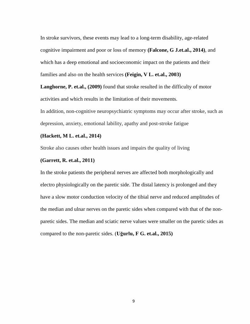
Palsy~clinical at eMedicine] Nutrition is essential for the proper growth and
development for a child with spastic hemiplegia.
Figure :1 Pathophysiology of Hemiplegia
3.1.3 Pathogenesis
Damage to the pyramidal tracts produces impairment or loss of voluntary movement
from interruption of the conduction of motor impulses.
3.1.4 Hemiplegia Diagnosis
Faced with a hemiplegic patient, the doctor performs a neurological exam to assess the
extent of the invasion. It tests the presence of a Babinski sign by stimulating the outer
edge of the sole from the heel to midfoot: if the person suffers from hemiplegia, the
great toe by an extension, which is indicative a lesion of the pyramidal pathway.

14
There are situations where the diagnosis is not obvious because the hemiplegia may
present with subtle signs such as clumsiness or fatigability of muscles on one side of
the body. So the doctor performs a series of examinations, and in particular the
operation of Barre highlighting deficits on one leg or one arm or both. In the case of an
unconscious person, the hemiplegia was found from different specific maneuvers.
Imaging studies, CT or magnetic resonance imaging (MRI) can detect the causes
(aneurysm, tumor embolism) cause brain damage. An electro encephalogram may give
information about brain injury and their importance. A neuropsychological
examination can identify possible cognitive impairment such as aphasia, common in
cases of hemiplegia.
A nerve conduction study (NCS) is a medical diagnostic test commonly used to
evaluate the function, especially the ability of electrical conduction, of the motor and
sensory nerves of the human body. Nerve conduction studies are used mainly for
evaluation of paresthesias (numbness, tingling, burning) and/or weakness of the arms
and legs.
15
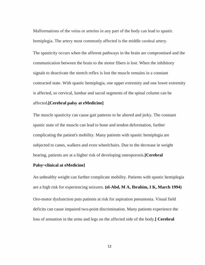
Fig: 2 Graphical representation of the Motor Nerve Conduction
Velocity
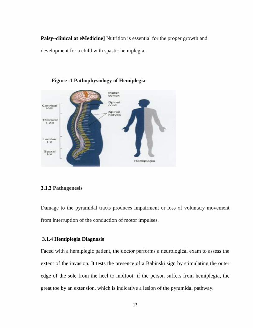
Fig: 3 Median motor nerve conduction
16
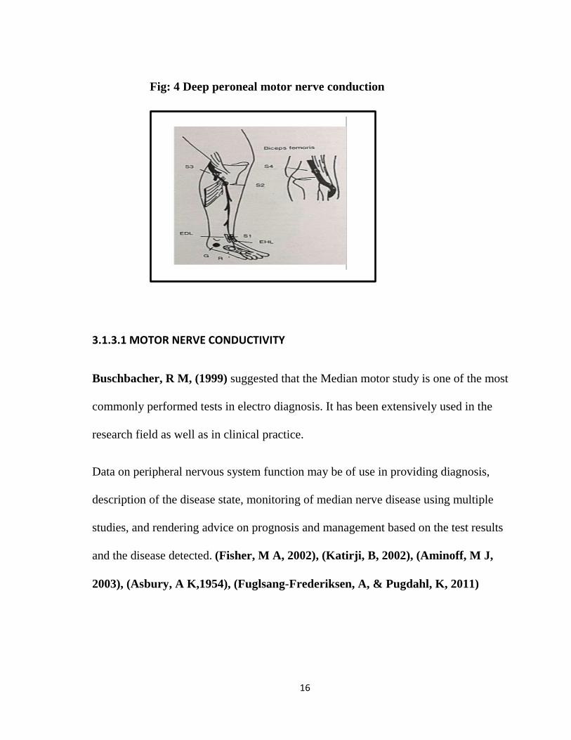
Fig: 4 Deep peroneal motor nerve conduction
3.1.3.1 MOTOR NERVE CONDUCTIVITY
Buschbacher, R M, (1999) suggested that the Median motor study is one of the most
commonly performed tests in electro diagnosis. It has been extensively used in the
research field as well as in clinical practice.
Data on peripheral nervous system function may be of use in providing diagnosis,
description of the disease state, monitoring of median nerve disease using multiple
studies, and rendering advice on prognosis and management based on the test results
and the disease detected. (Fisher, M A, 2002), (Katirji, B, 2002), (Aminoff, M J,
2003), (Asbury, A K,1954), (Fuglsang-Frederiksen, A, & Pugdahl, K, 2011)

17
It is obviously preferable in a clinical setting to have reference data derived from a
sample population that approximates, as closely as possible, the demographic
characteristics of the patient being tested. (Robinson, L R, Rubner, D E, 1994)
The Western and Middle East countries have published many studies from normative
data of the median nerve. (Hennessey, W J. et.al., 1994), (Falco, F J. et.al., 1992),
(Hennessey, W J. et.al, 1994), (Kumar, B R, & Gill, H S, 1985), (Correa Pérez, M,
1986), (Cochran, W G, 1977)
According to a study conducted on Median Nerve Conduction in Healthy Nigerians:
Normative Data, the reference range for median motor nerve velocity is 49.48 – 66.92
msec (L F Owolabi, et.al., 2016)
Uğurlu, F G. et.al., (2015) did an Ultrasonographic evaluation of the median and sciatic
nerves in 33 hemiplegic patients after stroke out of which 18 were women and 15
were men. They found that prolonged distal latency and slowed motor conduction
velocity of the tibial nerve as well as reduced amplitudes of the median and ulnar
nerves were observed on the paretic sides when compared with those of the nonparetic
sides (all P < 0.05). The median and sciatic nerve cross-sectional area values were
found to be smaller on the paretic sides when compared with the nonparetic sides (all P
< 0.05).
18
KOJI SHIGENO, (1972) did a study on hemiparesis patients due to Cerebrovascular
disease with varying degrees and revealed the relationship of hemiplegic amyotrophy
to severity of motor impairment which suggests that (in addition to the possible close
proximity of the vasomotor and central trophic systems to the pyramidal tract) lower
motor neurons, involved by upper motor neurons, may possibly induce denervation
atrophy
Neurographic studies in hemiplegic patients with spastic hemiparesis after stroke done
by Ivan, G, Milanov, (1995) on the motor conduction velocities, were evaluated in
median, ulnar, peroneal and tibial nerves which revealed normal motor conduction
velocities
Dr. Zainab, M M, Chatriwala1 et.al., (2016) conducted an observational study on
the Common Peroneal Nerve Conduction Velocity (NCV) in Post Stroke Patients
where the Distal latency were prolonged. The Motor NCV and CMAP amplitude were
reduced in the affected lower limbs compared to the unaffected lower limbs. The study
in stroke individuals also concluded that the spasticity of ankle plantar flexors and/or
weakness of ankle dorsi-flexors could cause electrophysiological changes in the
Common Peroneal Nerve.
19
K, Takebe, M G, Narayan, et.al., (1975) did a study on nerve conduction velocities
of ulnar and peroneal nerves bilaterally in 27 hemiplegic patient where they found that
the upper motor lesion affects the function of the lower motor neuron. The causes
demonstrated were, the lower skin temperature, atrophic thinning of the fibers and
other less plausible factors.
S, Chokroverty, and J, Medina, (1978) did Electrophysiological study on 13
hemiplegic patients and where they showed that Motor nerve conduction velocities of
the ulnar and common peroneal but not the median nerves were substantially reduced
in the affected limbs of hemiplegic patients. The reduction of skin temperature in the
hemiplegic limbs was related to the slowing of conduction velocity of the common
peroneal nerve.
Gender has a definite effect on NCS (Nerve Conduction Study) variables. The tests
were done on JAVA RMS Aleron-201 series. (Abhishek Kumar & Anjali Prasad,
2016)
Thakur et.al., (2010) found an increase in all component of CMAP(Compound
Muscle Action Potential) for male as compared to female and was statistically
significant for ante-cubital fossa and right popliteal fossa.
20
The age factor had been negatively correlated to the amplitude in MNC conducted by
Huang et.al., (2009). This study also showed that the conduction velocity was slightly
more in the upper limb than the lower limb which could be due to the length of the
nerve.
Study conducted by Shailja Tiwari et.al., (2015) showed that as on increasing the
temperature from 290 C to 390C there was significant increase in nerve conduction
velocity by 1.0 to 1.4 m/second per degree rise in temperature.
3.1.4. CLINICAL FEATURES
Hemiplegia Sign
Hemiplegia caused by a lesion of the pyramidal tract. This is the main neural pathway
that carries the motor orders. It is therefore a set of neurons involved in voluntary
movement.
The pyramidal pathway begins in the brain at an area of nerve cells of pyramidal shape
and joined with other nerve cells of the spinal cord. Pyramidal tract neurons then
transmit their orders to LMN which carry them to the muscles. Before reaching the
spinal cord, brain stem, the pyramidal tract changes sides. This explains that a lesion is
localized on the side opposite the affected limb: left brain injury causes a right
hemiplegia and vice versa.

21
Observed with different depending on the location of the injury.
When the lesion in the brain cortex, this causes a disproportionate hemiplegia: the
face and arms are predominantly affected.
When the lesion is located in the white matter of the brain, this causes a
proportional hemiplegia: arm and leg are affected similarly deficient.
A brainstem lesion, it causes a paralysis of one side of the body and involvement of the
face on the other side.
Hemiplegia Symptoms
In some cases the lesions, arm and leg are affected, in others only the arm or only the
face. When hemiplegia is partial and that movements are still possible, there is a
decrease in muscle strength and mobility impaired, as manifested by clumsiness,
trouble walking accompanied by a great tiredness and falls of one side. When
hemiplegia is total, even the reflexes are abolished. However, the Babinski sign is
present: when you touch the outside of the foot, it causes an extension of the big toe. In
a healthy person, this stimulation leads to a bending of the big toe. Hemiplegia is
accompanied by changes in muscle tone: the muscles are stiff and overly contracted
any (spastic hemiplegia) or conversely soft and flabby (flaccid hemiplegia). On the
face, the damage to the muscles can result in a drooping eyelid or an asymmetric smile.
22
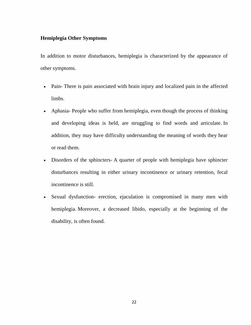
Hemiplegia Other Symptoms
In addition to motor disturbances, hemiplegia is characterized by the appearance of
other symptoms.
Pain- There is pain associated with brain injury and localized pain in the affected
limbs.
Aphasia- People who suffer from hemiplegia, even though the process of thinking
and developing ideas is held, are struggling to find words and articulate. In
addition, they may have difficulty understanding the meaning of words they hear
or read them.
Disorders of the sphincters- A quarter of people with hemiplegia have sphincter
disturbances resulting in either urinary incontinence or urinary retention, fecal
incontinence is still.
Sexual dysfunction- erection, ejaculation is compromised in many men with
hemiplegia. Moreover, a decreased libido, especially at the beginning of the
disability, is often found.
23
Hemiplegia Complication
The immobility of paralysis arising Member is responsible for complications that
specialists in physical medicine and rehabilitation at trying to prevent the initial
management. The main complication remains on the loss of autonomy: everything
must be done to try to recover mobility as complete as possible.
Among the complications that can occur after hemiplegia
Pain in joints of immobilized different: the shoulder is often affected with a
stiffening of the muscles (spasticity) and local inflammation.
Moreover, the bones of people with hemiplegia are weakened and lose bone density
(osteopenia) as the brain give rise to abnormal vascularization of bone. Finally, sitting
in a wheelchair or bedridden status may promote pressure sores (skin necrosis at the
points of support) and problems such as venous disorders of venous circulation, the
risk of phlebitis and edema. The sphincter disturbances can cause infectious
complications.

24
3.1.5 Conventional Management
3.1.5.1 Pharmacotherapy
A study conducted by Feeney, D M. et.al., (1993) on Nor-adrenergic
pharmacotherapy, intra cerebral infusion and adrenal transplantation showed
improvement in the functional recovery after cortical damage which revealed that the
widespread reduction of glycolytic and oxidative metabolism, produced by focal
cortical injury, is normalized by the same treatment which alleviates symptoms and is
worsened by drugs which exacerbate deficits. The data support the hypothesis that
providing SRE (symptom relevant experience) during a period of enhanced NA
(noradrenergic) synaptic activity produces an enduring functional recovery after
cortical injury by attenuating remote functional depression.
Pharmacotherapy administered to enhance the Cognitive and Motor Recovery
following Stroke were especially the antidepressants, acetyl-cholinesterase inhibitors
and memantine for aphasia. But, clinical trials are needed to address the shortcomings
of stroke management. (Xabier Beristain, 2015)

25
3.1.5.2 Surgical Approach
Surgical approaches in case of Hemiplegia include tendon transfer, muscle
lengthening, and arthrodesis. These procedures are considered to be permanent to fix
the solutions: in case of arthrodesis, where the overall range of movement is reduced.
Achieving a more functional hand position and improving the appearance or hygiene of
the arm and hand are the goals of the treatment. Robust studies of long-term outcome
are only few that are available, but there is evidence of benefit (Smitherman, J A.
et.al., 2011), (Eliasson, A C. et.al., 1998), (Skold, A. et.al., 1999)
The long-term benefit in terms of function and cosmesis from the patients aspect were
perceived by many Research scholars. (Skold, A. et.al., 2007), (Johnstone, B R.
et.al.,2003)
3.2.1 Spastic Hemiplegia and Autonomic Dysfunction
Patients with various cerebrovascular diseases commonly have disturbances of the
autonomic nervous system. The damage occurs in the fronto-parietal cortical areas and
in the brain stem mainly in the central autonomic network. It may be also be because of
disruption of the autonomic pathways. The autonomic pathway descends through the
mesencephalon, pons, and medulla to the spinal cord from the hypothalamus.
Abnormalities in heart rate and blood pressure regulation, reflects the cardiovascular
autonomic dysfunction. Asymmetric sweating with cold hemiplegic limbs, reflects the

26
changes in the sudomotor and vasomotor regulatory systems. These are the most
common clinical problems in case of stroke.
Some may have complaints like bladder and bowel dysfunction or incontinence and
impotency after stroke. In the acute phase of stroke there occurs increased sympathetic
activity resulting in Cardiovascular autonomic dysfunction and abnormal sweating.
These changes may be irreversible attimes. This autonomic imbalance also contributes
to abnormalities in the parasympathetic nervous system. Quantitative analysis methods
are inevitable for detecting the autonomic dysfunctioning, because these disturbances
are disabling and uncomfortable for the subjects. It may also be prognostically
unfavorable. (Korpelainen, J T. et.al., 1999)
Juha T Korpelainen et.al., (2017) concluded that hyperhidrosis or profuse sweating
on the paretic side of the body was observed in 55% of the patients at baseline, after 5
minutes of heating in 74%, and after 10 minutes of heating in 77%.
Hyperhidrosis observed throughout the body correlated with the severity of paresis,
which is because of the reduced muscle tone, and the extensor plantar response. This
sweating disturbance might be because of the lesion of a putative sympathoinhibitory
pathway controlling sweating. The failure of this pathway could also be related to other
manifestations of sympathetic hyperfunction, e.g., cardiac complications

27
3.2.2 Yoga and Health
Yoga is one of the six systems of Indian Vedic philosophy (Darshan). Maharishi
Patanjali, rightly known as the “Father of Yoga”, compiled and refined various
aspects of yoga systematically in his “Yoga Sutras” (aphorisms), wherein he
advocated the eight-fold path known as “Ashtanga Yoga” for an all-around
development of human personality. These include - Yama [moral odes],
Niyama [self-purification and study], Asana [posture], Pranayama [breath
control], Pratyahara [sense control], Dharana [concentration], Dhyana [meditation],
and Samadhi [super contemplation]. These are formulated on the basis of multifarious
psychological understanding of human personality.
Other aspects of yoga philosophies are broadly classified into four streams namely
Work, Worship, Philosophy, and Psychic control. “Karma Yoga,” the path of work,
promotes pleasure in labor without indulging in thoughts of success or failure. A free
mind allows the task to be done in a skillful manner. “Bhakti Yoga,” the path of
worship, is a systematic method of engaging the mind in the practice of divine love.
This attitude of love softens our emotions and tranquilizes our mind. “Gyana Yoga,”
the path of philosophy, is a systematic way of enlightening the mind about the realities
of life by contemplation. This will strip off the garb of Avidya (ignorance) from our
mind as it goes to its natural state of rest.

28
“Raja Yoga,” the path of psychic control, is a systematic process of culturing the
mind. It is based on the eight-fold path set by Patanjali.
Studies of neurological disorders, like epilepsy, have shown improvements attributed
to yoga. However, it is important to recognize that behavioral modification and altered
lifestyle accounts for the improvement in the outcomes.
A study carried out on 60 students doing their First-year MBBS were assigned to two
groups: the yoga and control group (30 each). The yoga group practiced integrated
yoga practices for 35 min daily for a period of 12weeks under the guidance of a trained
yoga teacher. The control group were not assigned for any kind of yoga practice at all
for stress management. It was found that the serum IFN- γ decreased with examination
stress. Decreased serum IFN- γ levels is an indication of the decline in Cellular
immunity. The decrease in serum IFN- γ was less significant in the yoga group than the
control group which indicated a decline in cellular immunity with examination stress
which was found to be more among the control group than the yoga group students.
(Gopal, A. et.al., 2011)
The study by Sirven et.al., (2003) exhibited the percentage of individuals who
benefited from yoga was among the highest in all CAM (Complementary and
Alternative Medicine) modalities.

29
Bastille et.al., (2004) demonstrated the benefits of yoga among post-stroke patients.
In the study conducted by Oken et.al., (2004) they concluded that yoga and aerobic
exercise were effective in relieving fatigue in MS patients.
One study has shown that psychological benefits of an aerobic exercise intervention in
a group of healthy young adults could be increased simply by informing subjects that
the exercise program was specifically designed to improve psychological well-being.
(Desharnais, R. et.al., 1993)
The study for CTS, the yoga-based regimen was more effective than wrist splinting and
no supplementary treatment control in relieving some symptoms and signs (Garfinkel,
M S. et.al., 1998)
La forge, (1997) revealed that Mind-body practice with existing health promotion and
cardiac rehabilitation services can improve the self-efficacy and long-term adherence
to healthy behaviors. It can as well improve the personal stress management skills.
There are also numerous primary and secondary preventive indications for
cardiovascular disease (CVD) where the mind-body exercise plays a primary or
complementary role.
Barnes et.al., (2001) demonstrated that transcendental meditation program had
improvements in cardiovascular reactivity. He also observed the significant reductions
in resting systolic blood pressure (SBP) in adolescents with high normal BP.
30
The beneficial effects of yoga at rest and during stressful states were reflected as
reduction in the reactivity of blood pressure, heart rate, and cardiac output to simulated
stressors. Successful cardiovascular risk factor modification using a “Kriya” yoga
program was also achieved by Schmidt, T. et.al., (1997)
Ives, J C. et.al., (2000) suggested that mind-body exercise methods are now used
widely in the health, fitness, and rehabilitation fields. Yoga helps to reduce stress,
decrease hypertension, and also exerts cardiorespiratory benefits.
Yank et.al., (2007) reviewed papers to find out the effects of yoga intervention
especially on the common risk factors of chronic diseases like overweight,
hypertension, high glucose level and high cholesterol. A systematic search yielded 32
articles published between 1980 and april 2007 which revealed that the yoga
interventions are generally effective in reducing body weight, blood pressure, glucose
level and high cholesterol.
Kiecolt, G.et.al., (2010) observed that the stress factors like serum interleukin (IL)-6
levels and C-reactive protein (CRP) are higher in the novices than the yoga experts. It
is found that IL-6 promotes CRP Production. The yoga experts produced less
lipopolysaccharide-stimulated IL-6 in response to the stressor than novices.

31
Smith et.al., (2011) concluded that yoga when practiced in a more integrated form,
i.e., with an ethical and spiritual component provides additional benefits over yoga
than when practiced as an exercise regimen.
Innes, K E. et.al., (2007) study suggests that chronic stress and related psychosocial
factors also play an important role in the development and progression in the
pathogenesis of cardio vascular diseases. Integrated psychological and physiological
components of health, yoga and other traditional mind-body therapies may offer
particular promise in both the primary and secondary prevention of CVD.
Yoga and meditation has also been commonly used for muscle relaxation. (Ghoncheh,
S, & Smith, J C, 2004).
Yoga can be performed by most people, including young people and cardiac patients
(Ades, P A. et.al., 2003),( Tran, M. et.al., 2001), (Raub, J, 2002) (Dash, M, & Telles, S,
2001). Yoga builds up a core stability during and after pregnancy (Berk, B, 2001) and
it increases the creativity and reduces stress, (Khasky, A D, Smith, J C, 1999) as well as
to improve muscle power, dexterity, visual perception, (Raghuraj, P, & Telles, S, 1997)
and reaction time (Madanmohan et.al.,1993) while strength, endurance, and muscle
reaction times which have been quantified previously, but little has been done to
quantify the muscle use during yoga practice (Narayan, R. et.al., 1990), (Dostalek, C.
et.al., 1979)

32
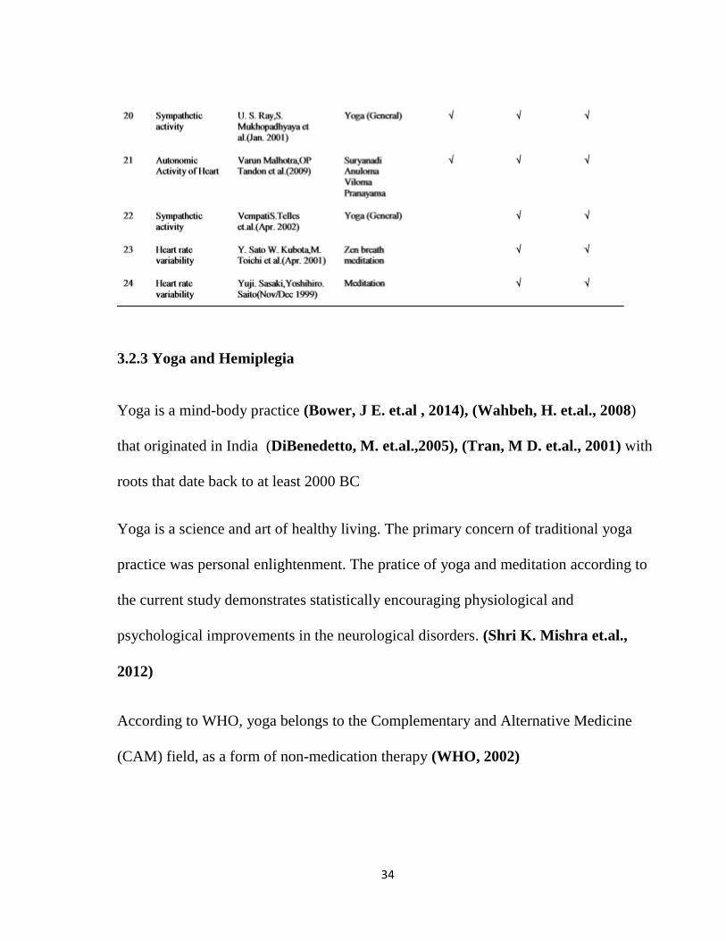
Table-1 Distribution of Studies on Autonomic Variable and Yoga for Wellbeing

33
34
3.2.3 Yoga and Hemiplegia
Yoga is a mind-body practice (Bower, J E. et.al , 2014), (Wahbeh, H. et.al., 2008)
that originated in India (DiBenedetto, M. et.al.,2005), (Tran, M D. et.al., 2001) with
roots that date back to at least 2000 BC
Yoga is a science and art of healthy living. The primary concern of traditional yoga
practice was personal enlightenment. The pratice of yoga and meditation according to
the current study demonstrates statistically encouraging physiological and
psychological improvements in the neurological disorders. (Shri K. Mishra et.al.,
2012)
According to WHO, yoga belongs to the Complementary and Alternative Medicine
(CAM) field, as a form of non-medication therapy (WHO, 2002)

35
Recent evidence highlights the positive effects of yoga for people with an increased
risk of cardiovascular disease (Cramer, H. et.al.,2013) and as an add-on therapy for
treating the carpal tunnel syndrome (O'Connor, D. et.al., 2003), depression,
(Uebelacker, L A. et.al., 2010), primary prevention of cardiovascular disease.
(Hartley, L. et.al., 2012)
A recent non-Cochrane systematic review concluded that, in stroke rehabilitation, yoga
can be used as a self-administered practice, due to its alleged effect of relieving the
mind and body from stress. Yoga was found to act on both the psychological and
physical levels, and immense improvements were noted in self-efficacy and confidence
level. These changes may lead to a change in the behaviour with improvement in
health. (Lazaridou, A. et.al., 2013)
Julie, V, Bastill & Kathleen, M, Gill-Body, (2004) conducted a study on the effect of
Yoga on poststroke hemiparesis which revealed improvement in TMB(Timed
Movement Battery) Scores and BBS (Berg Balance Scale) Scores confirming that yoga
may be beneficial to people who have had a stroke.
It is also known that yoga is a good training technique for muscle relaxation. It also
reduces anxiety Platania-Solazzo, A. et.al., (1992) and has been shown to decrease
neurological reaction time and improve muscle strength and endurance of the
expiratory and abdominal muscles (Madanmohan et.al., 1993)

36
3.2.4 Sukshma Vyayama (Joint exercises)
The pawanmuktasana series is one of the most important series of practices that has a
very profound effect on the human body and mind and is thus a most useful tool for the
yogic management of various disorders and maintenance of health. It is one of the
special contributions of Bihar School of Yoga and the teachings of Paramahamsa
Satyananda.
In Sanskrit these practices are referred to as Sukshma vyayama which means 'subtle
exercise'. The Sanskrit word pawan means 'wind' or 'prana'; mukta means 'release' and
asana means 'pose' or ‘posture’. Thus, pawanmuktasana are a group of asanas which
helps mainly to remove any blockages that prevent the free flow of energy in the nadis
or channels in the body and also through the energy channels of mind. Sometimes, the
energy becomes blocked due to wrong or bad posture, disturbed bodily functions,
psychological or emotional problems or an imbalanced or unhealthy lifestyle. Some of
the major discomforts are stiffness, muscular tension, lack of proper blood flow and
some minor functional defects. If however, these blockages become chronic, it may
lead to the malfunctioning or disease of the limb, joint or physical 21 organ. Regular
practice of pawanmuktasana promotes total health, regulating and stabilising the flow
of energy throughout the body.

37
Pawanmuktasana is divided into three distinct groups of asanas: the anti-rheumatic
group, the digestive/abdominal group and the shakti bandha or energy block group. All
three groups supplement each other, stimulating and encouraging a free flow of energy
throughout the body.
This group of asanas is concerned with loosening up the joints of the body. It is
excellent for those with rheumatism, arthritis, high blood pressure, heart problems or
other ailments where vigorous physical exercise is not advised. It is particularly useful
for eliminating energy blockages in the joints and outer extremities of the physical
body, and works on the pranic and mental bodies as well.
The practices may be performed in three ways:
With awareness of the actual physical movement
With awareness and integrated breathing
With awareness of the movement of prana in the body
After every two or three movements, sit quietly in the base position with the eyes
closed and be aware of the natural breath, of the part or parts of the body that have just
been moved, and of any thoughts or feelings that come into the mind. After a minute or
so continue the practice. This will not only rest the body but will also develop
awareness of the internal energy patterns, and the mental and emotional processes.

38
Some of the Sukshma Vyayama Practices are:
Padanguli Naman (toe bending)
Goolf Naman (ankle bending)
Goolf Chakra (ankle rotation)
Janu Naman (knee bending)
Mushtika Bandhana (hand clenching)
Manibandha Naman (wrist bending)
Manibandha Chakra (wrist joint rotation)
Kehuni Naman (elbow bending)
Skandha Chakra (shoulder socket rotation)
Greeva Sanchalana (neck movements)
Benefits:
All the asanas given for foot and calf helps in returning the stagnant lymph and
venous blood, thus relieving the tiredness and cramp, and also prevents venous
thrombosis especially in the bedridden and post-operative patients.
The knee joint is an important weight bearing joint of the body which as such
has no strong muscles for its support, is the most vulnerable joint for injuries,
sprains and osteoarthritis. Thus all the knee asanas strengthen the quadriceps
muscle and the ligaments around the knee joint. These practices rejuvenate and

39
revitalize the joint by activating the healing energies by hastening a proper
blood supply and drainage.
Neck is the region through which all the nerves connecting the different organs
and limbs of the body pass by. Therefore, strain or tension gets lodged or
accumulated in the muscles of the neck and shoulders, especially after a
prolonged desk work or continuous sitting or other bad postures. The yoga
practices done for the neck, shoulders and arms release tension, heaviness and
stiffness in these areas. (Swami Satyananda saraswati, 2002, P 21- 44)
The study conducted by Shambhu Prasad Shaw & Ravi Kulkarni, (2009) on the
Effect of Sukshma-vyayama on School Children revealed that the important tool to
improve the strength and concentration of students is by practicing the Yogic
Sukshma-Vyayama. 64 students were allotted to two groups, one as the experimental
group while the other as control group. The 32 students in the experimental group
practiced yogic sukshma-vyayama in sequence along with some basic yogic practices
while the 32 students followed their usual school routine programs and not the
Sukshma-vyayama practice in the control group. This training program lasted
continuously for 21 days. The yoga group followed some sukshma-vyayama practices
related to hand-grip strength where there was a significant change in the right hand
grip-strength (p=0.018, paired samples t-test); while no change observed in the hand
grip-strength in the left hand (p=0.274). For control group, there showed no changes in

40
both the hand grip-strength; while p=0.536 for the right hand and p=0.419 for the left
hand (Paired sample t-test).
3.2.5. Nadishodhana Pranayama (NSP)
Patanjali, the father of yoga, described pranayama as the gradual unforced cessation or
expansion or exercising of breath. Pranayama is derived from two sanskrit words-
prana (life) or ayama (control). Pranayama or control of prana or life force controls
ones heart beat pulse and mind (Sri Paramhansa Yogananda, 2002, P 496 - 507).
The process of pranayama involves systematic and disciplined inspiration and
expiration with retention of breath or holding of breath in specific proportion and
specific manner for a specific duration. (Nalini Sharma and Shri Ram Sharma,
2008)
According to the verse in Patanjali Yoga Sutra (2:49) Pranayama means,
“Tasmin sati shvasa prashvasa yorgati vicchedah pranayama” which means that
once harmony with the physical body has been achieved, through interruption of the
movement engendered by inhaling and exhaling one attempts to harmonize the energy.
In ancient Indian yoga books, lots of pranayamas are described to various benefits.
These are bhastrika, kapalbhati, anuloma viloma, bhramari, ujjayi, sheetali, sheetkari,
nadi shodhana, etc.

41
By knowing to control the breath, one can gain control over the emotions and other
mental states as well. By becoming aware of the breath, one can gradually become
more sensitive to the subtle objects like our mind and to the flow of energy throughout
the body and a stronger energy awareness develops within us. How one breathes has an
impact on the heart, brain and nervous system. There exists a direct correlation
between the breath and anxiety or well-being. During stress, the breath is shorter, more
frequent and quite shallow which maintains a level of arousal. Slower and deeper
breathing results in a more relaxed state via autonomic reflexive stimulation and
decreases the partial pressure of carbon dioxide in the lungs and bloodstream resulting
in a corresponding increase in the pH of the blood, where it becomes less acidic
hastening an effective blood oxygen synthesis. It also benefits in the metabolism and
functioning of brain. For example, deep breathing practices increases the levels of
noradrenalin, a compound that functions as a hormone and as a neurotransmitter in the
nervous system. (A, Jr Stancak and M, Kuna, 1994)
Nadi Shodhana Pranayama or Alternate nostril breathing, which activates and
harmonizes ida and pingala nadis. Shodhana means ‗to purify‘.
In English this breathing practice is called as Nadi purification pranayama.
42
Baddhapadmāsano yogī prānam chandrena pūrayet |
Dhārayitvā yathāśakti bhūyah sūryena rechayet ||
Prānam sūryena chākrshya pūrayedudaram śanaih |
Vidhivatkumbhakam krtvā punaśchandrena rechayet ||
Yena tyajettena pītvā dhārayedatirodhatah |
Rechayechcha tatoanyena śanaireva na veghatah ||
Hatha yoga pradipika (2/7, 8, 9)
Meaning: Adopting baddha padmasana, the yogi inhales through the left nostril and
holds the breath to their capacity, and then exhales through the right nostril. Then
inhaling gently through the right nostril, gradually fill the abdomen, performing
kumbhaka as before, and then exhaling completely through the left nostril.
(Swami Muktibodhananda, 2008)
Nadi shodhana pranayama (alternate nostril breathing) affects the cerebral
hemisphericity of brain by alternately stimulating the right-brain and then the left-brain
passing through the corpus callosum. The process is done by the action of the air
flowing through the nostrils that stimulates the contra-lateral (opposite) side of the
brain through the nerve endings underneath the nasal mucosa in the nostrils.

43
Each side of the body is governed by nerves originating in the opposite side of the
brain, and stimulating the airflow in one nostril increasing nervous activity in the brain
on the opposite side to that nostril. Each side of the brain performs different activities
and processes, the autonomic nervous system will also be stimulated and relaxed
correspondingly via the Nadishodhana pranayama.
(A, Jr, Stancak and M, Kuna, 1994)
Increasing the flow of air through the right nostril stimulates the sympathetic nervous
system thereby increasing the heart rate, producing more sweat, dilating the pupils and
opening up the lungs, i.e. the fight or flight reaction. Increasing the flow of air through
the left nostril stimulates the parasympathetic nervous system thereby increasing the
digestion, lowering the heart rate and relaxing the body. So, by practising the nadi
shodhan pranayam, there occurs a balance between both of these systems and helps in
balancing the brain activity. (D, Shannahoff - Khalsa and B, Kennedy, 1993)
Dandekar Pradnya Deepak, 2013 conducted a study on the short-term training of
Anulom-Vilom in Healthy Volunteers and found that few minutes daily practice of
Anulom-Vilom Pranayam had significant effect on Systolic Blood Pressure, good
positive effect on digestive power and mental freshness and also helps to distress
humans at their work places and also help in maintaining better physical and mental
health. (Dandekar Pradnya Deepak, 2013)

44
Pallav Sengupta, (2016), postulates that mind-body exercise such as yoga couples
sustained muscular activity with internally directed focus, producing a temporary self-
contemplative mental state. This triggers the neuro-hormonal mechanisms that brings
about healthier benefits, evidenced by the sympathetic activity suppression. Thus, yoga
reduces stress and anxiety, improves autonomic and higher neural center functioning
and even improves physical health of cancer patients. He concluded that yoga can be
beneficial in the prevention and cure of diseases.
3.2.6. AUM Chanting
The AUM chanting practice in a rhythmic way by concentrating on the breath is a
traditional way of the powerful means in calming down the mind and helping in
enhancing the memory. According to Upanishads, Om is the name or symbol of God
(Chinmayanada Swami, 2002). AUM is a combination of three letters, namely, A, U,
and M. These syllables represent the past, the present, and the future and also the three
planes of being. (Sivananda Swami, 2005).
AUM is the force behind all positive thoughts or vibrations and chanting or even
thinking about AUM will bring about the inner silence or a quiet mental state or peace.
(Sanjay Kumar et.al., 2010)

45
Bhagavad Gita describes AUM as the Brahman or consciousness and he who
remembers it always, attains the supreme goal (Madhusudhan Saraswati &
Gambhiranada Swami, 1998).
Patanjali’s Yoga Sutra (PYS), which is a world famous, reputed and a legendary
classical yoga text explained AUM as the Pranava or Pranava mantra and that is Iswara
(Taimini, I K, 1986).
One of the most common diseases in the world is hypertension and persistent
hypertension causes cardiovascular diseases. (Kearney, P M. et.al., 2005)
It was reported that, the chanting of ‘AUM’ reduces heart rate, blood pressure, mental
agitation and the skin resistance (Telles, S. et.al., 1995) Telles, S. et.al., 1998). Earlier
studies reported that, effective 'AUM' chanting causes resonance or vibration around
the ears, which is transmitted through the auricular branch of the vagus nerve and
stimulates it thereby relaxing the body and mind (Kraus, T. et.al., 2007). Vagal nerve
stimulation is one of the most common treatment for depression. (Nahas, Z. et.al.,
2005) (Jobst, B C, 2010) Some studies performed earlier reported that Om chanting
deactivates limbic system. ( Kalyani, B G. et.al., 2011)
Medical treatment of hypertension is not always effective to achieve blood pressure
control but there are some other practices which can actually do that . (Saxena, T &
Saxena, M, 2009)

46
Yogic mantras and prayers have been found beneficial for maintaining many
physiological and psychological functions of the body (Bernardi, L. et.al., 2001). Om
chanting is an important exhalation exercise, (Saxena, T & Saxena, M, 2009) and
significantly improves pulmonary functions in healthy subjects. (A, Mooventhan and
Vitthal Khode, 2014)
A study conducted on the Beneficial effects of AUM chanting on depression, anxiety,
stress and cognition in elderly women with hypertension by Arati Amin et.al., (2016)
where they found that, by following six months of AUM chanting practice, the systolic
and diastolic pressure, pulse rate, depression, anxiety and stress decreased
significantly. MMSE (The Mini Mental State Examination) scores improved
significantly followed by Om chanting.
During the AUM chanting practice, our mind focuses only on the repetition of AUM
chanting which helps us to reach a state of mental steadiness and provides calm and
peace to the stressed mind. (Ajay Anil Gurjar et.al., 2016)
3.2.7. Nadishudhana Pranayama and AUM Chanting
Dr. Kanchan Joshi, (2012) conducted a study on the Effect of Nadishodhana
Prayayama and Om chanting on Memory Enhancement of College Students. He
concluded that the yoga package with nadishodhana Pranayama & Om chanting
caused significant memory enhancement of college students.

47
3.2.8. Deep Relaxation Technique (DRT)
It is a part by part relaxation by directing the attention of mind on different parts of the
body starting from the toes and ending with the head, a feeling of relaxation is
propagated. Total time duration is 6 minutes. It works at all levels namely; physical,
pranic, mental, emotional, intellectual and spiritual (Dr.H R, Nagendra & Dr.R,
Nagarathna , 1986)
Ghanshyam Singh Thakur et.al., (2009) did a study to find the Effect of DRT (Deep
Relaxation Technique) on the capacity to influence REG (Random Event Generator)
and found that DRT and SR (Supine Rest) will not invoke the psycho kinetic power in
human beings.
48
MATERIALS
AND
METHODS
49
4.0 MATERIALS AND METHODS
4.1 Subjects
A total of 75 subjects of spastic hemiplegic men with ages ranging between 30 – 60
years participated in the study.
4.1.1 Description of the subjects including the selection of samples:
The study subjects were conveniently recruited from the Government Yoga & Nature
Cure Hospital, Arumbakkam, Chennai District of Tamilnadu State
in India. The Subjects were recruited from the above mentioned hospital
through screening done to assess diagnostic criteria, inclusion and exclusion
criteria. Seventy-five participants were screened through a routine medical check-up
and those satisfying the Diagnostic criteria for Obesity were recruited for the
study.
50
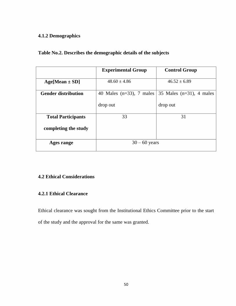
4.1.2 Demographics
Table No.2. Describes the demographic details of the subjects
Experimental Group Control Group
Age[Mean ± SD] 48.60 ± 4.86 46.52 ± 6.89
Gender distribution 40 Males (n=33), 7 males
drop out
35 Males (n=31), 4 males
drop out
Total Participants
completing the study
33
31
Ages range 30 – 60 years
4.2 Ethical Considerations
4.2.1 Ethical Clearance
Ethical clearance was sought from the Institutional Ethics Committee prior to the start
of the study and the approval for the same was granted.
51
4.2.2 Written Informed Consent
Subjects who fulfilled inclusion criteria were appraised about the purpose of
the study and their rights as research subjects. Informed consent form was
administered in English. As all the subjects were almost illiterates, there
was a requirement of translating the signed informed consent form into
native language i.e., Tamil. Adequate time was given to each
patient to go through the information sheet and their queries were answered.
Their right to withdraw anytime from the study and the need for willingness to
participate voluntarily in the study was explained. All the subjects expressed
their willingness to participate in the study by giving a signed informed
consent. After obtaining informed consent, they will be subjected to a battery of Motor
Nerve Conduction test using RMS Aleron Electromyography, (Version 1.1, April
2014) in the Institute of Government Yoga and Naturopathy Medical College.

52
Fig: 5 RMS Aleron Electromyography (Version 1.1, April 2014)
Fig: 6 Screen showing the graph of Motor Nerve Conductivity

53
Fig: 7 Getting Informed Consent from the Hemiplegic Patient
(A sample of information sheet and consent form is enclosed as Annexure)
4.3 Screening of Subjects
4.3.1 Criteria for Diagnosis:
The Motor Nerve Conduction test is to measure the nerve conduction velocity of the
Median nerve and Peroneal nerve. The distance between the recording electrodes is
divided by the difference in the latency between the dorsal and the base response to
calculate conduction velocity.
54
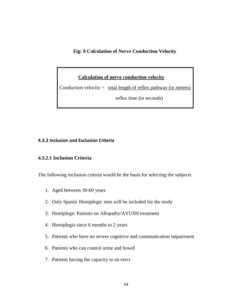
Fig: 8 Calculation of Nerve Conduction Velocity
Calculation of nerve conduction velocity
Conduction velocity = total length of reflex pathway (in metres)
reflex time (in seconds)
4.3.2 Inclusion and Exclusion Criteria
4.3.2.1 Inclusion Criteria
The following inclusion criteria would be the basis for selecting the subjects
1. Aged between 30-60 years
2. Only Spastic Hemiplegic men will be included for the study
3. Hemiplegic Patients on Allopathy/AYUSH treatment
4. Hemiplegia since 6 months to 2 years
5. Patients who have no severe cognitive and communication impairment
6. Patients who can control urine and bowel
7. Patients having the capacity to sit erect
55
4.3.2.2 Exclusion Criteria
Participants will be excluded if they had:
1. Accident history
2. Current alcoholic and smoker
3. Peripheral Neuropathy
4. Facial Palsy
4.4 Design
4.4.1 Type of the design
A Prospective Controlled Trial with control group
4.4.2 Convenient Sampling
The patients who satisfied the Inclusion and Exclusion criteria were
conveniently grouped into Experimental and Control groups as per the
patient availability and period of stay.

56
4.4.3 Allocation of patients into study & control groups
The patients were allocated conveniently to Study group or Wait Control group.
Seventy five subjects were initially screened and assigned to two groups
i.e., Experimental group (n=40) and Wait list control group (n=35)
4.4.4 Data Points
The data collection was done before (day 1), and after (day 56) the intervention.
Figure 9: Illustration of Data Points

57
4.4.5 Trial Profile
The trail profile of the study is presented as Figure 4 which illustrates the
study plan; flow of patients across data points and reasons for the drop out.
Figure 10: Trail Profile

58
4.5 Assessments
The baseline and post-intervention assessments consisted of:
Table 3: List of Primary and Secondary outcome variables
4.5.1.1 Nerve Conduction Velocity
Nerve conduction velocity is an important aspect of studying about the nerve
conduction. It is the speed or velocity at which an electrochemical impulse propagates
down a neural pathway. Some factors like age, sex, and various medical conditions
affect the Conduction velocities.
PRIMARY OUTCOME
VARIABLES
SECONDARY OUTCOME
VARIABLES
Motor Nerve Conductivity of
Median Nerve
Resting Cardio-Vascular
Parameters
Motor Nerve Conductivity of
Deep Peroneal Nerve
BMI

59
The Conduction velocities vary to each individual and depend largely on an axon's
diameter and the degree to which that axon is myelinated, but the majority of 'normal'
individuals fall within defined ranges.[Nerve conduction velocity, National Institutes
of Health, 31 October 2013, Retrieved 13 November 2013]
Nerve impulses are extremely slow compared to the speed of electrical impulses which
are on the order of 50–99% of the speed of light, however, very fast compared to the
speed of blood flow, with some myelinated neurons conducting at speeds up to 120 m/s
(432 km/h or 275 mph).
Normal impulses in peripheral nerves of the legs travel at 40–45 m/s, and 50–65 m/s in
peripheral nerves of the arms (Parry, Gareth, J, 2007). Largely generalized, normal
conduction velocities for any given nerve will be in the range of 50–60 m/s.[Nerve
Conduction Study, Johns Hopkins Medicine, Retrieved 17 November 2013]
4.5.1.1.1 Testing Methods
Nerve Conduction Velocity is just one of many measurements commonly made during
a nerve conduction study (NCS). The purpose of these studies is to determine whether
nerve damage is present and how severe that damage may be.

60
Nerve conduction studies are performed as follows: [Nerve Conduction Study, Johns
Hopkins Medicine, Retrieved 17 November 2013]
The active & passive electrodes are placed on the subject's skin over the nerve to be
tested.
The stimulator passes the electrical impulses which are sent through one electrode
to stimulate the nerve.
The second electrode records the impulse which are then sent through the nerve as
a result of stimulation.
The time difference between the stimulation from the first electrode and pick-up by
the downstream electrode is known as the latency period. Nerve conduction
latencies are typically on the order of milliseconds.
The conduction velocity itself is not directly measured, but by calculating the
conduction velocities from NCS measurements is trivial. The distance between the
stimulating and receiving electrodes is divided by the latency period which gives the
value of nerve conduction velocity.
The functional unit of the muscle contraction is a motor unit, comprised of a single
alpha motor neuron and all the fibers it enervates. This muscle fiber contracts when the
action potentials (impulse) of the motor nerve which supplies it reaches a
depolarization threshold. The depolarization generates an electromagnetic field and the

61
potential is measured as a voltage. The depolarization, which spreads along the
membrane of the muscle, is a muscle action potential (Basmajian, J V. et.al., 1975).
The motor nerve is stimulated atleast at two points, or at the sites along its course. The
pulse is adjusted to record a compound muscle action potential (CAMP). The surface
recording electrodes are commonly used and placed in belly tendon of the muscle;
keeping the active electrode close to the motor point and the reference one close to the
tendon. Ground electrode is placed between stimulating and recording electrodes.
Surface stimulation of healthy nerve with an intensity of 5 – 40mA is given. However,
in a diseased nerve, the nerve excitability is reduced and the current requirement may
be much higher than normal. Filter setting for motor nerve conduction study is 5Hz to
10 kHz and sweep speed 2 to 5ms/division.
For Median motor nerve conduction study, the recording electrode is placed close to
the motor point of abductor pollicis brevis and the reference electrode about 3 Cm
distal from the first metacarpophalangeal joint. A supra maximal stimulation is given at
wrist (3Cm proximal to the distal wrist crease) and at elbow (near the volar crease of
the brachial pulse). The distal latency, nerve conduction velocity of different segments,
and compound muscle action potential amplitudes are measured at different levels
following the stimulation. The normal values of median motor nerve conduction are;
57.7 ± 4.9 m/s in Elbow according to Kimura and 58.52 ± 3.76 m/s in Elbow.

62
For Deep peroneal motor nerve conduction study, the active surface electrode is
placed over the digitorum Brevis, reference surface electrode is placed over the
base of little toe and ground surface electrode is placed over the dorsum aspect of
foot. The Nerve Conduction Velocity of Common peroneal nerve below the knee
segment is between 48.3 ± 3.9 m/s (U K, Misra & J, Kalita, 2006)
Fig: 11 Median Motor Nerve Conduction Test

63
Fig: 12 Peroneal Motor Nerve Conduction Test
4.5.2 Secondary outcome variables
4.5.2.1 Heart rate
The R waves from the electrocardiogram are detected, to obtain a point event
series of successive R-R intervals, from which the beat to beat heart rate series
are computed. The heart rate is obtained based on R-R inter-beat interval
analysis. The heart rate in beats per minute (bpm) was obtained by continuously
counting the QRS complexes in successive 60 s periods.

64
4.5.2.2 Respiratory Rate
Respiration was recorded using a volumetric pressure transducer fixed around
the trunk at the level of the lower costal margin as the subject sit erect. Care is
taken to adjust the strap such that full inhalation is not restricted. Breath rate is
calculated in cycles per minute (cpm) by counting the breath cycles in 60
second epochs, continuously.
4.5.2.3 Blood pressure
Blood pressure was recorded before and after the cold spinal bath by using a
standard mercury sphygmomanometer, auscultating over the right brachial
artery. The systolic pressure was noted as the first clear tapping sound
(korotkoff sounds) and diastolic pressure was noted as the reading at which the
korotkoff sounds appeared muffled.

65
4.6 Intervention
4.6.1 Test Intervention
The patients in the experiment group were trained in the proper practice of
Sukshma Vyayama, Nadi Shodhana Pranayama, AUM Chanting and DRT on first day
and was made to practice for a period of 56 days once daily. (Preferably morning or
evening)
4.6.1.1 Procedure of Test Intervention
4.6.1.1.a. Sukshma Vyayama
Siting in a comfortable pose one should perform the 10 practices of Sukshma
Vyayama slowly without haste and in coordination with the breathing. Regular
practice of pawanmuktasana series-1 removes the so-called energy blockages from
the body and prevents new ones from forming. In this way, it promotes total health,
regulating and stabilizing the flow of energy throughout the body (Swami
Satyananda saraswati ,2002, P- 21&22)

66
4.6.1.1.b Nadi Shodhana Pranayama
Sit in a comfortable posture preferably on the floor over a yoga mat. With the right
hand thumb, close the right nostril and breathe in slowly and fully through the left
nostril, exhale through the right, inhale through the right and exhale through the left.
This is one round. Subjects should perform at the ratio of 1:1. (For example, breathe in
for a count of 5 and breathe out for a count of 5). While practicing, be sure that the
head and body are on the same line and erect. If the right arm becomes tired, support
the elbow in the left palm. Practice ten rounds. (Swami Muktibodhanada, 2008)
4.6.1.1.c. General description about Nada anusaandhana
Here different sounds like ‘A’, ‘U’, ‘M’ and ‘AUM’ are produced loudly so that they
generate a fine vibration or resonance all over the body. This resonance sounds act as
stimulations and the post-resonance silence deepens the awareness and releases even
the very subtle tensions (Dr.H R, Nagendra, Dr.R, Nagarathna, 2011)

67
4.6.1.1.d. General description about Deep Relaxation Technique (DRT)
It is part by part relaxation by directing the attention of mind on different parts of
the body starting from the toes and ending with the head, a feeling of relaxation is
Propagated. Total time duration is 6 minutes. It works at all levels namely;
physical, pranic, mental, emotional, intellectual and spiritual (Dr.H R, Nagendra,
Dr.R Nagarathna, 1986)
4.6.1.1.e. Counseling
It is the act of helping the client to see things more clearly, possibly from a different
view-point which enables one to focus on feelings, experiences or behavior, with a
goal to facilitate positive change with trust. It is the activity of giving mental support,
assistance, advice and guidance, especially on personal problems and difficulties.

68
Table 4: Intervention Chart
S.NO YOGIC PRACTICE DURATION
1. Sukshma Vyayama 1. Padanguli Naman (toe stretch)
2. Goolf Naman (ankle bending)
3. Goolf Chakra (ankle rotation)
4. Janu Nanam (Knee Stretch)
5. Mushtika Bandana (Finger Stretch)
6. Mani Bandha Nanam (wrist bending)
7. Mani Bandha Chakra (Wrist Rotation)
8. Kehuni Nanam (elbow Stretch)
9. Skandha Chakra (Shoulder Rotation)
10. GreevaSanchalana (Neck movement)
10 rounds of each practice for 3 mins. (30 minutes totally)
2. Pranayama Nadishodhana Pranayama 15 rounds (5 mins)
3. Nada anusaadhan
‘A’, ‘U’, ‘M’ & ‘AUM’ chanting 10 chantings of each mantra (5 mins totally)
4. Relaxation Technique Deep Relaxation Technique 10 to 20 mins
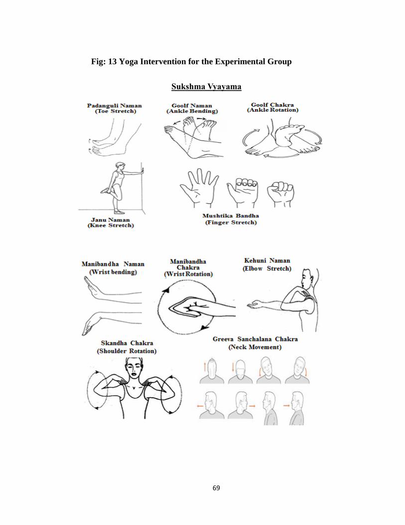
69
Fig: 13 Yoga Intervention for the Experimental Group
Sukshma Vyayama

70

71
4.6.2 Control Intervention
The control group were under other AYUSH treatments except Yoga for 56 days. They
were also offered to be trained in the experiment intervention if interested after the
study.
4.7 Data extraction & analysis
4.7.1 Data Extraction
The data was collected as self-reported observations using primary outcomes
and secondary outcome variables. The assessments were done on the first day
(baseline data) and end of 56th day (post data). The data was organized in Microsoft
Excel Sheets (Version 2010).
4.6.2 Data Analysis
Data expressed as Mean and SD. Inter group and intra group comparison of mean was
done by paired and unpaired t test using R statistical software version 3.1.1.
72
RESULTS
AND
OBSERVATIONS
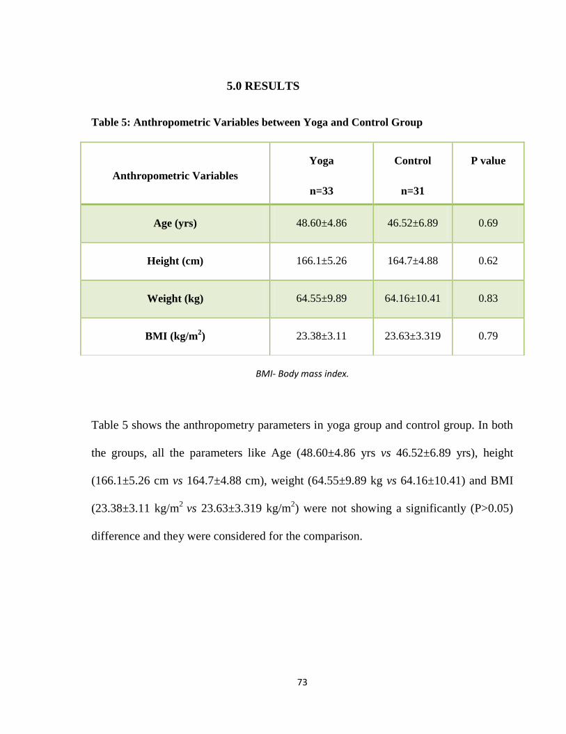
73
5.0 RESULTS
Table 5: Anthropometric Variables between Yoga and Control Group
BMI- Body mass index.
Table 5 shows the anthropometry parameters in yoga group and control group. In both
the groups, all the parameters like Age (48.60±4.86 yrs vs 46.52±6.89 yrs), height
(166.1±5.26 cm vs 164.7±4.88 cm), weight (64.55±9.89 kg vs 64.16±10.41) and BMI
(23.38±3.11 kg/m2
vs 23.63±3.319 kg/m2) were not showing a significantly (P>0.05)
difference and they were considered for the comparison.
Anthropometric Variables
Yoga
n=33
Control
n=31
P value
Age (yrs) 48.60±4.86 46.52±6.89 0.69
Height (cm) 166.1±5.26 164.7±4.88 0.62
Weight (kg) 64.55±9.89 64.16±10.41 0.83
BMI (kg/m2) 23.38±3.11 23.63±3.319 0.79

74
Table 6: Resting Cardiovascular Parameters before and after between Yoga and Control
Group
Cardiovascular
parameters
Yoga
P value
Control
P value
Before After Before After
HR (bpm) 78.0±2.23 78.06±2.09 0.9 78.52±3.86 76.39±3.32 0.03
SBP (mmHg) 122.1±14.53 117.6±10.23 0.01 125.8±15.23 125.2±12.88 0.69
DBP (mmHg) 78.79± 9.27 77.58±7.91 0.42 80.32±11.10 79.68±6.57 0.67
PP (mmHg) 43.33±10.21 40.0±7.07 0.12 45.48±10.68 45.89±8.50 0.98
MAP (mmHg) 93.23±10.22 90.91±8.13 0.07 95.48±11.60 94.84±8.24 0.57
RPP (bpm-mmHg) 9525±1160 9178±835 0.03 9890±1377 95554±1025 0.05
DoP (bpm-mmHg) 5403±1832 4748±1137 0.04 5829±1912 5787±1568 0.87
Data Expressed Mean±SD. In depended and paired t.test was used to compare the intergroup and intra group differences. SBP, Systolic blood pressure; DBP, diastolic blood pressure; HR, heart rate; PP, pulse pressure; MAP, mean arterial pressure; RPP, rate pressure product; DoP, double product.
There was no significant difference found in control group patients. After 56 days of
intervention, Yoga group patients showed a significant (P<0.05) reduction in the
resting cardiovascular parameters such as SBP from 122.1±14.53 to 117.6±10.23
mmHg, RPP from 9525±1160 to 9178±835 bpm-mmHg and DoP from 5403±1832 to
4748±1137 bpm mmHg. It clearly showed they were shifted to the status of
parasympathetic domination.

75
Table 7: Motor Nerve Conductivity before and after between Yoga and Control Group
Nerve Conduction
velocity (NVC)
Yoga (n=31)
P
value
Control (n=33)
P
value
Before After Before After
Median nerve (msec) 55.63±5.05 53.09±5.61* 0.02 50.64±16.13 50.75±9.18 0.97
Peroneal nerve(msec) 45.52±7.89 41.59±5.47** 0.05 48.26±5.57 46.41±5.34 0.14
* compared with after yoga and control. * P<0.05, ** P<0.01, *** P<0.001.
Yoga group patients showed a significant (P<0.05) shortening of nerve conduction
velocity after 56 days of yoga intervention compared to control group patients
(P>0.05). In yoga group, median nerve conductivity was significantly reduced from
55.63±5.05 ms to 53.09±5.61 ms reflecting a positive improvement. Peroneal nerve
conductivity also shortened from 45.52±7.89 to 41.59±5.47 msec in yoga group
patients after yoga intervention but there was no significant changes found in control
group patients.
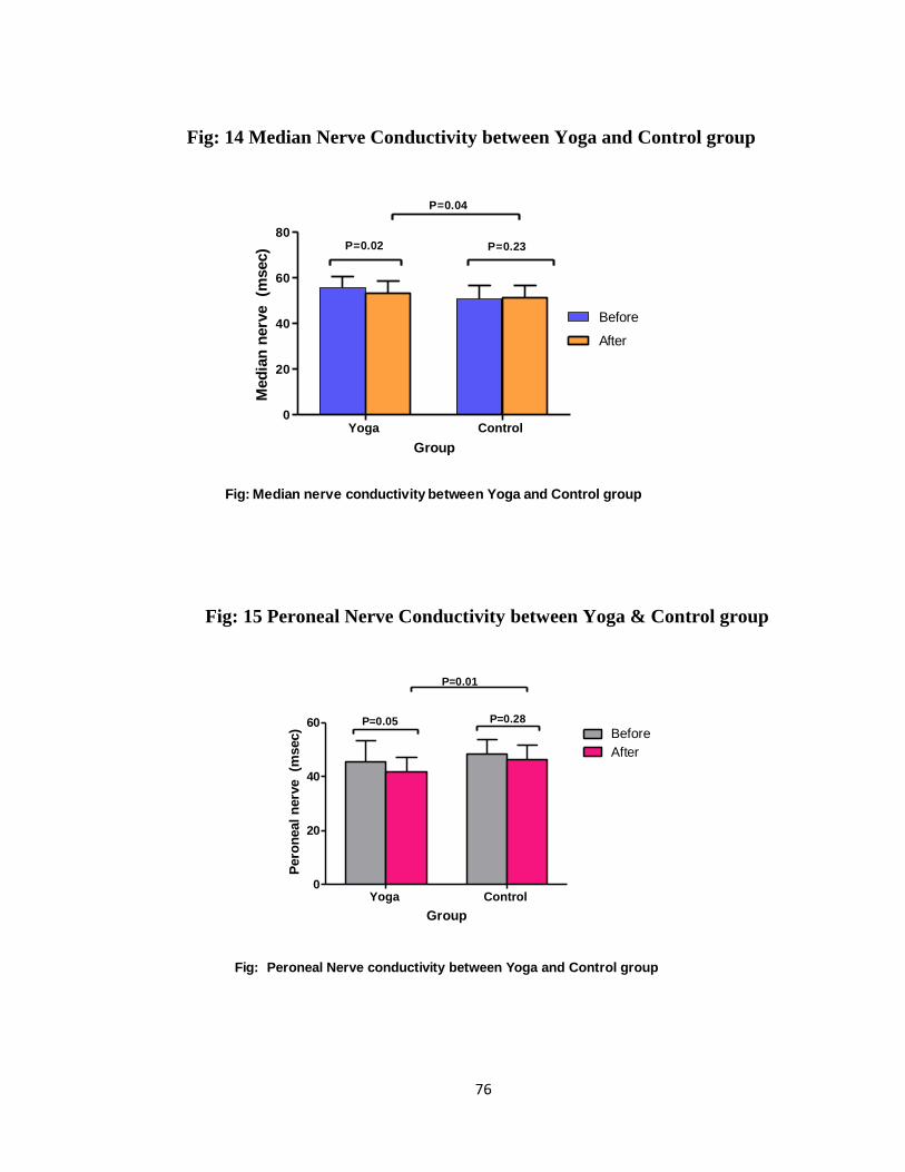
76
Fig: 14 Median Nerve Conductivity between Yoga and Control group
Fig: Median nerve conductivity between Yoga and Control group
Yoga Control 0
20
40
60
80
Before
After
P=0.02 P=0.23
P=0.04
Group
Med
ian
nerv
e
(msec)
Fig: 15 Peroneal Nerve Conductivity between Yoga & Control group
Fig: Peroneal Nerve conductivity between Yoga and Control group
Yoga Control 0
20
40
60Before
After
P=0.05 P=0.28
P=0.01
Group
Pero
neal
nerv
e
(msec)
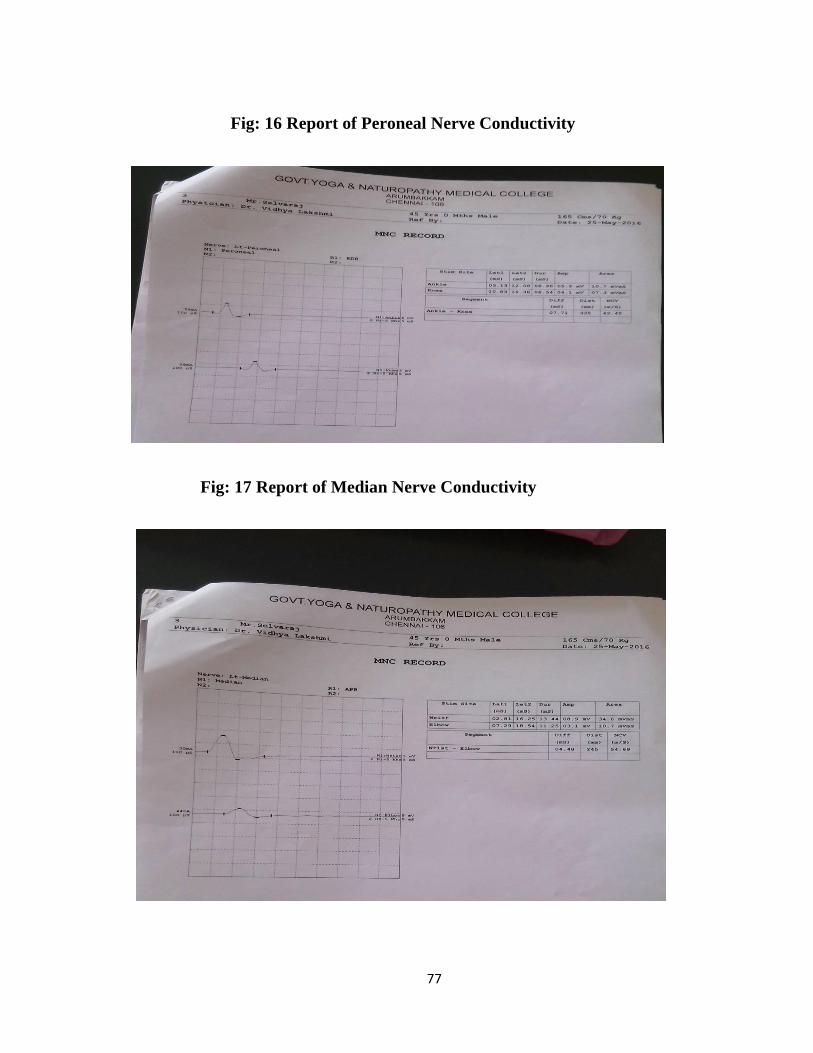
77
Fig: 16 Report of Peroneal Nerve Conductivity
Fig: 17 Report of Median Nerve Conductivity

78
DISCUSSION

79
6.0 DISCUSSION
This Prospective Controlled study was designed to look for differences in the Motor
Nerve Conductivity of Median nerve and Peroneal nerve and Cardio-respiratory
parameters between Spastic Hemiplegic Men in Government Yoga and Naturopathy
Hospital, Chennai. The Result of this study revealed that a combined practice of
Sukshma Vyayama, Nadishodhana pranayama, AUM chanting and DRT improved the
Motor nerve conductivity and Cardio-respiratory parameters in the experimental group
significantly in comparison to that of the control group. In this study there is a
significant difference (p<0.05) within the experimental group in the MNC of Median
Nerve (p<0.02) and Peroneal Nerve (p<0.05) at the end of the 56th day in comparison
to their baseline data. Yoga group patients showed a significant (P<0.05) reduction in
the resting cardiovascular parameters such as SBP, RPP and DoP. It clearly showed
they were shifted to the status of parasympathetic domination.
Similar results were found in another study conducted in the Management of Chronic
Hemiplegia patients through Clinical Yoga techniques which revealed improvement in
memory recall, proprioception, motor function in both limbs and in expression in
activity of Daily Living. Clinical Yoga helps in opening up some of the motor areas
which were idle due to lack of stimulation. (Dr K V, Dilipkumar & Dr P K,
Narayana Sarma, 2000)

80
Electrophysiological investigations done in hemiplegic patients were well documented
in many reports (Meredith J et.al, 1981), (Ring H et.al, 1993), (Chino N, 1981),
(Ring H et.al, 1993)
Milanov, I, (1995) who evaluated the motor conductivity in median, ulnar, peroneal
and tibial nerves found that the mean wave amplitudes were significantly decreased for
each nerve study, in both the upper and lower limbs of the paralyzed limbs, when
compared to the healthy side. In contrast, the mean motor conduction velocities were
not reduced in the unaffected limbs. In our study, both the upper limb and lower limb
showed improvement after the intervention. Wasting of muscles in patients after
lesions of the upper motor neuron, can be a major cause for reduced conduction
velocity (Takebe, K. et.al., 1975)
Mc Comas, A J. et.al., (1971) described a possible mechanism for muscle atrophy
following upper motor neuron lesions. We believe that a decreased diameter of the
nerve fiber as a result or cause of muscle atrophy, could lead to a decreased nerve
conduction velocity in the hemiplegic patients.

81
Basal HR is mainly determined by parasympathetic nervous system (PNS) and DBP is
depends on the status of peripheral vascular resistance (PVR) which is mainly altered
by sympathetic nervous system (SNS). MAP refers to mean pressure throughout the
cardiac cycle and it is determined by both SNS and PNS. Decrease DBP, and MAP
represents increase in parasympathetic and decrease in sympathetic activity in yoga
group.
RPP and DoP represent sympathetic activity and they are indirect measures of O2
consumption and work load on heart. Rise in PP with decreased RPP indicates that the
tissue perfusion is increased with decreased work load on heart in yoga group
hemiplegic patients.
Takebe et.al., (1975) did a study on 27 hemiplegic patients and noted the significant
slowing of peroneal nerve conduction velocities in the affected extremities. A
significant difference in skin temperature between the extremities of the two sides was
also noted at the same time. They found that muscle atrophy due to a decreased
diameter of the nerve fiber could lead to decreased nerve conduction velocity.
Chokroverty, S and Medina, J, (1978) measured bilaterally the motor nerve
conduction velocities of the CPN in 44 hemiplegic patients and found a statistically
significant difference between the two limbs. In 63% of the patients skin temperature
was reduced in the hemiplegic limbs.
82
We shall adopt the hypothesis that continuous inversion position, as in hypotonic drop
foot, may affect the electro-physiological properties of the nerve. It most probably
results from nerve traction and compression at the level of the peroneal neck, causing
demyelinization and even axonopathy. Myelin loss results in slowing of nerve
conduction through the area involved. When compression is severe, ischemic changes
occur and cause secondary axonal damage, expressed by reduction of CMAP
amplitude. Nonetheless, we must take into consideration that the slowing of conduction
velocities of the CPN in the hemiplegic limbs may be related also to the lowering of
the skin temperature and nerve axonal compression in the same limbs.
A study conducted on the experience of stroke which can have a negative impact on
both psychological and physical health and on quality of life showed that Yoga and
relevant practices were promising therapies that have been used with patients with a
variety of conditions. Yoga and mindfulness are the self- administered intervention
options for stroke rehabilitation and proven to be clinically valuable.
(Lazaridou, A. et.al., 2013)
The practice of yoga and meditation demonstrates statistically encouraging physical
function, physiological and psychological improvements in the neurological disorders
and thus yoga is an integrative, alternative, and complementary therapy (Mishra, S
K. et.al., 2012)

83
Another study conducted by (Mishra, S K. et.al., 2012) confirmed that Yoga resulted
in significant improvements in exercise capacity and a mean improvement in HRQL
(Health Related Quality of Life) in Chronic diseases and which may be an useful
adjunct to formal rehabilitation programs
Many people who have had a stroke reported a reduced level of activity resulting in an
impaired health status. Yoga offers a gentle alternative exercise program which can be
easily practiced by people who have had a stroke. The effects of a yoga-based exercise
program showed growing evidence with improvements in chronic post stroke
hemiparesis people with mobility limitations (Julie, V, Bastille and Kathleen, M,
Gill-Body, 2016)
A therapeutic yoga intervention following stroke may improve multiple aspects of
physical functioning. Thus, Yoga intervention is complementary to the traditional
rehabilitation (Schmid, A A. et.al., 2014)
Overall, the practice of IAYT for 56 days among spastic hemiplegic men has resulted
in the improvement of motor function in both the limbs. It improved their quality of
living and they gained confidence. Thus Yoga practices helped to activate the
autonomic changes.

84
CONCLUSION

85
7.0 CONCLUSION
This study showed that 56 days of IAYT improves the Motor Nerve Conductivity of
both the Median nerve and Peroneal nerve in hemiplegic patients. This revealed that
yoga practice has significant role in improvement in the neuronal activity which is
altered in spastic paralysis patients. Decreases Systolic Blood Pressure, Rate Pressure
Product & Double Product showed that increase in the dominance of parasympathetic
activity in spastic hemiplegic men. However, the practice of IAYT on Body
Composition and Heart Rate has not been much appreciable in these subjects. Further
research need in this field with a larger sample size and duration is warranted to reveal
accurate changes in this field.

86
SUMMARY

87
8.0 SUMMARY
The aim of this study is to establish the effect of yoga on the motor nerve conductivity
in the spastic hemiplegic men.
The Autonomic Nervous System has been found to be an important factor in regulating
the maintenance of the constant energy storage and utilization. Hemiplegics were
found to be stressed, depressed, with insecured feeling and dependency on others for
their daily activities causing disturbance in the Autonomic functioning.
Yoga is frequently used as a lifestyle intervention to reduce stress, restore autonomic
nervous system balance and other therapeutic purposes. This study has used the
integrated approach of yoga therapy as a tool to assess if it had any influence on the
autonomic activity in spastic Hemiplegic men.
The disease was found predominant among the non-vegetarians, alcoholics, smokers,
hypertensive and hyperlipidemic subjects. It was also found commonly in people of
low socio-economic status due to their unhealthy lifestyle and stressful job.
In this study, the yogic intervention was given for 6 days a week for 56days, once daily
for an hour. 75 hemiplegics were examined clearly by modern aspects and their age
group was between 30-60 years who were trained with the yoga practices. The
practices included, 30 mins of Sukshma vyayama, 5 mins of Nadishodhana pranayama,
5 mins of AUM chanting and 10-20 mins of Deep Relaxation Technique. The yoga
88
group had 40 subjects while 35 were in the control group who did not undergo the
Yogic intervention for those 56days.
The 75 subjects were evaluated for baseline and post-interventional using variables like
Motor Nerve Conductivity of Median and Peroneal nerves, Cardio-respiratory
parameters and BMI.
Statistical Analysis of the data collected was done by the method of unpaired ‘t’ test.
Following the analysis of the data, it was shown that there were significant changes in
both the Median and Peroneal Motor nerve conduction and reductions in the Systolic
Blood Pressure (SBP), Rate Pressure Product (RPP) and Double Product (DoP). No
significant changes observed in the Heart rate (HR), Diastolic Blood Pressure (DBP)
and Pulse Pressure (PP)
Overall, this study has shown that an integrated practice of Yoga for 56 days promotes
the sympathovagal balance or activity.
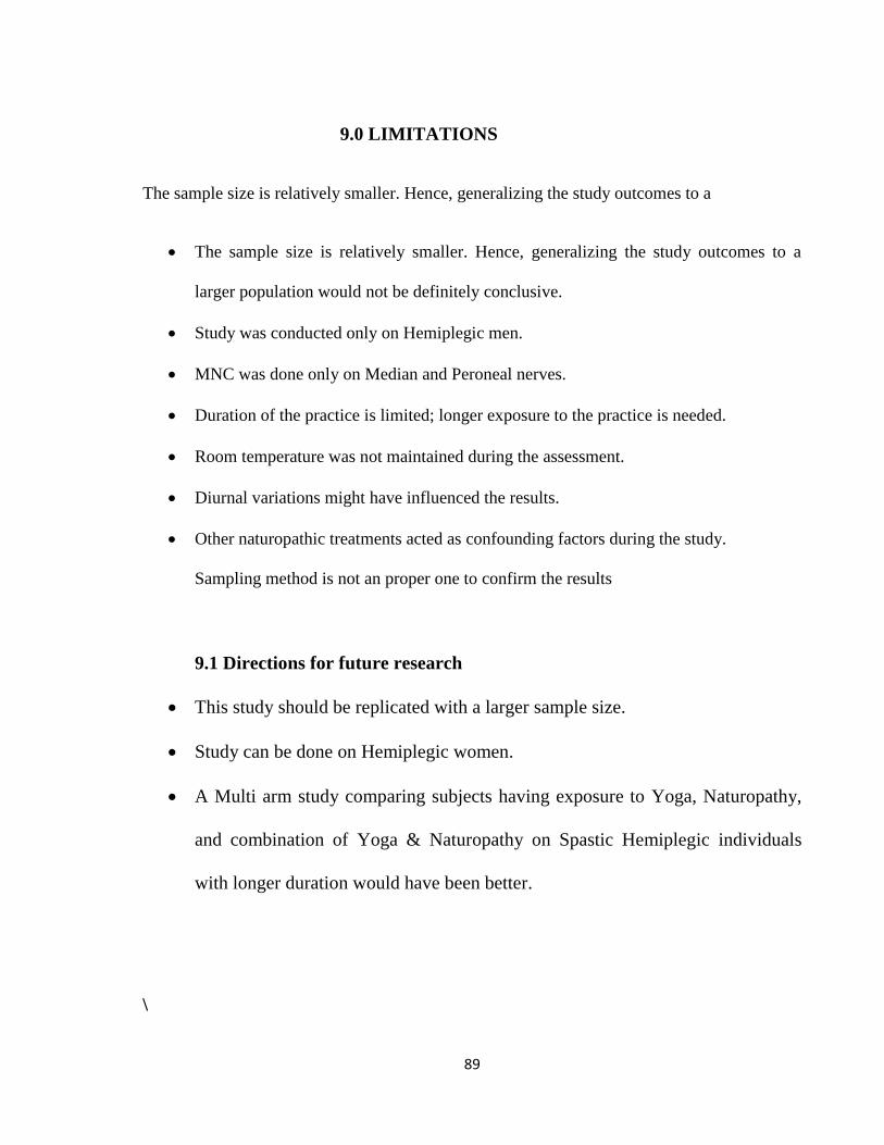
89
9.0 LIMITATIONS
The sample size is relatively smaller. Hence, generalizing the study outcomes to a
The sample size is relatively smaller. Hence, generalizing the study outcomes to a
larger population would not be definitely conclusive.
Study was conducted only on Hemiplegic men.
MNC was done only on Median and Peroneal nerves.
Duration of the practice is limited; longer exposure to the practice is needed.
Room temperature was not maintained during the assessment.
Diurnal variations might have influenced the results.
Other naturopathic treatments acted as confounding factors during the study.
Sampling method is not an proper one to confirm the results
9.1 Directions for future research
This study should be replicated with a larger sample size.
Study can be done on Hemiplegic women.
A Multi arm study comparing subjects having exposure to Yoga, Naturopathy,
and combination of Yoga & Naturopathy on Spastic Hemiplegic individuals
with longer duration would have been better.
\

90
10.0 BIBLIOGRAPHIC REFERENCES
Abhishek Kumar and Anjali Prasad 2016, ‘Nerve conduction velocity in median nerve
and tibial nerve of healthy adult population with respect to gender’, National Journal of
Physiology, Pharmacy and Pharmacology, April 17, 2016.
A Jr Stancak and M Kuna 1994, ‘EEG Changes during Forced Alternate Nostril
Breathing’, Int-JPsychophysiol, Oct 1994, 18(1): 75-9
A Mooventhan and Vitthal Khode 2014, ‘Effect of Bhramari pranayama and OM
chanting on pulmonary function in healthy individuals: A prospective randomized
control trial’, Int J Yoga. 2014 Jul-Dec;7(2):104–110
Ades, P A, Savage, P D, Cress, M E, et al.2003, ‘Resistance training on physical
performance in disabled older female cardiac patients’, Med Sci Sports Exerc. 35
(2003) 1265-1270
Ajay Anil Gurjar, Siddharth, A, Ladhake 2016, ‘Analysis and Dissection of Sanskrit
Divine Sound “OM” Using Digital Signal Processing to Study the Science Behind
“OM” Chanting’, 2016 7th International Conference on Intelligent Systems, Modelling
and Simulation
Arati Amin, Sai Sailesh Kumar, Archana Rajagopalan, Supriya Rajan, Soumya Mishra,
Udaya Kumar Reddy, Joseph Kurien Mukkadan P K2016, ‘Beneficial effects of OM
chanting on depression, anxiety, stress and cognition in elderly women with
hypertension’, Indian Journal of Clinical Anatomy and Physiology, July-September
2016;3(3);253-255
Arch Neurol 1978;35:360–3
Asbury, A K 1954, ‘Approach to the patient with peripheral neuropathy’, Harrison's
Principles of Internal Medicine. Part 15: Neurologic Disorders. Nerve and Muscle
Disorders. Electrodiagnosis. Ch. 363. Sec. 3. Blakiston, New York: 1954.
Barnes, V A, Treiber, F A, Davis, H 2001, ‘Impact of Transcendental Meditation on
cardiovascular function at rest and during acute stress in adolescents with high normal
blood pressure’, J Psychosom Res. 2001;51:597–605 [PubMed]
Basmajian, J V, Gregory, S, Rash 2006, ‘Ed Computers in Electromyography’,
Butterworths, 1975.Electromyography Fundamentals
Bastille JV, Gill-Body KM. A Yoga-Based Exercise Program for People with Chronic
Poststroke Hemiparesis. Phys Ther. 2004;84:33–48. [PubMed]

91
Berk, B 2001, ‘Yoga for moms, Building core stability before, during and after
pregnancy’, Midwifery Today Int Midwife. 59 (2001)27-29
Bernardi, L, Sleight, P, Bandinelli, G, Cencetti, S, Fattorini, L, WdowczycSzulc, J, et
al. 2001, ‘Effect of rosary prayer and yoga mantras on autonomic cardiovascular
rhythms: Comparative study’, Br Med J. 2001;323:22–9)
Bower, J E, Greendalee, G, Crosswell, A D, Garet, D, Sternlieb, B, Ganz, P A, et
al.2014, ‘Yoga reduces inflammatory signaling in fatigued breast cancer survivors: a
randomized controlled trial’, Psychoneuroendocrinology 2014;43:20–9. [PUBMED:
24703167
Brashear, Allison 2010, ‘Spasticity: Diagnosis and Management’. New York: Demos
Medical]
Buschbacher, R M 1999, ‘Median nerve motor conduction to the abductor pollicis
brevis’, Am J Phys Med Rehabil. 1999; 78(6 Suppl):S1–8. [PubMed]
C F, et al.2010, ‘Stress, inflammation, and yoga practice’, Psychosom Med. 2010
Cerebral palsy at eMedicine
Cerebral Palsy~clinical at eMedicine
Chan, W, Immink M A, Hillier, S 2012, ‘Yoga and exercise for symptoms of
depression and anxiety in people with post stroke disability: a randomized, controlled
pilot trial’, Altern Ther Health Med 2012 MayJun;18 (3):3443
Chinmayanada Swami 2005, ‘Katha Upanisad’, Mumbai: Central Chinmayananda
Mission; 2002.
Chinmayananda Swami 1984, ‘Chinmayananda Swami Mandukya Upanishad’,
Mumbai: Sachin publishers; 1984
Chino, N 1981, ‘Electrophysiological investigation on shoulder subluxation
Chokroverty, S, Medina, J, 1978, ‘Electrophysiological study of hemiplegia. Motor
nerve conduction velocity, brachial plexus latency, and electromyography’ 1978,
Archives of Neurology, The reference range for median nerve (motor) velocity, distal
latency, and amplitude were 49.48–66.92, 1.95–4.52, and 4.3–11.3
Cochran, W G 1977, ‘Sampling techniques’, 3rd ed. New York: John Wiley & Sons;
1977
conduction velocity in hemiplegia: possible factors’, Arch Phys Med Rehabil 1975,
56:285-289
92
Correa Pérez, M, Sosa, A, López Acevedo, C E 1986, ‘Nerve conduction velocities:
Normal values for median and ulnar nerves’, Bol Asoc Med PR. 1986;78:191–
6. [PubMed]
Courtenay, W H 2001, ‘Counseling men in medical settings: The six-point HEALTH
plan’ In G. R. Brooks & G. E. Good (Eds.), ‘The new handbook of psychotherapy and
counseling with men’ pp. 59-91, San Francisco, CA: Jossey-Bass
Cramer, H, Lauche, R, Klose, P, Lange, S, Langhorst, J, Dobos, G J 2013, ‘Yoga for
women diagnosed with breast cancer’, Cochrane Database of Systematic
Reviews 2013, Issue 10. [DOI: 10.1002/14651858.CD010802]
D Shannahoff - Khalsa and B Kennedy 1993, ‘The Effects of Unilateral Forced Nostril
Breathing on the Heart’, Int-J-Neurosci., Nov 1993, 73(1-2): 47-60
Dandekar Pradnya Deepak , dept of Kriya Sharir, Mahatma Gandhi Ayurved College
Hospital and Research Centre, Maharashtra, India 2013, ‘Impact of Short Term
Training of Anulom Vilom Pranayama on Blood Pressure and Pulse Rate in Healthy
Volunteers’, Int. J. Res. Ayurveda Pharm. 4(2), Mar-Apr 2013
Dash, M, Telles, S 2001, ‘Improvement in hand grip strength in normal volunteers and
rheumatoid arthritis patients following yoga training’, Indian J Physiol Pharmacol. 45
(2001) 355-360
Desharnais, R, Jobin, J, Cote, C, Levesque L, Godin, G 1993, ‘Aerobic exercise and
the placebo effect: A controlled study’, Psychosom Med. 1993;55:149–54. [PubMed]]
DiBenedetto, M, Innes, K E, Taylor, A G, Rodeheaver, P F, Boxer, J A, Wright, H J, et
al.2005, ‘Effect of a gentle Iyengar yoga program on gait in the elderly: an exploratory
study’, Archives of Physical Medicine and Rehabilitation 2005;86(9):1830–7.
[PUBMED: 16181950]
Dostalek, C, Faber, J, Krasa, H, et al. 1979, ‘Yoga meditation effect on the EEG and
EMG activity’, Act Nerv Super (Praha). 21(1) (1979) 41
Dr K V, Dilipkumar M D (Ay) and Dr P K, Narayana Sarma M D (Ay) 2000, ‘A study
on Clinical Yoga techniques in the Management of Chronic Hemiplegia patients’,
International Conference on Yoga, 2000
Dr. Kanchan Joshi, Department of Yoga, H.N.B. Govt. P.g. College, Khatima,
Uttarakhanda E mail: [email protected], 2012, ‘Effect of Nadishodhan
Prayayama and Om chanting on Memory Enhancement of College Students’,
International Journal of Yoga and Allied Sciences (ISSN: 2278 – 5159) Volume: 1,
Issue: 1

93
Dr. Zainab, M M, Chatriwala1 , Dr. Mahendra Shende, Post Graduate Student,
Associate Professor, Department of Neurosciences Physiotherapy, Dr. A P J Abdul
Kalam College of Physiotherapy, PIMS, Loni, Maharashtra, India, 2016, ‘Nerve
Conduction Velocity of Common Peroneal Nerve in Post Stroke Patients”-An
Observational Study’, IOSR Journal of Sports and Physical Education (IOSR-JSPE) e-
ISSN: 2347-6737, p-ISSN: 2347-6745, Volume 3, Issue 2 (Mar. – Apr. 2016), PP 50-
55
Dr.H R,Nagendra, Dr.R, Nagarathna 2011 , ‘Integrated Approach Of Yoga Therapy for
Positive Health’ , Published by-Swami Vivekananda Yoga Prakashana, ISBN 978-81-
87313-08-3, Page – 168, 2011
Dr.H R.Nagendra, Dr.R Nagarathna 1986, ‘New Perspective in Stress Management’,
Published by - Swami Vivekananda Yoga Prakashana, , ISBN 978-81-87313-01-4,
Page – 63
Dr.H,R.Nagendra, Dr.R,Nagarathna 1986,’New Perspective in Stress Management’,
Published by - Swami Vivekananda Yoga Prakashana, , ISBN 978-81-87313-01-4,
Page – 63, 1986
dysfunction in patients with hemiplegic atrophy’, Nat New Biol 1971, 233:21-23
el- Abd, M A, Ibrahim, I K 1994, ‘Impaired afferent control in patients with spastic
hemiplegia at different recovery stages: contribution to gait disorder’, Arch Phys Med
Rehabil. 75 (3): 312–7. PMID 8129585. doi:10.1016/0003-9993(94)90035-3
Eliasson, A C, Ekholm, C, Carlstedt, T 1998, ‘Hand function in children with cerebral
palsy after upper-limb tendon transfer and muscle release’, Dev Med Child
Neurol (1998) 40:612–21.10.1111/j.1469-8749.1998.tb15427.x [PubMed]
(EMG) follow-up of hemiplegic's shoulder’, Eur J Phys Med Rehabil 1993, 3:137-140.
Falco, F J, Hennessey, W J, Braddom, R L, Goldberg, G 1992, ‘Standardized nerve
conduction studies in the upper limb of the healthy elderly’, Am J Phys Med
Rehabil. 1992;71:263–71. [PubMed]
Falcone, G J, Malik, R, Dichgans, M, Rosand, J 2014, ‘Current concepts and clinical
applications of stroke genetics’, Lancet Neurology 2014, 13(4):405-18 [PUBMED:
24646874]
Feb;72(2):113–21
Feeney, D M, Weisend, M P, Kline, AE 1993, ‘Noradrenergic pharmacotherapy,
intracerebral infusion and adrenal transplantation promote functional recovery after
cortical damage’, J Neural Transplant Plast, 1993 Jul-Sep;4(3):199-213..
PMID:8018752 PMCID: PMC2565261 DOI:10.1155/NP.1993.199
94
Feigin, V L, Lawes, C M, Bennett, D A, Anderson, C S 2003, ‘Stroke epidemiology: a
review of population-based studies of incidence, prevalence, and case-fatality in the
late 20th century’, Lancet Neurology 2003, 2:43–53 [PUBMED: 12849300]
Feuerstein, G 2000, ‘Toward a definition of yoga therapy’, International Journal of
Yoga Therapy, 2000, pp. 10:5–10
Fisher, M A 2002, ‘H reflexes and F waves. Fundamentals, normal and abnormal
patterns’, Neurol Clin. 2002;20:339–60 [PubMed]
Fuglsang-Frederiksen, A, Pugdahl, K 2011, ‘Current status on electrodiagnostic
standards and guidelines in neuromuscular
disorders’, Clinneurophys. 2011;122:440455. [PubMed]
Garfinkel, M S, Singhal, A, Katz, W A, Allan, D A, Reshetar, R, Schumacher, H R
1998, ‘Yoga-Based Intervention for Carpal Tunnel Syndrome: A Randomized
Trial’, JAMA. 1998;280:1601–3. [PubMed]
Garrett, R, Immink, M A, Hillier, S 2011, 'Becoming connected: the lived experience
of yoga participation after stroke’, Disability and Rehabilitation 2011;33(25-26):2404–
15. [PUBMED: 19623830]
Ghanshyam Singh Thakur, Nagendra, H R & Nagarathna, R 2009, ‘Effect of deep
relaxation technique on the capacity to influence REG – a randomized control trial’,
Swami Vivekananda Yoga Research Foundation, Indian Journal of Traditional
Knowledge. Vol.8 (3), July 2009,pp 459-463
Ghoncheh, S, Smith, J C 2004, ‘Progressive muscle relaxation, yoga stretching, and
ABC relaxation theory’, J Clin Psychol.60 (2004) 131-136
Gopal, A, Mondal, S, Gandhi, A, Arora, S, Bhattacharjee, J 2011, ‘Effect of integrated
yoga practices on immune responses in examination stress – A preliminary study’, Int J
Yoga. 2011; 4:26-32 [PMC free article] [PubMed]
Goyeche, J R M 1979, ‘Yoga as therapy in psychosomatic medicine’, Psychotherapy
and Psychosomatics, 31(1–4): pp. 373–381 PubMed
Hackett, M L, Köhler S, O'Brien, J T, Mead, GE 2014, ‘ Neuropsychiatric outcomes of
stroke’, Lancet Neurology 2014, 13(5):525-34. [PUBMED: 24685278]
Hartley, L, Dyakova, M, Holmes, J, Clarke, A, Rees, K 2012, ‘Yoga for the primary
prevention of cardiovascular disease’, Cochrane Database of Systematic
Reviews 2012, Issue 9. [DOI: 10.1002/14651858.CD010072]

95
Hatano, S 1976, ‘Experience from a multicentre stroke register: a preliminary
report’, Bulletin of the World Health Organization, 1976, 54:541–53 [PUBMED:
1088404]
Hennessey, W J, Falco, F J, Braddom, R L 1994, ‘Median and ulnar nerve conduction
studies: Normative data for young adults’, Arch Phys Med Rehabil. 1994;75:259–
64. [PubMed]
Hennessey, W J, Falco, F J, Goldberg, G, Braddom, R L 1994, ‘Gender and arm
length: Influence on nerve conduction parameters in the upper limb’, Arch Phys Med
Rehabil. 1994;75:265–9. [PubMed]
Huang CR, Chang WN, Chang HW, Tsai NW, Lu CH. Effects of age, gender, height,
and weight on late responses and nerve conduction study parameters. Acta Neurol
Taiwan. 2009;18(9):242-9.
Immink, M A , Hillier, S , Petkov, J 2014, ‘Randomized controlled trial of yoga for
chronic poststroke hemiparesis: motor function, mental health, and quality of life
outcomes’, 2014 May Jun;21(3):25671.doi: 10.1310/tsr2103256
in hemiplegics’, Scand J Rehabil Med 1981, 13(1):17-21
Innes, K E, Vincent, H K, Taylor, A G 2007, ‘Chronic stress and insulin resistance
related indices of cardiovascular disease risk, part 2: a potential role for mind body
therapies’, Altern Ther Health Med. 2007;13(5):44–51
Ivan, G, Milanov 1995, Medical University of Sofi, ‘Neurographic studies in
hemiplegic patients’, March 1995 Pubmed)
Ives, J C, Sosnoff, J 2000, ‘Beyond the mind-body exercise hype’, Phys Sportsmed.
2000 Mar;28(3):67–81
Jobst, B C 2010, ‘Electrical stimulation in epilepsy: Vagus nerve and brain
stimulation’, Curr Treat Options Neurol 2010;12:443-53
Johnstone, B R, Richardson, P W, Coombs, C J, Duncan, J A 2003, ‘Functional and
cosmetic outcome of surgery for cerebral palsy in the upper limb’ Hand
Clin (2003) 19:679–86.10.1016/S0749-0712(03)00031-[PubMed]
Juha T Korpelainen, MD, Kyosti A Sotaniemi, MD, and Vilho V Myllyla, MD, 2017,
‘Hyperhidrosis as a Reflection of Autonomic Failure in Patients With Acute
Hemispheral Brain Infarction An Evaporimetric Study’, stroke.ahajournals.org,juli
2,2017

96
Julie V Bastill and Kathleen M Gill-Body 2004, ‘A Yoga-Based Exercise Program for
People With Chronic Poststroke Hemiparesis’, February 2004,DOI:
10.1093/ptj/84.1.33 · Source: PubMed
Julie V Bastille and Kathleen M Gill-Body 2016, ‘A Yoga Based Exercise Program for
People With Chronic Poststroke Hemiparesis’,1/6/2016 [Pubmed]
K, Takebe, M G, Narayan, Carl kukulka, J V, Basmajian 1975, ‘Slowing of nerve
conduction velocity in hemiplegia: possible factors’, August 1975 [Pubmed]
Kalyani, B G, Venkatasubramanian, G, Arasappa, R, Rao, N P, Kalmady, S V, Behere,
R V, Rao, H, Vasudev, M K, Gangadhar, BN 2011, ‘Neurohemodynamic correlates of
‘OM’ chanting: A pilot functional magnetic resonance imaging study’, Int J Yoga.
2011;4:3-6
Katirji, B 2002, ‘The clinical electromyography examination. An overview’, Neurol
Clin. 2002;20:291–303[PubMed]
Kearney, P M, Whelton, M, Reynolds, K, Muntner, P, Whelton, P K, He J 2004,
‘Global burden of hypertension: analysis of worldwide data’, Lancet. 2005,365:217-
223, Swedish Council on Technology Assessment in Health Care, Moderately Elevated
BloodPressure. 2013, [http:// www.sbu.se/upload/Publikationer/Content0/1/Blodtryck_
2004/Hypertoni_vol2.pdf] (Published October 2004
Khasky, A D, Smith, J C 1999, ‘Stress, relaxation states, and creativity’, Percept Mot
Skills. 88 (1999) 409-416
Kiecolt-Glaser, J K, Christian, L, Preston, H, Houts, C R, Malarkey, W B, Emery,
Koji shigeno 1972, ‘Hemiplegic amyotrophy and motor nerve conduction velocity in
hemiplegic patients’, keio j. med. 21: 73-88, 1972
Kong, K H, & Yang, S Y 2006, ‘Health-related quality of life among chronic stroke
survivors attending a rehabilitation clinic’,Singapore Medical Journal,47(3), pp. 213-
218
Korpelainen, J T , Sotaniemi, K A, Myllylä, V V 1999, ‘Autonomic nervous system
disorders in stroke’, Clin Auton Res. 1999 Dec;9(6):325-33. PMID:10638806
Kraus, T, Hosl, K, Kiess, O, Schanze, A, Kornhuber, J, Forster, C 2007, ‘BOLD fMRI
deactivation of limbic and temporal brain structures and mood enhancing effect by
transcutaneous vagus nerve stimulation’, J Neural Transm 2007;114:1485-93
Kumar, B R, Gill, H S 1985, ‘Motor nerve conduction velocities amongst healthy
subjects', J Assoc Physicians India. 1985;33:345–8. [PubMed]

97
La Forge, R 1997, ‘Mind-body fitness: encouraging prospects for primary and
secondary prevention’, J Cardiovasc Nurs. 1997 Apr;11(3):53–65.]
Langhorne, P, Sandercock, P, Prasad, K 2009, ‘Evidence-based practice for
stroke’, Lancet Neurology 2009, 8:308–9 [PUBMED: 19296917]
Lazaridou, A , Philbrook, P, Tzika, A 2013, ‘A Yoga and mindfulness as therapeutic
interventions for stroke rehabilitation: a systematic review’,2013;2013:357108. doi:
10.1155/2013/357108. Epub 2013 May 27
Lazaridou, A , Philbrook, P, Tzika, A A 2013, ‘Yoga and mindfulness as therapeutic
interventions for stroke rehabilitation: a systematic review’, Evid Based Complement
Alternat Med, 2013:357108, doi: 10.1155/2013/357108, Epub 2013 May 27
Lazaridou, A, Phaethon, P, Tzika, A A 2013, ‘Yoga and mindfulness as therapeutic
intervention for stroke rehabilitation: a systematic review’, HPC Evidence-Based
Complementary and Alternative Medicine 2013;2013(357108):9 pages. [PUBMED:
23781259]
Madanmohan, Thombre, D P, Balakumar, B, et al.1993, ‘Effect of yoga training on
reaction time, respiratory endurance and muscle strength’, Indian J Physiol Pharmacol.
36 (1993) 229-233
Madanmohan, Thombre, D P, Balakumar, B, et al.1993, ‘Effect of yoga training on
reaction time, respiratory endurance and muscle strength’, Indian J Physiol Pharmacol.
36 (1993) 229-233
Madhusudhan Saraswati, Gambhiranada Swami 1998, ‘Bhagavad Gita’, Himalaya:
Advaita Ashrama; 1998
malalignment in the hemiplegic's shoulder at admission for rehabilitation: a preventive
approach’, Eur J Phys Med Rehabil 1993, 3:199-203
Mc Comas, A J, Sica, R E P, Upton, A R M, Aguilera, N, Currie, S 1971, ‘Motoneuron
Meredith, J, Taft, G, Kaplan, P 1981, ‘Diagnosis and treatment of the hemiplegic
patient with brachial plexus injury’, Am J Occup Ther 1981, 35:656-660
Milanov, I 1995, ‘Neurographic studies in hemiplegic patients’, Functional Neurology
1995, 10:77-82
Mishra, S K, Singh, P, Bunch, S J, Zhang, R 2012, ‘The therapeutic value of yoga in
neurological disorders’, Ann Indian Acad Neurol. 2012 Oct;15 (4):24754.doi:
10.4103/09722327.104328.
98
Nahariya , Israel , Atzmon Tsur M D 2007, Department of Rehabilitation, Western
Galilee Hospital, ‘Common Peroneal Neuropathy in Patients after First-Time Stroke’,
Vol 9, December 2007
Nahas, Z, Marangell, L B, Husain, M M, Rush, A J, Sackeim, H A, Lisanby, S H, et
al.2005, ‘Two-year outcome of vagus nerve stimulation (VNS) for treatment of major
depressive episodes’, J Clin Psychiatry 2005;66:1097-104
Nalini Sharma and Shri Ram Sharma 2008, ‘Holistic Insight into Health Promotion and
Healing in Yoga’, Journal of Clinical and Diagnostic Research, [serial online] February
2008 [cited: 2008 Feb 4 ]; 2:690-695, Available from
http://www.jcdr.net/back_issues.asp?issn=0973709x and year=2008andmonth=
Februaryandvolume =2and issue= 1andpage=690-695andid=164
Narayan, R, Kamat, A, Khanolkar, M, et al.1990, ‘Quantitative evaluation of muscle
relaxation induced by Kundalini yoga with the help of EMG integrator’, Indian J
Physiol Pharmacol. 34 (1990) 279-281
nerve conduction velocity in hemiplegia: possible factors’, Arch Phys Med Rehabil
1975;56:285–9
O’Connor, D, Marshall, S C, Massy-Westropp, N, Pitt, V 2003, ‘Non-surgical
treatment (other than steroid injection) for carpal tunnel syndrome’, Cochrane
Database of Systematic Reviews 2003, Issue 1. [DOI: 10.1002/14651858.CD003219]
Oken, B S, Kishiyama, S, Zajdel, D, Bourdette, D, Carlsen, J, Haas, M, et al.2004,
‘Randomized controlled trial of Yoga and exercise in multiple
sclerosis’, Neurology. 2004;62:2058–64. [PubMed]
Owolabi, L F, Adebisi, S S, Danborno, B S, and Buraimoh, A A 2016, ‘Annals of
Medical & Health Sciences Research’, 2016 Mar-Apr,6(2);85-89.doi:10.4.103/2141-
9248.181839
Pallav Sengupta 2016, ‘Health Impacts of Yoga and Pranayama : A State –of-the-Art
Review’, Pubmed 8/20/2016
Parry, Gareth, J 2007, ‘Guillain–Barré Syndrome’, New York, NY: Demos Medical
Publishing. pp. 1–9. ISBN 978-1-932603-56-9
Pedretti, L W , Smith, J A , & Pendleton, H M 2001, ‘Cerebral vascular accident’. In
L.W. Pedretti & M. B. Early (Eds.), ‘Occupational therapy: Practice skills for physical
dysfunction’ (5th ed., pp. 785-806). St. Louis, MO: Mosby
99
Platania-Solazzo, A, Field TM, Blank, J, et al. 1992, ‘Relaxation therapy reduces
anxiety in child and adolescent psychiatric patients’, Acta Paedopsychiatr. 55 (1992)
115-120
Raghuraj, P ,Telles, S 1997, ‘Muscle power, dexterity skill and visual perception in
community home girls trained in yoga or sports and in regular school girls’, Indian J
Physiol Pharmacol. 41(1997) 409-415
Raub, J 2002, ‘Psychophysiologic effects of Hatha yoga on musculoskeletal and
cardiopulmonary function: a literature review’, J Altern Complement Med. 8 (2002)
797-812
Ring, H, Feder, M, Berchadsky, R, Samuels, G 1993, ‘Prevalence of pain and
Ring, H, Tsur, A, Vashdi, Y 1993, ‘long-term clinical and electromyographical
Robinson, L R, Rubner, D E 1994, ‘Statistical considerations for the development and
use of reference values as applied to nerve conduction studies’, Phys Med Rehabil Clin
N Am. 1994;5:531–40
Sanjay Kumar , H R Nagendra, N K Manjunath, K V Naveen, and Shirley Telles 2010,
‘Meditation on OM: Relevance from ancient texts and contemporary science’, Int J
Yoga. 2010 Jan-Jun;3(1):2–5
Savinelli, R., Timm, M., & Montgomery, J 1978, ‘Therapy evaluation and
management of patients with hemiplegia’, Clinical Orthopaedics and Related
Research, 131, pp. 15-29
Saxena, T, Saxena, M 2009, ‘The effect of various breathing exercises (pranayama) in
patients with bronchial asthma of mild to moderate severity’, Int J Yoga. 2009;2:22–5.
Schmid, A A , Miller, K K , Van Puymbroeck, M , DeBaunSprague, E 2014 , ‘Yoga
leads to multiple physical improvements after stroke, a pilot study’, 2014
Dec;22(6):9941000.doi:10.1016/j.ctim.2014.09.005. Epub 2014 Oct 7.
Schmid, A A , Van Puymbroeck, M, Altenburger, P A, Schalk, N L, Dierks, T A,
Miller, K K, Damush, T M, Bravata, D M, Williams, L S 2012, ‘Poststroke balance
improves with yoga: a pilot study’, 2012 Sep;43(9):24027.
doi:10.1161/Strokeaha.112.658211. Epub 2012 Jul 26
Schmidt, T, Wijga, A, Von Zur Muhlen, A, Brabant, G, Wagner, T O 1997, ‘Changes
in cardiovascular risk factors and hormones during a comprehensive residential three
month Kriya Yoga training and vegetarian nutrition’, Acta Physiol Scand
Suppl. 1997;640:158–62. [PubMed]

100
Shailja Tiwari, Ashutosh Garg, S.P. Garg ,Kiran patel 2015, ‘A study of effect of
temperature on conduction velocity of median sensory and motor nerve in normal
subjects’, Indian Journal of Basic and Applied Medical Research; March 2015: Vol.-4,
Issue- 2, P. 250-253
Shambhu Prasad Shaw & Ravi Kulkarni 2009, Swami Vivekananda Yoga Research
Foundation, Bengaluru, India, ‘Effect of Sukshma-vyayama on School Children’
[email protected]. December 22, 2009
Shri K Mishra, Parampreet Singh, Steven J Bunch, and Ray Zhang 2012, ‘The
therapeutic value of yoga in neurological disorders’, Ann Indian Acad Neurol. 2012
Oct-Dec; 15(4): 247–254.doi: 10.4103/0972-2327.104328
Sims, N R, Muyderman, H 2009, ‘Mitochondria, oxidative metabolism and cell death
in stroke’, Biochimica et Biophysica Acta2009;1802(1):80–91 [PUBMED: 19751827].
Sirven, J I, Drazkowski, J F, Zimmerman, R S, Bortz, J J, Shulman, D L, Macleish, M
2003, ‘Complementary/alternative medicine for epilepsy in
Arizona’, Neurology. 2003;61:576–7. [PubMed]
Sivananda Swami 2005, ‘Japa Yoga A comprehensive treatise on Mantra-Sastra’,
Himalayas, India: A Divine Life Society Publication; 2005
Skold, A, Ekholm, C, Eliasson, A C 1999, ‘Improved hand function remains after
upper-limb tendon transfer and muscle release in children with hemiplegia’, Dev Med
Child Neurol (1999) 41:284–510.1017/S0012162299210602 [PubMed]
Skold, A, Josephsson, S, Fitinghoff , H, Eliasson, A C 2007, ‘Experiences of use of the
cerebral palsy hemiplegic hand in young persons treated with upper extremity
surgery’, J Hand Ther (2007) 20:262–72.10.1197/j.jht.2007.04.003 [PubMed]
Smith, J A, Greer, T, Sheets, T, Watson, S 2011, ‘Is there more to yoga than
exercise?’, Altern Ther Health Med. 2011;17(3):22–9
Smitherman, J A, Davids, J R, Tanner, S, Hardin, J W, Wagner, L V, Peace, L C, et al
2016, ‘Functional outcomes following single-event multilevel surgery of the upper
extremity for children with hemiplegic cerebral palsy’, J Bone Joint Surg
Am (2011) 93:655–61.10.2016/JBJS.J.00295 [PubMed]
Sri Paramhansa Yogananda 2002, ‘God Talks with Arjuna. The Bhagavad Gita Royal
Science of GodRealization’, The Immortal Dialogue between Soul and Spirit, A New
Translation and Commentary, 2002, chapter IV Verse 29 pp. 496-507
Swami Muktibodhanada 2008, ‘Hatha Yoga Pradipika’, 3rd ed. Bihar, India: Yoga
Publication Trust Munger; 2008. ISBN:81-85787387, Page No:166-89

101
Swami Muktibodhanada 2008, ‘Hatha Yoga Pradipika’, 3rd ed. Munger, Bihar, India:
Yoga Publication Trust. 2008; ISBN:81-85787-38-7. Page No:166
Swami Satyananda saraswati 2002, ‘Asana Pranayama Mudra Bandha’ Published by
Bihar School of Yoga, Yoga Publications Trust, Munger and Bihar, India. ISBN: 81-
86336-14-1. Pages – 21 & 22
Swami Satyananda saraswati 2002, ‘Asana Pranayama Mudra Bandha’ Published by
Bihar School of Yoga, Yoga Publications Trust,2002 Munger and Bihar, India. ISBN:
81-86336-14-1. Page-21 to 44
Swami Satyananda saraswati, 2002, ‘Asana Pranayama Mudra Bandha’, Published by
Bihar School of Yoga, Yoga Publications Trust, Munger and Bihar, India. ISBN: 81-
86336-14-1. pp. 1
Tager, D, Good, G E, & Morrison, J B 2006, ‘Our Bodies, Ourselves revisited: Male
body image and psychological well-being’, International Journal of Men’s Studies,
5(3), pp. 228-237
Taimini, I K 1986, ‘The Science of Yoga’, Madras, India: The Theosophical
Publishing House; 1986
Takebe, K, Narayan, M G, Kukulka, C, Basmajian, J V 1975, ‘Slowing of
Takebe, K, Narayan, M G, Kukulka, C, Basmajian, J V 1975, ‘Slowing of nerve
Tardieu, C, Huet de la Tour, E, Bret, M D, Tardieu, G 1982, ‘Muscle
hypoextensibility in children with cerebral palsy: I. Clinical and experimental
observations’, Arch Phys Med Rehabil. 63(3): 97–102. PMID 7073456
Tardieu, G, Tardieu, C, Colbeau-Justin, P, Lespargot, A 1982, ‘Muscle
hypoextensibility in children with cerebral palsy: II Therapeutic implications’, Arch
Phys Med Rehabil. 63 (3): 103–7. PMID 7073451
Telles, S, Nagarathna, R, Nagendra, H R 1995, ‘Autonomic changes during ‘OM’
meditation’, Indian J Physiol Pharmacol. 1995;39:418–20
Telles, S, Nagarathna, R, Nagendra, H R 1998, ‘Autonomic changes while mentally
repeating two syllables-one meaningful and the other neutral’, Indian J Physiol
Pharmacol. 1998;42:57–63
Thakur D, Pandel BH, Bajaj BK, Jha CB. Nerve conduction study in healthy
individual: A gender based study. Health Renaissance. 2010;8(3):169-75
102
Tran, M D, Holly, R G, Lashbrook, J, Amsterdam, E A 2001, ‘Effects of hatha yoga
practice on the health-related aspects of physical fitness’, Preventive
Cardiology 2001;4(4):165–70. [PUBMED: 11832673]
Tran, M, Holly, R, Lashbrook, J, Amsterdam, E 2001, ‘Effects of Yoga practice on the
health-related aspects of physical fitness’, Prevent Cardiol. 4 (2001) 165-170,)
U K, Misra, J, Kalita 2006, ‘Clinical Neurophysiology’, Second Edition, Chapter 3,
Pages - 25,31&78
Uebelacker, L A, Epstein-Lubow, G, Gaudiano, B A, Tremont, G, Battle, C L, Miller, I
W 2010, ‘Hatha yoga for depression: a critical review of the evidence for efficacy,
plausible mechanisms of action, and directions for future research’, Journal of
Psychiatric Practice2010; 16(1):22-33. [PUBMED: 20098228]
Uğurlu, F G , Tiftik, T, Kara, M, Türkkan, C, Ersöz, M, Akkuş, S, Özçakar, L 2015,
‘Ultrasonographic evaluation of the median and sciatic nerves in hemiplegic patients
after stroke’, Am J Phys Med Rehabil, 2015 Jun;94(6):42935.doi:
10.1097/PHM.0000000000000207
Uğurlu, F G, Tiftik, T, Kara, M, Türkkan, C, Ersöz, M, Akkuş, S, Özçakar, L 2015,
‘Ultrasonographic evaluation of the median and sciatic nerves in hemiplegic patients
after stroke’, Am J Phys Med Rehabil. 2015 Jun;94(6):429-35. doi: 10.1097
Veronika Egen-Lappe, Ingrid Köster, and Ingrid Schubert 2013, ‘Incidence estimate
and guideline-oriented treatment for post-stroke spasticity: an analysis based on
German statutory health insurance data’, Int J Gen Med. 2013; 6: 135–144.Published
online 2013 Mar 13. doi: 10.2147/IJGM.S36030 PMCID: PMC3601044
Wahbeh, H, Elsas, S M, Oken, B S 2008, ‘Mind-body interventions: applications in
neurology’, Neurology 2008;70(24):2321-8. [PUBMED: 18541886]
World Health Organization. WHO Traditional Medicine Strategy 2002–2005. Geneva:
World Health Organization, 2002
Xabier Beristain 2015 from Loyola University Medical Center, Esteban
Golombievski, Article on ‘Pharmacotherapy to Enhance Cognitive and Motor
Recovery Following Stroke’, October 2015 with 16 Reads DOI: 10.1007/s40266-015-
0299-0)
Yang, K 2007, ‘A review of yoga programs for four leading risk factors of chronic
diseases’, Evid Based Complement Alternat Med. 2007 Dec;4(4):487–91
Zenobia, C.Y. Chan, 2012 The Hong Kong Polytechnic University, Hong Kong,
‘Hemiplegia in Men: A Case Study’, The Qualitative Report 2012, Vol. 17, Article 44,
pp. 1-17

103
ANNEXURES

104
CERTIFICATES
105

106
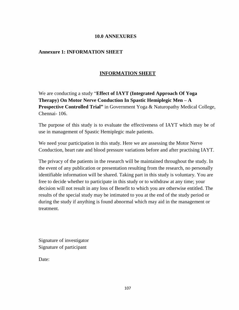
107
10.0 ANNEXURES
Annexure 1: INFORMATION SHEET
INFORMATION SHEET
We are conducting a study “Effect of IAYT (Integrated Approach Of Yoga
Therapy) On Motor Nerve Conduction In Spastic Hemiplegic Men – A
Prospective Controlled Trial” in Government Yoga & Naturopathy Medical College,
Chennai- 106.
The purpose of this study is to evaluate the effectiveness of IAYT which may be of
use in management of Spastic Hemiplegic male patients.
We need your participation in this study. Here we are assessing the Motor Nerve
Conduction, heart rate and blood pressure variations before and after practising IAYT.
The privacy of the patients in the research will be maintained throughout the study. In
the event of any publication or presentation resulting from the research, no personally
identifiable information will be shared. Taking part in this study is voluntary. You are
free to decide whether to participate in this study or to withdraw at any time; your
decision will not result in any loss of Benefit to which you are otherwise entitled. The
results of the special study may be intimated to you at the end of the study period or
during the study if anything is found abnormal which may aid in the management or
treatment.
Signature of investigator
Signature of participant
Date:

108
Annexure 2: INFORMED CONSENT FORM
INFORMED CONSENT FORM
Title of the study: “Effect of IAYT (Integrated Approach Of Yoga Therapy)
On Motor Nerve
Conduction In Spastic Hemiplegic Men – A Prospective
Controlled Trial”
Name of the Participant:
Name of the Principal Investigator: Dr.R.Vidhyalakshmi
Name of the Institution: Government Yoga and Naturopathy Medical College,
Arumbakkam, Chennai – 600 106.
Documentation of the informed consent
I _____________________________ have read the information in this form (or it has
been read to me). I was free to ask any questions and they have been answered. I am
over 18 years of age and, exercising my free power of choice, hereby give my consent
to be included as a participant in “Effect of IAYT (Integrated Approach Of Yoga
Therapy) On Motor Nerve Conduction In Spastic Hemiplegic Men – A
Prospective Controlled Trial”
1. I have read and understood this consent form and the information provided to me.
2. I have had the consent document explained to me.
3. I have been explained about the nature of the study.
4. I have been explained about my rights and responsibilities by the investigator.
5. I have been informed the investigator of all the treatments I am taking or have taken
in the past ________ months including any native (alternative) treatment.
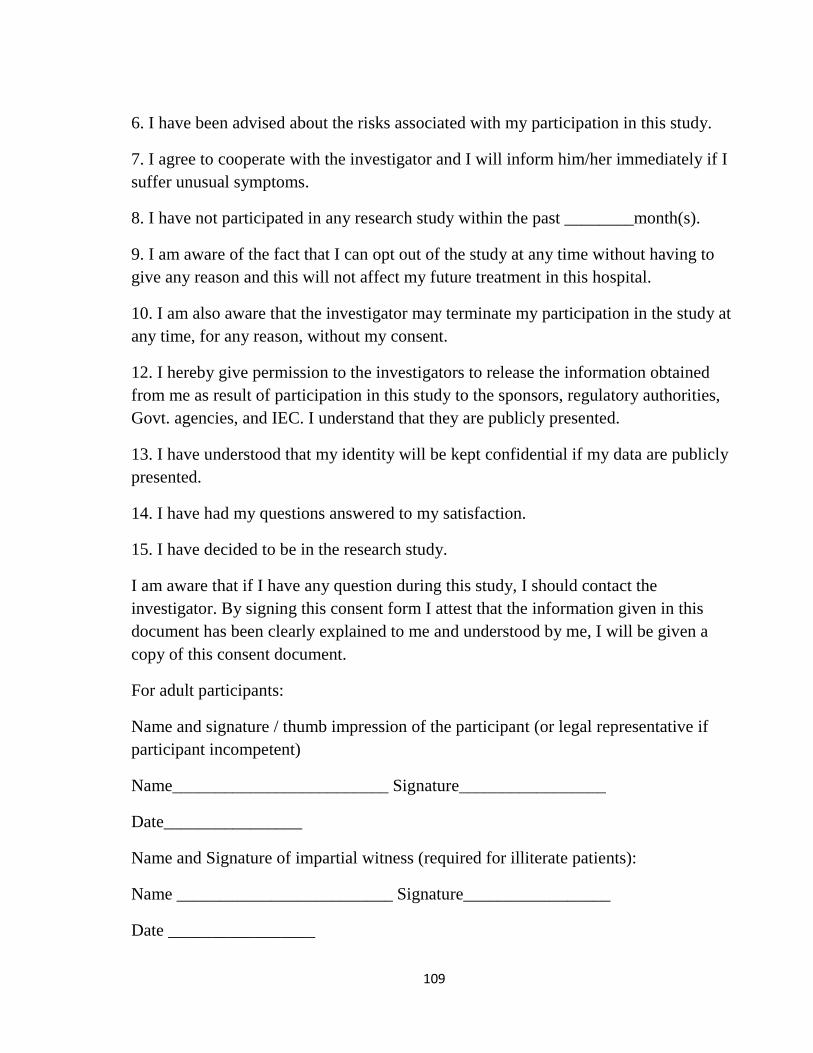
109
6. I have been advised about the risks associated with my participation in this study.
7. I agree to cooperate with the investigator and I will inform him/her immediately if I
suffer unusual symptoms.
8. I have not participated in any research study within the past ________month(s).
9. I am aware of the fact that I can opt out of the study at any time without having to
give any reason and this will not affect my future treatment in this hospital.
10. I am also aware that the investigator may terminate my participation in the study at
any time, for any reason, without my consent.
12. I hereby give permission to the investigators to release the information obtained
from me as result of participation in this study to the sponsors, regulatory authorities,
Govt. agencies, and IEC. I understand that they are publicly presented.
13. I have understood that my identity will be kept confidential if my data are publicly
presented.
14. I have had my questions answered to my satisfaction.
15. I have decided to be in the research study.
I am aware that if I have any question during this study, I should contact the
investigator. By signing this consent form I attest that the information given in this
document has been clearly explained to me and understood by me, I will be given a
copy of this consent document.
For adult participants:
Name and signature / thumb impression of the participant (or legal representative if
participant incompetent)
Name_________________________ Signature_________________
Date________________
Name and Signature of impartial witness (required for illiterate patients):
Name _________________________ Signature_________________
Date _________________

110
Address and contact number of the impartial witness:
Name and Signature of the investigator or his representative obtaining consent:
Name _________________________Signature____________________
Date __________________
INFORMATION TO PARTICIPANTS
Investigator: Dr.R.Vidhyalakshmi, B.N.Y.S
Name of Participant:
Title: “Effect of IAYT (Integrated Approach Of Yoga Therapy) On Motor Nerve
Conduction In Spastic Hemilegic Men – A Prospective Controlled Trial”
You are invited to take part in this research/ study /procedures. The information in this
document is meant to help you decide whether or not to take part. Please feel free to
ask if you have any queries or concerns.
You are being asked to participate in this study being conducted in
Government Yoga and Naturopathy Medical College,
Arumbakkam,
Chennai – 600 106.

111
What is the Purpose of the Research?
Hemiplegia is a condition that causes half of the body to be paralyzed (unable to
move). It's caused by damage to one half of the brain; specifically, when that damage
affects the parts of the brain responsible for motor movements. The half of the body
affected depends on which half of the brain has been damaged.
To determine whether the yoga based lifestyle program has a beneficial effect on the
management of Hemiplegia.
To determine the effect of IAYT on Motor Nerve Conduction on Median and Deep
peroneal nerve.
The Study Design
Seventy-five Male patients with Spastic Hemiplegia of age group between 30-60yrs
will participate in the study.
Study Procedures
The study involves assessment of Motor Nerve Conduction Velocity.
You will be required to visit the hospital during the study.
(The test involves Electro Myogram / EMG study before the commencement of
therapies and after 56 days.)
You may have to come to the hospital (study site) for examination and investigations
apart from your scheduled visits, if required.
Possible Risks to you - Nil
Possible benefits to you- Motor Nerve dysfunction can be diagnosed at an early stage
so that proper intervention can be taken.
Possible benefits to other people
The result of the research may provide benefits to the society in terms of advancement
of medical knowledge and/or therapeutic benefits to future patients.

112
Confidentiality of the information obtained from you
You have the right to confidentiality regarding the privacy of your medical information
(personal details, results of physical examinations, investigations, and your medical
history). By signing this document, you will be allowing the research team
investigators, other study personnel, sponsors, IEC and any person or agency required
by law like the Drug Controller General of India to view your data, if required.
The information from this study, if published in scientific journals or presented at
scientific meetings, will not reveal your identity.
How will your decision to not participate in the study affect you?
Your decisions to not to participate in this research study will not affect your medical
care or your relationship with investigator or the institution. Your doctor will still take
care of you and you will not lose any benefits to which you are entitled.
Can you decide to stop participating in the study once you start?
The participation in this research is purely voluntary and you have the right to
withdraw from this study at any time during course of the study without giving any
reasons.
However, it is advisable that you talk to the research team prior to stopping the
treatment

113
Annexure 3 : ஆராய்ச்சி தகவல் தாள்
சசன்னை அரசு யயாகா மற்றும் இயற்னக மருத்துவமனைக்கு வரும் பக்கவாத
யநாயாளிகளிடம் யமாட்டார் நரம்பு கடத்துதல் என்னும் பாியசாதனை பற்றிய
ஒரு ஆராய்ச்சி இங்கு நனடசபற்று வருகின்றது.
பக்கவாதத்தில் பற்பல நரம்புகள் பாதிக்கபடும். அதில் குறிப்பாக,மீடியன் மற்றும்
டீப்-சபயராைியல் நரம்புகனள ஆராய்வயத இந்த ஆராய்ச்சியின் யநாக்கமாகும்.
நீங்களும் இந்த ஆராய்ச்சியில் பங்யகற்க நாங்கள் விரும்புகியறாம். இந்த
ஆராய்ச்சியில் உங்களுனடய யமாட்டார் நரம்பு கடத்துதல் பற்றிய பாியசாதனை
சசய்யப்பட்டு தகவல்கனள ஆராய்யவாம். அதைால் தங்களது யநாயின்
ஆய்வறிக்னகக்யகா அல்லது சிகிச்னசக்யகா பாதிப்பு ஏற்படாது என்பனதயும்
சதாிவித்துக்சகாள்கியறாம்.
முடிவுகனள அல்லது கருத்துக்கனள சவளியிடும்யபாயதா அல்லது
ஆராய்ச்சியின்யபாயதா தங்களது சபயனரயயா அல்லது அனடயாளங்கனளயயா
செளியிடமாட்யடாம் என்பனதயும் சதாிவித்துக்சகாள்கியறாம்.
இந்த ஆராய்ச்சியில் பங்யகற்பது தங்களுனடய விருப்பத்தின் யபாில்தான்
இருக்கிறது. யமலும் நீங்கள் எந்யநரமும் இந்த ஆராய்ச்சியிலிருந்து
பின்வாங்கலாம் என்பனதயும் சதாிவித்துக்சகாள்கியறாம்.
இந்த சிறப்பு பாியசாதனைகளின் முடிவுகனள ஆராய்ச்சியின்யபாது அல்லது
ஆராய்ச்சியின் முடிெின்யபாது தங்களுக்கு அறிவிப்யபாம்
என்பனதயும்சதாிவித்துக்சகாள்கியறாம்.
ஆராய்ச்சியாளர் னகசயாப்பம் பங்யகற்பாளர்னகசயாப்பம்
யததி :

114
ஆராய்ச்சி ஒப்புதல் கடிதம்
ஆராய்ச்சி தனலப்பு :
சபயர் : யததி :
வயது : உள் யநாயாளி
எண் :
பால் : ஆராய்ச்சி
யசர்க்னக எண் :
இந்த ஆராய்ச்சியின் விவரங்களும் அதன் யநாக்கங்களும் முழுனமயாகவும்
எைக்கு சதளிவாகவும் விளக்கப்பட்டது.
எைக்கு விளக்கப்பட்ட விஷங்கனள நான் புாிந்துசகாண்டு நான் எைது
சம்மதத்னதத் சதாிவிக்கியறன்.
எைக்கு யமாட்டார் நரம்பு கடத்துதல் பாியசாதனை சசய்துசகாள்ள சம்மதம்.
இந்த ஆராய்ச்சியில் பிறாின் நிற்பந்தமின்றி என் சசாந்த விருப்பத்தின் யபாில்
தான் பங்கு சபறுகியறன் மற்றும் நான் இந்த ஆராய்ச்சியிலிருந்து எந்யநரமும்
பின்வாங்கலாம் என்பனதயும் அதைால் எந்த பாதிப்பும் ஏற்படாது என்பனதயும்
நான் புாிந்துசகாண்யடன்.
நான் யமாட்டார் நரம்பு கடத்துதல் பாியசாதனை பற்றிய விவரங்கனளயும்
ஆராய்ச்சியாளர் மூலம் சதாிந்துக்சகாண்யடன்.
நான் என்னுனடய சுயநினைவுடன் மற்றும் முழு சுதந்திரத்துடன் இந்த மருத்துவ
ஆராய்ச்சியில் என்னை யசர்த்துக்சகாள்ள சம்மதிக்கியறன்.
யமற்கண்ட யமாட்டார் நரம்பு கடத்துதல் பாியசாதனையின்யபாது யலசாை
மின்சார அதிர்ச்சி ஏற்படலாம் எை மருத்துவர் மூலம் சதாிந்துக்சகாண்யடன்.
னகசயாப்பம்
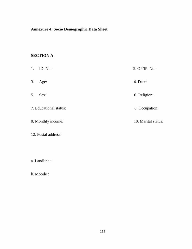
115
Annexure 4: Socio Demographic Data Sheet
SECTION A
1. ID. No: 2. OP/IP. No:
3. Age: 4. Date:
5. Sex: 6. Religion:
7. Educational status: 8. Occupation:
9. Monthly income: 10. Marital status:
12. Postal address:
a. Landline :
b. Mobile :

116
SECTION B: ILLNESS DETAILS
13. Duration of illness:
14. History of chief complaints:
15. Treatment:
16. Any medical illness?
Diabetes mellitus/Systemic Hypertension/Seizures/any other specify:
17. Presence of medical illness in the family:
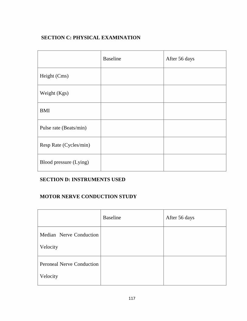
117
SECTION C: PHYSICAL EXAMINATION
Baseline After 56 days
Height (Cms)
Weight (Kgs)
BMI
Pulse rate (Beats/min)
Resp Rate (Cycles/min)
Blood pressure (Lying)
SECTION D: INSTRUMENTS USED
MOTOR NERVE CONDUCTION STUDY
Baseline After 56 days
Median Nerve Conduction
Velocity
Peroneal Nerve Conduction
Velocity
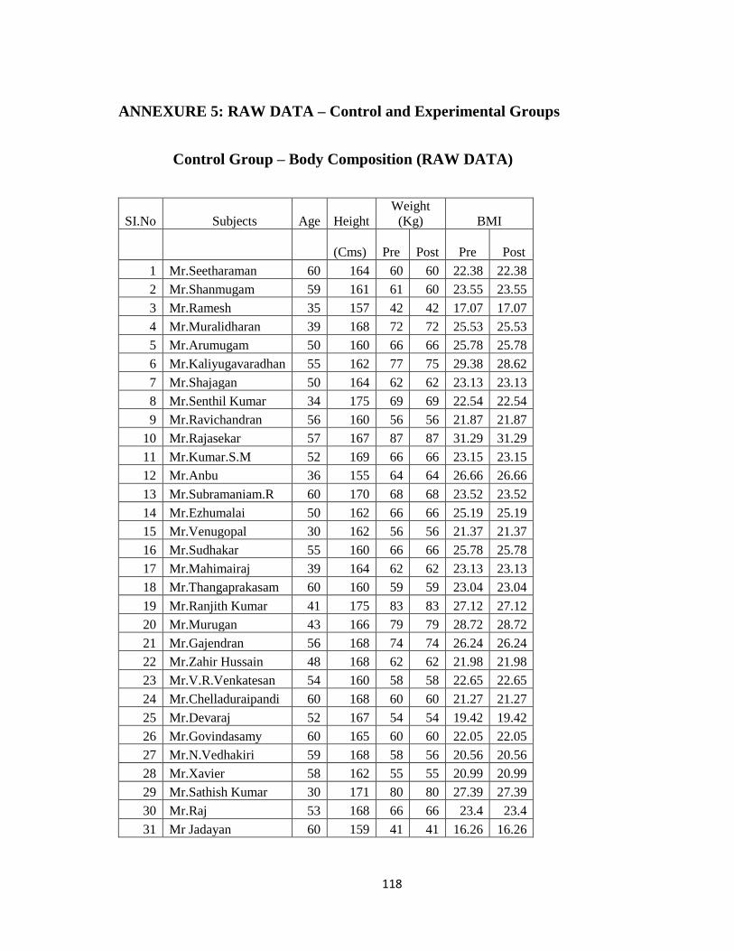
118
ANNEXURE 5: RAW DATA – Control and Experimental Groups
Control Group – Body Composition (RAW DATA)
SI.No Subjects Age Height
Weight
(Kg) BMI
(Cms)
Pre
Post Pre Post
1 Mr.Seetharaman 60 164 60 60 22.38 22.38
2 Mr.Shanmugam 59 161 61 60 23.55 23.55
3 Mr.Ramesh 35 157 42 42 17.07 17.07
4 Mr.Muralidharan 39 168 72 72 25.53 25.53
5 Mr.Arumugam 50 160 66 66 25.78 25.78
6 Mr.Kaliyugavaradhan 55 162 77 75 29.38 28.62
7 Mr.Shajagan 50 164 62 62 23.13 23.13
8 Mr.Senthil Kumar 34 175 69 69 22.54 22.54
9 Mr.Ravichandran 56 160 56 56 21.87 21.87
10 Mr.Rajasekar 57 167 87 87 31.29 31.29
11 Mr.Kumar.S.M 52 169 66 66 23.15 23.15
12 Mr.Anbu 36 155 64 64 26.66 26.66
13 Mr.Subramaniam.R 60 170 68 68 23.52 23.52
14 Mr.Ezhumalai 50 162 66 66 25.19 25.19
15 Mr.Venugopal 30 162 56 56 21.37 21.37
16 Mr.Sudhakar 55 160 66 66 25.78 25.78
17 Mr.Mahimairaj 39 164 62 62 23.13 23.13
18 Mr.Thangaprakasam 60 160 59 59 23.04 23.04
19 Mr.Ranjith Kumar 41 175 83 83 27.12 27.12
20 Mr.Murugan 43 166 79 79 28.72 28.72
21 Mr.Gajendran 56 168 74 74 26.24 26.24
22 Mr.Zahir Hussain 48 168 62 62 21.98 21.98
23 Mr.V.R.Venkatesan 54 160 58 58 22.65 22.65
24 Mr.Chelladuraipandi 60 168 60 60 21.27 21.27
25 Mr.Devaraj 52 167 54 54 19.42 19.42
26 Mr.Govindasamy 60 165 60 60 22.05 22.05
27 Mr.N.Vedhakiri 59 168 58 56 20.56 20.56
28 Mr.Xavier 58 162 55 55 20.99 20.99
29 Mr.Sathish Kumar 30 171 80 80 27.39 27.39
30 Mr.Raj 53 168 66 66 23.4 23.4
31 Mr Jadayan 60 159 41 41 16.26 16.26

119
Control Group – Motor Nerve Conduction Velocity (RAW DATA)
SI.No Subjects Age Median MNC Peroneal MNC
Pre Post Pre Post
1 Mr.Seetharaman 60 49.9 50.98 46.28 45.2
2 Mr.Shanmugam 59 52.24 48.04 43.79 42.01
3 Mr.Ramesh 35 53.49 52.35 52.16 44.43
4 Mr.Muralidharan 39 37.85 52.51 45.45 48.04
5 Mr.Arumugam 50 12 11.78 50.35 46.39
6 Mr.Kaliyugavaradhan 55 52.69 47.97 53.33 51.88
7 Mr.Shajagan 50 56.77 55.44 42.77 49.88
8 Mr.Senthil Kumar 34 56 54.9 48 40.81
9 Mr.Ravichandran 56 13.71 46.13 41.14 37.7
10 Mr.Rajasekar 57 92.53 32.42 40.07 41.27
11 Mr.Kumar.S.M 52 49 50.1 46.24 30.23
12 Mr.Anbu 36 60 53.96 55.79 51.23
13 Mr.Subramaniam.R 60 58.04 56.77 42.05 47.47
14 Mr.Ezhumalai 50 59.95 55.8 44.51 43.5
15 Mr.Venugopal 30 58.04 54.28 51.72 51.03
16 Mr.Sudhakar 55 55.04 53.78 49.88 46.84
17 Mr.Mahimairaj 39 56.21 52.4 52.25 47.46
18 Mr.Thangaprakasam 60 64.77 61.43 46.84 43.27
19 Mr.Ranjith Kumar 41 54.9 53.74 45.54 53.18
20 Mr.Murugan 43 11.78 45.38 42.47 46.99
21 Mr.Gajendran 56 51.96 57.86 48.51 38.43
22 Mr.Zahir Hussain 48 61.64 63.323 54.5 58.08
23 Mr.V.R.Venkatesan 54 48.96 47 48.01 53.18
24 Mr.Chelladuraipandi 60 54 51.932 47.47 45.54
25 Mr.Devaraj 52 59.95 52.19 49.87 47.39
26 Mr.Govindasamy 60 52 50.88 44.71 46.33
27 Mr.N.Vedhakiri 59 38.57 43.16 46.78 45.62
28 Mr.Xavier 58 55.44 55.56 48.65 46.8
29 Mr.Sathish Kumar 30 55.56 54.49 47.46 50.95
30 Mr.Raj 53 56.77 55.56 49.62 48.56
31 Mr Jadayan 60 29.93 51.17 69.72 49.1

120
Control Group – Resting Cardiovascular Variables (RAW DATA)
No Subjects Age HR SBP DBP PP MAP RPP Dop
Pre Post Pre Post Pre Post Pre Post Pre Post Pre Post Pre Post
1 Mr.Seetharaman 60 78 76 130 130 80 80 50 50 96.66666667 96.66666667 10140 9880 6500 6500
2 Mr.Shanmugam 59 76 80 90 110 60 70 30 40 70 83.33333333 6840 8800 2700 4400
3 Mr.Ramesh 35 78 82 100 100 60 60 40 40 73.33333333 73.33333333 7800 8200 4000 4000
4 Mr.Muralidharan 39 78 78 110 100 80 70 30 30 90 80 8580 7800 3300 3000
5 Mr.Arumugam 50 70 78 130 140 90 90 40 50 103.3333333 106.6666667 9100 10920 5200 7000
6 Mr.Kaliyugavaradhan 55 84 80 140 130 90 80 50 50 106.6666667 96.66666667 11760 10400 7000 6500
7 Mr.Shajagan 50 78 72 130 140 100 90 30 50 110 106.6666667 10140 10080 3900 7000
8 Mr.Senthil Kumar 34 78 82 150 140 90 80 60 60 110 100 11700 11480 9000 8400
9 Mr.Ravichandran 56 78 74 140 140 90 80 50 60 106.6666667 100 10920 10360 7000 8400
10 Mr.Rajasekar 57 82 72 130 130 70 80 60 50 90 96.66666667 10660 9360 7800 6500
11 Mr.Kumar.S.M 52 82 78 130 130 80 80 50 50 96.66666667 96.66666667 10660 10140 6500 6500
12 Mr.Anbu 36 80 74 120 120 70 80 50 40 86.66666667 93.33333333 9600 8880 6000 4800
13 Mr.Subramaniam.R 60 86 74 110 120 70 80 40 40 83.33333333 93.33333333 9460 8880 4400 4800
14 Mr.Ezhumalai 50 72 80 100 100 60 70 40 30 73.33333333 80 7200 8000 4000 3000
15 Mr.Venugopal 30 80 74 130 130 80 80 50 50 96.66666667 96.66666667 10400 9620 6500 6500
16 Mr.Sudhakar 55 82 78 140 140 90 90 50 50 106.6666667 106.6666667 11480 10920 7000 7000
17 Mr.Mahimairaj 39 78 80 130 140 100 90 30 50 110 106.6666667 10140 11200 3900 7000
18 Mr.Thangaprakasam 60 78 76 140 140 100 90 40 50 113.3333333 106.6666667 10920 10640 5600 7000
19 Mr.Ranjith Kumar 41 80 78 110 110 80 80 30 30 90 90 8800 8580 3300 3300
20 Mr.Murugan 43 76 78 130 130 80 80 50 50 96.66666667 96.66666667 9880 10140 6500 6500
21 Mr.Gajendran 56 68 74 120 120 70 80 50 40 86.66666667 93.33333333 8160 8880 6000 4800
22 Mr.Zahir Hussain 48 76 68 120 120 80 80 40 40 93.33333333 93.33333333 9120 8160 4800 4800
23 Mr.V.R.Venkatesan 54 80 74 130 130 80 80 50 50 96.66666667 96.66666667 10400 9620 6500 6500
24 Mr.Chelladuraipandi 60 80 76 130 130 80 80 50 50 96.66666667 96.66666667 10400 9880 6500 6500
25 Mr.Devaraj 52 76 72 110 110 80 70 30 40 90 83.33333333 8360 7920 3300 4400
26 Mr.Govindasamy 60 74 76 130 130 90 80 40 50 103.3333333 96.66666667 9620 9880 5200 6500
27 Mr.N.Vedhakiri 59 82 76 140 120 80 80 60 40 100 93.33333333 11480 9120 8400 4800
28 Mr.Xavier 58 80 72 130 130 80 80 50 50 96.66666667 96.66666667 10400 9360 6500 6500
29 Mr.Sathish Kumar 30 82 78 130 120 70 80 60 40 90 93.33333333 10660 9360 7800 4800
30 Mr.Raj 53 80 78 160 140 90 80 70 60 113.3333333 100 12800 10920 11200 8400
31 Mr Jadayan 60 82 80 110 110 70 80 40 30 83.33333333 90 9020 8800 4400 3300
HR- Heart Rate, SBP- Systolic Blood Pressure, DBP- Diastolic Blood Pressure, PP- Pulse Pressure,
MAP- Mean Arterial Pressure, RPP- Rate Pressure Product, DoP- Double Product
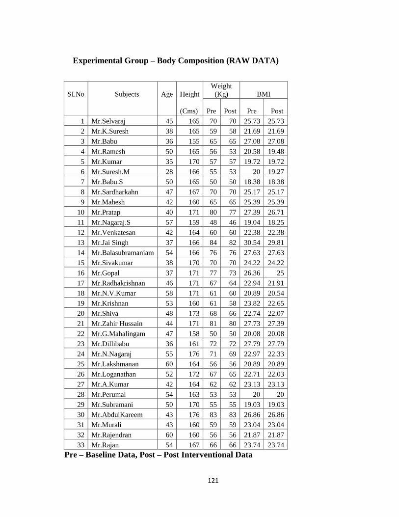
121
Experimental Group – Body Composition (RAW DATA)
SI.No Subjects Age Height
Weight
(Kg) BMI
(Cms)
Pre
Post Pre Post
1 Mr.Selvaraj 45 165 70 70 25.73 25.73
2 Mr.K.Suresh 38 165 59 58 21.69 21.69
3 Mr.Babu 36 155 65 65 27.08 27.08
4 Mr.Ramesh 50 165 56 53 20.58 19.48
5 Mr.Kumar 35 170 57 57 19.72 19.72
6 Mr.Suresh.M 28 166 55 53 20 19.27
7 Mr.Babu.S 50 165 50 50 18.38 18.38
8 Mr.Sardharkahn 47 167 70 70 25.17 25.17
9 Mr.Mahesh 42 160 65 65 25.39 25.39
10 Mr.Pratap 40 171 80 77 27.39 26.71
11 Mr.Nagaraj.S 57 159 48 46 19.04 18.25
12 Mr.Venkatesan 42 164 60 60 22.38 22.38
13 Mr.Jai Singh 37 166 84 82 30.54 29.81
14 Mr.Balasubramaniam 54 166 76 76 27.63 27.63
15 Mr.Sivakumar 38 170 70 70 24.22 24.22
16 Mr.Gopal 37 171 77 73 26.36 25
17 Mr.Radhakrishnan 46 171 67 64 22.94 21.91
18 Mr.N.V.Kumar 58 171 61 60 20.89 20.54
19 Mr.Krishnan 53 160 61 58 23.82 22.65
20 Mr.Shiva 48 173 68 66 22.74 22.07
21 Mr.Zahir Hussain 44 171 81 80 27.73 27.39
22 Mr.G.Mahalingam 47 158 50 50 20.08 20.08
23 Mr.Dillibabu 36 161 72 72 27.79 27.79
24 Mr.N.Nagaraj 55 176 71 69 22.97 22.33
25 Mr.Lakshmanan 60 164 56 56 20.89 20.89
26 Mr.Loganathan 52 172 67 65 22.71 22.03
27 Mr.A.Kumar 42 164 62 62 23.13 23.13
28 Mr.Perumal 54 163 53 53 20 20
29 Mr.Subramani 50 170 55 55 19.03 19.03
30 Mr.AbdulKareem 43 176 83 83 26.86 26.86
31 Mr.Murali 43 160 59 59 23.04 23.04
32 Mr.Rajendran 60 160 56 56 21.87 21.87
33 Mr.Rajan 54 167 66 66 23.74 23.74
Pre – Baseline Data, Post – Post Interventional Data

122
Experimental Group – Motor Nerve Conduction Velocity (RAW DATA)
SI.No Subjects Age
Median
MNC
Peroneal
MNC
Pre Post Pre Post
1 Mr.Selvaraj 45 54.69 61.87 43.45 51.07
2 Mr.K.Suresh 38 54 63.08 47.47 49.09
3 Mr.Babu 36 53.38 60.34 34.28 33.95
4 Mr.Ramesh 50 48.94 56.92 44.43 51.54
5 Mr.Kumar 35 56.92 52.04 46.21 46.21
6 Mr.Suresh.M 28 50 57.08 46.04 55.91
7 Mr.Babu.S 50 58.23 63.01 46.68 43.25
8 Mr.Sardharkahn 47 51.2 53.78 44.5 50.28
9 Mr.Mahesh 42 53.78 64.38 48.04 47.99
10 Mr.Pratap 40 41.53 50 39.21 46.36
11 Mr.Nagaraj.S 57 48 48.98 37.52 45.36
12 Mr.Venkatesan 42 38.23 51 43.76 43.25
13 Mr.Jai Singh 37 52.15 53.24 42.03 36.6
14 Mr.Balasubramaniam 54 50 51.04 37.67 35.8
15 Mr.Sivakumar 38 55.88 58.16 39.91 35.01
16 Mr.Gopal 37 55 64.4 37.84 44.63
17 Mr.Radhakrishnan 46 68.83 54.08 41.22 41.67
18 Mr.N.V.Kumar 58 49.04 56.92 59.01 49.08
19 Mr.Krishnan 53 52.4 48.98 45.31 44.77
20 Mr.Shiva 48 51.82 52.84 37.93 49
21 Mr.Zahir Hussain 44 55.21 57.86 49.71 47.41
22 Mr.G.Mahalingam 47 56.21 50.1 49.12 49.76
23 Mr.Dillibabu 36 55.29 58.08 48.59 48.03
24 Mr.N.Nagaraj 55 60.5 53 46.93 44.89
25 Mr.Lakshmanan 60 53.57 51.17 59.11 48.57
26 Mr.Loganathan 52 57.57 60.27 50.86 46.94
27 Mr.A.Kumar 42 48.98 59.11 47.96 46.42
28 Mr.Perumal 54 43.97 46.23 32.72 34.48
29 Mr.Subramani 50 55.1 57.57 73.17 46.44
30 Mr.AbdulKareem 43 50.11 48.02 49.25 48
31 Mr.Murali 43 57.57 54 48.52 49.09
32 Mr.Rajendran 60 55.04 60.88 47.39 53.33
33 Mr.Rajan 54 58.89 57.38 36.24 40.21
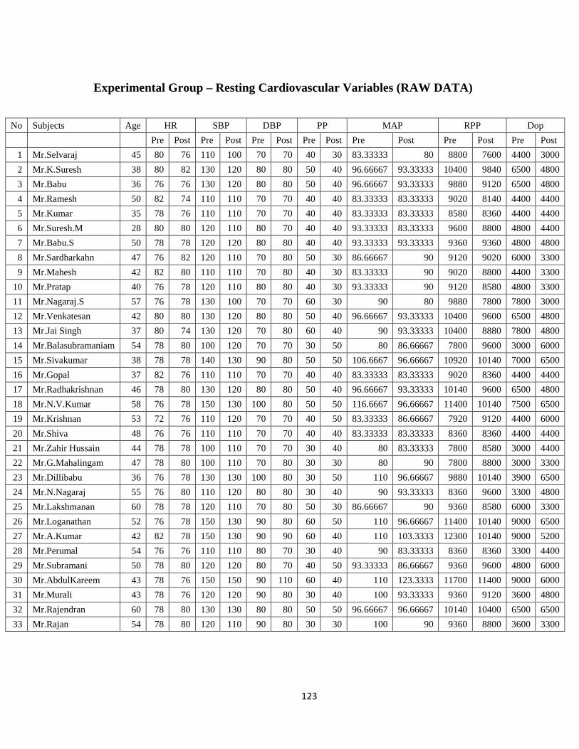
123
Experimental Group – Resting Cardiovascular Variables (RAW DATA)
No Subjects Age HR SBP DBP PP MAP RPP Dop
Pre Post Pre Post Pre Post Pre Post Pre Post Pre Post Pre Post
1 Mr.Selvaraj 45 80 76 110 100 70 70 40 30 83.33333 80 8800 7600 4400 3000
2 Mr.K.Suresh 38 80 82 130 120 80 80 50 40 96.66667 93.33333 10400 9840 6500 4800
3 Mr.Babu 36 76 76 130 120 80 80 50 40 96.66667 93.33333 9880 9120 6500 4800
4 Mr.Ramesh 50 82 74 110 110 70 70 40 40 83.33333 83.33333 9020 8140 4400 4400
5 Mr.Kumar 35 78 76 110 110 70 70 40 40 83.33333 83.33333 8580 8360 4400 4400
6 Mr.Suresh.M 28 80 80 120 110 80 70 40 40 93.33333 83.33333 9600 8800 4800 4400
7 Mr.Babu.S 50 78 78 120 120 80 80 40 40 93.33333 93.33333 9360 9360 4800 4800
8 Mr.Sardharkahn 47 76 82 120 110 70 80 50 30 86.66667 90 9120 9020 6000 3300
9 Mr.Mahesh 42 82 80 110 110 70 80 40 30 83.33333 90 9020 8800 4400 3300
10 Mr.Pratap 40 76 78 120 110 80 80 40 30 93.33333 90 9120 8580 4800 3300
11 Mr.Nagaraj.S 57 76 78 130 100 70 70 60 30 90 80 9880 7800 7800 3000
12 Mr.Venkatesan 42 80 80 130 120 80 80 50 40 96.66667 93.33333 10400 9600 6500 4800
13 Mr.Jai Singh 37 80 74 130 120 70 80 60 40 90 93.33333 10400 8880 7800 4800
14 Mr.Balasubramaniam 54 78 80 100 120 70 70 30 50 80 86.66667 7800 9600 3000 6000
15 Mr.Sivakumar 38 78 78 140 130 90 80 50 50 106.6667 96.66667 10920 10140 7000 6500
16 Mr.Gopal 37 82 76 110 110 70 70 40 40 83.33333 83.33333 9020 8360 4400 4400
17 Mr.Radhakrishnan 46 78 80 130 120 80 80 50 40 96.66667 93.33333 10140 9600 6500 4800
18 Mr.N.V.Kumar 58 76 78 150 130 100 80 50 50 116.6667 96.66667 11400 10140 7500 6500
19 Mr.Krishnan 53 72 76 110 120 70 70 40 50 83.33333 86.66667 7920 9120 4400 6000
20 Mr.Shiva 48 76 76 110 110 70 70 40 40 83.33333 83.33333 8360 8360 4400 4400
21 Mr.Zahir Hussain 44 78 78 100 110 70 70 30 40 80 83.33333 7800 8580 3000 4400
22 Mr.G.Mahalingam 47 78 80 100 110 70 80 30 30 80 90 7800 8800 3000 3300
23 Mr.Dillibabu 36 76 78 130 130 100 80 30 50 110 96.66667 9880 10140 3900 6500
24 Mr.N.Nagaraj 55 76 80 110 120 80 80 30 40 90 93.33333 8360 9600 3300 4800
25 Mr.Lakshmanan 60 78 78 120 110 70 80 50 30 86.66667 90 9360 8580 6000 3300
26 Mr.Loganathan 52 76 78 150 130 90 80 60 50 110 96.66667 11400 10140 9000 6500
27 Mr.A.Kumar 42 82 78 150 130 90 90 60 40 110 103.3333 12300 10140 9000 5200
28 Mr.Perumal 54 76 76 110 110 80 70 30 40 90 83.33333 8360 8360 3300 4400
29 Mr.Subramani 50 78 80 120 120 80 70 40 50 93.33333 86.66667 9360 9600 4800 6000
30 Mr.AbdulKareem 43 78 76 150 150 90 110 60 40 110 123.3333 11700 11400 9000 6000
31 Mr.Murali 43 78 76 120 120 90 80 30 40 100 93.33333 9360 9120 3600 4800
32 Mr.Rajendran 60 78 80 130 130 80 80 50 50 96.66667 96.66667 10140 10400 6500 6500
33 Mr.Rajan 54 78 80 120 110 90 80 30 30 100 90 9360 8800 3600 3300