Embed Size (px)
Citation preview

J. Cent. South Univ. (2013) 20: 2954−2959 DOI: 10.1007/s11771-013-1818-y
Effect of Fe2O3/SiO2 ratio on maghemite-silica particulate nanocomposites
ANG Bee Chin1, YAACOB Iskandar Idris2, NURDIN Irwan1
1. Department of Mechanical, University of Malaya, Kuala Lumpur 50603, Malaysia; 2. Department of Manufacturing and Materials Engineering, Kulliyyah of Engineering, International
Islamic University Malaysia, Kuala Lumpur 50728, Malaysia
© Central South University Press and Springer-Verlag Berlin Heidelberg 2013
Abstract: Maghemite-silica particulate nanocomposites were prepared by modified 2-step sol-gel process. Superparamagnetic maghemite nanoparticles were successfully produced using Massart’s procedure. Nanocomposites consisting of synthesized maghemite nanoparticles and silica were produced by dispersing the as-synthesized maghemite nanoparticles into the silica particulate form. The system was then heated at 140 °C for 3 d. A variety of mass ratios of Fe2O3/SiO2 was investigated. Moreover, no surfactant or other unnecessary precursor was involved. The nanocomposites were characterized using XRD, BET and AGM. The XRD diffraction patterns show the reflection corresponding to maghemite nanoparticles and a visible wide band at 2θ from 20° to 35° which are the characteristics of the amorphous phase of the silica gel. The patterns also exhibit the presence of only maghemite and SiO2 amorphous phase, which indicates that there is no chemical reaction between the silica particulate gel and maghemite nanoparticles to form other compounds. The calculated crystallite size for encapsulated maghemite nanoparticles is smaller than the as-synthesized maghemite nanoparticles indicating the dissolution of the nanoparticles. Very high surface area is attained for the produced nanocomposites (360–390 m2/g). This enhances the sensitivity and the reactivity of the nanocomposites. The shapes of the magnetization curves for nanocomposites are very similar to the as-synthesized maghemite nanoparticles. Superparamagnetic behaviour is exhibited by all samples, indicating that the size of the maghemite nanoparticles is always within the nanometre range. The increase in iron content gives rise to a small particle growth. Key words: maghemite nanoparticles; nanocomposites; sol-gel; silica
1 Introduction
There has been a dramatic increase in interest for nanocrystalline maghemites due to their magnetic properties which are strongly dependent on the particle and crystallite sizes. This is especially so when the particle size reaches nanometer scale [1]. If the particle size is sufficiently small, the magnetic properties of nanoparticles transform from ferromagnetic to superparamagnetic. Moreover, maghemite nanoparticles have very high surface areas compared with bulk materials. Additionally, maghemite nanoparticles have higher reactivity [2]. The properties of magnetic nanoparticles are a function of particle size, shape and surface chemistry [3−4]. Among these, maghemite nanoparticles with a size range of 2−10 nm are of particular importance.
The reason for maghemite nanoparticles being a material of interest is due to their novel properties and potential applications in many areas such as high-density recording media [5−6], catalysis [7], bio-medical
separation process [8], matrices for magneto-optical devices [9], magnetic refrigeration [10] and controlled drug delivery systems [11].
The most critical problem associated with maghemite nanoparticles is their tendency to agglomerate. Most of their unique properties will no longer be retained when the particles aggregate to micron size. This problem needs to be solved in order to maximize the useful nature of the individual particles. Therefore, a number of researches are developed towards finding solutions to prevent the agglomeration and aggregation problem.
Much effort has been made in the synthesis of maghemite nanoparticles due to their superior magnetic properties. The methods included are precipitation, impregnation [12], a special precipitation technique known as Massart process [13], mechanical crushing of powder [14], sol-gel preparation [15], polymer matrix mediated synthesis [16], usage of lauric acid as a non-aqueous medium [17] and using water in oil microemulsion [18−19]. However, the agglomeration and aggregation problem still occur sometimes. In
Foundation item: Project(RP021-2012C) supported by University of Malaya under the UMRG Fund, Malaysia Received date: 2012−08−01; Accepted date: 2013−01−15 Corresponding author: ANG Bee Chin, Lecturer, PhD; Tel: +60−379675258; E-mail: [email protected]

J. Cent. South Univ. (2013) 20: 2954−2959
2955
addition, these methods require tedious washing procedure in order to remove residual surfactants.
Several attempts have been made to disperse the maghemite phase in a variety of matrix materials such as silica [9, 20], porous glass [21] and polymer [22−23]. This technique is used to block the long-range magnetic dipole-dipole interactions among the nanoparticles even in the absence of an applied magnetic field [24] using a physical barrier, which in turn, minimizes the agglomeration.
The generation of maghemite nanocomposites can be classified into two types, namely, the 1-step and 2-step procedures. In the 1-step procedure, maghemite nanocomposites are produced in a single step, whereby maghemite nanoparticles and silica gel are produced simultaneously. For this procedure, various methods have been used such as microemulsion [18, 25−26], sol-gel [15], arc-discharge [27], reactor [28] and low-pressure flames [29].
In the 2-step procedure, maghemite nanoparticles or silica gels are produced first and the encapsulation process is performed later. The 2-step procedure have been used by MORNET et al [30], SARTORATTO et al [31] and ZHANG et al [32] for producing iron oxide nanocomposites.
Although the 1-step procedure is more common compared to the 2-step procedure, the former method suffers from a major disadvantage, the size and shape of the nanoparticles are difficult to control. In addition, 1-step procedure usually uses surfactants, reactors or stabilizers to help the dispersion process. It is known that such chemicals could not be completely removed [25] and the products are therefore unsuitable for bio-applications. Achieving particle size control in nanocomposites is certainly more difficult. Therefore, most of the works are devoted to a careful investigation of the relationship between particle size and magnetic properties rather than searching for the appropriate preparation conditions which favor the particle size control.
In this research, maghemite nanoparticles were produced by Massart’s procedure. This enables good monitoring and control of the size and homogeneity of the as-synthesized maghemite nanoparticles. In addition, the method retains the particles particular identities without serious kinetic exchange involved. The size of the maghemite nanoparticles was further controlled by encapsulating them into a matrix by sol-gel technique. In this process, the particulate form of silica matrix was used as the matrix. This is a novel approach for synthesizing these materials. This method is a two-step procedure to obtain homogeneous and well-coated particles.
2 Experimental
The first stage involved the synthesis of maghemite nanoparticles using Massart’s procedure. The raw chemicals used for preparation of maghemite nanoparticles were ferrous chloride hexahydrate (Sigma), ammonium hydroxide (Fisher Chemicals), ferric chloride (Fisher Chemicals), ferric nitrate (AJAX Chemicals), hydrochloric acid (AJAX Chemicals) and nitric acid (Merck).
Maghemite nanoparticles were synthesized by chemical coprecipitation (Massart’s procedure) of ferric and ferrous ions in alkaline solution. Aqueous solutions of ferrous chloride (stabilized with addition of a few drops of HCl) and ferric chloride were mixed at a molar ratio of Fe3+: Fe2+ of 2:1. The excess amount of ammonium hydroxide (NH4OH) was then added to the solution to ensure the complete precipitation process. The addition of NH4OH caused instantaneous formation of the black precipitates. After the precipitate was settled to the bottom, the clear supernatant liquid was decanted. The precipitate was then washed with deionized water. This process was repeated several times. The precipitate was then stirred in nitric acid solution. The nitric acid was used as an oxidation agent to oxidize the remaining iron oxides into maghemite. The particles were then recovered by centrifugation at 6 000 r/min for 15 min. They were then oxidized completely to maghemite at 90 C in ferric nitrate solution. The particles were isolated and then peptized in deionized water. Powder specimens were obtained by drying the suspension in an oven at slightly elevated temperature, which were labeled as M1.
The second stage of this work involved the preparation of maghemite nanocomposites in particulate form of silica matrix. The modified sol-gel method was also used for the preparation of the nanocomposites. The raw materials used were tetraethyl orthosilicate, TEOS (Aldrich), ethanol (Ajax Chemical), ammonium hydroxide (Fisher Chemicals) and deionized water. Deionized water with a resistivity of approximately 16−18 MΩ/cm was obtained using ELGA ultra analytic deionizer, and used for the preparation of the solutions. All chemicals were of analytical grade and used without any further purification.
The maghemite nanocomposites consisting of as-synthesized maghemite nanoparticles and particulate form of SiO2 matrix were prepared using modified sol-gel procedure. A typical sol-gel precursor mixture consisting of TEOS and deionized water with mass ratio of 5:3 was used. The sol-gel mixture was stirred for 15

J. Cent. South Univ. (2013) 20: 2954−2959
2956
min in ultrasonic bath. The maghemite nanoparticle powders were then dispersed in ethanol (6 mL) followed by dispersion in the aged sol-gel mixture for an additional 1 h in ultrasonic bath. The pH of the solution was retained at 10 to achieve the particulate form of silica matrix. In this analysis, ammonium hydroxide solution was used to control the pH level. Following this, the system was heated at 140 °C for a period of 3 d. The product was washed with deionized water by centrifugation at 6 000 r/min for 5 min to remove un-encapsulated maghemite nanoparticles.
A number of selected mass ratios of Fe2O3/SiO2 were prepared and investigated. The mass ratios of Fe2O3/SiO2 investigated were 0.35, 0.7 and 1.4 and labelled as MNP025, MNP050 and MNP100, respectively.
This method is a promising alternative technique for fabrication of nanocomposites as it is simple, manufacturable, inexpensive and fast. Additionally, it is prepared at room temperature and the composition, crystalline distribution and properties of maghemite nanoparticles and maghemite nanocomposite can be controlled. Moreover, no surfactants or other unnecessary precursors are involved.
3 Characterization
The structure and phase of the nanoparticles were examined by X-ray diffraction (XRD) using Cu Kα radiation (Philips X-Pert MPD PW 3040). The X-ray diffraction patterns were taken from 20° to 80° (2θ value). The mean crystallite size was deduced from the full width at the half maximum (FWHM) of 3 main peaks of the X-ray diffraction pattern using Scherrer’s relation. The actual lattice parameter of the samples was also determined using the data extracted from the XRD patterns. HRTEM micrograph and EELS results were obtained from a Zeiss Libra high resolution transmission electron microscope with an acceleration voltage of 200 kV. N2-physisorption measurements were collected on a Sorptomatic 1990 system (Thermo Finnigan). Before the analysis was carried out, all samples were out-gassed at 120 °C for 8 h. The specific surface area (SBET) and the total pore volume (Vp) were estimated by BET method [33−34]. The average pore width (dp) was calculated from specific surface area and total pore volume [35]. The magnetic properties of the samples were measured using an alternating gradient magnetometer (MicroMag, model 2900), with maximum applied fields of ±10 kOe at room temperature and the magnetic size was calculated based on the formula quoted in ANG and YAACOB [19].
4 Results and discussion
Figure 1 shows the XRD diffraction patterns for as-prepared nanocomposites. The XRD diffraction patterns for samples MNP025, MNP050 and MNP100 clearly show the reflection corresponding to maghemite nanoparticles (ICDD PDF Card No. 39−1346) and a visible wide band at 2θ from 20° to 35° which are characteristics of the amorphous phase of the silica gel. The patterns also exhibit the presence of only maghemite and SiO2 amorphous phases, which indicates that there are no chemical reactions between the silica particulate gel and maghemite nanoparticles to form other compounds.
Fig. 1 XRD patterns for samples
The results are confirmed further by calculating the lattice parameter of the samples. The actual lattice parameter of the samples is also determined using the data extracted from the XRD patterns, as listed in Table 1. The lattice parameters calculated are within the range of 8.33−8.35 Ǻ. It is noted that the lattice parameters are 8.33 and 8.396 Ǻ for bulk maghemite and magnetite, respectively. Besides that, as shown in Fig. 2, the EELS is carried out and the result obtained from the cores clearly shows the presence of Fe-L3 signals, which proves that the embedded particles are iron-based compounds. Therefore, the values obtained indicate that the particles are more likely to be maghemite rather than magnetite.
The broadening of the (311), (511) and (440) reflections increases in the order of MNP100, MNP050 and MNP025. The observed trend suggests a possible decrease in crystallite size when the concentration of maghemite nanoparticles decreases, or in other words, with decreasing mass ratio of Fe2O3/SiO2. The broadening and low intensities of the peaks indicate that the crystallite sizes of the samples are within nanometre scale for all samples. The average crystallite sizes are calculated from the major peaks using Scherrer’s

J. Cent. South Univ. (2013) 20: 2954−2959
2957
equation and the details are listed in Table 2. The calculated crystallite sizes are 4.07, 4.82 and
5.45 nm for samples MNP025, MNP050 and MNP100, respectively. Comparison of the average crystallite size shows that the encapsulated maghemite nanoparticles have a smaller average crystallite size. This could be due to a slight dissolution of maghemite nanoparticles in the silica particulate matrix. These results agree well with the broadening trend observed previously. The surface areas of the MNP samples were studied by N2 gas absorption desorption method. Figure 3 shows the N2 gas absorption desorption isotherms for samples.
The unique features are as follows: (1) very high adsorption at p/po→0, which indicates that a high volume of micropores is present for all samples; (2) a relatively moderate increase in the adsorbed amount of N2 within the p/po range of 0−0.95; (3) a sharp increase in adsorbed N2 within the p/po range of 0.95−1.0; (4) the hysteresis loop is rather small and horizontally oriented; (5) the isotherms do not exhibit plateau at p/po→1.0 but they asymptotically approach the y-axis. These features correspond relatively well to the Type IV isotherm. The calculated specific surface area calculated from the BET method is shown in Table 3.
Table 1 Lattice parameter for samples
Sample 2θ/(°) d-spacing/Å Miller indices (hkl) Lattice constant/Å Average lattice constant/Å
M1 35.575 2.520 311 8.36
8.35 62.851 1.477 440 8.35
MNP025 35.666 2.514 311 8.34
8.34 62.986 1.473 8 440 8.34
MNP050 35.767 2.507 1 311 8.32
8.30 63.51 1.462 9 440 8.28
MNP100 35.738 2.509 1 311 8.32
8.33 63.045 1.472 6 440 8.33
Fig. 2 HRTEM micrograph (a) and EELS result (b) of MNP025
Table 2 Crystallite sizes for samples
Sample 2θ/(°) θ/rad cos θ Wb(sample) Ws(standard) Crystallite size/
nm Arerage crystallite size/
nm
M1 35.579 2 0.310 53 0.952 17 1.245 0 0.053 1 6.09
6.14 62.850 7 0.548 55 0.853 28 1.106 6 0.161 8 6.20
MMP025 35.666 2 0.311 29 0.951 94 0.053 1 0.053 1 5.06
4.07 62.986 2 0.549 73 0.852 67 0.161 8 0.161 8 3.09
MNP050 35.767 5 0.312 17 0.951 67 0.053 1 0.053 1 7.39
4.82 63.510 5 0.554 31 0.850 27 0.161 8 0.161 8 2.26
MNP100 35.738 2 0.311 92 0.951 75 0.053 1 0.053 1 6.66
5.45 63.045 6 0.550 25 0.852 39 0.161 8 0.161 8 4.25
J. Cent. South Univ. (2013) 20: 2954−2959
2958
Fig. 3 N2 gas adsorption desorption isotherm for samples Table 3 Specific surface area and pore specific volume of
samples
Sample Specific surface area/
(m2·g−1) Pore specific
volume/(cm3·g−1)
M1 154.374 7 0.023 3
MNP025 369.91 0.220 5
MNP050 370.97 0.216 4
MNP100 382.90 0.224 4
The result shows that increasing the amount of maghemite nanoparticles leads to a larger surface area for the nanocomposites. This shows that the particles do not fill the pores between the silica spherical particles. Rather, the maghemite particles are embedded within the silica particles. The results show good agreement with HRTEM observations as shown in Fig. 2.
Sample M1 obtains a typical mesoporous isotherm without any micropores behaviour because the adsorption at relative pressure p/po0 is nearly null. The existence of hysteresis loops in the isotherms is due to the capillary condensation of N2 gas occurring in the mesopores material.
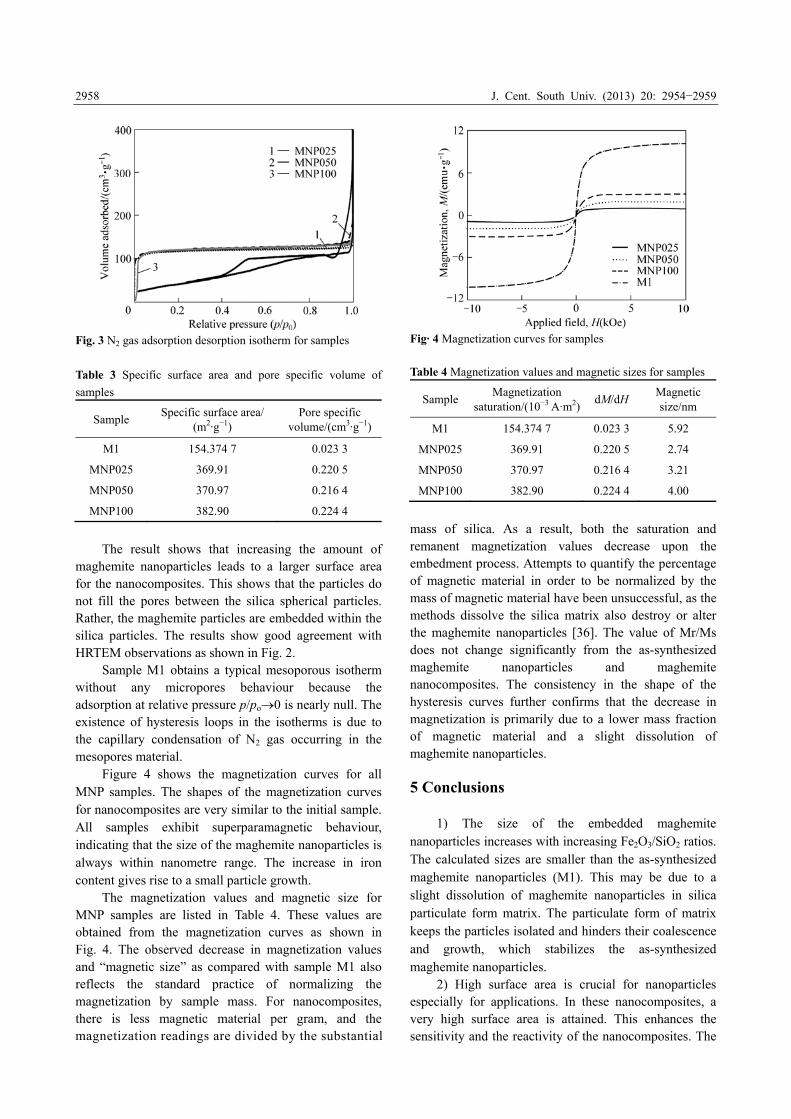
Figure 4 shows the magnetization curves for all MNP samples. The shapes of the magnetization curves for nanocomposites are very similar to the initial sample. All samples exhibit superparamagnetic behaviour, indicating that the size of the maghemite nanoparticles is always within nanometre range. The increase in iron content gives rise to a small particle growth.
The magnetization values and magnetic size for MNP samples are listed in Table 4. These values are obtained from the magnetization curves as shown in Fig. 4. The observed decrease in magnetization values and “magnetic size” as compared with sample M1 also reflects the standard practice of normalizing the magnetization by sample mass. For nanocomposites, there is less magnetic material per gram, and the magnetization readings are divided by the substantial
Fig· 4 Magnetization curves for samples
Table 4 Magnetization values and magnetic sizes for samples
Sample Magnetization
saturation/(10−3 A·m2) dM/dH
Magnetic size/nm
M1 154.374 7 0.023 3 5.92
MNP025 369.91 0.220 5 2.74
MNP050 370.97 0.216 4 3.21
MNP100 382.90 0.224 4 4.00
mass of silica. As a result, both the saturation and remanent magnetization values decrease upon the embedment process. Attempts to quantify the percentage of magnetic material in order to be normalized by the mass of magnetic material have been unsuccessful, as the methods dissolve the silica matrix also destroy or alter the maghemite nanoparticles [36]. The value of Mr/Ms does not change significantly from the as-synthesized maghemite nanoparticles and maghemite nanocomposites. The consistency in the shape of the hysteresis curves further confirms that the decrease in magnetization is primarily due to a lower mass fraction of magnetic material and a slight dissolution of maghemite nanoparticles.
5 Conclusions
1) The size of the embedded maghemite nanoparticles increases with increasing Fe2O3/SiO2 ratios. The calculated sizes are smaller than the as-synthesized maghemite nanoparticles (M1). This may be due to a slight dissolution of maghemite nanoparticles in silica particulate form matrix. The particulate form of matrix keeps the particles isolated and hinders their coalescence and growth, which stabilizes the as-synthesized maghemite nanoparticles.
2) High surface area is crucial for nanoparticles especially for applications. In these nanocomposites, a very high surface area is attained. This enhances the sensitivity and the reactivity of the nanocomposites. The

J. Cent. South Univ. (2013) 20: 2954−2959
2959
modified sol-gel process gives the advantages of nanocomposites with high surface areas, good control of maghemite nanoparticles, no required surfactants and monodispersed nanoparticles. Therefore, the aggregation and agglomeration problem is solved. These nanocomposites would be very useful for the bio-application. References [1] MORALES M P, PECHARROMAN C, GONZALEZ T, SEMA C J.
Structural characteristics of uniform γ-Fe2O3 particles with different
axial (length/width) ratios [J]. J Solid State Chem, 1994, 108:
158−163.
[2] ASLAM M, FU L, LI S, VINAYAK P D. Silica encapsulation and
magnetic properties of FePt nanoparticles [J]. J of Coll & Inter Sci,
2005, 290: 444−449.
[3] KROLL E, WINNIK F M, ZIOLO R. In situ preparation of
nanocrystalline γ-Fe2O3 in iron (II) cross-linked alginate gels [J].
Chem Mater, 1996, 8: 1594−1596.
[4] VOLLATH D, SZABO D V, TAYLOR R D, WILLIS J O,
SICKAFUS K E. Synthesis and properties of nanocrystalline
superparamagnetic γ-Fe2O3 [J]. Nanostruct Mater, 1995, 6: 941−944.
[5] MARTIN J I, NOGUES J, LIU K, VICENT J I, SCHULLER I K.
Ordered magnetic nanostructures: Fabrication and properties [J]. J
Magn Magn Mater, 2003, 256: 449−501.
[6] BATE G. Magnetic recording materials since 1975 [J]. J Magn Magn
Mater, 1999, 100: 413−424.
[7] IDA T, TSUIKI H, UENO A. Characterization of iron oxide in
Fe2O3/SiO2 catalyst [J]. J Catal, 1987, 106: 428−439.
[8] GUPTA A J, GUPTA M. Synthesis and surface engineering of iron
oxide nanoparticles for biomedical applications [J]. J Biomaterials,
2005, 26: 3995−4021.
[9] ENNAS G, MARONGIU G, MUSINU A, FALQUI A, BALLIRANO
P, CAMINITI R. Characterization of Nanocrystalline γ–Fe2O3
Prepared by Wet Chemical Method [J]. Mater Res, 1998, 14(4):
1570.
[10] MCMICHAEL R D, SHULL R D, SWARTZENDRUBER L J,
BENNETT L H, WATSON R E. Magnetocaloric effect in
superparamagnets [J]. J Magn Magn Mater, 1992, 111(1/2): 29−33.
[11] BHATNAGAR S P, ROSENSWEIG R E. Introduction to the
magnetic fluids bibliography [J]. J Magn Magn Mater, 1995, 149:
198.
[12] SATTERFIELD C N. Heterogeneous catalyst in industrial practice
[M]. Singapore: McGraw Hill, Inc. 1991: 85−110.
[13] BEE A, MASSART R, NEVEU S. Synthesis of very fine maghemite
particles [J]. J of Magn Mat, 1995, 149: 6−9.
[14] GOMEZ-VILLACIEROS R, HERNAN L, MORALES J.
Mechanochemical preparation and thermal stability of gamma-Fe2O3
derived from gamma FeOOH [J]. Mat Res Bulletin, 1987, 22:
513−520.
[15] CANNAS C, CONCAS G, GATTESCHI D, FALQUI A, MUSINU A,
PICCALUGA G, SANGREGORIO C, SPANO G. Superparamagnetic
behaviour of γ-Fe2O3 nanoparticles dispersed in a silica matrix [J].
Phys Chem Phys, 2001, 3: 832−838.
[16] HOH J C, YAACOB I I, TEH C L. Cobalt ferrite magnetic
nanoparticle by polymer matrix template synthesis for high magnetic
field bioseparation [J]. Key Engineering Materials, 2004, 206:
1201−1205.
[17] JING Z H, WU S H. Synthesis, characterization and magnetic
properties of -Fe2O3 nanoparticles via a non-aqueous medium [J].
Journal of Solid State Chem, 2004, 177: 1213−1218.
[18] YANG H H, ZHANG S Q, CHEN X L, ZHUANG Z X, XU J G,
WANG X R. Magnetite-containing spherical silica nanoparticles for
biocatalysis and bioseparation [J]. Anal Chem, 2004, 76: 1316−1321.
[19] ANG B C, YAACOB I I. Synthesis and characterization of iron
oxides nanoparticles [J]. Key Engineering Materials, 2006, 306/308:
1115−1120.
[20] MONTE F D, MORALES M P, LEVY D, FERNANDEZ A,
OCANA M, ROIG A, MOLINS E, OGRADY K, SERNA C J.
Formation of γ-Fe2O3 isolated nanoparticles in a silica matrix [J].
Langmuir, 1997, 13: 3627−3634.
[21] BORELLI N F, MORSE D L, SCHREURS J W H. Magnetic
properties of iron oxide photolytically produced from Fe(CO)5
impregnated porous glass [J]. J Appl Phys, 1983, 54: 3344−3350.
[22] ZIOLO R F, GIANNELIS E P, WEINTEIN B A, OHORO M P,
GANGULY B N, MEHROTRA V, RUSSELL M W, HUFFMAN D R.
Matrix-mediated synthesis of nanocrystalline γ-Fe2O3: a new
optically transparent magnetic material [J]. Advanced Materials,
1992, 257: 219−222.
[23] NGUYEN M T, DIAZ A F. A novel method for the preparation of
magnetic nanoparticles in a polypyrrole powder [J]. Adv Mater, 1994,
6: 858−860.
[24] VEKAS L, DOINA B, OANA M. Magnetic nanofluids stabilized
with various chain length surfactants [J]. Romanian Reports in
Physics, 2006, 58(3): 257−267.
[25] SANTRA S, TAPEC R, THEODOROPOULOU N, DOBSON J,
HEBARD A, TAN W H. Synthesis and characterization of
silica-coated iron oxide nanoparticles in microemulsion: the effect of
non-ionic surfactants [J]. Langmuir, 2001, 17: 2900−2906.
[26] MARIA F C, CORRIAS A, PASHINA G. Iron-oxide-silica aerogel
and xerogel nanocomposite materials [J]. J Non-Cryst Solids, 2001,
293−295: 25−31.
[27] PACHECO R F, ARRUEBO M, MARQUINA C, IBARA R,
ARBIOL J, SANTAMARIA J. Highly magnetic silica-coated iron
nanoparticles prepared by the arc-discharge method [J].
Nanotechnology, 2006, 17: 1188−1192.
[28] BARRADO E, RODRIGUEZ J A, PRIETO F, MEDINA J.
Characterization of iron oxides embedded in silica gel obtained by
two different methods [J]. J Non Crys Solids, 2005, 351: 906− 914.
[29] JANZEN C, KNIPPING J, RELLINGHAUS B, ROTH P. Formation
of silica-embedded iron oxide nanoparticles in low-pressure flames
[J]. Journal of Nanoparticles Res, 2003, 5: 589−596.
[30] MORNET S, GRASSET E, PORTIER J, DUGUET E.
Maghemite@silica nanoparticles for biological applications [J].
European Cells and Materials, 2002, 3(2): 110−113.
[31] SARTORATTO P P C, CAIADO K L, PEDROZA R C, SILVA S W,
MORAIS P C. The thermal stability of maghemite-silica
nanocomposites: An investigation using X-ray diffraction and Raman
spectroscopy [J]. Journal of Alloys and Compounds, 2007, 434−435:
650−654.
[32] ZHANG L, GEORGIA C, PAPAEFTHYMIOU Z R F, YING J Y.
Novel γ-Fe2O3/SiO2 magnetic nanocomposites via sol-gel matrix
mediated synthesis [J]. Nanostruct Mater, 1997, 9: 185−188.
[33] GREGG S J, SING K S W. Adsorption, Surface Area and Porosity
(2nd ed) [M]. United State: Academic Press Inc, 1982: 42.
[34] BRUNAUER S, EMMETT P H, TELLER E. Adsorption of gases in
multimolecular layers [J]. Journal of the American Chemical Society,
1938, 60: 309−19.
[35] KAMAL M S K, MAKHLOUF S A. High surface area thermally
stabilized porous iron oxide/silica nanocomposites via a formamide
modified sol-gel process [J]. Appl Surface Sci, 2008, 254:
3767−3773.
[36] VESTAL C R, ZHANG Z J. Synthesis and magnetic characterization
of Mn and Co spinel ferrite-silica nanoparticles with tunable
magnetic core [J]. Nanoletters, 2003, 3(12): 1739−1743.
(Edited by FANG Jing-hua)