Embed Size (px)
Citation preview

fbioe-08-00964 August 24, 2020 Time: 22:13 # 1
ORIGINAL RESEARCHpublished: 26 August 2020
doi: 10.3389/fbioe.2020.00964
Edited by:Ridha Hambli,
Polytech Orléans, France
Reviewed by:Jim Richards,
University of Central Lancashire,United Kingdom
Nicola Lovecchio,University of Milan, Italy
*Correspondence:Anne Focke
Specialty section:This article was submitted to
Biomechanics,a section of the journal
Frontiers in Bioengineering andBiotechnology
Received: 19 December 2019Accepted: 24 July 2020
Published: 26 August 2020
Citation:Focke A, Steingrebe H, Möhler F,Ringhof S, Sell S, Potthast W and
Stein T (2020) Effect of Different KneeBraces in ACL-Deficient Patients.Front. Bioeng. Biotechnol. 8:964.
doi: 10.3389/fbioe.2020.00964
Effect of Different Knee Braces inACL-Deficient PatientsAnne Focke1* , Hannah Steingrebe1,2, Felix Möhler1, Steffen Ringhof1,3, Stefan Sell2,4,Wolfgang Potthast5,6 and Thorsten Stein1
1 BioMotion Center, Institute of Sports and Sports Science, Karlsruhe Institute of Technology (KIT), Karlsruhe, Germany,2 Sports Orthopedics, Institute of Sports and Sports Science, Karlsruhe Institute of Technology (KIT), Karlsruhe, Germany,3 Department of Sport and Sport Science, University of Freiburg, Freiburg, Germany, 4 Joint Center Black Forest, Neuenbürg,Germany, 5 Institute of Biomechanics and Orthopaedics, German Sport University Cologne, Cologne, Germany, 6 ARCUSClinics Pforzheim, Pforzheim, Germany
Knee braces are often used during rehabilitation after ACL injury. There are two mainconcepts, rigid and soft braces, but studies comparing the two show conflicting results.Most studies used movement tasks with low translational or rotational loads and didnot provide joint kinematics. Therefore, the purpose of this study was to investigatethe influence of two different knee braces (rigid vs. soft) on knee joint kinematics inACL-deficient patients compared to an unbraced control condition using two tasks(walking and 180◦ cutting) provoking knee movements in the frontal and transverseplanes. 17 subjects with ACL-deficient knees participated in this study. 3D knee jointkinematics were recorded. To provoke frontal plane knee joint motion a laterally tiltingplate was applied during a walking task. Both braces reduced the maximum valgusangle compared to the unbraced condition, stabilizing the knee joint against excessivevalgus motion. Yet, no differences in peak abduction angle between the two braceswere found. However, a significant extension deficit was observed with the rigid brace.Moreover, both braces increased transverse plane RoM and peak internal rotationangle, with the effects being significantly larger with the rigid brace. These effects havebeen associated with decreased knee stability and unphysiological cartilage loading.Therefore, the soft brace seems to be able to limit peak abduction with a lesserimpact on physiological gait compared to the rigid brace. The cutting task was selectedto provoke transverse plane knee movement and large external knee rotation wasexpected. However, none of the braces was able to reduce peak external knee rotation.Again, an increase in transverse plane RoM was observed with both braces. Based onthese results, no brace outmatched the other in the second task. This study was the firstattempt to clarify the effect of brace design for the stabilization of the knee joint duringmovements with frontal and transverse plane loading. However, to provide physiciansand patients with a comprehensive guideline for brace usage, future studies will have toextent these findings to other daily or sportive movement tasks.
Keywords: rigid, soft, walking, cutting, kinematics, 3D, knee joint
Frontiers in Bioengineering and Biotechnology | www.frontiersin.org 1 August 2020 | Volume 8 | Article 964

fbioe-08-00964 August 24, 2020 Time: 22:13 # 2
Focke et al. Braces in ACL-Deficient Patients
INTRODUCTION
The knee joint is of great importance for human locomotion andis one of the most complex joints of the entire body (Tittel, 2003).Its anatomy does not provide bony guidance for rotation andextreme translation between the tibia and femur; therefore theligamentous apparatus of the knee is not only very important, butalso vulnerable in these situations (Tittel, 2003). Ruptures of theanterior cruciate ligament (ACL) are the most common ligamentinjury in the knee (Palm et al., 2012). Approximately 200,000ACL injuries occur each year in the United States (Woo et al.,2006b; Escamilla et al., 2012; Strutzenberger et al., 2012). Mostof these injuries are caused by non-contact situations (Spindlerand Wright, 2008; Levine et al., 2013), and females show a higherinjury rate than males (Woo et al., 2006a; Spindler and Wright,2008). The highest injury rates are seen in sports which includestop-and-go actions, jumps, rotations and fast changes of velocityor direction such as football, handball, basketball, volleyball,skiing and tennis (Lam et al., 2009; Levine et al., 2013). ACLruptures typically occur during movements with high knee valgusmoments in combination with internal or external rotations ofthe tibia (Hughes and Watkins, 2006).
Consequences of ACL ruptures are biomechanical andneuromuscular changes with impact on the kinematics of theknee joint. Previous studies showed a higher anterior shift of thetibia in the ACL-deficient knee (Beynnon et al., 2003) as well as ahigher variability of knee kinematic patterns (Dennis et al., 2005).Besides these mechanical effects, the proprioceptive capacity ofthe knee joint is also reduced. Due to a decrease in afferent input,reaction times to external disturbances, and thereby posturalcontrol mechanisms, are negatively affected (Lysholm et al.,1998; Lee et al., 2009; Palm et al., 2012). Altogether, thesechanges in biomechanics and the sensorimotor system can leadto compensation mechanisms which result in altered patternsof muscular activity (Théoret and Lamontagne, 2006; Robertset al., 2007), elevated risk of secondary injuries (Wiggins et al.,2016) and chronic diseases of the overloaded structures (e.g.,osteoarthritis) (Andriacchi et al., 2004).
Irrespective of the treatment method (surgical or conservativetherapy), braces as a simple and cost-effective aid are often usedin order to immobilize the knee joint, to prevent excessive jointmovements and to improve stability during activity and thusto prevent secondary injuries. There are several different braceconcepts. Traditional knee braces are designed as rigid shellswith a hinge joint and straps to mechanically guide and supportthe knee joint during motion. Previous studies investigating themechanical effects of such braces showed conflicting results.On the one hand, a reduction of anteroposterior laxity inthe knee was observed for low-load conditions (Wojtys et al.,1996; Beynnon et al., 2003). On the other hand, no positiveeffects of braces on knee stability could be found in morecomplex conditions or in sports with higher loads (Ramseyet al., 2001; Beynnon et al., 2003). Additionally, functionalknee bracing with rigid braces seemed to impact the gaitpattern (DeVita et al., 1998). Finally, the subjective perceptionof comfort differed among patients: while some patientsreported discomfort using rigid braces (Risberg et al., 1999;
Singer and Lamontagne, 2008), other patients reported benefitssuch as a higher sense of stability or increased performance(Swirtun et al., 2005; Birmingham et al., 2008).
Due to the conflicting results regarding the effectivenessof rigid braces, alternative brace concepts are broughtinto focus. Besides pure mechanical stabilization, recentapproaches included sensorimotor aspects to potentially enhancestabilization during dynamic situations. This approach wasbased on previous studies showing that bandages improvedsensorimotor control by increasing the proprioception of themuscles surrounding the knee (Beynnon et al., 1999, 2002; Selfeet al., 2008, 2011; Baltaci et al., 2011; Bodendorfer et al., 2019).For patients with ACL ruptures, the disadvantage of bandagesseems to be an insufficient mechanical stabilization compared torigid braces (Luber et al., 1998). Therefore, an alternative to bothbandages and rigid braces might be soft braces: these comprisestretchable stocking fabric (similar to bandages) with additionallateral rigid rails (Giotis et al., 2013; Pierrat et al., 2015). Softbraces, comprising bandage fabrics and rigid elements, mighttherefore combine the benefits of a mechanically stable rigidbrace with the proprioceptive advantages of a bandage.
Yet, previous studies comparing rigid and soft braces forthe treatment of ACL-deficient subjects show conflicting results.Strutzenberger et al. (2012) found a higher rate of forcedevelopment in counter-movement jumps and a reduced swaypath length during single leg stance on an unstable, laterallyperturbed platform with a soft compared to a rigid brace.Beynnon et al. (2003) compared two rigid braces and one softbrace and found a significant reduction in anteroposterior laxityduring tests with the Vermont Knee Laxity Device for all threebraces. However, positive effects were only found during weight-bearing and non-weight-bearing postures and not for the loadacceptance phase. Mortaza et al. (2013) compared a rigid brace,a soft brace and a bandage and found no significant differencesin jump distance, peak torque and power between the threeconditions during functional (cross-over hop and single legvertical jump) and isokinetic tests.
The abovementioned studies used movement tasks such asjump, balance or strength tests with low translational and/orrotational loads. Therefore, they do not strain the knee jointin the frontal and transverse planes. These motions, however,are of particular relevance as the function of the ACL is torestrict excessive motion in these planes. Additionally, excessivevalgus moments and external and internal rotations of the tibiaare known to be the main causes of ACL injuries (Hughesand Watkins, 2006). Consequently, knowledge of the abilityof different brace concepts to provide stability of the kneejoint during dynamic situations with high frontal and rotationalloads is of great interest. The aforementioned studies quantifiedbrace effects mostly using the performance in functional orstrength tests and did not provide joint kinematics of the lowerextremities. Yet, kinematic data is needed to understand themode of action of different brace concepts, how they affect gaitpatterns and to evaluate whether one brace concept providesbetter knee stabilization effects.
Therefore, the purpose of this study was to investigate theinfluence of two different braces (rigid vs. soft) on knee joint
Frontiers in Bioengineering and Biotechnology | www.frontiersin.org 2 August 2020 | Volume 8 | Article 964

fbioe-08-00964 August 24, 2020 Time: 22:13 # 3
Focke et al. Braces in ACL-Deficient Patients
kinematics in ACL-deficient patients using two movement tasksprovoking knee movements in the frontal and transverse planes.It was hypothesized that both braces would stabilize the kneejoint, in terms of decreased peak abduction and rotation angles,compared to an unbraced control condition.
MATERIALS AND METHODS
SubjectsDuring subject recruitment 118 potential subjects were screenedfor eligibility. Thereof 41 fulfilled all defined inclusion criteria andwere invited for the first test session. During this session a total of17 subjects with ACL-deficient knees demonstrated an unstableknee joint and subsequently participated in this study (10 females,7 males; age: 44.4 ± 11.5 years; height: 1.68 ± 0.08 m; mass: 77.6± 11.5 kg). Subjects were sportively active for 198± 117 min perweek with focus on either team sports (e.g., handball, football) orrecreational sportive activities (e.g., running, cycling, hiking, orswimming). The time interval between injury and biomechanicaldata collection was between 0.25 and 32 years (11.8± 12.6 years).Although some of the ACL ruptures had occurred several yearsago, all subjects showed symptoms of an unstable knee. Kneeinstability was defined as fulfilling at least two of the followingthree criteria: (a) side-to-side difference in knee laxity ≥ 3 mmevaluated by use of the KT-1000TM arthrometer (MEDmetric,San Diego, CA, United States), (b) limb symmetry score below85% during both the single hop test for distance, and (c) thetimed hop test (Noyes et al., 1991). Besides knee instability,additional inclusion criteria were: (a) unilateral rupture of theACL without surgical reconstruction, (b) age between 18 and60 years, (c) moderate sport activity, (d) absence of injuries ofthe posterior cruciate ligament or other structures in the knee, (e)no gonarthrosis of grade 2–4 (Kellgren and Lawrence, 1957), and(f) contralateral side free of injuries.
The study design was approved by the Ethics Board of the StateMedical Association of Baden-Württemberg. All patients wereinformed about the procedures of the study and gave their writteninformed consent prior to study participation.
Experimental ProtocolAll subjects were tested on two occasions. During the firstsession, patients were informed about the study procedure andwere screened regarding the inclusion criteria via questionnaires.Then, knee instability was tested using the abovementionedtests. Subjects included in the study were provided with both asoft brace (SofTec Genu; Bauerfeind Inc., Zeulenroda-Triebes,Germany) and a rigid brace (4Titude Donjoy; ORMED GmbH,Freiburg, Germany). Both braces were fitted individually tothe injured knee by an experienced orthopedic technician, andsubjects were instructed regarding the correct positioning ofthe braces. Subsequently, patients were familiarized with themovement tasks to reduce learning effects. Subjects then woreboth braces in alternation during their everyday activities fora period of at least four weeks to avoid habituation effectsduring measurement.
During the second session, subjects completed a standardized5 min warm-up on a bicycle ergometer (intensity: 50% bodymass in Watt, 60 RPM). They then performed two movementtasks: (a) walking over a suddenly tilting plate and (b) 180◦cutting. These tasks were chosen to provoke external momentsin the frontal and transverse planes, respectively, and thereforeto induce instability and relative motion between femur andtibia. Each patient performed both movement tasks under threedifferent conditions: injured leg with rigid brace, injured legwith soft brace, and injured leg without brace. The order of thethree conditions within each movement task was randomizedfor all subjects.
Walking over tiltable plate: The subjects were instructedto walk across a 7.5 m walkway at a prescribed speed of5 km/h (± 5%), verified using infrared timing gates (Figure 1A).A custom-made tiltable plate (60 × 60 cm) was embedded in themiddle of the walkway, which had to be struck by the subjectswith their injured leg. Hydraulic controlled tilting of the plate by9◦ either to the left or to the right side, provoking a supination orpronation at the ankle, was triggered by another infrared timinggate positioned at the beginning of the walkway. The time delaybetween triggering and onset of tilting was set by a custom-made software program allowing for two different conditions:the plate was either already completely tilted before being struck(predictive condition) or the plate tilted when it was struck(reactive condition). Both time delay (predictive or reactive)and tilting direction (pronation or supination) were randomizedwithin each of the three brace conditions. Hence, subjects wereunaware of the tilting condition prior to movement initiation.For each test condition, three valid trials were recorded. Thus,a total of 36 successful trials were recorded: 2 time delay × 2tilting directions × 3 brace conditions × 3 trials. However, sincevalgus movements are more important than varus movements inthe context of ACL injuries, only pronation trials (18 trials) wereanalyzed in this paper.
Cutting of 180◦: Subjects performed a cutting movement of180◦ after walking with a prescribed approach speed of 7 km/h(± 5%). The experimental setup (Figure 1B) was very similarto the walking task, however, the plate embedded in the middleof the walkway was fixed in a straight position rather thantilting. The distance from initial position to the plate was 3.5 m.Two timing gates were positioned 1 and 2 m in front of theplate and were used to control approach speed. The cuttingmovement was performed on the plate in a step turn mannerusing the injured leg. Three valid trials were recorded for eachtest condition resulting in a total of 9 recorded trials (3 braceconditions× 3 trials).
Data CollectionA motion capture system (10 cameras; 200 Hz; Vicon MotionSystems; Oxford Metrics Group, Oxford, United Kingdom)was used to capture 42 spherical retro-reflective markers(14 mm) placed on predefined anatomical landmarks ofthe subject (Figures 2A,B). In addition 22 anthropometricmeasurements were taken manually according to thealaska Dynamicus Handbook [Advanced Lagrangian Solverin kinetic Analysis, Insys GmbH, Chemnitz, Germany
Frontiers in Bioengineering and Biotechnology | www.frontiersin.org 3 August 2020 | Volume 8 | Article 964

fbioe-08-00964 August 24, 2020 Time: 22:13 # 4
Focke et al. Braces in ACL-Deficient Patients
FIGURE 1 | Experimental setup of movement tasks: (A) walking, and (B) 180◦ cutting. Plate contact occurred always with injured leg. Cutting movement (B) wasperformed in a step turn manner.
FIGURE 2 | Marker positions in frontal (A) and dorsal (B) view.
(Härtel and Hermsdorf, 2006)]. A static trial was recordedduring which the participants stood in a neutral position,with their feet shoulder-width apart, toes pointing anteriorlyand hip and knee joints in full extension. This static trial wasused to adapt the multi-body-model (alaska Dynamicus) toeach subject. Three dimensional ground reaction forces werecaptured with the custom-made tiltable plate embedded in themiddle of the walkway.
Knee Marker ReconstructionWhen the braces were applied, the knee markers at the injuredleg were removed during the dynamic trials. Therefore, additionalclusters of four markers were attached to the thigh and shank to
reconstruct the knee markers. During the initial static referencetrial, the actual knee markers were applied in addition to thecluster markers. The cluster marker positions defined a referenceframe, which was embedded rigidly to the shank (shank referenceframe, SF). This frame was also used to set up reference vectorsfrom the SF origin to the respective knee marker b̃ and to eachcluster marker x̃i (i = 1, . . . , 4). Both b̃ and x̃i can be assumed tobe time-invariant in SF.
Once the reference vectors were determined by a referencetrial, knee markers were removed. For the trials without the kneemarkers a least-squares algorithm (Cappozzo et al., 1997) wasused to calculate the optimized position and orientation of SF. Atfirst, SF was set up identically to the reference trial at each time
Frontiers in Bioengineering and Biotechnology | www.frontiersin.org 4 August 2020 | Volume 8 | Article 964

fbioe-08-00964 August 24, 2020 Time: 22:13 # 5
Focke et al. Braces in ACL-Deficient Patients
instant. Optimization was achieved by minimizing the deviationei between each instantaneous cluster marker position xi in thecurrent SF in relation to the corresponding reference vector x̃i:
ei = x̃i − xi.
To transform this equation from SF to the laboratory frame (LF),a translation vector p and a rotation matrix R were introduced,so that
ei = Rx̃i −(yi − p
)with yi as the coordinates of the cluster markers in the LF andp as the vector pointing from the origin of the LF to the origin
of the SF. Solving minp,R
4∑i=1||ei||2 allowed calculation of p and R
by a singular value decomposition (Hanson and Norris, 1981).Together p and R transformed SF to an optimized SF.’ Therequired knee marker position c in the LF could be calculated ateach time instant by a retransformation of the reference vector b̃(time-invariant in SF’) to LF:
c = Rb̃ + p.
This procedure was done for both medial and lateral kneemarkers for all three brace conditions.
Data Processing and AnalysisKinematic data were analyzed during stance phase on the plate.Heel-strike and toe-off of the injured leg on the plate weredetermined via force sensors embedded in the plate using athreshold of 10 N (Tirosh and Sparrow, 2003). Force data werefiltered with a third-order Butterworth low-pass filter with a cut-off frequency of 50 Hz. Three-dimensional marker trajectorieswere filtered using a second-order Butterworth low-pass filterwith a cut-off frequency of 6 Hz for the walking conditionand 10 Hz for the cutting condition (Kirtley, 2006). An inversekinematics approach using the multi-body model Dynamicus(Härtel and Hermsdorf, 2006) was used to calculate 3D kneeangles as objective parameters suggested in the literature to beindicators for knee joint stability (Schrijvers et al., 2019). Basedon the preprocessed data, peak joint angles (minimum andmaximum), ranges of motion (RoM), joint angles at touch down(TD) and at resultant peak ground reaction force (Peak GRF)in the sagittal, frontal and transverse planes were calculated forthe knee joint during the stance phases of walking and cutting.The RoM was used to eliminate possible errors of absolutemeasures (Giotis et al., 2013), TD was used to indicate phaseof load transfer and Peak GRF was taken as the time of peakload, as previously used (Ewing et al., 2016; Alirezaei Noghondarand Bressel, 2017; Duffell et al., 2017). For all parameters themain focus was on the frontal and transverse plane as ACL-deficient knees are particularly vulnerable to forces in theseplanes (Hughes and Watkins, 2006; Levine et al., 2013). Thevalues acquired from the three valid trials were averaged foreach test condition.
StatisticsAll statistical tests were performed using IBM SPSS Statistics24.0 (IBM Corporation, Armonk, NY, United States). The
effect of braces (rigid, soft, without) on knee angles wasinvestigated by use of one-way repeated measures ANOVAtests, separately run for the walking conditions (predictive,reactive) and the 180◦ cutting movement. If sphericity wasviolated, Greenhouse-Geisser estimates were used to correctfor these violations. The significant main effects for thebraced conditions were analyzed in post-hoc comparisonswith Holm-Bonferroni corrections to adjust for multiplecomparisons. Effect sizes were determined using partial etasquared (small effect: η2
p = 0.01; medium effect: η2p = 0.06;
large effect η2p = 0.14) (Cohen, 1988; Richardson, 2011).
For all statistical tests, the level of significance was seta priori to 0.05.
RESULTS
Figure 3 shows the knee joint kinematics during predictive andreactive walking as well as 180◦ cutting in the sagittal, frontaland transverse planes during stance phase. Tables 1–3 presentthe mean values of knee joint angles during stance phase of allthree movement tasks. Data are given in all planes for all braceconditions with respective p-values and effect sizes.
WalkingAs tilting of the plate was intended to induce perturbationsin the frontal plane, knee valgus and varus angles (particularlymaximum valgus angles) were considered the main outcomeparameters for walking.
Maximum valgus angle mainly occurred at the beginning ofstance phase and was therefore similar to the valgus angle atTD. Both braces significantly reduced the maximum valgus anglecompared to the unbraced condition for both walking conditions(predictive and reactive) (Tables 1, 2). The valgus angle at TDwas also significantly smaller with both braces for predictivewalking, and a comparable tendency was found for reactivewalking (Tables 1, 2).
Significantly different results between the braced andunbraced conditions were also found in the sagittal andtransverse planes. Patients generally walked with a more flexedknee in the braced conditions compared to the unbracedcondition, whereby mainly the rigid brace showed significantand larger differences to the unbraced condition than the softbrace (Tables 1, 2).
In the transverse plane, the maximum external rotation angleand the rotation angle at Peak GRF occurred at similar timesand were significantly smaller for the rigid than the unbracedcondition in predictive and reactive walking (Tables 1, 2). Thesoft brace significantly reduced only the maximum externalrotation angle in predictive walking compared to the unbracedcondition (Tables 1, 2). The rotation angle at TD was significantlysmaller for the rigid brace than the unbraced condition inpredictive but not reactive walking (Tables 1, 2). With both bracesa significant increase in transverse plane RoM and peak internalrotation occurred compared to the unbraced condition. Thesealterations were significantly larger with the rigid brace comparedto the soft brace.
Frontiers in Bioengineering and Biotechnology | www.frontiersin.org 5 August 2020 | Volume 8 | Article 964

fbioe-08-00964 August 24, 2020 Time: 22:13 # 6
Focke et al. Braces in ACL-Deficient Patients
FIGURE 3 | Knee joint kinematics of all three movement tasks in the sagittal, frontal, and transverse planes during stance phase (mean ± sd). Red = without brace,black = rigid brace, blue = soft brace.
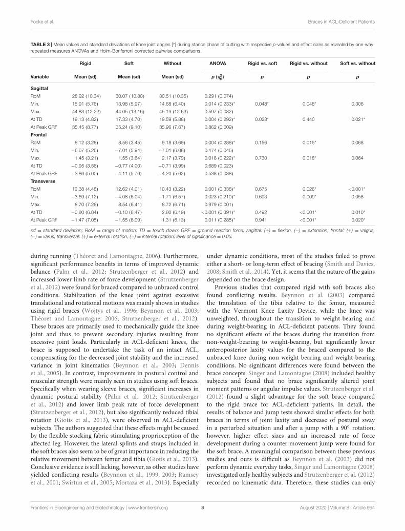
Cutting of 180◦
During cutting, stabilization of the knee joint in the transverseplane is of particular relevance. Specifically, at the beginningof the stance phase significant differences between braced andunbraced conditions were found in the transverse plane. Externalrotation angle at TD and Peak GRF were significantly smaller forboth braces than the unbraced condition (Table 3). Maximumexternal rotation angle occurred at the end of stance phase andwas similar for all three conditions. Again, both braces increasedthe observed RoM and additionally, a significant increase in peakinternal rotation occurred with the rigid brace compared to thecontrol condition.
DISCUSSION
The present study investigated the effects of two different kneebrace designs, soft and rigid, on knee joint kinematics in ACL-deficient patients. In summary, results showed that both braces
induced changes in knee joint kinematics during walking andcutting when compared to an unbraced control condition.
With regard to the walking task, in which the tilted plate wasused to disturb joint stability in the frontal plane, both braceswere able to reduce the maximum valgus angle as comparedto the unbraced condition, stabilizing the knee joint againstan excessive valgus motion during stance phase. Despite theirdiffering concepts, both rigid and soft brace demonstrated thatthey provide their desired effects. However, both braces revealeda higher RoM in transverse plane caused by a more pronouncedinternal rotation compared to the unbraced condition. This effectwas more pronounced in the rigid compared to the soft brace.
The cutting task was conducted to provoke relative rotationbetween the proximal and distal segments of the ACL-deficientknee joint and therefore, to evaluate the compensatory capacitiesof the braces in the transverse plane. In this regard, none of thebraces was able to reduce peak external knee rotation. Yet, againa significant increase in transverse plane RoM was observed withboth braces, caused by a more pronounced internal rotation.
Frontiers in Bioengineering and Biotechnology | www.frontiersin.org 6 August 2020 | Volume 8 | Article 964
fbioe-08-00964 August 24, 2020 Time: 22:13 # 7
Focke et al. Braces in ACL-Deficient Patients
TABLE 1 | Mean values and standard deviations of knee joint angles [◦] during stance phase of predictive walking (force plate tilted before the step) with respectivep-values and effect sizes as revealed by one-way repeated measures ANOVAs and Holm-Bonferroni corrected pairwise comparisons.
Rigid Soft Without ANOVA Rigid vs. soft Rigid vs. without Soft vs. without
Variable Mean (sd) Mean (sd) Mean (sd) p (η2p) p p p
Sagittal
RoM 27.34 (3.86) 27.16 (4.38) 28.18 (3.67) 0.084 (0.144)
Min. 9.04 (4.10) 8.85 (4.06) 6.87 (4.06) <0.001 (0.468)* 0.636 0.002* <0.001*
Max. 36.38 (3.02) 36.01 (3.38) 35.05 (3.13) 0.044 (0.178)* 0.527 0.045* 0.148
At TD 15.01 (4.52) 13.96 (4.60) 13.92 (4.10) 0.048 (0.193)* 0.160 0.048* 0.924
At Peak GRF 29.77 (3.37) 29.58 (4.17) 28.97 (4.41) 0.255 (0.082)
Frontal
RoM 4.47 (2.07) 5.03 (1.92) 5.06 (1.89) 0.083 (0.162)
Min. −4.40 (4.52) −5.08 (4.59) −4.24 (4.70) 0.006 (0.271)* 0.044* 0.548 0.009*
Max. 0.07 (4.19) −0.05 (3.66) 0.83 (3.73) 0.005 (0.278)* 0.699 0.016* 0.006*
At TD −0.57 (3.49) −0.66 (3.13) 0.09 (3.16) 0.034 (0.191)* 0.818 0.039* 0.039*
At Peak GRF −1.75 (5.06) −2.62 (4.73) −1.76 (5.04) 0.003 (0.301)* 0.030* 0.971 0.030*
Transverse
RoM 9.42 (2.52) 8.43 (2.50) 7.56 (2.24) <0.001 (0.487)* 0.014* <0.001* 0.019*
Min. −4.33 (6.03) −1.72 (6.34) 0.69 (6.35) <0.001 (0.631)* 0.008* <0.001* 0.008*
Max. 5.09 (5.48) 6.72 (5.60) 8.26 (5.35) <0.001 (0.400)* 0.061 <0.001* 0.040*
At TD 2.20 (5.57) 3.42 (6.16) 4.17 (5.82) 0.027 (0.202)* 0.258 0.033* 0.262
At Peak GRF 4.45 (5.67) 6.23 (5.85) 7.85 (5.39) <0.001 (0.413)* 0.056 <0.001* 0.056
sd = standard deviation; RoM = range of motion; TD = touch down; GRF = ground reaction force; sagittal: (+) = flexion, (−) = extension; frontal: (+) = valgus,(−) = varus; transversal: (+) = external rotation, (−) = internal rotation; level of significance = 0.05.
TABLE 2 | Mean values and standard deviations of knee joint angles [◦] during stance phase of reactive walking (force plate tilted during the step) with respectivep-values and effect sizes as revealed by one-way repeated measures ANOVAs and Holm-Bonferroni corrected pairwise comparisons.
Rigid Soft Without ANOVA Rigid vs. soft Rigid vs. without Soft vs. without
Variable Mean (sd) Mean (sd) Mean (sd) p (η2p) p p p
Sagittal
RoM 26.52 (5.72) 27.43 (4.47) 27.15 (4.00) 0.533 (0.039)
Min. 8.22 (4.97) 7.34 (4.19) 6.36 (4.55) 0.002 (0.334)* 0.089 0.012* 0.018*
Max. 34.74 (3.73) 34.78 (3.61) 33.51 (3.63) 0.202 (0.095)
At TD 14.01 (4.14) 13.04 (4.24) 12.91 (4.14) 0.036 (0.187)* 0.156 0.042* 0.765
At Peak GRF 28.88 (3.21) 29.12 (3.11) 28.06 (3.58) 0.064 (0.158)
Frontal
RoM 4.36 (1.59) 5.19 (1.90) 4.86 (1.69) 0.018 (0.221)* 0.051 0.182 0.184
Min. −4.21 (4.40) −5.11 (4.56) −4.07 (4.58) 0.007 (0.264)* 0.044* 0.643 0.030*
Max. 0.15 (4.10) 0.09 (3.51) 0.79 (3.66) 0.024 (0.208)* 0.837 0.030* 0.030*
At TD −0.45 (3.47) −0.43 (3.07) 0.16 (3.11) 0.059 (0.162)
At Peak GRF −1.88 (5.08) −2.48 (4.67) −1.73 (4.85) 0.024 (0.208)* 0.126 0.490 0.063
Transverse
RoM 9.10 (2.52) 8.16 (2.42) 7.46 (2.20) <0.001 (0.444)* 0.028* <0.001* 0.034*
Min. −4.78 (6.41) −2.07 (6.17) 0.03 (6.12) <0.001 (0.614)* 0.006* <0.001* 0.006*
Max. 4.32 (5.42) 6.08 (5.74) 7.49 (5.46) 0.001 (0.364)* 0.064 0.003* 0.064
At TD 1.72 (6.00) 2.81 (5.97) 3.50 (5.93) 0.055 (0.166)
At Peak GRF 3.67 (5.54) 5.54 (5.85) 7.10 (5.53) <0.001 (0.381)* 0.068 <0.001* 0.068
sd = standard deviation; RoM = range of motion; TD = touch down; GRF = ground reaction force; sagittal: (+) = flexion, (−) = extension; frontal: (+) = valgus,(−) = varus; transversal: (+) = external rotation, (−) = internal rotation; level of significance = 0.05.
Embedding of the Results Into theCurrent State of ResearchDue to large differences between the current study andprevious work in terms of the selected movement tasks, appliedmethods, calculated parameters, included subjects and braces,
a comparison of the results is extremely difficult. Previousstudies observed a reduction in anteroposterior knee jointlaxity (Wojtys et al., 1996; Beynnon et al., 2003; Strutzenbergeret al., 2012; Pierrat et al., 2015) for low-load conditions andlower ranges of motions in the frontal and transverse planes
Frontiers in Bioengineering and Biotechnology | www.frontiersin.org 7 August 2020 | Volume 8 | Article 964
fbioe-08-00964 August 24, 2020 Time: 22:13 # 8
Focke et al. Braces in ACL-Deficient Patients
TABLE 3 | Mean values and standard deviations of knee joint angles [◦] during stance phase of cutting with respective p-values and effect sizes as revealed by one-wayrepeated measures ANOVAs and Holm-Bonferroni corrected pairwise comparisons.
Rigid Soft Without ANOVA Rigid vs. soft Rigid vs. without Soft vs. without
Variable Mean (sd) Mean (sd) Mean (sd) p (η2p) p p p
Sagittal
RoM 28.92 (10.34) 30.07 (10.80) 30.51 (10.35) 0.291 (0.074)
Min. 15.91 (5.76) 13.98 (5.97) 14.68 (6.40) 0.014 (0.233)* 0.048* 0.048* 0.306
Max. 44.83 (12.22) 44.05 (13.16) 45.19 (12.63) 0.597 (0.032)
At TD 19.13 (4.82) 17.33 (4.70) 19.59 (5.88) 0.004 (0.292)* 0.028* 0.440 0.021*
At Peak GRF 35.45 (8.77) 35.24 (9.10) 35.96 (7.67) 0.862 (0.009)
Frontal
RoM 8.12 (3.28) 8.56 (3.45) 9.18 (3.69) 0.004 (0.288)* 0.156 0.015* 0.068
Min. −6.67 (5.26) −7.01 (5.94) −7.01 (6.08) 0.474 (0.046)
Max. 1.45 (3.21) 1.55 (3.64) 2.17 (3.79) 0.018 (0.222)* 0.730 0.018* 0.064
At TD −0.95 (3.56) −0.77 (4.00) −0.71 (3.99) 0.689 (0.023)
At Peak GRF −3.86 (5.00) −4.11 (5.76) −4.20 (5.62) 0.538 (0.038)
Transverse
RoM 12.38 (4.48) 12.62 (4.01) 10.43 (3.22) 0.001 (0.338)* 0.675 0.026* <0.001*
Min. −3.69 (7.12) −4.08 (6.04) −1.71 (6.57) 0.023 (0.210)* 0.693 0.009* 0.058
Max. 8.70 (7.26) 8.54 (6.41) 8.72 (6.71) 0.979 (0.001)
At TD −0.80 (6.84) −0.10 (6.47) 2.80 (6.19) <0.001 (0.391)* 0.492 <0.001* 0.010*
At Peak GRF −1.47 (7.05) −1.55 (6.09) 1.31 (6.13) 0.011 (0.285)* 0.941 <0.001* 0.020*
sd = standard deviation; RoM = range of motion; TD = touch down; GRF = ground reaction force; sagittal: (+) = flexion, (−) = extension; frontal: (+) = valgus,(−) = varus; transversal: (+) = external rotation, (−) = internal rotation; level of significance = 0.05.
during running (Théoret and Lamontagne, 2006). Furthermore,significant performance benefits in terms of improved dynamicbalance (Palm et al., 2012; Strutzenberger et al., 2012) andincreased lower limb rate of force development (Strutzenbergeret al., 2012) were found for braced compared to unbraced controlconditions. Stabilization of the knee joint against excessivetranslational and rotational motions was mainly shown in studiesusing rigid braces (Wojtys et al., 1996; Beynnon et al., 2003;Théoret and Lamontagne, 2006; Strutzenberger et al., 2012).These braces are primarily used to mechanically guide the kneejoint and thus to prevent secondary injuries resulting fromexcessive joint loads. Particularly in ACL-deficient knees, thebrace is supposed to undertake the task of an intact ACL,compensating for the decreased joint stability and the increasedvariance in joint kinematics (Beynnon et al., 2003; Denniset al., 2005). In contrast, improvements in postural control andmuscular strength were mainly seen in studies using soft braces.Specifically when wearing sleeve braces, significant increases indynamic postural stability (Palm et al., 2012; Strutzenbergeret al., 2012) and lower limb peak rate of force development(Strutzenberger et al., 2012), but also significantly reduced tibialrotation (Giotis et al., 2013), were observed in ACL-deficientsubjects. The authors suggested that these effects might be causedby the flexible stocking fabric stimulating proprioception of theaffected leg. However, the lateral splints and straps included inthe soft braces also seem to be of great importance in reducing therelative movement between femur and tibia (Giotis et al., 2013).Conclusive evidence is still lacking, however, as other studies haveyielded conflicting results (Beynnon et al., 1999, 2003; Ramseyet al., 2001; Swirtun et al., 2005; Mortaza et al., 2013). Especially
under dynamic conditions, most of the studies failed to proveeither a short- or long-term effect of bracing (Smith and Davies,2008; Smith et al., 2014). Yet, it seems that the nature of the gainsdepended on the brace design.
Previous studies that compared rigid with soft braces alsofound conflicting results. Beynnon et al. (2003) comparedthe translation of the tibia relative to the femur, measuredwith the Vermont Knee Laxity Device, while the knee wasunweighted, throughout the transition to weight-bearing andduring weight-bearing in ACL-deficient patients. They foundno significant effects of the braces during the transition fromnon-weight-bearing to weight-bearing, but significantly loweranteroposterior laxity values for the braced compared to theunbraced knee during non-weight-bearing and weight-bearingconditions. No significant differences were found between thebrace concepts. Singer and Lamontagne (2008) included healthysubjects and found that no brace significantly altered jointmoment patterns or angular impulse values. Strutzenberger et al.(2012) found a slight advantage for the soft brace comparedto the rigid brace for ACL-deficient patients. In detail, theresults of balance and jump tests showed similar effects for bothbraces in terms of joint laxity and decrease of postural swayin a perturbed situation and after a jump with a 90◦ rotation;however, higher effect sizes and an increased rate of forcedevelopment during a counter movement jump were found forthe soft brace. A meaningful comparison between these previousstudies and ours is difficult as Beynnon et al. (2003) did notperform dynamic everyday tasks, Singer and Lamontagne (2008)investigated only healthy subjects and Strutzenberger et al. (2012)recorded no kinematic data. Therefore, these studies can only
Frontiers in Bioengineering and Biotechnology | www.frontiersin.org 8 August 2020 | Volume 8 | Article 964

fbioe-08-00964 August 24, 2020 Time: 22:13 # 9
Focke et al. Braces in ACL-Deficient Patients
be used to a limited extent to make statements on theappropriate use of braces in everyday life and sports in ACL-deficient patients.
The current study was the first to contrast the effects ofa rigid and a soft brace on knee joint kinematics in ACL-deficient patients during dynamic situations with high frontaland rotational loads. The chosen tasks provoked movementsin the frontal and transverse planes and thus challengedthe braces in the planes ACL-deficient knees are particularlyvulnerable to (Hughes and Watkins, 2006; Levine et al., 2013);in contrast to many previous studies, which applied only staticor simple dynamic tests to assess the effects of functionalknee braces in ACL-deficient subjects (Wojtys et al., 1996;Beynnon et al., 1999, 2003; Ramsey et al., 2001; Swirtunet al., 2005; Théoret and Lamontagne, 2006; Palm et al.,2012; Strutzenberger et al., 2012; Mortaza et al., 2013; Pierratet al., 2015). Additionally, we analyzed brace effects in anACL-deficient subject population, which is important as manyprevious studies included only healthy (Singer and Lamontagne,2008; Baltaci et al., 2011; Giotis et al., 2011; Hanzlíková et al.,2016; Bodendorfer et al., 2019) or ACL-reconstructed subjects(DeVita et al., 1998; Risberg et al., 1999; Birmingham et al., 2008;Smith and Davies, 2008; Giotis et al., 2011, 2016; Hanzlíkováet al., 2019). As knee joint instability in these populations isprevented by the original or reconstructed ACL, it is difficultto transfer the results on brace stabilization effects to ACL-deficient patients. Finally, we recorded 3D knee joint kinematicsas these have been suggested to indicate knee joint stability(Schrijvers et al., 2019).
Considering the abovementioned results, the present studyprovides some additional evidence for the functional effectivenessof brace designs when applied to ACL-deficient patients. First,it was shown that during walking tasks with frontal disturbancebracing may decrease peak abduction and external rotationin the knee joint. Therefore, braces could provide additionalstability when patients return to sports after ACL ruptures.Second, the present study revealed that both rigid and softbraces had similar effects on joint angles in the frontal andtransverse planes. However, the rigid brace showed a strongerreduction of the external rotation compared to the soft bracein the walking condition. This might be explained by thedifferent concepts of the braces. The mechanically guiding rigidbrace has shells, straps and hinge joints to stabilize the kneejoint against excessive joint motions. Due to its rigid thighand shank cuffs connecting the two lateral joint splints, theexternal rotation of the tibia could be reduced mechanicallyand thus to a greater extend compared to the soft bracewith only two lateral splints. Nevertheless, there were alsosignificant reductions in external tibia rotation in the soft brace.Thus, the lateral joint splints combined with the stretchablestocking fabric might provide both mechanical stabilization andimproved proprioception and neuromuscular control (Beynnonet al., 1999, 2002; Baltaci et al., 2011; Strutzenberger et al.,2012; Giotis et al., 2013). The latter effects might be ofparticular relevance, as ACL injury affects not just the mechanicsof the knee joint, but also the proprioceptive capacity; andas a consequence, postural control and reaction times to
external disturbances (Lysholm et al., 1998; Lee et al., 2009;Palm et al., 2012).
Besides the reduction in external rotation an increase intransverse plane RoM caused by a greater internal tibia rotationwas observed for both braces compared to an unbraced controlcondition during both movement tasks (walking task: softbrace <1◦; rigid brace ≈ 1–2◦; cutting task: soft brace ≈2◦; rigid brace ≈ 2◦). Increased knee RoM of about 3◦has been interpreted as an increase in knee instability insubjects with patellofemoral pain syndrome (Richards et al.,2018). Yet, as comparable studies especially during cuttingmovements are missing future investigations are needed toconfirm this effect. Thereby, particular focus should be paidto changes in muscular stabilization with brace application asalterations in muscle activity due to brace application havebeen emphasized in a review by Brisson and Lamontagne(2014). In addition, during a 3-month follow-up investigationafter ACL reconstruction, usage of a rigid brace has shownto significantly increase thigh atrophy compared with patientsin an unbraced group (Risberg et al., 1999). However, itshould be noted that no soft brace condition was includedin this study. Therefore, altered muscle activity patterns andlong-term usage of a rigid brace might cause a decrease inmuscular joint stabilization which might not be the case witha soft brace, enhancing neuromuscular control. Concerningthe internal tibia rotation, previous studies showed that ACL-deficient subjects tend to show greater internal tibia rotation(Georgoulis et al., 2005). An increase of this deviation causedby brace usage might reinforce the effect of a load shift topreviously less loaded cartilage areas and thus, could increasethe risk for the development of knee OA (Andriacchi et al.,2006; van de Velde et al., 2009). In future studies, attentionshould be paid to this issue. Thereby, it is important to checkwhether these findings can be replicated and are also evident inother movement tasks.
Lastly, wearers of rigid braces often report discomfort andchanges in gait patterns (DeVita et al., 1998; Risberg et al., 1999;Singer and Lamontagne, 2008), which also becomes apparent inthe extension deficiency observed particularly with the rigid bracein the present study.
LimitationsIt is important to note, that our study has some limitations, whichwill be discussed in the following in order to give a balancedview of our results.
One limitation of the study is that the magnitudes of thedifferences between the braced and the unbraced conditions werequite small. They ranged between 1◦ and 3◦ and might thereforenot be clinically relevant. Actually, there is no previous studyreporting minimal clinically important differences (MCID) forknee joint kinematics in the transverse and frontal plane forlocomotion tasks. Di Stasi and Snyder-Mackler (2012) reporta MCID of 3◦ for the sagittal plane representing about 10%of the RoM. Transferring this percentage to the smaller RoMobserved in the frontal and transverse plane, an alterationof 1◦ would already exceed this threshold. From a clinicalperspective, Andriacchi et al. (2006) demonstrated that an
Frontiers in Bioengineering and Biotechnology | www.frontiersin.org 9 August 2020 | Volume 8 | Article 964

fbioe-08-00964 August 24, 2020 Time: 22:13 # 10
Focke et al. Braces in ACL-Deficient Patients
increase in tibia internal rotation resulted in cartilage thinning,potentially leading to secondary knee OA. Even though itcannot be inferred from this data, small kinematic changes of1◦ can become crucial for joint health due to the repetitivenature of movements like running and walking, which isaccompanied by daily thousand-fold cartilage loading duringthese activities.
The fact that the knee markers were reconstructed usingclusters during the dynamic trials certainly leads to greaterinaccuracies in the calculation of knee angles. However, placingthe knee markers on the braces would not adequately reproducethe underlying relative movement between the tibia and femur.Also, cutting out the braces would not have been possible dueto the lateral rigid splints and potential alterations of the braces’properties. Therefore, calculation using clusters seemed to usto be the most appropriate and accurate method. Additionally,differences in other studies were of similar magnitude (Singerand Lamontagne, 2008; Giotis et al., 2013), which increases theplausibility of our data.
The interpretation of knee angles in the transverse planecan certainly be regarded as critical due to the multi-bodymodeling applied. However, movements in the transverse planeare significantly involved in the injury mechanism of an ACLrupture (Hughes and Watkins, 2006; Levine et al., 2013) andshould therefore not simply be disregarded as in most previousstudies. Ramsey et al. (2001) analyzed knee kinematics of ACL-deficient subjects with and without a rigid brace using bonepins and reported changes in frontal and transverse plane kneeRoM of similar magnitude as shown in the present study. Eventhough their study results vary partly between subjects and themethodology allowed only a small sample size, this similaritysuggests that the detected effects are consistent across differentmeasurement and modeling methods.
Additionally, time post injury varied strongly between subjectsand partially exceeded several years. In order to maintain asufficiently large sample size we were unable to limit inclusioncriteria on this point increasing the risk for confounding changesthat might have occurred over time. Nevertheless, we think thatthe population investigated in this study is representative ofthe population for which brace usage is a potential treatmentmethod in order to increase knee stability as the perceived kneeinstability apparently persist even after 32 years post injury(Mihelic et al., 2011).
Summary of Results, PracticalImplications and Future ResearchThe aim of our study was to analyze, if the two different kneebraces are able to stabilize the knee joint, in terms of decreasedpeak abduction and rotation angles, compared to an unbracedcontrol condition. To provoke frontal plane knee joint motiona laterally tilting plate was applied during walking. The resultsconfirm our hypothesis, that both braces are able to limit the peakabduction angle compared to an unbraced control condition. Inaddition, our results revealed no differences in peak abductionangle between the two braces. However, a significant extensiondeficit was observed during walking with the rigid brace. Despite
the fact that the movement task was not designed to provokelarge knee rotation, both braces led to increased transverse planeRoM caused by an increased peak internal rotation angle, withthe effects being significantly larger with the rigid brace. Asdescribed previously, these effects have been associated withdecreased knee stability and unphysiological cartilage loading.Therefore, for moderate intensity movement tasks with mainlyfrontal plane knee loading, the soft brace seems to be ableto stabilize ACL relevant peak abduction with a lesser impacton physiological gait compared to the rigid brace. The cuttingmovement of 180◦ performed in this study was selected toprovoke transverse plane knee movement. As it was performedin a step turn manner especially large external knee rotation wasexpected (Taylor et al., 2005). In contrast to our expectations,none of the braces was able to reduce peak external knee rotation.Yet, again a significant increase in transverse plane RoM wasobserved with both braces caused by a more pronounced internalrotation. Based on these results, no brace outmatched the otherin the second task.
Future studies will have to address the replication of theobserved kinematic changes as well as their extension toother movement tasks like running or stair climbing. Thereby,special attention should be paid to the increased internalrotation of the tibia and, if confirmed, its implications forthe effects of long-term usage of knee braces. Additionally,the underlying effects that may cause the observed kinematicchanges should be clarified by analyzing the impact ofdifferent brace types on neuromuscular activation patters.Thereby, once again, insights on long-term effects of braceusage on muscular strength and subsequent muscular jointstabilization are needed.
Finally, it should be mentioned that improved knee stabilitycould also be obtained through reconstruction of the ACL,but the incidence of osteoarthritis 11 years after operativemanagement is much higher than after conservative treatment(Kessler et al., 2008). In consequence, conservative therapy of theknee including bracing might be a cheap and efficient alternative,to (a) reduce the loss of proprioception and mechanical stability,(b) maintain physical activity levels and (c) prevent secondaryinjuries like meniscus tears or osteoarthritis. The results of thepresent study, therefore, are of particular relevance, not just forscientists and therapists, but also for the health care system, bracecompanies, and the patients themselves.
DATA AVAILABILITY STATEMENT
The datasets generated for this study are available on request tothe corresponding author.
ETHICS STATEMENT
The studies involving human participants were reviewed andapproved by Ethics Board of the State Medical Association ofBaden-Württemberg. The patients/participants provided theirwritten informed consent to participate in this study.
Frontiers in Bioengineering and Biotechnology | www.frontiersin.org 10 August 2020 | Volume 8 | Article 964

fbioe-08-00964 August 24, 2020 Time: 22:13 # 11
Focke et al. Braces in ACL-Deficient Patients
AUTHOR CONTRIBUTIONS
AF designed the study, collected and analyzed the data,evaluated the literature, and also wrote the initial draft ofthe manuscript. HS collected and analyzed the data andrevised the manuscript. FM analyzed the data and revisedthe manuscript. SR evaluated the literature and revised themanuscript. SS revised the manuscript. WP was responsiblefor funding acquisition, designed the study, and revised themanuscript. TS was responsible for resources, supervision, andproject administration, designed the study, and revised the
manuscript. HS, FM, SR, SS, WP, and TS approved the finalversion of the manuscript. All authors contributed to the articleand approved the submitted version.
FUNDING
Bauerfeind AG provided financial and material support for thisstudy. The funder had no role in study design, data collection andanalysis, decision to publish, or preparation of the manuscript.We acknowledge support by the KIT-Publication Fund of theKarlsruhe Institute of Technology.
REFERENCESAlirezaei Noghondar, F., and Bressel, E. (2017). Effect of shoe insole density on
impact characteristics and performance during a jump-landing task. FootwearSci. 9, 95–101. doi: 10.1080/19424280.2017.1305003
Andriacchi, T. P., Briant, P. L., Bevill, S. L., and Koo, S. (2006). Rotational changesat the knee after ACL injury cause cartilage thinning. Clin. Orthop. Relat. Res.442, 39–44. doi: 10.1097/01.blo.0000197079.26600.09
Andriacchi, T. P., Mündermann, A., Smith, R. L., Alexander, E. J., Dyrby, C. O., andKoo, S. (2004). A framework for the in vivo pathomechanics of osteoarthritisat the knee. Ann. Biomed. Eng. 32, 447–457. doi: 10.1023/b:abme.0000017541.82498.37
Baltaci, G., Aktas, G., Camci, E., Oksuz, S., Yildiz, S., and Kalaycioglu,T. (2011). The effect of prophylactic knee bracing on performance:balance, proprioception, coordination, and muscular power. Knee Surg.Sports Traumatol. Arthrosc. 19, 1722–1728. doi: 10.1007/s00167-011-1491-3
Beynnon, B. D., Fleming, B. C., Churchill, D. L., and Brown, D. (2003). Theeffect of anterior cruciate ligament deficiency and functional bracing ontranslation of the tibia relative to the femur during nonweightbearing andweightbearing. Am. J. Sports Med. 31, 99–105. doi: 10.1177/03635465030310012801
Beynnon, B. D., Good, L., and Risberg, M. A. (2002). The effect of bracing onproprioception of knees with anterior cruciate ligament injury. J. Orthop. SportsPhys. Ther. 32, 11–15. doi: 10.2519/jospt.2002.32.1.11
Beynnon, B. D., Ryder, S. H., Konradsen, L., Johnson, R. J., Johnson, K., andRenström, P. A. (1999). The effect of anterior cruciate ligament trauma andbracing on knee proprioception. Am. J. Sports Med. 27, 150–155. doi: 10.1177/03635465990270020601
Birmingham, T. B., Bryant, D. M., Giffin, J. R., Litchfield, R. B., Kramer, J. F.,Donner, A., et al. (2008). A randomized controlled trial comparing theeffectiveness of functional knee brace and neoprene sleeve use after anteriorcruciate ligament reconstruction. Am. J. Sports Med. 36, 648–655. doi: 10.1177/0363546507311601
Bodendorfer, B. M., Arnold, N. R., Shu, H. T., Leary, E. V., Cook, J. L., Gray,A. D., et al. (2019). Do neoprene sleeves and prophylactic knee braces affectneuromuscular control and cutting agility? Phys. Ther. Sport 39, 23–31. doi:10.1016/j.ptsp.2019.05.007
Brisson, N., and Lamontagne, M. (2014). The efficacy of functional knee bracesin stabilizing anterior cruciate ligament deficient knees: a review. J. Orthop. 6,1–12.
Cappozzo, A., Cappello, A., Della Croce, U., and Pensalfini, F. (1997). Surface-marker cluster design criteria for 3-D bone movement reconstruction. IEEETrans. Biomed. Eng. 44, 1165–1174. doi: 10.1109/10.649988
Cohen, J. (1988). Statistical Power Analysis for the Behavioral Sciences. Hillsdale,NJ: L. Erlbaum Associates.
Dennis, D. A., Mahfouz, M. R., Komistek, R. D., and Hoff, W. (2005).In vivo determination of normal and anterior cruciate ligament-deficient kneekinematics. J. Biomech. 38, 241–253. doi: 10.1016/j.jbiomech.2004.02.042
DeVita, P., Lassiter, T., Hortobagyi, T., and Torry, M. (1998). Functionalknee brace effects during walking in patients with anterior cruciate
ligament reconstruction. Am. J. Sports Med. 26, 778–784. doi: 10.1177/03635465980260060701
Di Stasi, S. L., and Snyder-Mackler, L. (2012). The effects of neuromuscular trainingon the gait patterns of ACL-deficient men and women. Clin. Biomech. 27,360–365. doi: 10.1016/j.clinbiomech.2011.10.008
Duffell, L. D., Jordan, S. J., Cobb, J. P., and McGregor, A. H. (2017). Gait adaptationswith aging in healthy participants and people with knee-joint osteoarthritis.Gait Posture 57, 246–251. doi: 10.1016/j.gaitpost.2017.06.015
Escamilla, R. F., Macleod, T. D., Wilk, K. E., Paulos, L., and Andrews, J. R. (2012).Anterior cruciate ligament strain and tensile forces for weight-bearing andnon-weight-bearing exercises: a guide to exercise selection. J. Orthop. SportsPhys. Ther. 42, 208–220. doi: 10.2519/jospt.2012.3768
Ewing, K. A., Begg, R. K., Galea, M. P., and Lee, P. V. S. (2016). Effects ofprophylactic knee bracing on lower limb kinematics, kinetics, and energeticsduring double-leg drop landing at 2 heights. Am. J. Sports Med. 44, 1753–1761.doi: 10.1177/0363546516637178
Georgoulis, A. D., Ristanis, S., Moraiti, C., Mitsou, A., Bernard, M., and Stergiou,N. (2005). Three-dimensional kinematics of the tibiofemoral joint in ACL-deficient and reconstructed patients shows increased tibial rotation. Operat.Tech. Orthop. 15, 49–56. doi: 10.1053/j.oto.2004.10.006
Giotis, D., Paschos, N. K., Zampeli, F., Pappas, E., Mitsionis, G., and Georgoulis,A. D. (2016). Bracing can partially limit tibial rotation during stressfulactivities after anterior crucial ligament reconstruction with a hamstringgraft. Orthop. Traumatol. Surg. Res. 102, 601–606. doi: 10.1016/j.otsr.2016.04.005
Giotis, D., Tsiaras, V., Ristanis, S., Zampeli, F., Mitsionis, G., Stergiou, N., et al.(2011). Knee braces can decrease tibial rotation during pivoting that occurs inhigh demanding activities. Knee Surg. Sports Traumatol. Arthrosc. 19, 1347–1354. doi: 10.1007/s00167-011-1454-8
Giotis, D., Zampeli, F., Pappas, E., Mitsionis, G., Papadopoulos, P., and Georgoulis,A. D. (2013). The effect of knee braces on tibial rotation in anterior cruciateligament-deficient knees during high-demand athletic activities. Clin. J. SportMed. 23, 287–292. doi: 10.1097/jsm.0b013e31827ee800
Hanson, R. J., and Norris, M. J. (1981). Analysis of measurements based on thesingular value decomposition. SIAM J. Sci. Stat. Comput. 2, 363–373.
Hanzlíková, I., Richards, J., Hébert-Losier, K., and Smékal, D. (2019). Theeffect of proprioceptive knee bracing on knee stability after anterior cruciateligament reconstruction.Gait Posture 67, 242–247. doi: 10.1016/j.gaitpost.2018.10.026
Hanzlíková, I., Richards, J., Tomsa, M., Chohan, A., May, K., Smékal, D., et al.(2016). The effect of proprioceptive knee bracing on knee stability during threedifferent sport related movement tasks in healthy subjects and the implicationsto the management of Anterior Cruciate Ligament (ACL) injuries. Gait Posture48, 165–170. doi: 10.1016/j.gaitpost.2016.05.011
Härtel, T., and Hermsdorf, H. (2006). Biomechanical modelling and simulation ofhuman body by means of DYNAMICUS. J. Biomech. 39:S549.
Hughes, G., and Watkins, J. (2006). A risk-factor model for anterior cruciateligament injury. Sports Med. 36, 411–428. doi: 10.2165/00007256-200636050-00004
Kellgren, J. H., and Lawrence, J. S. (1957). Radiological assessment of osteo-arthrosis. Ann. Rheum Dis. 16, 494–502. doi: 10.1136/ard.16.4.494
Frontiers in Bioengineering and Biotechnology | www.frontiersin.org 11 August 2020 | Volume 8 | Article 964

fbioe-08-00964 August 24, 2020 Time: 22:13 # 12
Focke et al. Braces in ACL-Deficient Patients
Kessler, M. A., Behrend, H., Henz, S., Stutz, G., Rukavina, A., and Kuster, M. S.(2008). Function, osteoarthritis and activity after ACL-rupture: 11 years follow-up results of conservative versus reconstructive treatment. Knee Surg. SportsTraumatol. Arthrosc. 16, 442–448. doi: 10.1007/s00167-008-0498-x
Kirtley, C. (2006). Clinical Gait Analysis: Theory and Practice. Churchill: Elsevier.Lam, M.-H., Fong, D. T., Yung, P. S., Ho, E. P., Chan, W.-Y., and Chan, K.-M.
(2009). Knee stability assessment on anterior cruciate ligament injury: clinicaland biomechanical approaches. Sports Med. Arthrosc. Rehabil. Ther. Technol.1:20.
Lee, H.-M., Cheng, C.-K., and Liau, J.-J. (2009). Correlation betweenproprioception, muscle strength, knee laxity, and dynamic standing balance inpatients with chronic anterior cruciate ligament deficiency. Knee 16, 387–391.doi: 10.1016/j.knee.2009.01.006
Levine, J. W., Kiapour, A. M., Quatman, C. E., Wordeman, S. C., Goel, V. K.,Hewett, T. E., et al. (2013). Clinically relevant injury patterns after an anteriorcruciate ligament injury provide insight into injury mechanisms. Am. J. SportsMed. 41, 385–395. doi: 10.1177/0363546512465167
Luber, M., Binder, E., and Schaff, P. (1998). Stabilisierungseigenschaftenvon hartrahmenorthesen und bandagen mit gelenkschienen gegen vordereschublade – erste ergebnisse. Orthopädie Technik 5, 350–354.
Lysholm, M., Ledin, T., Ödkvist, L. M., and Good, L. (1998). Postural control- a comparison between patients with chronic anterior cruciate ligamentinsufficiency and healthy individuals. Scand. J. Med. Sci. Sports 8, 432–438.doi: 10.1111/j.1600-0838.1998.tb00464.x
Mihelic, R., Jurdana, H., Jotanovic, Z., Madjarevic, T., and Tudor, A. (2011).Long-term results of anterior cruciate ligament reconstruction: a comparisonwith non-operative treatment with a follow-up of 17–20 years. Int. Orthop. 35,1093–1097. doi: 10.1007/s00264-011-1206-x
Mortaza, N., Abu Osman, N. A., Jamshidi, A. A., and Razjouyan, J. (2013).Influence of functional knee bracing on the isokinetic and functional tests ofanterior cruciate ligament deficient patients. PLoS One 8:e64308. doi: 10.1371/journal.pone.0064308
Noyes, F. R., Barber, S. D., and Mangine, R. E. (1991). Abnormal lower limbsymmetry determined by function hop tests after anterior cruciate ligamentrupture. Am. J. Sports Med. 19, 513–518. doi: 10.1177/036354659101900518
Palm, H.-G., Brattinger, F., Stegmueller, B., Achatz, G., Riesner, H.-J., and Friemert,B. (2012). Effects of knee bracing on postural control after anterior cruciateligament rupture. Knee 19, 664–671. doi: 10.1016/j.knee.2011.07.011
Pierrat, B., Oullion, R., Molimard, J., Navarro, L., Combreas, M., Avril, S., et al.(2015). Characterisation of in-vivo mechanical action of knee braces regardingtheir anti-drawer effect. Knee 22, 80–87. doi: 10.1016/j.knee.2014.12.001
Ramsey, D. K., Lamontagne, M., Wretenberg, P. F., Valentin, A., Engström, B., andNémeth, G. (2001). Assessment of functional knee bracing: an in vivo three-dimensional kinematic analysis of the anterior cruciate deficient knee. Clin.Biomech. 16, 61–70. doi: 10.1016/s0268-0033(00)00065-6
Richards, J. D., Selfe, J., Kelly, S., Callaghan, M., and Atkins, L. (2018). Are theredifferences in knee stability between patients with patellofemoral pain andhealthy subjects during a slow step descent task? PRM 1, 78–81.
Richardson, J. T. E. (2011). Eta squared and partial eta squared as measures of effectsize in educational research. Educ. Res. Rev. 6, 135–147. doi: 10.1016/j.edurev.2010.12.001
Risberg, M. A., Holm, I., Steen, H., Eriksson, J., and Ekeland, A. (1999). The effectof knee bracing after anterior cruciate ligament reconstruction. A prospective,randomized study with two years’ follow-up. Am. J. Sports Med. 27, 76–83.
Roberts, D., Ageberg, E., Andersson, G., and Fridén, T. (2007). Clinicalmeasurements of proprioception, muscle strength and laxity in relation tofunction in the ACL-injured knee. Knee Surg. Sports Traumatol. Arthrosc. 15,9–16. doi: 10.1007/s00167-006-0128-4
Schrijvers, J. C., van den Noort, J. C., van der Esch, M., Dekker, J., and Harlaar, J.(2019). Objective parameters to measure (in)stability of the knee joint duringgait: A review of literature. Gait Posture 70, 235–253. doi: 10.1016/j.gaitpost.2019.03.016
Selfe, J., Richards, J., Thewlis, D., and Kilmurray, S. (2008). The biomechanics ofstep descent under different treatment modalities used in patellofemoral pain.Gait Posture 27, 258–263. doi: 10.1016/j.gaitpost.2007.03.017
Selfe, J., Thewlis, D., Hill, S., Whitaker, J., Sutton, C., and Richards, J. (2011). Aclinical study of the biomechanics of step descent using different treatment
modalities for patellofemoral pain. Gait Posture 34, 92–96. doi: 10.1016/j.gaitpost.2011.03.019
Singer, J. C., and Lamontagne, M. (2008). The effect of functionalknee brace design and hinge misalignment on lower limb jointmechanics. Clin. Biomech. 23, 52–59. doi: 10.1016/j.clinbiomech.2007.08.013
Smith, S. D., Laprade, R. F., Jansson, K. S., Arøen, A., and Wijdicks, C. A. (2014).Functional bracing of ACL injuries: current state and future directions. KneeSurg. Sports Traumatol. Arthrosc. 22, 1131–1141. doi: 10.1007/s00167-013-2514-z
Smith, T. O., and Davies, L. (2008). A systematic review of bracing followingreconstruction of the anterior cruciate ligament. Physiotherapy 94, 1–10. doi:10.1016/j.physio.2007.04.007
Spindler, K. P., and Wright, R. W. (2008). Clinical practice.Anterior cruciate ligament tear. N. Engl. J. Med. 359,2135–2142.
Strutzenberger, G., Braig, M., Sell, S., Boes, K., and Schwameder, H. (2012). Effectof brace design on patients with ACL-ruptures. Int. J. Sports Med. 33, 934–939.doi: 10.1055/s-0032-1304634
Swirtun, L. R., Jansson, A., and Renstrm, P. (2005). The effects of a functionalknee brace during early treatment of patients with a nonoperated acute anteriorcruciate ligament tear. Clin. J. Sport Med. 15, 299–304. doi: 10.1097/01.jsm.0000180018.14394.7e
Taylor, M. J. D., Dabnichki, P., and Strike, S. C. (2005). A three-dimensionalbiomechanical comparison between turning strategies during the stancephase of walking. Hum. Mov. Sci. 24, 558–573. doi: 10.1016/j.humov.2005.07.005
Théoret, D., and Lamontagne, M. (2006). Study on three-dimensional kinematicsand electromyography of ACL deficient knee participants wearing a functionalknee brace during running. Knee Surg. Sports Traumatol. Arthrosc. 14, 555–563.doi: 10.1007/s00167-006-0072-3
Tirosh, O., and Sparrow, W. A. (2003). Identifying heel contact and toe-off using forceplate thresholds with a range of digital-filter cutofffrequencies. J. Appl. Biomech. 19, 178–184. doi: 10.1123/jab.19.2.178
Tittel, K. (2003). Beschreibende und funktionelle Anatomie desMenschen. München:Urban & Fischer.
van de Velde, S. K., Bingham, J. T., Hosseini, A., Kozanek, M., DeFrate, L. E.,Gill, T. J., et al. (2009). Increased tibiofemoral cartilage contact deformation inpatients with anterior cruciate ligament deficiency. Art. Rheum. 60, 3693–3702.doi: 10.1002/art.24965
Wiggins, A. J., Grandhi, R. K., Schneider, D. K., Stanfield, D., Webster, K. E.,and Myer, G. D. (2016). Risk of secondary injury in younger athletes afteranterior cruciate ligament reconstruction: a systematic review and meta-analysis. Am. J. Sports Med. 44, 1861–1876. doi: 10.1177/0363546515621554
Wojtys, E. M., Kothari, S. U., and Huston, L. J. (1996). Anterior cruciate ligamentfunctional brace use in sports. Am. J. Sports Med. 24, 539–546. doi: 10.1177/036354659602400421
Woo, S. L.-Y., Abramowitch, S. D., Kilger, R., and Liang, R. (2006a). Biomechanicsof knee ligaments: injury, healing, and repair. J. Biomech. 39, 1–20. doi: 10.1016/j.jbiomech.2004.10.025
Woo, S. L.-Y., Wu, C., Dede, O., Vercillo, F., and Noorani, S. (2006b).Biomechanics and anterior cruciate ligament reconstruction. J. Orthop. Surg.Res. 1:2.
Conflict of Interest: The authors declare that the research was conducted in theabsence of any commercial or financial relationships that could be construed as apotential conflict of interest.
Copyright © 2020 Focke, Steingrebe, Möhler, Ringhof, Sell, Potthast and Stein. Thisis an open-access article distributed under the terms of the Creative CommonsAttribution License (CC BY). The use, distribution or reproduction in other forumsis permitted, provided the original author(s) and the copyright owner(s) are creditedand that the original publication in this journal is cited, in accordance with acceptedacademic practice. No use, distribution or reproduction is permitted which does notcomply with these terms.
Frontiers in Bioengineering and Biotechnology | www.frontiersin.org 12 August 2020 | Volume 8 | Article 964