Embed Size (px)
Citation preview
EFFECT OF CONSUMPTION OF BEE PRODUCTS ON TELOMERE
LENGTH AND LONGEVITY OF LIFE IN BEEKEEPERS
NURUL FATIHAH BINTI MOHAMAD NASIR
UNIVERSITI SAINS MALAYSIA
2015
i
EFFECT OF CONSUMPTION OF BEE PRODUCTS ON TELOMERE
LENGTH AND LONGEVITY OF LIFE IN BEEKEEPERS
by
NURUL FATIHAH BINTI MOHAMAD NASIR
Thesis submitted in fulfillment of requirements
for the degree of
Master of Science
October 2015
ii
ACKNOWLEDGMENTS
First and foremost, I would like to praise Allah S.W.T for His uncounted blessings.
Special appreciation goes to my main supervisor, Assoc. Prof. Dr. T.P. Kannan for
his enormous support, expertise and enthusiastic guidance during my study. I had
learnt a lot from him, not only about the subject itself but also about life. For that, I
am grateful to be supervised under him. He has my full respect, not only as my
supervisor, but also as a mentor.
I would like to express my sincere gratitude to my co-supervisor, Assoc. Prof. Dr.
Shaharum Shamsuddin for his support as well as for teaching me on how to do
hybridization (Southern blot). His help during the trouble-shooting of Southern blot
is also appreciated. I would also like to extend my gratitude to my other co-
supervisors, Prof. Dr. Siti Amrah Sulaiman and Dr. Azlina Ahmad for their
encouragement and effort during this study.
A special thanks to my dear husband, Dr. Che Muhammad Nur Hidayat Che Nawi
for his full support and understanding during my study. He has always been with me
through thick and thin. Once he told me this when I felt devastated after failing many
times in my experiment, “Honey, do you know that Thomas Edison had failed
thousands times before he successfully invented the light bulb? Instead of giving up,
he said, “I haven’t failed. I have just found 10,000 ways that won’t work” and that is
how I found my courage again.
iii
I take this opportunity to thank my dearest daughter, Che Fatimah Az-zahrah Che
Muhammad Nur Hidayat who has always been my strength, my little angel sent by
God. She always accompanies me until late night whenever I had to finish writing
my manuscripts and thesis and there are times when she would fall asleep on my lap.
To mommy’s little angel, I always love you.
I am also thankful to my family especially my mother, Mrs. Noriah Mohammed and
my siblings who have always supported me during this study period. They helped me
to take care of my daughter whenever I had to go home late due to lab work and
always there to cheer me up when I lost my way.
I would like to thank my postgraduate friends, Hani, Wani, Siti, Izyan, Han, Yati, Ili,
Ain, Dayat, Aizat and Marini for their kindness and moral support during this course
of study. Our friendship and memories will always be cherished.
I am grateful to the staff of Human Genome Centre, INFORMM, Craniofacial
Science Laboratory (School of Dental Sciences) and Molecular Biology Lab (School
of Health Sciences) for their friendly environment and being helpful at times of need.
I sincerely thank the Academic Staff Training Scheme (ASTS) Universiti Sains
Malaysia and Kementerian Pengajian Tinggi Malaysia for providing the scholarship
to do my Master programme. Last but not least, I thank the USM Short Term Grant
(304/PPSG/61312032) for funding this research.
iv
LIST OF CONTENTS
CONTENTS
TITLE PAGE i
ACKNOWLEDGMENTS ii
LIST OF CONTENTS iv
LIST OF APPENDICES viii
LIST OF TABLES ix
LIST OF FIGURES x
LIST OF ABBREVIATIONS xi
ABSTRAK xv
ABSTRACT xvi
CHAPTER 1 – INTRODUCTION 1
1.1 Background of the study 1
1.2 Problem statement 2
1.3 Justification of the study 4
1.4 Objectives of the Study Objectives of the Study 4
1.4.1 General objective 4
1.4.2 Objectives of the Study Specific objectives 4
1.5 Research hypothesis 4
CHAPTER TWO - LITERATURE REVIEW 5
2.1 Beekeeping Beekeeping 5
2.2 Telomere and its regulation Telomere and its regulation 8
2.2.1 Telomere 8
2.2.2 Telomerase structure 10
2.2.3 Interplay between telosome and telomerase in telomere
maintenance
11
v
2.3 2.3 Telomeres and ageing 2.3 Telomeres and ageing Telomeres and ageing Telomeres and ageing 20
2.3.1 Replicative ageing 20
2.3.2 Replicative senescence 22
2.4 Telomeres and longevity of life Telomeres and longevity of life 24
2.5 Telomeres and oxidative stress 25
2.5.1 Oxidative stress 25
2.5.2 How oxidative stress causes telomere shortening? 26
2.6 Diet, lifestyle and telomere length 28
2.7 Anti-oxidants and telomere length 30
2.7.1 Vitamin B12 30
2.7.2 Vitamins C and E 30
2.7.3 Polyphenols 31
2.8 Bee products 31
2.8.1 Composition of bee products 31
2.8.2 Biological and pharmacological activity of bee products 33
2.9 Methods on measuring telomere length 36
2.9.1 Southern blot 37
2.9.2 Quantitative Polymerase Chain Reaction 37
2.9.3 Quantitative Fluorescence in situ 38
CHAPTER 3 - MATERIALS AND METHODS 40
3.1 Study design and flow chart of study 40
3.2 Ethical approval 42
3.3 Sample size calculation 42
3.4 Inclusion and exclusion criteria 43
3.5 Materials 44
3.5.1 Blood sample collection 44
3.6 Reagents 45
vi
3.6.1 DNA extraction reagents 45
3.6.2 TeloTAGGG Telomere Length Assay kit 45
3.6.3 Electrophoresis reagents 46
3.6.4 Southern blot reagents 47
3.6.5 Developing film reagents 47
3.7 Methods 48
3.7.1 Protocol for DNA extraction using QIAamp® DNA
Blood Mini Kit
48
3.7.2 Concentration and purity measurements of extracted
DNA
50
3.7.3 Evaluation of DNA integrity 50
3.7.4 Genomic DNA digestion 51
3.7.5 Agarose gel electrophoresis 52
3.7.6 Southern blot analysis 55
3.7.7 Developing film 59
3.7.8 TRF Length Analysis 60
3.7.9 Statistical analyses 60
CHAPTER 4 - RESULTS 61
4.1 Demographic analyses 61
4.2 Evaluation of DNA integrity 65
4.3 DNA digestion and gel electrophoresis 65
4.4 Southern blotting 65
4.5 TRF length analysis 69
CHAPTER 5 – DISCUSSION 72
5.1 Baseline characteristics 72
5.2 Inclusion and exclusion criteria 73
5.2.1 Healthy individuals 73
vii
5.2.2 Age 30 years and above 73
5.2.3 Males 74
5.2.4 Beekeeping experience of a minimum of 5 years 74
5.2.5 Non-consumption of bee products and non-involvement
of beekeeping related activities among non-beekeepers
75
5.3 Telomere 75
5.4 Beekeepers have significantly longer telomere length
than non-beekeepers
76
5.5 Period of bee products consumption and frequency of
eating bee products are independently predictive of
increasing telomere length
78
5.5.1 Telomeres and oxidative stress 79
5.5.2 Bee products are rich with antioxidants 80
5.5.3 Telomeres and antioxidants 81
5.5.4 Anti-oxidative capacity of bee products 82
5.5.5 Bee products may protect telomere by reducing the level
of 8-oxod-G and modulating inflammatory process
84
5.6 Southern blot 86
5.7 Limitations of the study 88
5.8 Future prospects 90
CHAPTER 6 - CONCLUSIONS 91
REFERENCES 92
APPENDICES 115
LIST OF PUBLICATIONS AND PRESENTATIONS 132
viii
LIST OF APPENDICES
Page
Appendix A Ethical approval letter 115
Appendix B Consent form for subjects 116
Appendix C Description of reagents used for DNA extraction 119
Appendix D Description of reagents used for DNA digestion 121
Appendix E Description of reagents used for gel electrophoresis 123
Appendix F Preparation of reagents used for Southern blot 124
Appendix G Preparation of reagents used for hybridization 126
Appendix H Preparation of reagents used for developing film 127
Appendix I Mean TRF length of beekeepers 128
Appendix J Mean TRF length of non-beekeepers 130
ix
LIST OF TABLES
Page
Table 2.1 Advantages and disadvantages of three methods used to
measure telomeres in epidemiological settings
39
Table 3.1 Reaction mixture for DNA digestion with RsaI and
HinfI restriction enzymes
54
Table 4.1 Mean (SD) baseline characteristics of beekeepers and
non-beekeepers
64
Table 4.2 Difference between mean TRF length of beekeepers and
non-beekeepers
70
Table 4.3 Multiple regression results for relative telomere length 71
x
LIST OF FIGURES
Page
Figure 2.1 Smoking the hive to reduce the electroantennograph
response of the guard bees
8
Figure 2.2 Telomere replication 18
Figure 3.1 Flow chart of the study 41
Figure 3.2 Recognition sites of RsaI and HinfI restriction enzymes 53
Figure 3.3 Arrangement of Southern blot 57
Figure 4.1 Origin distribution of beekeepers 62
Figure 4.2 Race distribution of beekeepers 63
Figure 4.3 Gel electrophoresis showing the DNA integrity 66
Figure 4.4 Digested DNA samples 67
Figure 4.5 A Southern blot analysis 68
xi
LIST OF ABBREVIATIONS
°C Degree Celsius
% Percent
µl Microlitre
µg Microgram
A260/A280 Ratio of 260 absorbance over 280 absorbance
ATM Ataxia telangiectasia mutated
ATR Ataxia telangiectasia and Rad3-related protein
BRCT BRCA1 C-terminus
BTBD12 BTBD12 domain contains protein 12
Buffer AL Lysis buffer AL
Buffer AW1 Wash buffer 1
Buffer AW2 Wash buffer 2
Buffer TAE Tris-acetate buffer
CAPE Caffeic acid phenethyl ester
CAT Catalase
Cm Centimeter
cm2 Centimeter square
COX-1 Cyclooxygenase-1
COX-2 Cyclooxygenase-2
ddH2O Deionized distilled water
DIG Digoxigenin
DNA Deoxyribonucleic Acid
xii
Exo1 Exonuclease 1
GAR1 GAR1 ribonucleoprotein
GPx Glutathione reductase
GSH Glutathione
G Gram
H2O Water
HCl Hydrochloric acid
hnRNPA1 Heterogenous nuclear ribonucleoprotein A1
HSCs Hematopoietic stem cells
iNOS Inducible nitric oxide synthase
Kbp Kilo base pair
Li Length of TRF at position 𝑖
MEFs Mouse embryonic fibroblasts
Ml Millilitre
mJ Millijoule
MreII Double strand break repair protein MreII
Myb Myeloblastosis
MW Molecular weight
NaOH Sodium hydroxide
NaCl Sodium chloride
NADPH Nicotinamide adenine dinucleotide phosphate oxidase
NHEJ Non-homologous end joining
NHP2 NHP2 ribonucleoprotein
Nm Nanometer
NOP10 NOP10 ribonucleoprotein
xiii
OB Oligonucleotide/oligosaccharide-binding
OBFC1/Stn1 OB Fold-containing Protein 1/Stn 1
ODi Chemiluminescent signals
PARPI Poly [ADP-ribose] polymerase I
POT1 Protection of telomere 1
RAP1 Repressor/activator protein 1
RCT C-terminal domain
RNA Ribonucleic acid
RNP Ribonucleoprotein complex
ROS Reactive oxygen species
RPA Replication protein A
SANT Swi 3, Ada 2, N-Cor and TFIIIB
SB Southern blot
SSC Saline-sodium citrate
SOD Superoxide dismutase
TERT Telomerase reverse transcriptase
TERC Telomerase RNA component
TCAB1 Telomerase Cajal body protein 1
TRF1 Telomere repeat binding protein 1
TRF2 Telomere repeat binding protein 2
TIN2 TRF-1 interacting protein 2
TPP1 Telomere protection protein 1
TRFH TRF homolog
TERRA Telomeric repeat-containing RNA
TNF-α Tumour necrosis factor alpha

xiv
TRFs Terminal Restriction Fragments
U/µl Unit per microliter
UV Ultra violet
V/cm Volt per centimeter
V Volt
Wt/vol Weight over volume

xv
KESAN PENGAMBILAN PRODUK LEBAH TERHADAP PANJANG
TELOMER DAN PENINGKATAN JANGKA HAYAT PENTERNAK LEBAH
ABSTRAK
Kepercayaan bahawa penternak lebah hidup lebih lama berbanding orang lain telah
wujud sejak berkurun lamanya. Namun, tiada kajian telah dibuat bagi mendalami isu
peningkatan jangka hayat penternak lebah. Kajian yang lepas menunjukkan telomer
berkait dengan peningkatan jangka hayat. Justeru, kajian ini dibuat untuk
menganalisa telomer 30 orang penternak lebah dan 30 orang bukan penternak lebah
lelaki dan mengaitkan dengan peningkatan jangka hayat. Analisis Southern Terminal
Restriction Fragment Length (TRFs) telah dibuat dengan mencernakan DNA dengan
HinfI/RsaI dengan menggunakan kit TeloTAGGG Telomere Length Assay.
Menariknya, kajian mendapati panjang telomer penternak lebah lelaki adalah lebih
panjang berbanding bukan penternak lebah lelaki dengan nilai p kurang daripada
0.05, mencadangkan bahawa penternak lebah mungkin hidup lebih lama berbanding
bukan penternak lebah. Kajian ini juga mendapati bahawa pengambilan produk lebah
dalam jangka masa yang lama dan kekerapan pengambilan produk lebah untuk setiap
hari berkait dengan panjang telomer. Satu peningkatan tahun dalam pengambilan
produk lebah berkait dengan peningkatan panjang telomer sebanyak 0.258 kbp. Di
samping itu, setiap peningkatan frekuensi dalam pengambilan produk lebah setiap
hari berkait dengan peningkatan panjang telomer sebanyak 2.66 kbp. Hasil kajian ini
mencadangkan bahawa produk lebah mungkin memainkan peranan dalam
mengekalkan panjang telomer.
xvi
EFFECT OF CONSUMPTION OF BEE PRODUCTS ON TELOMERE
LENGTH AND LONGEVITY OF LIFE IN BEEKEEPERS
ABSTRACT
The belief that beekeepers live longer than anyone else is present since ages and no
research has been done to explore their longevity. Research has shown that telomere
is associated with the longevity of life. Hence, this study aimed to investigate the
telomere length in 30 male beekeepers and 30 male non-beekeepers and associate
them with the longevity of life. Southern blot analysis of terminal restriction
fragments (TRFs) was carried out by HinfI/RsaI digestion of human genomic DNA
using TeloTAGGG Telomere Length Assay. Interestingly, the present study found
that the telomere length of male beekeepers was significantly longer than those of
male non-beekeepers with a p-value of less than 0.05, suggesting that beekeepers
may have longer life compared to non-beekeepers. It was further found that the
consumption of bee products for a long period and frequent consumption of bee
products per day are associated with telomere length. A year increase in consuming
bee products is associated with a mean increase in telomere length of 0.258 kbp. In
addition, an increase in frequency of consuming bee products per day was also
associated with a mean increase of 2.66 kbp in telomere length. These results suggest
that bee products might play a role in telomere length maintenance.
1
CHAPTER 1
INTRODUCTION
1.1 Background of the study
“There is nothing in the world that could beat honey as an aid to defy old age. Keep
bees and eat honey if you want to live long. Beekeepers live longer than anybody
else.”
-John Anderson
There are many examples in history which confirm the belief that beekeepers seem to
live longer than anyone else. One of the examples was Anacreon, who died at the age
of 115. He credited his long life to the daily use of honey. Other example includes
Johann Dzierzon who was the Father of Modern Beekeeping, lived until he was 95
years old. Lorenzo Lorreine Langstroth, who was described as Father of American
Beekeeping, died at the age of 85 years (Health, 2014). This observation is thought to
be contributed by the great consumption and inhalation of honey by beekeepers.
Bees have been of human interest for more than 5000 years ago due to the benefits of
honey (Association, 2005). Ancient Egypt for example, highly valued the honey and
bees. The pharaoh had used the title of Bee King and the Gods were also associated
with bees. In addition, bees were also chosen as a symbol for the country. They kept
bees and honey in temples and named them as Mansion of Bee (Crane, 1999). These
events suggest that the beekeeping activity has existed for a very long time.
Interestingly, honey has been suggested as a significant food item in human
evolution (Crittenden, 2011; Wrangham, 2011). Recently, it is thought that the ability
2
of human to climb trees mainly stems from the desire to collect honey (Kraft et al.,
2014). Honey is extremely high in energy (~3.0 kcal g-1) and nutritions (Bogdanov
et al., 2008). Besides that, it has many functional properties preferred by humans
such as long preservation time (Nagai et al., 2006), anti-microbial, antiviral, anti-
parasitory, antioxidant effects and anti-inflammatory (Bogdanov et al., 2008).
Propolis and royal jelly, which are the other bee products are also widely known for
such properties (Viuda‐Martos et al., 2008). Hence, it is unsurprising that bee
products could play such a vital role in human evolution.
1.2 Problem statement
Although history has proven that beekeepers had lived longer than anyone else, there
is dearth of research and information in exploring if this belief is only the “old wives
tales” or vice versa? The quest for the ‘miracle’ to longevity of life has been longing
by human race since long time ago. The desire for longevity of life can be seen from
the market growth of anti-ageing products. According to Global Industry Analyst
report, anti-ageing market is projected to be worth USD 291.5 billion by 2015
(WorldHealth.Net, 2009). They continued that consumers spending on anti-ageing
products are also expected to reach $291.9 billion by 2015 (Mitteness, 2013). Thus,
seeking an answer to this belief might be a good opportunity to probably solve some
of the puzzles into longevity of life that might benefit human beings rather than
leaving to be a mere belief.
3
Telomere length has been suggested to be a marker of biological ageing (Mather et
al., 2011). Telomeres are the tandem repeat sequence of TTAGGG (Blackburn,
1991; Lu et al., 2013) and associated with telomere-associated proteins called
shelterin (Lu et al., 2013). Telomeres shorten with every cell division (Harley et al.,
1990). This is because the DNA replication machinery is unable to copy the ends of
the linear molecules (Olovnikov, 1970). Shorter telomere length has been associated
with ageing as well as human ageing associated diseases like cancer, cardiovascular
diseases and obesity (Blackburn, 2010; Codd et al., 2013). In simpler thought,
shorter telomere length might indicate shorter life. In this connection, telomere
length can be a good indicator of measuring the longevity of life biologically.
In addition, there is lack of research in exploring the association between bee
products on telomere length as well. To date, people have studied the antioxidant
capacity of honey on cells (Beretta et al., 2007). The study demonstrated that honey
may lower the risks and effects of acute and chronic free radical induced pathologies
in vivo by reducing and lowering reactive oxygen species (ROS). The association
between telomere length and other antioxidants such as β-carotene, vitamin C or E
and omega 3 had been established (Shen et al., 2009; Paul, 2011). However, there is
lack of study on the association between telomere length and bee products. Thus, the
focus of this research is to throw light on this problem and to provide the answer to
this question.
4
1.3 Justification of the study
This research is aims to provide an insight into the longevity of life in beekeepers by
measuring and comparing the mean terminal restriction fragment length (TRF) of
telomere between beekeepers and non-beekeepers and associate with longevity of
life. Besides that, we hope to shed some light on the factors that may influence the
longevity of life in beekeepers. It is also hoped that this research would offer a base
for further studies in identifying independent beekeeping related factors such as bee
sting or using bee products as food causative agent for longevity of life and finally,
lead to the utilisation of bee products as agents for longevity of life.
1.4 Objectives of the study
1.4.1 General objective
To study the association between telomere length and longevity of life in beekeepers.
1.4.2 Specific objectives
1. To determine the Terminal Restriction Fragment length of telomere among
beekeepers and non-beekeepers.
2. To statistically evaluate the Terminal Restriction Fragment length of telomere
between the above two groups.
3. To determine the association between consumption of bee products and
telomere length variations.
1.5 Research hypothesis
Beekeeping and consumption of bee products influence telomere length variations.
5
CHAPTER TWO
LITERATURE REVIEW
2.1 Beekeeping
Beekeeping or apiculture is the maintenance and study of honey bee colonies,
commonly in hives, by humans (Crane, 2009; Columbia, 2011). A beekeeper or
apiarist keeps bees so that they could collect honey and other bee products like
propolis, pollen, beeswax and royal jelly. Other purposes of beekeeping are
to pollinate crops and to produce bees for sale to other beekeepers (Crane, 2009).
Generally, each colony of bees is kept in a hive although some may build their nests
in the open. Other type of beekeeping involves certain non-social bees that are reared
to pollinate crops (Crane, 2009).
Nowadays, bees are kept in movable-frame hives. This is because the hives need not
be destroyed in order to collect the honey. Another reason is because bee products
are also in their specific level of frames. These reasons make the work of harvesting
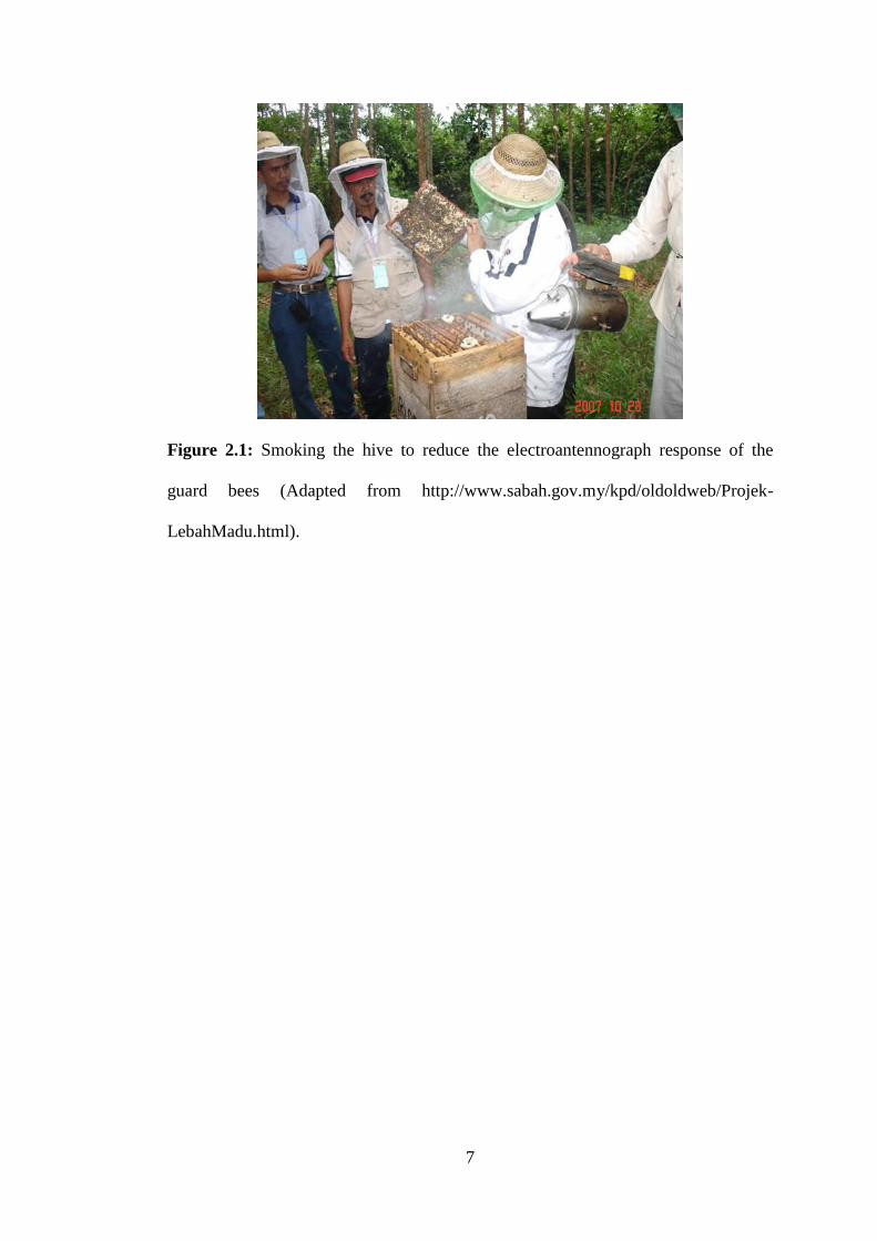
honey or other bee products to become more effective (Crane, 2009). During the
harvesting of honey or other bee products, the beekeepers smoke the bees (Figure
2.1) to reduce the electroantennograph response of the guard bees, who otherwise
would release a volatile alarm odour pheromone (Boch, 1962; Visscher et al., 1995).
When the smoke enters the hive, the antennae receptors of the guard bees are dulled
and they fail to sound the alarm. When exposed to smoke, bees are dramatically less
defensive and aggressive. As a result, the risk for engorgement and the tendency to
sting is reduced (Visscher et al., 1995).
6
Apis mellifera is a species of bees which has been used in most of the world’s
beekeeping. Other species include Apis dorsata and Apis cerana. Previously, bees
were kept mainly to produce honey and beeswax. Nowadays, beekeeping has been
tailored to different purposes like rearing queens or package bees for other
beekeepers who produce honey. Some may provide bee colonies for crops
pollinations and to produce royal jelly, pollen and bee venom since 1950s (Crane,
2009).
7
Figure 2.1: Smoking the hive to reduce the electroantennograph response of the
guard bees (Adapted from http://www.sabah.gov.my/kpd/oldoldweb/Projek-
LebahMadu.html).
8
2.2 Telomere and its regulation
2.2.1 Telomere
Telomeres are long repetitive DNA sequences located at the end of the linear
chromosomes (Blackburn, 1991) and bound by shelterin proteins or telosomes (Palm
and de Lange, 2008). Telosomes are the proteins which act as protection for the
telomere loop structure. This protection prevents the chromosome ends uncapped,
resemble a DNA break and activates DNA repair mechanism (Gomez et al., 2012).
In mammalian cells, telomere comprises double-stranded tandem repeats of
TTAGGG (Palm and de Lange, 2008). These repeat sequences do not encode for
proteins (Hodes, 1999). However, it consists of G-rich hexanucleotide repeats which
enable the single-stranded telomere G overhangs to form G-quadraplexes (Palm and
de Lange, 2008; Lipps and Rhodes, 2009), where each G base serves as both donor
and acceptor for hydrogen bond formation. In humans, telomeric G-quadraplex
structure is thought to contribute in telomere protection, suppression of
recombination and inhibition of telomerase-dependant telomere extension (Lipps and
Rhodes, 2009).
It is thought that telomere adopts the T-loop structure, where the telomere end folds
back on itself and the 3′ G strand overhang invades into the double-stranded DNA.
This structure formation is called D-loop (Palm and de Lange, 2008). Besides that, it
is believed that telomere structure can switch between a closed, protected state and
an open, extendable state, which allows the DNA terminus to undergo replication.
The protected state is necessary to safeguard the integrity of genomic material,
9
whereas the extendable state allows telomerase to extend short telomeres (Stewart et
al., 2012).
Telomeres protect the ends of linear chromosomes from breaking down and
degradation and in avoiding recognition and processing as double-strand breaks
(Kobryn and Chaconas, 2001). Studies carried out in yeast and other single
organisms have shown that the functions of telomeres include protection from the
chromosomal recombination, end-to-end fusion and recognition as damaged DNA,
determination of chromosomal localization within the nucleus and to regulate the cell
capacity for replication (Hodes, 1999).
Telomere length varies between chromosomes and between species. For instance,
mice have longer telomere length as compared to human. The shortest telomere
length is estimated to be 10 kbp. In human chromosomes, the telomere length is
between 0.5 and 15 kbp. In addition, telomere length is also dependent on the type of
tissue, age of the donor and the replicative history of the cells. For example,
chromosome 17p has shorter telomere length as compared to other chromosome
ends. Besides, it was observed that the average telomere length declines significantly
with increasing age in human nucleated blood cells (Aubert and Lansdorp, 2008).
Interestingly, rate of telomere attrition also varies markedly at different ages (Frenck
et al., 1998). An in vitro analysis of human fibroblast revealed that the telomere loss
is 50-100 bp per cell division (Allsopp et al., 1992).
10
2.2.2 Telomerase structure
Telomerase is a unique eukaryotic ribonucleoprotein (RNP) complex (Greider and
Blackburn, 1985; Blackburn, 1992; Bryan and Cech, 1999), which aids in the
stabilization of telomere length in human stem cells, reproductive cells (Wright et al.,
1996) and cancer cells (Kim et al., 1995; Shay and Bacchetti, 1997) by adding
TTAGGG repeats onto chromosomes ends. This addition is achieved using its
intrinsic RNA as a template for reverse transcription (Feng et al., 1995). There are
two conserved components of telomerase which are essential in the addition of
telomere repeat sequences. The first one is the core telomerase protein called
telomerase reverse transcriptase (TERT) and telomerase RNA component (TERC)
which complexes with TERT and provides the template for telomeric sequence
synthesis (Greider and Blackburn, 1989; Feng et al., 1995; Lingner et al., 1997). It is
thought that the human telomerase holoenzyme is assembled in the Cajal body,
where TERT and TERC form a RNP enzyme complex (Podlevsky and Chen, 2012).
While TERT and TERC are sufficient for the telomerase activity in vitro, other
proteins are also required for its assembly, trafficking and regulation (Blackburn and
Collins, 2011; Podlevsky and Chen, 2012).
Dyskerin is the most characterized mammalian telomerase accessory component
(Mitchell et al., 1999b). Dyskerin forms a core complex with three smaller proteins
NHP2 ribonucleoprotein (NHP2), NOP10 ribonucleoprotein (NOP10) and GAR1
ribonucleoprotein (GAR1). Dyskerin binds to an H/ACA box RNA structural motif
within TERC and to small nucleolar RNAs. This binding is essential for TERC
stability and telomerase function in vivo (Mitchell et al., 1999a; Mitchell et al.,
11
1999b; Chen et al., 2000). Another protein called telomerase Cajal body protein 1
(TCAB1) binds to TERC and regulates its trafficking (Tycowski et al., 2009;
Venteicher et al., 2009).
2.2.3 Interplay between telosome and telomerase in telomere maintenance
Telomere maintenance involves the interaction between telosome and telomerase. It
is the interplay between both which helps to maintain and protect the telomeres. Any
fault in either one would affect both the protection and maintenance of telomeres.
2.2.3.1 Telosome
The maintenance of telomere depends on the massive network of protein complexes
at the telomere. In this regard, telosome is central to this process. Telosome is
composed of six protein complexes which include telomeric repeat binding protein 1
and 2 (TRF 1 and TRF 2), the TRF-1 interacting protein 2 (TIN2),
Repressor/activator protein 1 homolog (RAP1), protection of telomeres 1 (POT1)
and telomere protection protein 1 (TPP1) (Liu et al., 2004a; de Lange, 2005). TRF1
and TRF2 have similar domain structure consisting of a C-terminal SANT/Myb
domain and an N-terminal TRF homology (TRFH) domain. These domains have
high binding specificity for the half site 5´-ÝTAGGGTTR-3´ in telomeric double-
stranded DNA (dsDNA) (De Lange, 2005). The two N-terminal
oligonucleotide/oligosaccharide-binding (OB) folds of POT1 are highly specific for
the 5´-TAGGGTTAG-3´ sequence of single-stranded G-overhangs (Lei et al., 2004).
TIN2 functions as a hub by binding to TPP1/POT1 heterodimer, TRF1 and TRF2
12
(Xin et al., 2008). TPP1 can also act together with POT1, TIN2 and telomerase
(Wang et al., 2007; Xin et al., 2008). Mammalian RAP1 is targeted to telomeric
DNA by directly interacting with TRF2. These six core proteins can act together as a
platform that recruits players from various pathways to the telomeres for
maintenance and protection (Lee et al., 2011). The details on the functions of each
telosome protein are described below.
2.2.3.1.1 Telomere repeat binding factor 1
TRF1 is the first double-stranded telomere DNA binding protein identified (Zhong et
al., 1992). It functions as a negative regulator for telomere length (Van Steensel and
de Lange, 1997). Study showed that the homozygous deletion of TRF1 in mice was
lethal to embryo during blastocyst stage with severe growth defects (Karlseder et al.,
2003). Apoptosis process was also accompanied these events suggesting that TRF1
plays vital roles that may be independent of telomere length regulation (Karlseder et
al., 2003). TRF1 expression is tightly regulated. As a consequence, it will lead to the
telomere homeostasis (Zeng et al., 2010).
13
2.2.3.1.2 Telomere repeat binding factor 2 and repressor/activator protein
1 homolog
TRF2 has appeared as an important player in maintaining the telomere length. It acts
as negative regulator for telomere length and contributes to telomere protection
(Smogorzewska et al., 2000). It acts as a hub by recruiting various factors for
telomere regulation. One of the ways of which TRF2 regulates telomere length is
through Ataxia telangiectasia mutated (ATM) mediated non-homologous end joining
(NHEJ) pathways. Study showed that TRF2-deficient mouse embryonic fibroblasts
(MEFs) had severe proliferation defects caused by enormous end-to-end fusions
facilitated by the NHEJ pathway (Celli and de Lange, 2005). Similar to TRF1,
homozygous inactivation of TRF2 in mice was embryonic lethal and cannot be
rescued by p53 abrogation. This means that different mechanisms are applied by
TRF1 and TRF2 to ensure survival during embryonic development (Celli and de
Lange, 2005).
The structure of RAP1 is highly conserved. It has a C-terminal (RCT) domain, a
BRCA1 C Terminus (BRCT) domain and Myb domain(s). Since mammalian RAP1
lacks telomere-binding capacity, it interacts with TRF2 for telomere localization (Li
et al., 2000; Palm and de Lange, 2008). Studies suggested that RAP1 repressed
homologous recombination (HR) at telomere. They found that TRF2/RAP1
complexes with DNA repair factor BTBD12 domain-containing protein 12
(BTBD12) and facilitates DNA damage response and Holliday junction processing.
In addition, number of DNA repair proteins has been found in the RAP1/TRF2
complex such as Rad50, Mre11, Poly [ADP-ribose] polymerase 1 (PARP1), and
14
Ku86/Ku70. In contrast to TRF1 and TRF2, RAP1-deficient mice appeared viable,
although with increased telomere recombination and fragility (Martinez et al., 2010).
2.2.3.1.3 Protection of telomeres 1
There are three main functions of POT1. The first one is to protect telomere ends
from ataxia telangiectasia and Rad3-related protein (ATR) dependent DNA damage
response. Other functions include to regulate telomerase-dependent telomere
elongation and controlling 5´-end resection at telomere termini (He et al., 2006;
Hockemeyer et al., 2006; Wu et al., 2006). Recently, Zou and team discovered that
TERRA (telomeric repeat-containing RNA), heterogeneous nuclear
ribonucleoprotein A1 (hnRNPA1), and POT1 could act together to remove
replication protein A (RPA) from telomeric ssDNA after DNA replication. RPA
exclusion is performed to support telomere end protection by inhibiting ATR-
mediated DNA damage signals (Flynn et al., 2011). Other than that, TPP1 can
interact directly with POT1 to enhance POT1 affinity for telomeric ssDNA (Wang et
al., 2007; Xin et al., 2007). Interestingly, TPP1 interacts directly with telomerase for
its recruitment to telomeres (Wang et al., 2007; Xin et al., 2007).
2.2.3.1.4 Telomere protection protein 1
Human TPP1 interacts with both TIN2 and POT1 (Liu et al., 2004b) by binding to
the c-terminus of POT1. In addition, TPP1 is required for POT1 to localize telomere
(Liu et al., 2004b; Kibe et al., 2010; Tejera et al., 2010). TPP1 interacts directly with
POT1 and enhances POT1 affinity for telomeric ssDNA (Wang et al., 2007; Xin et
15
al., 2007). Besides, TPP1 is recruited to telomere through its interaction with
telomerase (Wang et al., 2007; Xin et al., 2007). It has been shown that TPP1 null
MEFs and mice had decreased telomerase binding to telomeres and short telomeres
(Tejera et al., 2010). It had been suggested that telomere length is regulated through
the interaction between TPP1 OBFC1/Stn1, an OB-fold protein that directly binds to
ss-telomeric DNA (Wan et al., 2009). In support of this notion, recent study
discovered that OBFC1/Stn1-containing CTC1, STN1 and TEN1 (CST) complex is
involved in 5´-end resection for 3´-overhang generation. It was also found that the
depletion of OB Fold-containing Protein 1/Stn1 (OBFC1/Stn1) leads to telomere
elongation (Chen et al., 2012; Wu et al., 2012). Hence, these results showed that
TPP1 is crucial in both telomere end protection and length regulation.
2.2.3.1.5 TRF1-interacting protein 2
TIN2 interacts directly with TRF1, TRF2, and TPP1 (Xin et al., 2008) and acts as the
central component in the telosome complex (O'Connor et al., 2006). The disruption
of TIN2 leads to accumulation of RPA binding to telomere termini, significantly
decreased telomere localization of all telosome components, and increased ATR-
mediated DNA damage responses, similar with the results in POT1a/1b double
knockout mice (Takai et al., 2011). Presently, the only identified mutation in
telosome component in human diseases is TIN2. Patients with dyskeratosis congenita
(DC) have dysfunction in TIN2-dependent telomere length control. It is also believed
that TPP1-mediated telomerase recruitment might be interrupted. DC patients had
been found to express TIN2 with missense mutations which might justify the
16
telomere shortening phenotype observed in patients (Yang et al., 2011). Hence, TIN2
could be a possible target for therapeutic and diagnostic studies.
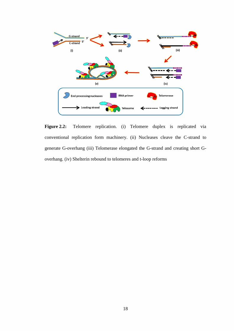
2.2.3.2 Telomere replication
Telomere replication involves multi-step processes (Figure 2.2). Firstly, the telomere
duplex is replicated via the conventional replication form machinery. In lagging
strand, telomere generated will gain a 3´ overhang automatically because RNA
primer has been removed on the terminal Okazaki fragment. In contrast to lagging
strand, overhang is not form on the telomere replicated by leading strand. Therefore,
telomere is synthesised by a series of DNA processing reactions. In budding yeast,
the processing steps are similar to those used to resect double-strand breaks during
DNA repair (Longhese et al., 2010). Initiation of resection requires recognition of the
DNA terminus by the Mre11-Rad50-Xrs2 (MRX) complex (MRN in humans) and
subsequent recruitment of the nucleases, exonuclease 1 (EXO1) and/or DNA
replication helicase/nuclease 2 (DNA2). These act in accordance with the helicase
Sgs1 to cleave the DNA 5´ strand thus creating the 3´ overhang. Although it is
unclear whether EXO1 and DNA2 play a similar role in human telomeres, genetic
analysis in mice has shown that another repair nuclease, Apollo/Snm1b is involved in
the overhang generation on leading strand telomeres (Chen et al., 2008; Lam et al.,
2010). Apollo can associate specifically with telomeres through an interaction with
TRF2 (Chen et al., 2008). DNA-processing to generate G-overhangs occurs
regardless of whether a cell expresses telomerase (Hemann and Greider, 1999). In
telomerase positive cells, the overhang is elongated by the addition of new repeats on
to the DNA terminus. Although the recruitment of telomerase to human telomere is
17
not fully understood, it is thought that the recruitment involves the trafficking of
telomerase to the telomere in association with Cajal bodies and interaction between
telomerase and TPP1 (Cristofari et al., 2007; Venteicher et al., 2009; Abreu et al.,
2010; Tejera et al., 2010). After this event, the complementary C-strand is filled-in to
leave an overhang that ranges in length from ∼40 to 400 nt (Huffman et al., 2000;
Zhao et al., 2009). Finally, the telomeres are rebound by telosome/shelterin and the t-
loop reforms.
18
Figure 2.2: Telomere replication. (i) Telomere duplex is replicated via
conventional replication form machinery. (ii) Nucleases cleave the C-strand to
generate G-overhang (iii) Telomerase elongated the G-strand and creating short G-
overhang. (iv) Shelterin rebound to telomeres and t-loop reforms
19
2.2.3.3 Telomere elongation by telomerase is tightly regulated
Telomere elongation by telomerase is tightly regulated. Telomerase elongates
telomeric DNA during S phase and into M phase. This means that the elongation is
cell-cycle-regulated (Diede and Gottschling, 1999; Marcand et al., 2000).
Telomerase favourably elongates the shortest telomeres. This is in accordance to cis-
regulatory mechanisms mediated through the telomere DNA–protein complex. As a
result, only a subset of telomeres may be elongated in any cell cycle (Teixeira et al.,
2004). Telomere elongation extent is very sensitive to the level of telomerase in cells.
This event is obvious in the study of the haploinsufficiency for genes encoding
telomerase components in yeast, mouse and human cells (Vulliamy et al., 2001;
Erdmann et al., 2004; Armanios et al., 2005; Hao et al., 2005; Yamaguchi et al.,
2005; Mozdy and Cech, 2006; Strong et al., 2011)
The reason behind this is probably due to imbalance stoichiometry between
telomerase and its substrates, in addition to other telomerase independent processes.
Although haematopoietic stem cells (HSCs) are naturally enriched with telomerase,
the effect of multiple cell division and ageing can be seen on their telomere length
(Vaziri et al., 1994; Chiu et al., 1996). Interestingly, telomere length in human male
germ cells remains stable or even elongate with age (Allsopp et al., 1992). Even so,
the mechanisms by which telomeres are maintained in germ cell lineages, which are
enriched for telomerase (Kim et al., 1994), are not fully understood.
20
Short telomeres have been suggested as having protective role as an innate tumour
suppressive mechanism in long-lived, multicellular organisms (Greider, 2006). The
reason behind this is thought to stem from the observation that in most cancer cells,
telomerase is upregulated to maintain their sustainability (Kim et al., 1994). Besides
that, limiting telomerase levels may also prevent unwanted telomere addition at DNA
double-strand-break sites, which could happen if telomerase competes with
appropriate DNA repair mechanisms (Zhou et al., 2000; Makovets and Blackburn,
2009).
2.3 Telomeres and ageing
2.3.1 Replicative ageing
While it is true that telomere shortening plays a protective role against cancer cells, it
appears that this decision has resulted in ageing consequence. Ageing is defined as a
process associated with the gradual decline in the performance of organ systems.
This decline has resulted in the loss of reserve capacity which in turn leads to an
increased chance of death (Gompertz, 1825). In some organ systems, this loss of
reserve capacity with increasing age can be attributed to the loss of cell function
(Martin et al., 1970).
The process by which most normal human cells "count" the number of times they
have divided and eventually undergoing a growth arrest, cellular senescence is
defined as replicative ageing. This process is dependent on telomere shortening
(Wright and Shay, 2005). The first observation that suggests the existence of internal
21
counting mechanism within the cell came from Hayflick. Hayflick observed that
cultured human fibroblasts have limited number of cell divisions (Hayflick and
Moorhead, 1961). The subsequent study then revealed that the telomeres shorten
with every cell division, suggesting that telomere loss is the molecular clock that
drives ageing (Harley et al., 1990; Hastie et al., 1990; Harley, 1991; Allsopp et al.,
1992).
To understand the reason for this limitation, it is best to appreciate the disposable
soma theory (Kirkwood, 1998). The disposable soma theory proposes that the rate at
which the species age is the balance between the energy devoted to reproduction
versus somatic repair. This means that if too much energy is invested in the repair of
somatic cells, less energy is left for reproduction and vice versa. Species that are
unable to survive very long due to the high mortality rate must invest most of their
energy in reproduction rather than cell repair.
For example, a mouse that sufficiently repaired itself for 20 years is making bad
investment since most mice will be eaten by its predators within 3 months.
Therefore, it is better for the mice to invest more energy in the early reproduction
and less in maintenance and repair (Wright and Shay, 2005). As humans have longer
average survival, we have been evolutionarily selected to invest more energy on
tissue maintenance and repair as compared to reproduction unlike mice. However,
the variety of tissue maintenance and repair processes such as the efficiency of DNA
repair, protection against oxidative damage and others limit the amount of energy
invested and contribute to ageing (Wright and Shay, 2005).
22
Apoptosis of damaged cells and replacing them with new ones are efficient ways of
keeping cells healthy. Besides that, replacing dying cells with new healthy cells can
dilute the build-up of ‘unrepairable and indigestible’ products that can contribute to
ageing. Nonetheless, using cell turnover to repair tissues may carry risk since
mistakes can occur during DNA replication. These mistakes can lead to harmful
mutations which will then lead to cancer (Vogelstein and Kinzler, 1993). Therefore,
by limiting the total number of times a cell could divide provides a powerful barrier
for the body from cancer formation (Wright and Shay, 1995).
There has been mounting evidences that the progressive loss of telomeric ends of
chromosomes is an important intrinsic timing mechanism in the ageing process, both
in cell culture and in vivo (Harley et al., 1990; Hastie et al., 1990). Based on the
analysis of cultured human fibroblasts and lymphocytes, the rate of loss of telomeres
is 50-100 bp per cell division (Allsopp et al., 1992). Short telomeres can induce anti-
proliferative signals that result in cellular senescence (Harley, 1991; Shay, 1995; Zou
et al., 2004). These events are discussed in detail below.
2.3.2 Replicative senescence
Telomere shortening can induce anti-proliferative signals which result in cellular
senescence (Harley, 1991; Shay, 1995; Zou et al., 2004). Cellular senescence
triggered by telomere shortening is termed replicative senescence. Replicative
senescence is caused by the ‘uncapping’ of critically shortened telomeres. This
happens when telomere-binding proteins are no longer protecting telomeres, making
telomeres recognized as single and lead to the breaking of the double-strand DNA.
23
As a result, DNA damage response pathway is activated like p53 pathway (Vaziri
and Benchimol, 1996; Takai et al., 2003) which will then lead to the growth arrest of
cells, apoptosis and senescence (Chin et al., 1999; Wong et al., 2003; Ferrón et al.,
2004; Flores and Blasco, 2009).
Interestingly, these senescent cells can remain viable for years (Shay and Wright,
2007). The accumulation of senescent cells is recognized as one of the two
mechanisms which probably contribute to ageing. The production of different
constellation of proteins as compared to those that are non-senescent but quiescent
adjacent cell during the accumulation of senescent cells is believed to change the
homeostasis of that tissue and lead to ageing (Shay and Wright, 2007). Studies
reported abundant senescent cells in telomerase null mice (Satyanarayana et al.,
2003). The senescent cells are usually marked using beta galactosidase staining and
these cells are always associated with changes in p53, p16 and p21 expression (Dimri
et al., 1995; Shelton et al., 1999; Oeseburg et al., 2009). The accumulation of
senescent cells may also lead to another mechanism of ageing which is the loss of
stem cell function (Collado et al., 2007). Stem cells are important because they
maintain the homeostasis of tissues by replenishing senescent and apoptotic cells.
Besides that, they repair damage that occurs throughout life (Rando, 2007). Various
studies reported the loss of stem cell function through telomere shortening in a
variety of tissues and experimental systems (Flores et al., 2006). The loss of stem
cell functions impair tissue repair and hence weaken the tissue functions and lead to
ageing (Collado et al., 2007).
24
2.4 Telomeres and longevity of life
It has been suggested that long telomeres may provide protection against cellular
senescence (Herbig et al., 2006). They could also be an indicator for unique genome
stability and cellular health (Epel et al., 2004). During the last 20 years, there are
rising evidences suggesting that telomere attrition may function as a key timing
mechanism during the ageing process in various species (López-Otín et al., 2013).
Shorter telomere length in humans is associated with many age related diseases such
as cardiovascular diseases, cancer, cognitive decline, diabetes and overall mortality
(Armanios, 2013). Interestingly, gender has also played its role in longevity of life
(Barrett and Richardson, 2011). Women are thought to live longer than men because
of oestrogen. Oestrogen has been shown to be associated with telomere length (Vina
et al., 2005).
Telomere attrition is negatively correlated with organismal life span (Haussmann et
al., 2003). Telomere length appeared to increase across its life span in long-lived
seabird, Oceanodromo leucorhoa (Haussmann et al., 2007). It has also found that
this species show little or no accumulation of short telomeres over time (Haussmann
and Mauck, 2008). Thus, the study on this species might offer the secret to longevity
and reproductive success.
The link between telomere length and human lifespan has been reported (Gomes et
al., 2011; Barrett et al., 2013). Study in the elderly aged more than 60 years showed
that telomere attrition is significantly associated with higher mortality rates, both
from infectious and cardiovascular diseases (Cawthon et al., 2003). Moreover,


![01/15/2021 09:58 am IP:[10.10.250.10] Packet No: 40772](https://img.pdfslide.us/doc/110x75/6250a73bf1f3a1706e63583b/01152021-0958-am-ip101025010-packet-no-40772-.jpg)
