Embed Size (px)
Citation preview

Basic Research—Technology
Effect of Combined Digital Imaging Parameterson Endodontic File MeasurementsMatheus Lima de Oliveira, DDS, MSc,* Geraldo Camilo de Souza Pinto, DDS,‡
Glaucia Maria Bovi Ambrosano, Agr Eng, MSc, PhD,†
and Guilherme Monteiro Tosoni, DDS, MSc, PhD‡
Abstract
Introduction: This study assessed the effect of thecombination of a dedicated endodontic filter, spatialresolution, and contrast resolution on the determinationof endodontic file lengths. Methods: Forty extractedsingle-rooted teeth were x-rayed with K-files (ISO size10 and 15) in the root canals. Images were acquiredusing the VistaScan system (D€urr Dental, Beitigheim-Bissingen, Germany) under different combining param-eters of spatial resolution (10 and 25 line pairs permillimeter [lp/mm]) and contrast resolution (8- and 16-bit depths). Subsequently, a dedicated endodontic filterwas applied on the 16-bit images, creating 2 additionalparameters. Six observers measured the length of theendodontic files in the root canals using the softwarethat accompanies the system. The mean values of theactual file lengths and the measurements of the radio-graphic images were submitted to 1-way analysis ofvariance and the Tukey test at a level of significanceof 5%. The intraobserver reproducibility was assessedby the intraclass correlation coefficient. Results: Allcombined image parameters showed excellent intraob-server agreement with intraclass correlation coefficientmeans higher than 0.98. The imaging parameter of 25lp/mm and 16 bit associated with the use of theendodontic filter did not differ significantly from theactual file lengths when both file sizes were analyzedtogether or separately (P > .05). When the size 15 filewas evaluated separately, only 8-bit images differedsignificantly from the actual file lengths (P # .05).Conclusions: The combination of an endodontic filterwith high spatial resolution and high contrast resolutionis recommended for the determination of file lengthswhen using storage phosphor plates. (J Endod2012;38:1404–1407)From the Departments of *Oral Diagnosis and †Social Dentis‡Department of Oral Diagnosis and Surgery, Araraquara Dental Sch
Financial support received from FAPESP, a research foundationAddress requests for reprints to Dr Matheus Lima de Oliveira, De
Box 52, 13414-903 Piracicaba, SP, Brazil. E-mail address: matheuso0099-2399/$ - see front matter
Copyright ª 2012 American Association of Endodontists.http://dx.doi.org/10.1016/j.joen.2012.06.006
1404 Oliveira et al.
Key WordsDigital dental radiography, endodontics, odontometry, software
Digital radiography dispenses with the processing of film and allows easy storage ofdynamic images that can be postprocessed by the observer to facilitate interpreta-
tion (1–3). Considering the wide clinical application of digital radiographic systems inthe health field and the advancement of these systems over the years, dentists need to becareful to purchase a system that best meets their goals and to select the best imagingparameter and postprocessing tool when given a specific task.
In the endodontic field, digital systems have established their usefulness becausethey have features that allow image enhancement for endodontic length determination(4). The success of endodontic treatment is closely related to the accuracy in deter-mining the actual length of the tooth by measuring the distance between the tip ofthe endodontic file and the radiographic apex (5). Digital systems seem to providemore accurate measurements with reduced subjective differences (3).
The VistaScan digital radiographic system (D€urr Dental, Beitigheim-Bissingen,Germany) uses storage phosphor plates as image receptors. It captures 8- and 16-bit images with spatial resolution of up to 25 line pairs permillimeter (lp/mm), allowingmore image details. In addition, a dedicated endodontic filter is available for imageenhancement. The contrast resolution or bit depth defines the range of shades ofgray in the radiographic image. Most of the digital dental systems provide 8-bit images(6), which hold 256 shades of gray. Sixteen-bit images have an increased scale ofcontrast, with 65,536 shades of gray. At this point, little is known in the scientific liter-ature about the combination of an endodontic filter and imaging parameters such asspatial resolution and contrast resolution for the determination of endodontic filelengths. The aim of this in vitro study was to assess the combination of a dedicatedendodontic filter, spatial resolution, and contrast resolution on the determination ofendodontic file lengths.
Materials and MethodsAfter institutional research ethics committee approval (protocol # 058/2009), 40
extracted human permanent single-rooted teeth with intact surfaces and a root canalfree of calcification were prepared following a previously described methodology(7). Size 10 and 15 K-type files (Dentsply Maillefer, Ballaigues, Switzerland) wereplaced at random lengths ranging 0 to 2 mm from the apical limit based on Ingle’sradiographic method (8). A ruler (Dentsply Maillefer) was used to measure the actualfile lengths with no magnification. The file lengths were not related to tooth lengths to
try, Piracicaba Dental School, State University of Campinas, Campinas, S~ao Paulo, Brazil; andool, S~ao Paulo State University, Araraquara, S~ao Paulo, Brazil.of the State of S~ao Paulo, Brazil (process no. 08/01715-5).partment of Oral Diagnosis, Piracicaba Dental School, State University of Campinas, UNICAMP, [email protected]
JOE — Volume 38, Number 10, October 2012
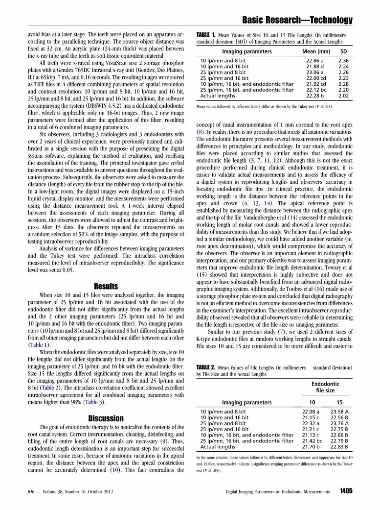
TABLE 1. Mean Values of Size 10 and 15 File Lengths (in millimeters �standard deviation [SD]) of Imaging Parameters and the Actual Lengths
Imaging parameters Mean (mm) SD
10 lp/mm and 8 bit 22.86 a 2.3610 lp/mm and 16 bit 21.88 d 2.2425 lp/mm and 8 bit 23.06 a 2.2625 lp/mm and 16 bit 22.00 cd 2.2310 lp/mm, 16 bit, and endodontic filter 21.92 cd 2.2825 lp/mm, 16 bit, and endodontic filter 22.12 bc 2.20Actual lengths 22.28 b 2.02
Mean values followed by different letters differ as shown by the Tukey test (P # .05).
TABLE 2. Mean Values of File Lengths (in millimeters � standard deviation)by File Size and the Actual Lengths
Imaging parameters
Endodonticfile size
10 15
10 lp/mm and 8 bit 22.08 a 23.58 A10 lp/mm and 16 bit 21.15 c 22.56 B25 lp/mm and 8 bit 22.32 a 23.76 A25 lp/mm and 16 bit 21.21 c 22.75 B10 lp/mm, 16 bit, and endodontic filter 21.13 c 22.66 B25 lp/mm, 16 bit, and endodontic filter 21.42 bc 22.79 BActual lengths 21.70 b 22.83 B
In the same column, mean values followed by different letters (lowercase and uppercase for size 10
and 15 files, respectively) indicate a significant imaging parameter difference as shown by the Tukey
test (P # .05).
Basic Research—Technology
avoid bias at a later stage. The teeth were placed on an apparatus ac-cording to the paralleling technique. The source-object distance wasfixed at 32 cm. An acrylic plate (24-mm thick) was placed betweenthe x-ray tube and the teeth as soft-tissue equivalent material.All teeth were x-rayed using VistaScan size 2 storage phosphorplates with a Gendex 765DC Intraoral x-ray unit (Gendex, Des Plaines,IL) at 65kVp, 7 mA, and 0.16 seconds. The resulting images were storedas TIFF files in 4 different combining parameters of spatial resolutionand contrast resolution: 10 lp/mm and 8 bit, 10 lp/mm and 16 bit,25 lp/mm and 8 bit, and 25 lp/mm and 16 bit. In addition, the softwareaccompanying the system (DBSWIN 4.5.2) has a dedicated endodonticfilter, which is applicable only on 16-bit images. Thus, 2 new imageparameters were formed after the application of this filter, resultingin a total of 6 combined imaging parameters.
Six observers, including 3 radiologists and 3 endodontists withover 2 years of clinical experience, were previously trained and cali-brated in a single session with the purpose of presenting the digitalsystem software, explaining the method of evaluation, and verifyingthe assimilation of the training. The principal investigator gave verbalinstructions and was available to answer questions throughout the eval-uation process. Subsequently, the observers were asked to measure thedistance (length) of every file from the rubber stop to the tip of the file.In a low-light room, the digital images were displayed on a 15-inchliquid crystal display monitor, and the measurements were performedusing the distance measurement tool. A 1-week interval elapsedbetween the assessments of each imaging parameter. During allsessions, the observers were allowed to adjust the contrast and bright-ness. After 15 days, the observers repeated the measurements ona random selection of 50% of the image samples, with the purpose oftesting intraobserver reproducibility.
Analysis of variance for differences between imaging parametersand the Tukey test were performed. The intraclass correlationmeasured the level of intraobserver reproducibility. The significancelevel was set at 0.05.
ResultsWhen size 10 and 15 files were analyzed together, the imaging
parameter of 25 lp/mm and 16 bit associated with the use of theendodontic filter did not differ significantly from the actual lengthsand the 2 other imaging parameters (25 lp/mm and 16 bit and10 lp/mm and 16 bit with the endodontic filter). Two imaging param-eters (10 lp/mm and 8 bit and 25 lp/mm and 8 bit) differed significantlyfrom all other imaging parameters but did not differ between each other(Table 1).
When the endodontic files were analyzed separately by size, size 10file lengths did not differ significantly from the actual lengths on theimaging parameter of 25 lp/mm and 16 bit with the endodontic filter.Size 15 file lengths differed significantly from the actual lengths onthe imaging parameters of 10 lp/mm and 8 bit and 25 lp/mm and8 bit (Table 2). The intraclass correlation coefficient showed excellentintraobserver agreement for all combined imaging parameters withmeans higher than 98% (Table 3).
DiscussionThe goal of endodontic therapy is to neutralize the contents of the
root canal system. Correct instrumentation, cleaning, disinfecting, andfilling of the entire length of root canals are necessary (9). Thus,endodontic length determination is an important step for successfultreatment. In some cases, because of anatomic variations in the apicalregion, the distance between the apex and the apical constrictioncannot be accurately determined (10). This fact contradicts the
JOE — Volume 38, Number 10, October 2012
concept of canal instrumentation of 1 mm coronal to the root apex(8). In reality, there is no procedure that meets all anatomic variations.The endodontic literature presents several measurement methods withdifferences in principles and methodology. In our study, endodonticfiles were placed according to similar studies that assessed theendodontic file length (3, 7, 11, 12). Although this is not the exactprocedure performed during clinical endodontic treatment, it iseasier to validate actual measurements and to assess the efficacy ofa digital system in reproducing lengths and observers’ accuracy inlocating endodontic file tips. In clinical practice, the endodonticworking length is the distance between the reference points in theapex and crown (4, 13, 14). The apical reference point isestablished by measuring the distance between the radiographic apexand the tip of the file. Vandenberghe et al (14) assessed the endodonticworking length of molar root canals and showed a lower reproduc-ibility of measurements than this study. We believe that if we had adop-ted a similar methodology, we could have added another variable (ie,root apex determination), which would compromise the accuracy ofthe observers. The observer is an important element in radiographicinterpretation, and our primary objective was to assess imaging param-eters that improve endodontic file length determination. Tewary et al(15) showed that interpretation is highly subjective and does notappear to have substantially benefited from an advanced digital radio-graphic imaging system. Additionally, de Toubes et al (16) made use ofa storage phosphor plate system and concluded that digital radiographyis not an efficient method to overcome inconsistencies from differencesin the examiner’s interpretation. The excellent intraobserver reproduc-ibility observed revealed that all observers were reliable in determiningthe file length irrespective of the file size or imaging parameter.
Similar to our previous study (7), we used 2 different sizes ofK-type endodontic files at random working lengths in straight canals.File sizes 10 and 15 are considered to be more difficult and easier to
Digital Imaging Parameters on Endodontic Measurements 1405

TABLE 3. Intraclass Correlation Coefficient Means and Minimum andMaximum Values
Imaging parameters Mean Minimum Maximum
10 lp/mm and 8 bit 0.983 0.934 0.99410 lp/mm and 16 bit 0.984 0.975 0.99425 lp/mm and 8 bit 0.982 0.973 0.99625 lp/mm and 16 bit 0.991 0.986 0.99610 lp/mm, 16 bit, and
endodontic filter0.993 0.979 0.999
25 lp/mm, 16 bit, andendodontic filter
0.992 0.987 0.995
Basic Research—Technology
visualize radiographically, respectively (17, 18). In addition to theendodontic file size, factors including image receptor type, scatteredradiation effects, differences in bone density, superimposition oftrabecular bone pattern, bone processes and roots, and selection ofthe optimal exposure time can influence radiographic discriminationof the file (18). When comparing the 2 file sizes within different imagingparameters, this study revealed that the observers were more accuratewith size 15 files than with size 10 because the former presented fewersignificant differences from the actual lengths. This could be becausethe greater thickness covers more pixels of the image, allowing bettervisualization (19).
The 2 spatial resolutions tested were observed within the imagingparameters that differed from the actual lengths. This leads us to believethat spatial resolution itself did not interfere in the study, and even the10-lp/mm images were of sufficient quality to enable the measurementsto be performed even with a K-type endodontic file size 10 (14). Theseresults are in agreement with Li et al (20), who found that higher spatialresolution images do not improve the detection of proximal caries ina digital phosphor plate system.
As far as we know, there is no other study in the literature that hasdirectly compared contrast resolutions in the determination ofendodontic file lengths. According to Vandenberghe et al (21), highercontrast resolution is helpful for periodontal diagnosis, and accordingto K€unzel et al (22), it improves image quality. These studies supportour findings that 16-bit images are more accurate than 8-bit imagesin determining file lengths irrespective of the file size.
Studies related to the detection of periapical lesions and endodontictreatment success have investigated the effect of image processing usingdigital tools for diagnosis including brightness and contrast adjustments(18, 23, 24). The proper application of these tools can improve diagnosisby showing subtle differences of radiodensity from surrounding tissuesand facilitating the visualization of endodontic file tips (14). Theendodontic filter of the VistaScan system has been designed for 16-bitimages, and its use in this in vitro study significantly improved the effi-cacy of file length determination, mainly of small-sized files. Kal et al (3)revealed that contrast/brightness, invert, and edge enhancement toolsmay be recommended for accurate file length measurements althoughprocessed phosphor plate digital images are equivalent to film for inter-preting root canal morphology and the final assessment of endodontictreatment. Unlike our study, Vandenberghe et al (14) showed that theuse of dedicated filtering does not influence the outcome of workinglength determination. This is probably because the measurementswere performed in curved canals that could have concealed the actualfile lengths on the radiographic image.
Despite the increasing availability of enhancement tools in soft-ware applications, it is important to check their efficacy objectively.Haiter-Neto et al (25), using the same digital system, assessed 3 differentimage filters for the detection of proximal caries and found that only thefilter ‘‘Fine’’ was accurate for diagnosis. According to Yalcinkaya et al
1406 Oliveira et al.
(26), specific filters can be used on the VistaScan system dependingon the diagnostic task. In conclusion, the combination of imagingparameters such as endodontic filter, high spatial resolution, andhigh contrast resolution is recommended for the determination of filelengths in a storage phosphor plate system.
AcknowledgmentsThe authors deny any conflicts of interest related to this study.
References1. Wenzel A, Gr€ondahl H. Direct digital radiography in the dental office. Int Dent J
1995;45:27–34.2. Wenzel A, Hintze H. Perception of image quality in direct digital radiography after
application of various image treatment filters for detectability of dental disease. Den-tomaxillofac Radiol 1993;22:131–4.
3. Kal B, Baksi B, D€undar N, Sen B. Effect of various digital processing algorithms onthe measurement accuracy of endodontic file length. Oral Surg Oral Med Oral PatholOral Radiol Endod 2007;103:280–4.
4. Woolhiser GA, Brand JW, Hoen MM, Geist JR, Pikula AA, Pink FE. Accuracy of film-based, digital, and enhanced digital images for endodontic length determination.Oral Surg Oral Med Oral Pathol Oral Radiol Endod 2005;99:499–504.
5. Certosimo FJ, Milos MF, Walker T. Endodontic working length determination–where does it end? Gen Dent 1999;47:281–6.
6. Wenzel A, Haiter-Neto F, Gotfredsen E. Influence of spatial resolution and bit depthon detection of small caries lesions with digital receptors. Oral Surg Oral Med OralPathol Oral Radiol Endod 2007;103:418–22.
7. Oliveira ML, Ambrosano GM, Almeida SM, Haiter-Neto F, Tosoni GM. Efficacy ofseveral digital radiographic imaging systems for laboratory determination ofendodontic file length. Int Endod J 2011;44:469–73.
8. Shanmugaraj M, Nivedha R, Mathan R, Balagopal S. Evaluation of working lengthdetermination methods: an in vivo / ex vivo study. Indian J Dent Res 2007;18:60–2.
9. Patel S, Ricucci D, Durak C, Tay F. Internal root resorption: a review. J Endod 2010;36:1107–21.
10. Herrera M, Abalos C, Planas AJ, Llamas R. Influence of apical constriction diameteron Root ZX apex locator precision. J Endod 2007;33:995–8.
11. Velders XL, Sanderink GC, van der Stelt PF. Dose reduction of two digital sensorsystems measuring file lengths. Oral Surg Oral Med Oral Pathol Oral Radiol Endod1996;81:607–12.
12. Ellingsen MA, Harrington GW, Hollender LG. Radiovisiography versus conventionalradiography for detection of small instruments in endodontic length determination.Part 1. In vitro evaluation. J Endod 1995;21:326–31.
13. Sheaffer JC, Eleazer PD, Scheetz JP, Clark SJ, Farman AG. Endodontic measurementaccuracy and perceived radiograph quality: effects of film speed and density. OralSurg Oral Med Oral Pathol Oral Radiol Endod 2003;96:441–8.
14. Vandenberghe B, Bud M, Sutanto A, Jacobs R. The use of high-resolution digitalimaging technology for small diameter K-file length determination in endodontics.Clin Oral Investig 2010;14:223–31.
15. Tewary S, Luzzo J, Hartwell G. Endodontic radiography: who is reading the digitalradiograph? J Endod 2011;37:919–21.
16. de Toubes KM, Cortes MI, Valadares MA, Fonseca LC, Nunes E, Silveira FF. Compar-ative analysis of accessory mesial canal identification in mandibular first molars byusing four different diagnostic methods. J Endod 2012;38:436–41.
17. Fuge KN, Stuck AM, Love RM. A comparison of digitally scanned radiographs withconventional film for the detection of small endodontic instruments. Int Endod J1998;31:123–6.
18. Friedlander LT, Love RM, Chandler NP. A comparison of phosphor-plate digitalimages with conventional radiographs for the perceived clarity of fine endodontic filesand periapical lesions. Oral Surg Oral Med Oral Pathol Oral Radiol Endod 2002;93:321–7.
19. Zinelis S, Magnissalis EA, Margelos J, Lambrianidis T. Clinical relevance of standard-ization of endodontic files dimensions according to the ISO 3630-1 specification.J Endod 2002;28:367–70.
20. Li G, Berkhout WER, Sanderink GC, Martins M, van der Stelt PF. Detection of in vitroproximal caries in storage phosphor plate radiographs scanned with different reso-lutions. Dentomaxillofac Radiol 2008;37:325–9.
21. Vandenberghe B, Bosmans H, Yang J, Jacobs R. A comprehensive in vitro study of imageaccuracy and quality for periodontal diagnosis. Part 2: the influence of intra-oral imagereceptor on periodontal measurements. Clin Oral Investig 2010;15:551–62.
22. K€unzel A, Weimar S, Willers R, Becker J. Diagnostic of tooth fractures with the Vis-tascan system. Dent Traumatol 2008;24:537–41.
JOE — Volume 38, Number 10, October 2012

Basic Research—Technology
23. Kullendorff B, Nilsson M. Diagnostic accuracy of direct digital dental radiography forthe detection of periapical bone lesions: II. Effects on diagnostic accuracy afterapplication of image processing. Oral Surg Oral Med Oral Pathol Oral Radiol Endod1996;82:585–9.
24. Li G, Sanderink GC, Welander U, McDavid WD, Nasstrom K. Evaluation of endodonticfiles in digital radiographs before and after employing three image processing algo-rithms. Dentomaxillofac Radiol 2004;33:6–11.
JOE — Volume 38, Number 10, October 2012
25. Haiter-Neto F, Casanova M, Frydenberg M, Wenzel A. Task-specific enhancement filters instorage phosphor images from the Vistascan system for detectionof proximal caries lesionsof known size. Oral Surg Oral Med Oral Pathol Oral Radiol Endod 2009;107:116–21.
26. Yalcinkaya S, K€unzel A, Willers R, Thoms M, Becker J. Subjective image quality ofdigitally filtered radiographs acquired by the D€urr Vistascan system comparedwith conventional radiographs. Oral Surg Oral Med Oral Pathol Oral Radiol Endod2006;101:643–51.
Digital Imaging Parameters on Endodontic Measurements 1407