Embed Size (px)
Citation preview
9454
Abstract. – OBJECTIVE: The pathogenesis of tongue cancer (TA) has not been fully illustrated. Cyclooxygenase-2 (COX-2) is correlated with the precancerous lesion of oral cavity mucosa and malignant transformation. The focal adhesion kinase (FAK) and gap junction protein connex-in 43 (Cx43) are involved in the occurrence and progression of tumors. This study aimed to in-vestigate the effect of celecoxib on the prolifer-ation, malignant transformation, and expression of FAK and Cx43 proteins.
MATERIALS AND METHODS: Healthy male Sprague-Dawley (SD) rats (4 months old) were divided into control, model and celecoxib group. 7,12-dimethylbenzanthracene (DMBA) was used to generate tongue mucosal carcinoma, coupled with celecoxib intervention. At 8, 12, 16, and 20 weeks after induction, the rat survival status, the tumor formation rate and the tongue tissue mor-phology were observed. Meanwhile, the expres-sion of FAK and Cx43 was also evaluated by us-ing immunohistochemistry (IHC).
RESULTS: Tumor occurrence rates after in-duction were 0, 26.67%, 66.67%, and 80% at 8, 12, 16, and 20 weeks, respectively. The celecox-ib treatment decreased such rats to 0, 0, 0, and 13.33%, respectively (p<0.05 compared to mod-el group). No significant change was observed in control group, whilst model group had mild to severe hyperplasia and squamous carcinoma with elongated time. Celecoxib treatment signifi-cantly improved the tissue morphology (p<0.05). The model group also had elevated FAK and de-pressed Cx43 protein expression (p<0.05). With elongated time, the FAK expression was further increased whilst Cx43 protein was depressed (p<0.05 compared to model group).
CONCLUSIONS: The focal application of cele-coxib effectively inhibited the DMBA-induced rat TA, possibly via regulating FAK and CX43 pro-tein expression, and inhibiting oral epidermal hyperplasia.
Key Words:Celecoxib, Tongue cancer, FAK, Cx43 protein.
Introduction
Oral cavity cancer has a relatively higher inci-dence. Among those, the most common is squa-mous cell carcinoma (SCC). Tongue cancer is one common malignant tumor in the oral cavity and has insignificant symptoms during onset. Most patients thus already have infiltration or metasta-sis at the time of confirmed diagnosis1,2. Abundant studies have been performed regarding etiology and diagnosis/treatment of tongue cancer, but still cannot clarify the pathogenesis mechanism. The effective measure for prevention thus is one focus for study3,4. The precancerous lesion is one risk factor for SCC. Abnormal hyperplasia is a common pathological manifestation of various precancerous lesions including mucosal erythe-ma, leukoplakia, and human papilloma. Various related factors participate in the progression from the abnormal proliferation of oral epidermal hy-perplasia to cancer. Cyclooxygenase-2 (COX-2) is related to the precancerous lesion of oral mucosa and pathogenesis of SCC. COX-2 expression is el-evated in human tongue SCC tissues with a more advanced malignancy. COX-2 participates in tu-mor progression via affecting the expression of the carcinogenic factor, inhibiting cell apoptosis and facilitating invasion/metastasis5,6. Selective COX-2 inhibitor celecoxib plays an important role in tumor prevention and treatment7 and can exert anti-tumor effects via facilitating cell apoptosis, inhibiting cell proliferation or tumor angiogene-sis, and modulating immunity. This in vitro study showed that celecoxib could inhibit the human tongue SCC Tca8113 cell growth and induce (the) apoptosis, plus the potentiation of the killing ef-fect by chemo-therapy drugs8. The focal adhesion kinase (FAK) is correlated with cell proliferation and apoptosis and participates in tumor progres-
European Review for Medical and Pharmacological Sciences 2019; 23: 9454-9463
B.-Z. SHAN1, B. GUO1, Y.-S. LI1, X.-F. SUN2
1Department of Stomatology, Jinan Central Hospital Affiliated to Shandong University, Jinan, P.R. China2Department of Intensive Care Unit, Jinan Central Hospital Affiliated to Shandong University, Jinan, P.R. China
Corresponding Author: Xufang Sun; MD; e-mail: [email protected]
Effect of celecoxib on protein expression of FAK and Cx43 in DMBA induced rat tongue carcinoma cells
Pathogenesis of tongue cancer
9455
sion and invasion/metastasis via multiple signal pathways such as tyrosine kinase receptor and G protein-coupled receptor9. Gap junction protein connexin 43 (Cx43) has decreased the expression level in precancerous lesion and malignant tumor tissues, with decreased expression level as ad-vanced differentiation grade of oral SCC10. This study established rat tongue carcinoma model via 7,12-dimethylbenzanthracene (DMBA) induc-tion, observing the effect of intervention by fo-cal application of COX-2 inhibitor celecoxib, in an attempt to observe the effect of celecoxib on FAK and Cx43 protein expression in abnormal hyperplasia and carcinoma tissues in the oral epi-dermal.
Materials and Methods
Experimental Animal GroupingHealthy male Sprague-Dawley (SD) rats (4
weeks age, body weight 140-160 g) were provid-ed by the Laboratory Animal Center, Chinese Medicine Academy (No. SYXK-2013-0025). Animals were kept in a specific pathogen free (SPF) grade facility with standard food and wa-ter. Animals were randomly divided into control group (n=20), model group (n=60) and celecoxib group (n=60). DMBA stimulus combined with trauma was used to induce rat tongue cancer model. The present investigation was approved by the Ethics Committee of Jinan Central Hospital, affiliated to Shandong University (Jinan, China).
Major Reagent and EquipmentThe celecoxib capsule (0.2 g) provided by
Pfizer Inc., Public (Brooklyn, NY, USA) was prepared for 6% of paste using matrix, including vaseline, hydroxypropyl methyl cellulose, liquid paraffin, polyethylene glycol 400, and lauro-capram (Kemiou, China). DMBA (≥95% purity, Sigma-Aldrich, St. Louis, MO, USA). Acetone (Kemiou, China) was prepared into 1% of solu-tion and was kept in the fridge at a tempera-ture of 4°C. Hydrate chloral, paraformaldehyde (Kemiou, China); mouse anti-rat FAK monoclo-nal antibody, rabbit anti-rat Cx43 polyclonal anti-body (Boster, Wuhan, China); Horseradish perox-idase labeled goat anti-rabbit secondary antibody (Cell Signaling Technology, Danvers, MA, USA). Diaminobenzidine (DAB) staining kit (ZSJQ BioTech., Beijing, China). The Image Pro Plus 6.0 software (Media Cybernetics, Inc., Bethesda, MD, USA) was used to analyze the images. The
inverted phase-contrast microscope (Olympus, Tokyo, Japan) was used to observe the images.
Animal Model PreparationRats were anesthetized with 10% of hydrate
chloral intraperitoneal injection (until the absence of corneal reflex) and 0-1 grade of limb muscle strength. The left edge mucous of the tongue was scratched using abrasive paper up to the hyper-emia, but without bleeding of the tongue mucosa. 1% of DMBA solution was applied on the tongue edge three times per week. Celecoxib group re-ceived a focal application of 6% paste daily. The animal survival rate and the tumor formation rate were observed.
Observation of General ConditionsBefore and after induction, rats were observed
for mental status, activity, appetite, and fecal. The oral mucosa was checked from reddish, erosion and neoplasia, whose location, size and growth conditions were recorded.
Hematoxylin-Eosin (HE) Staining for Tongue Tissue Pathology
At 8, 12, 16, and 20 weeks after induction, rats were sacrificed to observe the general conditions of the tongue. Tissues were fixed in paraformal-dehyde and sectioned into 5 μm coronal section. HE staining was then performed on paraffin sec-tions, followed by light filed microscopy to an-alyze the pathology of tongue SCC tissues. The Image-pro plus software was used to analyze tis-sue thickness of the epidermal layer, keratin, the abnormally proliferated epidermis and the degree of inflammatory infiltration. Average values were measured from 10 points of epidermal samples.
Immunohistochemistry (IHC) Method for FAK and Cx43 Protein Expression
At 8, 12, 16 and 20 weeks after induction, rats were sacrificed to collect tongue tissues, which were fixed in paraformaldehyde. Paraffin tissue sections (5 μm thickness) were prepared. After de-waxed, tissues slides were washed in phos-phate-buffered saline (PBS) pH7.4 for three times (3 min each), followed by 2 min antigen retriev-al. 3% of H2O2 was added on each slide for 10 min at room temperature incubation to block the activity of endogenous peroxidase. Primary anti-body (1:100 dilution) was added for 2 h incuba-tion at room temperature, followed by polymer enhancer for 20 min incubation at room tempera-ture. Enzyme-labeled anti-mouse/rabbit polymer
B.-Z. Shan, B. Guo, Y.-S. Li, X.-F. Sun
9456
was added for 30 min at room temperature incu-bation, followed by freshly made DAB substrate. The slide was observed under a microscope for 5 min, followed by hematoxylin counterstaining and 0.1% of HCl differentiation. Tap water was used to rinse tissue sections, which were dehy-drated by gradient ethanol (90%, 95% and abso-lute), and were immersed in xylene for mounting in neutral resin. The image-pro plus software was used to analyze data. The FAK positive expres-sion localizes in the cytoplasm as light yellow to brown color. Cx43 positive expression was on the membrane or cytoplasm as shown by dark yel-low granules. Five randomly selected fields under 40X objectives were recorded for positive stain-ing ratio (ImA), average light density of positive staining (ImIn) and IHC index (ImT). ImA = pos-itive staining area in cytoplasm (positive stain-ing area in cytoplasm + negative staining area of cytoplasm) × 100%. ImIn = light density value/staining area of cytoplasm. ImT = ImA X ImIn = light density value (positive area of cytoplasm + negative staining area of cytoplasm). The IHC staining strength was deduced as score 0 (less than 10% positive cells), score 1 (10%-40% posi-tive cells), score 2 (40%-70% positive) and score 3 (more than 70% positive cells). The summation of both scores was divided into weak expression (0-2 scores) and strong expression (3-6 scores).
Statistical AnalysisThe SPSS20.0 software (IBM, Armonk,
NY, USA) was used for statistical analysis. Measurement data were tested firstly for normal distribution. Those fitted normal distributions were presented as mean ± standard deviation (SD). The comparison among multiple groups was performed by the one-way analysis of variance (ANOVA),
which was validated by the Tukey’s Post-Hoc test. The LSD test was used in a paired comparison between groups. A statistical significance was de-fined when p<0.05.
Results
General Condition and Tumor Formation Rate
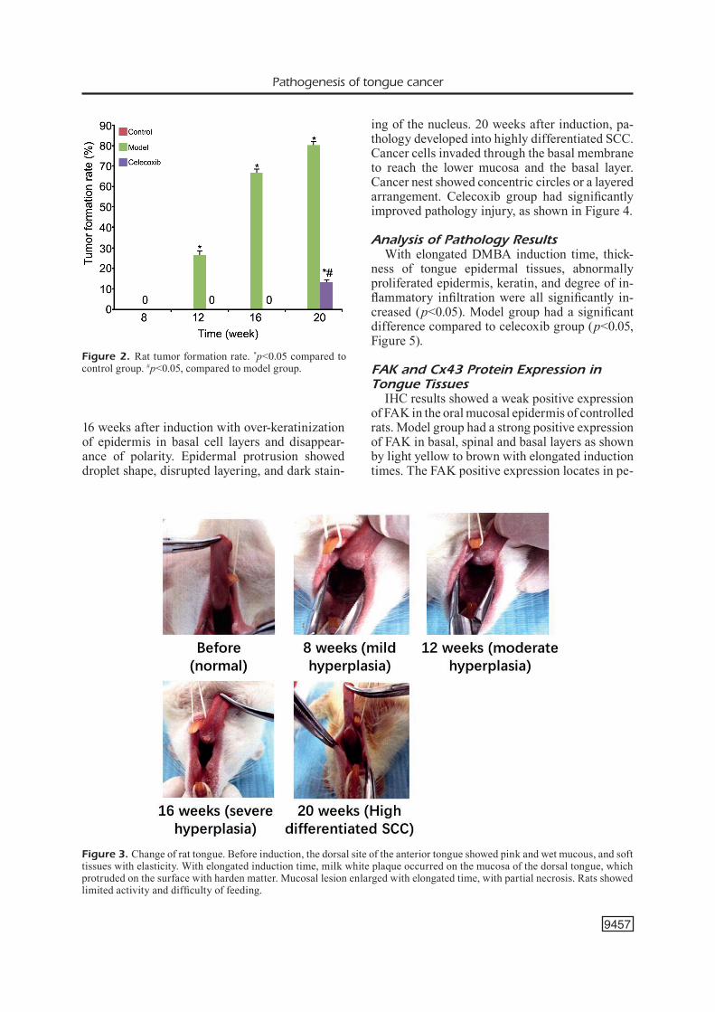
The model group had decreased activity and mental drooping. At the late phase of the exper-iment, due to the food intake difficulty caused by the lesion, rats had decreased body weight. Celecoxib group had improved food, activity and mental status compared to model group. By pathology observation, no tumor occurred in ce-lecoxib group 8 weeks after induction. The cu-mulative tumor rate at 12, 16, and 20 weeks after induction was 26.67%, 66.67%, and 80%, respec-tively in model group. With elongated induction time, the tumor formation rate was increased with extended induction time. The tumor formation rates in celecoxib group were 0, 0, and 13.33% at 12, 16 and 20 weeks after induction, respectively, with a significant difference of tumor formation rate between two groups (p<0.05). In general conditions, tumor formation rate and tongue were shown in Figures 1, 2, and 3.
Pathology of Tongue TissuesUnder light field microscopy, after HE stain-
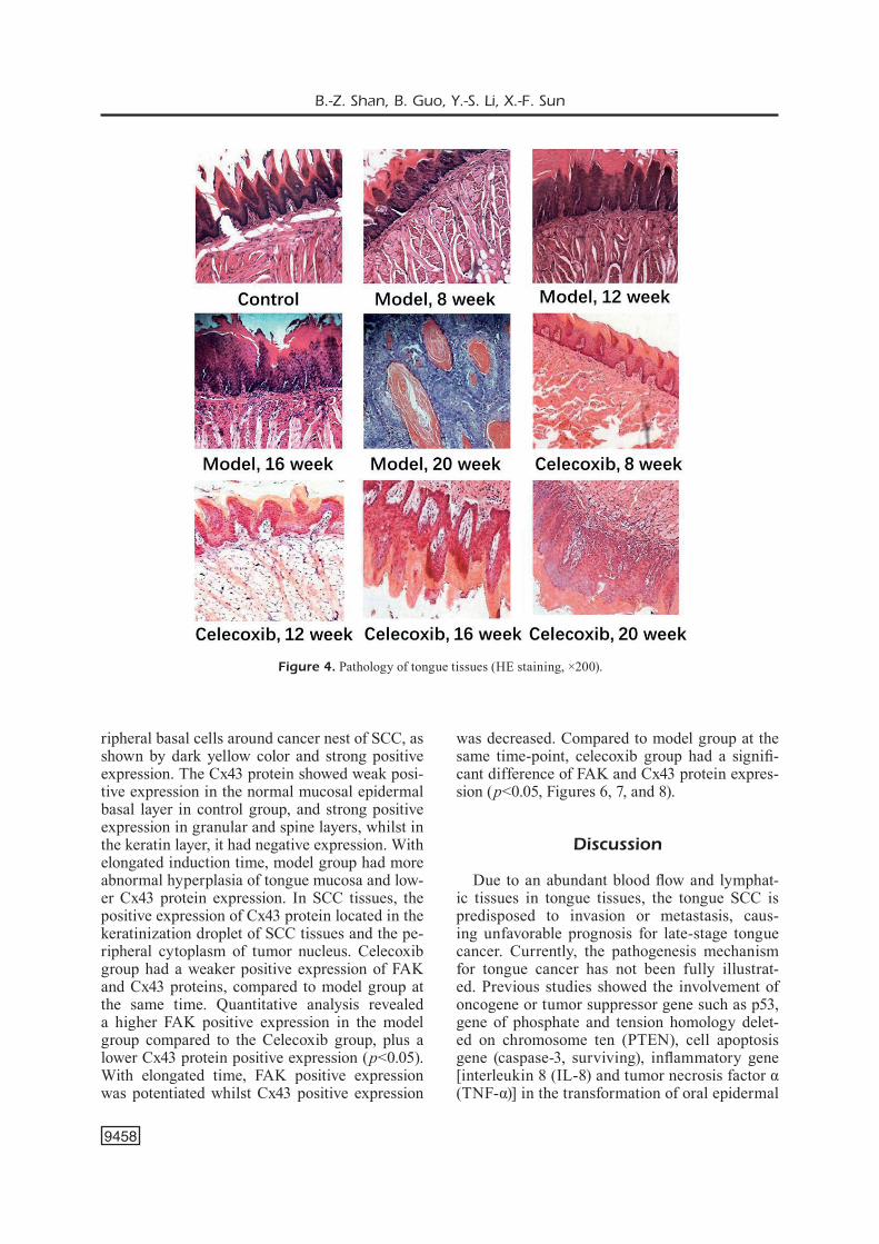
ing, no significant pathologic abnormality was observed in control group, with the regular ar-rangement of keratin epidermal on dorsal tongue, intact morphology, and no inflammatory cell in-filtration. Rats in model group had mild, moder-ate and severe abnormal hyperplasia at 8, 12, and
Figure 1. General conditions of all rats.
Pathogenesis of tongue cancer
9457
16 weeks after induction with over-keratinization of epidermis in basal cell layers and disappear-ance of polarity. Epidermal protrusion showed droplet shape, disrupted layering, and dark stain-
ing of the nucleus. 20 weeks after induction, pa-thology developed into highly differentiated SCC. Cancer cells invaded through the basal membrane to reach the lower mucosa and the basal layer. Cancer nest showed concentric circles or a layered arrangement. Celecoxib group had significantly improved pathology injury, as shown in Figure 4.
Analysis of Pathology ResultsWith elongated DMBA induction time, thick-
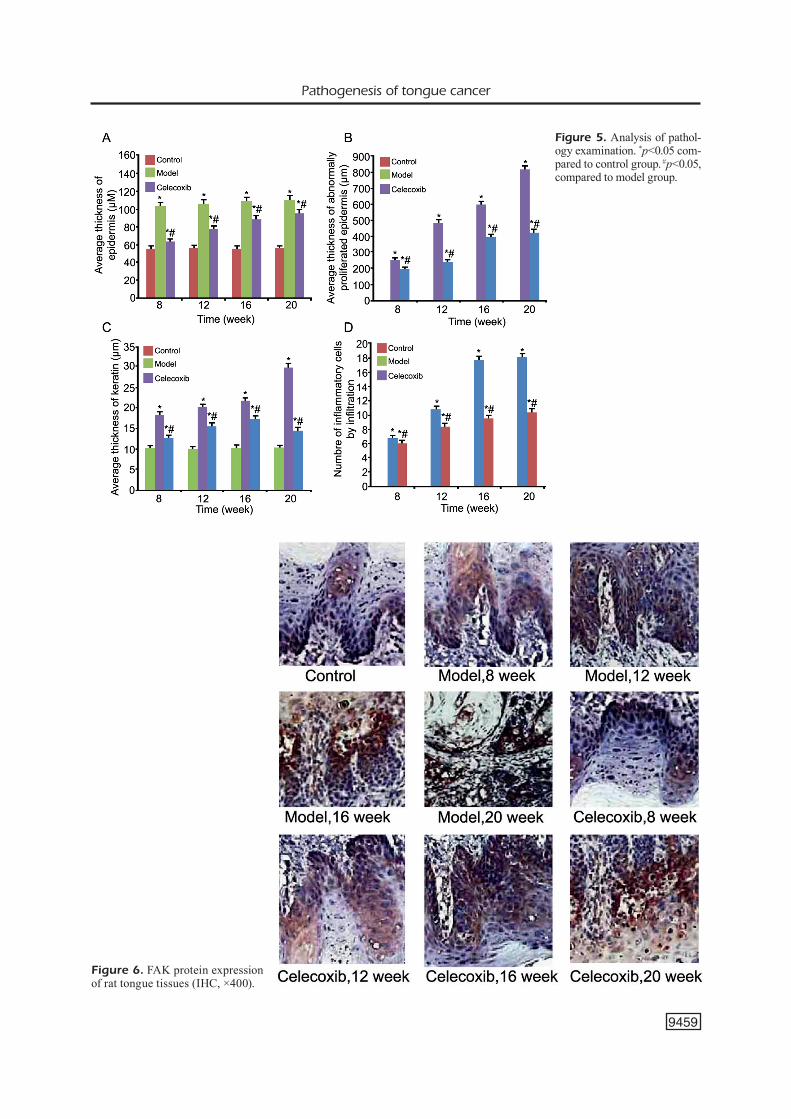
ness of tongue epidermal tissues, abnormally proliferated epidermis, keratin, and degree of in-flammatory infiltration were all significantly in-creased (p<0.05). Model group had a significant difference compared to celecoxib group (p<0.05, Figure 5).
FAK and Cx43 Protein Expression in Tongue Tissues
IHC results showed a weak positive expression of FAK in the oral mucosal epidermis of controlled rats. Model group had a strong positive expression of FAK in basal, spinal and basal layers as shown by light yellow to brown with elongated induction times. The FAK positive expression locates in pe-
Figure 2. Rat tumor formation rate. *p<0.05 compared to control group. #p<0.05, compared to model group.
Figure 3. Change of rat tongue. Before induction, the dorsal site of the anterior tongue showed pink and wet mucous, and soft tissues with elasticity. With elongated induction time, milk white plaque occurred on the mucosa of the dorsal tongue, which protruded on the surface with harden matter. Mucosal lesion enlarged with elongated time, with partial necrosis. Rats showed limited activity and difficulty of feeding.
B.-Z. Shan, B. Guo, Y.-S. Li, X.-F. Sun
9458
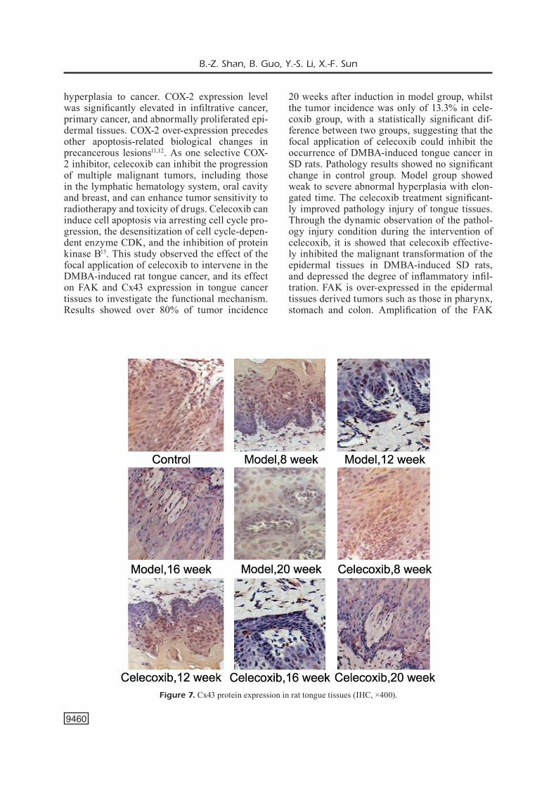
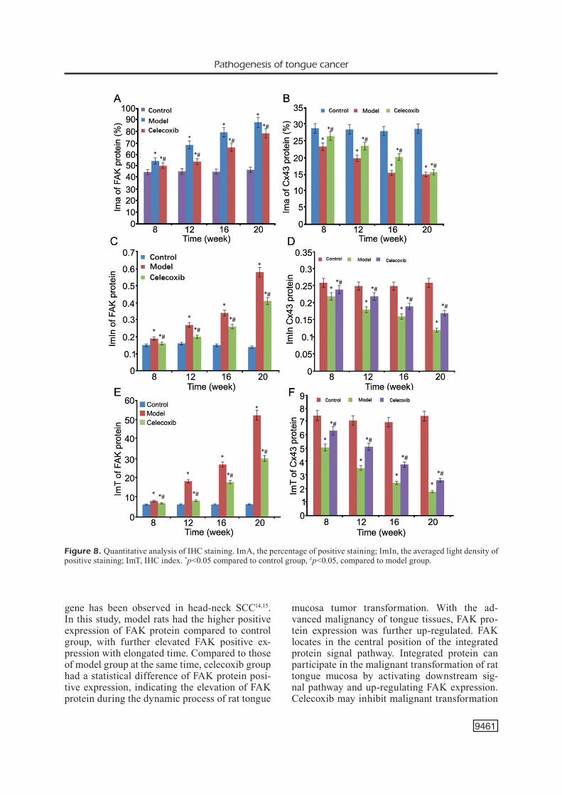
ripheral basal cells around cancer nest of SCC, as shown by dark yellow color and strong positive expression. The Cx43 protein showed weak posi-tive expression in the normal mucosal epidermal basal layer in control group, and strong positive expression in granular and spine layers, whilst in the keratin layer, it had negative expression. With elongated induction time, model group had more abnormal hyperplasia of tongue mucosa and low-er Cx43 protein expression. In SCC tissues, the positive expression of Cx43 protein located in the keratinization droplet of SCC tissues and the pe-ripheral cytoplasm of tumor nucleus. Celecoxib group had a weaker positive expression of FAK and Cx43 proteins, compared to model group at the same time. Quantitative analysis revealed a higher FAK positive expression in the model group compared to the Celecoxib group, plus a lower Cx43 protein positive expression (p<0.05). With elongated time, FAK positive expression was potentiated whilst Cx43 positive expression
was decreased. Compared to model group at the same time-point, celecoxib group had a signifi-cant difference of FAK and Cx43 protein expres-sion (p<0.05, Figures 6, 7, and 8).
Discussion
Due to an abundant blood flow and lymphat-ic tissues in tongue tissues, the tongue SCC is predisposed to invasion or metastasis, caus-ing unfavorable prognosis for late-stage tongue cancer. Currently, the pathogenesis mechanism for tongue cancer has not been fully illustrat-ed. Previous studies showed the involvement of oncogene or tumor suppressor gene such as p53, gene of phosphate and tension homology delet-ed on chromosome ten (PTEN), cell apoptosis gene (caspase-3, surviving), inflammatory gene [interleukin 8 (IL-8) and tumor necrosis factor α (TNF-α)] in the transformation of oral epidermal
Figure 4. Pathology of tongue tissues (HE staining, ×200).
Pathogenesis of tongue cancer
9459
Figure 5. Analysis of pathol-ogy examination. *p<0.05 com-pared to control group. #p<0.05, compared to model group.
Figure 6. FAK protein expression of rat tongue tissues (IHC, ×400).
B.-Z. Shan, B. Guo, Y.-S. Li, X.-F. Sun
9460
hyperplasia to cancer. COX-2 expression level was significantly elevated in infiltrative cancer, primary cancer, and abnormally proliferated epi-dermal tissues. COX-2 over-expression precedes other apoptosis-related biological changes in precancerous lesions11,12. As one selective COX-2 inhibitor, celecoxib can inhibit the progression of multiple malignant tumors, including those in the lymphatic hematology system, oral cavity and breast, and can enhance tumor sensitivity to radiotherapy and toxicity of drugs. Celecoxib can induce cell apoptosis via arresting cell cycle pro-gression, the desensitization of cell cycle-depen-dent enzyme CDK, and the inhibition of protein kinase B13. This study observed the effect of the focal application of celecoxib to intervene in the DMBA-induced rat tongue cancer, and its effect on FAK and Cx43 expression in tongue cancer tissues to investigate the functional mechanism. Results showed over 80% of tumor incidence
20 weeks after induction in model group, whilst the tumor incidence was only of 13.3% in cele-coxib group, with a statistically significant dif-ference between two groups, suggesting that the focal application of celecoxib could inhibit the occurrence of DMBA-induced tongue cancer in SD rats. Pathology results showed no significant change in control group. Model group showed weak to severe abnormal hyperplasia with elon-gated time. The celecoxib treatment significant-ly improved pathology injury of tongue tissues. Through the dynamic observation of the pathol-ogy injury condition during the intervention of celecoxib, it is showed that celecoxib effective-ly inhibited the malignant transformation of the epidermal tissues in DMBA-induced SD rats, and depressed the degree of inflammatory infil-tration. FAK is over-expressed in the epidermal tissues derived tumors such as those in pharynx, stomach and colon. Amplification of the FAK
Figure 7. Cx43 protein expression in rat tongue tissues (IHC, ×400).
Pathogenesis of tongue cancer
9461
gene has been observed in head-neck SCC14,15. In this study, model rats had the higher positive expression of FAK protein compared to control group, with further elevated FAK positive ex-pression with elongated time. Compared to those of model group at the same time, celecoxib group had a statistical difference of FAK protein posi-tive expression, indicating the elevation of FAK protein during the dynamic process of rat tongue
mucosa tumor transformation. With the ad-vanced malignancy of tongue tissues, FAK pro-tein expression was further up-regulated. FAK locates in the central position of the integrated protein signal pathway. Integrated protein can participate in the malignant transformation of rat tongue mucosa by activating downstream sig-nal pathway and up-regulating FAK expression. Celecoxib may inhibit malignant transformation
Figure 8. Quantitative analysis of IHC staining. ImA, the percentage of positive staining; ImIn, the averaged light density of positive staining; ImT, IHC index. *p<0.05 compared to control group, #p<0.05, compared to model group.
B.-Z. Shan, B. Guo, Y.-S. Li, X.-F. Sun
9462
of tongue tissues via down-regulating FAK pos-itive expression. In the normal basal layer of hu-man skin, Cx43 protein had a weak expression, with elevated Cx43 protein in spine layer, and negative expression in keratin layer16,17. Positive expression of Cx43 protein is probably related to the cell differentiation grade18,19. Cx43 had de-creased expression in various precancerous le-sions and tumor tissues including cervical cancer, papilloma, and SCC tissues. The reverse of Cx43 down-regulation can alleviate malignant pheno-type of certain tumors, and down-regulation of Cx43 is probably correlated with the malignant transformation of the epidermis18,19. This study observed the dynamic change of Cx43 protein in tumorigenesis of rat mucous. The result showed the lower positive expression of Cx43 protein in model group compared to control group, and lower positive expression with elongated time. Compared to those in model group at the same time, celecoxib group had a statistical difference of Cx43 protein positive expression. In tongue tu-morigenesis process of rats, the Cx43 expression gradually decreased, and showed decreased ex-pression in the mild hyperplasia tissues, indicat-ing the correlation between lower Cx43 protein expression and malignant transformation of rat oral cavity mucosal tumorigenesis. It can work as an early event of oral mucosal carcinoma. The IHC staining results showed that celecoxib might inhibit DMBA-induced tongue cancer of rats, decreasing the incidence of oral cavity SCC via regulating FAK and Cx43 protein expression as well as inhibiting abnormal proliferation of the oral epidermis. Some studies showed the partic-ipation of FAK gene in angiogenesis during ma-lignant transformation of the rat oral cavity. FAK gene knockout mice had abnormal angiogenesis during the embryonic stage, and FAK gene could facilitate the cancer cell invasion or metasta-sis20-22. This study observed the dynamic change of FAK and Cx43 protein expression in the rat tongue tumorigenesis as well as the intervention effect by celecoxib, leaving its detailed mecha-nism requiring further studies.
Conclusions
The focal application of celecoxib effectively inhibited the DMBA-induced tongue tissue tu-morigenesis of rats, possibly regulating the FAK and Cx43 protein expression and inhibiting ab-normal hyperplasia in oral cavity epidermis.
Conflict of InterestsThe Authors declare that they have no conflict of interests.
References
1) Goel V, Parihar PS, Parihar a, Goel aK, WaGhWani K, GuPta r, BhuteKar u. Accuracy of MRI in predic-tion of tumour thickness and nodal stage in oral tongue and gingivobuccal cancer with clinical cor-relation and staging. J Clin Diagn Res 2016; 10: TC01-TC05.
2) Fu S, Chen hh, ChenG P, ZhanG CB, Wu Y. MiR-155 regulates oral squamous cell carcinoma Tca8113 cell proliferation, cycle, and apoptosis via regulat-ing p27Kip1. Eur Rev Med Pharmacol Sci 2017; 21: 937-944.
3) SantoS hB, doS SantoS tK, PaZ ar, CaValCanti YW, nonaKa CF, GodoY GP, alVeS PM. Clinical findings and risk factors to oral squamous cell carcinoma in young patients: a 12-year retrospective anal-ysis. Med Oral Patol Oral Cir Bucal 2016; 21: e151-e156.
4) thanGaraj SV, ShYaMSundar V, KriShnaMurthY a, raMani P, GaneSan K, MuthuSWaMi M, raMShanKar V. Molecular portrait of oral tongue squamous cell carcinoma shown by integrative meta-analysis of expression orofiles with validations. PLoS One 2016; 11: e0156582.
5) Morita Y, Morita n, hata K, naKaniShi M, KiMoto n, oMata t, naKaMura Y, Yoneda t. Cyclooxygenase-2 expression is associated with vascular endotheli-al growth factor-c and lymph node metastasis in human oral tongue cancer. Oral Surg Oral Med Oral Pathol Oral Radiol 2014; 117: 502-510.
6) lindquiSt d, ahrlund-riChter a, tarján M, tot t, dalianiS t. Intense CD44 expression is a negative prognostic factor in tonsillar and base of tongue cancer. Anticancer Res 2012; 32: 153-161.
7) lee dY, liM jh, KiM Yj, KiM Sd, ParK SW, KWon SK, hah jh, KWon tK, KiM Kh, KiM Yh, SunG MW. Effect of celecoxib on survival of mobile tongue cancer. Anticancer Res 2015; 35: 4235-4241.
8) li WZ, WanG XY, li ZG, ZhanG jh, dinG Yq. Celecoxib enhances the inhibitory effect of cispla-tin on Tca8113 cells in human tongue squamous cell carcinoma in vivo and in vitro. J Oral Pathol Med 2010; 39: 579-584.
9) Xiao W, jianG M, li h, li C, Su r, huanG K. Knockdown of FAK inhibits the invasion and me-tastasis of Tca8113 cells in vitro. Mol Med Rep 2013; 8: 703-707.
10) BultYnCK G. The anti-metastatic micro-environ-ment of the bone: importance of osteocyte Cx43 hemichannels. Biochim Biophys Acta 2016; 1866: 121-127.
11) WanG Yh, Wu MW, YanG aK, ZhanG Wd, Sun j, liu tr, Chen YF. COX-2 gene increases tongue
Pathogenesis of tongue cancer
9463
cancer cell proliferation and invasion through VEGF-C pathway. Med Oncol 2011; 28 Suppl 1: S360-S366.
12) Wan SG, taCCioli C, jianG Y, Chen h, SMalleY Kj, huanG K, liu XP, FarBer jl, CroCe CM, FonG lY. Zinc deficiency activates S100A8 inflammation in the absence of COX-2 and promotes murine oral-esophageal tumor progression. Int J Cancer 2011; 129: 331-345.
13) li WZ, huo qj, WanG XY, Xu F. Inhibitive effect of celecoxib on the adhesion and invasion of human tongue squamous carcinoma cells to extracellular matrix via down regulation of MMP-2 expression. Prostaglandins Other Lipid Mediat 2010; 93: 113-119.
14) theoChariS S, KlijanienKo j, GiaGiniS C, aleXandrou P, PatSouriS e, SaStre-Garau X. FAK and Src expres-sion in mobile tongue squamous cell carcinoma: associations with clinicopathological parameters and patients survival. J Cancer Res Clin Oncol 2012; 138: 1369-1377.
15) ZhanG Zh, li j, luo F, WanG YS. Clinical significance of SCCRO (DCUN1D1) in prostate cancer and its proliferation-inhibiting effect on Lncap cells. Eur Rev Med Pharmacol Sci 2017; 21: 4283-4291.
16) SulKoWSKa u, FeBP aW, SulKoWSKi S. Association of STAT3 with Cx26 and Cx43 in human uterine en-dometrioid adenocarcinoma. Oncol Lett 2016; 11: 4134-4138.
17) Shi h, Shi d, Wu Y, Shen q, li j. Qigesan inhibits mi-gration and invasion of esophageal cancer cells via inducing connexin expression and enhancing gap junction function. Cancer Lett 2016; 380: 184-190.
18) CreSPin S, FroMont G, WaGer M, leVillain P, Cronier l, MonVoiSin a, deFaMie n, MeSnil M. Expression of a gap junction protein, connexin43, in a large pan-el of human gliomas: new insights. Cancer Med 2016; 5: 1742-1752.
19) dianati e, Poiraud j, WeBer-ouellette a, Plante i. ConneXinS, e-Cadherin, Claudin-7 and beta-caten-in transiently form junctional nexuses during the post-natal mammary gland development. Dev Biol 2016; 416: 52-68.
20) dai W, Sun C, huanG S, Zhou q. Carvacrol sup-presses proliferation and invasion in human oral squamous cell carcinoma. Onco Targets Ther 2016; 9: 2297-2304.
21) tanaKa t, KiMura M, iShiGuro h, MiZoGuChi K, taKeYaMa h. Connexin 43 expression is associat-ed with poor survival in patients with esophageal squamous cell carcinoma. Mol Clin Oncol 2016; 4: 989-993.
22) YanG l, Zhou q, Chen X, Su l, liu B, ZhanG h. Activation of the FAK/PI3K pathway is crucial for AURKA-induced epithelial-mesenchymal tran-sition in laryngeal cancer. Oncol Rep 2016; 36: 819-826.