Embed Size (px)
Citation preview

3561
Abstract. – OBJECTIVE: The aim of this pa-per is to investigate the effect of BDNF-TrKB pathway on AOH by accessing its regulatory role in retinal ganglion cell apoptosis.
MATERIALS AND METHODS: Acute ocular hypertension (AOH) model in rats was estab-lished by anterior chamber perfusion to increase intraocular tension. Rats were randomly divided into AOH group, control group and k252a group, with ten rats in each group. Rats were sacrificed 72 h after animal procedures and eyeballs were harvested. HE staining was used to observe ret-inal structural changes at different time points. Immunohistochemical staining was used to ob-serve the BDNF-positive cells in retinal tis-sues. TUNEL staining was conducted to mea-sure apoptosis of retinal ganglion cells. Reverse Transcriptase-Polymerase Chain Reaction (RT-PCR) and Western blot were performed to detect mRNA and protein levels of BDNF, TrKB, PI3K and ERK1 in retinal tissues, respectively.
RESULTS: HE and TUNEL staining showed significant pathological changes and abundant apoptotic cells in rat retina of AOH group and k252a group compared with those of the con-trol group (p<0.05). The number of survived ret-inal ganglion cells in the AOH group was lower than that of the control group (p<0.05). K252a group had the lowest number of survived reti-nal ganglion cells. Immunohistochemical results showed that BDNF was rarely expressed in rat retinal tissues of the control group, which was remarkably pronounced in the AOH group and k252a group. The number of BDNF-positive cells in the k252a group was higher than that in the AOH group (p<0.05). RT-PCR and Western blot indicated that mRNA and protein levels of rela-tive genes in BDNF-TrKB and PI3K/ERK1 path-ways were upregulated in AOH group (p<0.05), but were significantly downregulated in k252a group (p<0.05).
CONCLUSIONS: BNDF-TrKB pathway exerts a protective effect on retina against acute ocular hypertension by reducing retinal cell apoptosis.
Key Words: BDNF, Glaucoma, Ganglion cells, Apoptosis.
Introduction
Glaucoma is an ophthalmic emergency char-acterized by elevated intraocular pressure. It is a common blinding eye disease following myopia and cataract. About 65% of glaucoma patients may eventually lead to irreversible unilateral or bilateral blindness if timely treatment is lacked1,2. Optic nerve damage is the most serious patholog-ical change in glaucoma, mainly manifesting as apoptosis of retinal ganglion cells (RGCs), which will eventually cause the irreversible damage of the optic nerve3,4. The pathogenesis of glaucoma has not been comprehensively elucidated. Lucas et al5 found that excessive glutamate accumula-tion results in selective damage to retinal inner cells. Russo et al6 found that massive accumula-tion of glutamate in retina induced by neuronal glutamate transporters would lead to retinal isch-emia7. Bagnis et al8 suggested that intraocular pressure elevation may increase the concentration of glutamic acid in the vitreous body of patients with primary open angle glaucoma, thereafter leading to the apoptosis of ganglion cells.
BDNF is mainly synthesized in the brain. Perez et al9 found that BDNF is expressed in the rat retinal ganglion cell layer, proximal end of the fibroblast layer and the proximal end of the core layer. The ex-pression of high-affinity tyrosine kinase receptor B (TrkB) can be observed in the inner retina, retinal nerve fiber layer and optic nerve in embryonic rats. RGCs exert a protective response when the optic nerve is damaged, accompanied by the upregulated expressions of BDNF and its receptor10. Iwabe et al11
European Review for Medical and Pharmacological Sciences 2019; 23: 3561-3568
Y. LIANG, Y.-H. YU, H.-J. YU, L.-S. MA
Department of Ophthalmology, the Affiliated Yantai Yuhuangding Hospital of Qingdao University, Yantai, China
Yan Liang and Yonghong Yu contributed equally to this work
Corresponding Author: Lusheng Ma, MD; email: [email protected]
Effect of BDNF-TrKB pathway on apoptosis of retinal ganglion cells in glaucomatous animal model

Y. Liang, Y.-H. Yu, H.-J. Yu, L.-S. Ma
3562
detected that blockage of BDNF transport promotes the survival rate of ganglion cells in high intraocular pressure model in dogs. Hirsch et al12 observed that 2 days after complete transection of rat optic nerves, the amount of RGCs increases by 2 times, and the amount of TrkB-expressing RGCs also increases by 50%. Pease et al13 showed that the interruption of BDNF retrograde transport and accumulation of TrkB receptor of optic nerve papilla are the ma-jor pathogenic factors of RCGs death in acute and chronic high intraocular pressure rat models. Mo et al14 indicated that overexpressed BDNF by lenti-virus injection in the ganglion cell layer increases the survival rate of RGCs and reduces the apoptot-ic rate. Therefore, we believed that BDNF plays an important role against glaucoma-induced apoptosis.
In this study, AOH rat model was established by anterior chamber perfusion to elevate intraoc-ular pressure. After k252a intervention, we de-tected pathological lesions and cell apoptosis in retinal tissues. Subsequently, the regulatory effect of BDNF-TrKB pathway on AOH was explored, to provide the basis for the treatment of AOH.
Materials and Methods
Animals and GroupsThirty healthy male Sprague-Dawley (SD) rats
weighing 180-250 g without eye diseases were housed in SPF (specific pathogen-free) environ-ment. Rats had free access to water and food. The relative humidity of the feeding room was 55%, and the temperature was 22°C. Rats were kept un-der a standard 12/12 light-dark circle; they were randomly divided into control group, AOH group and k252a group, with 10 rats in each group. This study was approved by the Animal Ethics Com-mittee of Qingdao University Animal Center.
Preparation of Animal ModelAfter fasting 12 h, rats were anesthetized by
intraperitoneal injection. Compound Tropicamide Eye Drops were applied on their right eyes to di-late the pupils. The surface of the right eyes was anesthetized with benoxinate. The anterior cham-ber of the rat was injected with normal saline. The puncture needle was fixed by the glue cloth and the infusion bottle was raised to 150 cm above. Disconnection of the fundus blood vessels, reti-nal ischemia, red reflector of the fundus and pale conjunctiva were observed under an ophthalmo-scope. The recovery of blood supply in the reti-na can be observed after pulling out the puncture
needle. The right eyes were coated with tetracy-cline ointment to prevent infection.
After the procedures of eyeball pressure, pupil dilation was performed with atropine. 10 pmol/μL k252a solution or isodose saline was slowly injected into the vitreous cavity under the micro-scope using a 10 μL syringe needle. The needle was pulled out 30 s after injection to prevent drug spillover.
Sample CollectionRats were sacrificed by exsanguination from ab-
dominal aorta (weighed 200-280 g at the time of sacrifice) under 10% chloral hydrate (0.4 g/kg, i.p.) anesthesia. About 10 ml of blood was extracted by exsanguination. No heart beating indicated the an-imal death. No rat exhibited signs of peritonitis fol-lowing the administration of 10% chloral hydrate. The eyeball was quickly extirpated by cutting off fascia of the outer canthus in the right eye. 0.5 mm optic nerve was retained and the eyeball was re-moved completely. The eyeballs were rinsed with saline and then soaked in the eyeball fixative.
Paraffin Embedded Retinal Tissues and Hematoxylin and Eosin (HE) Staining
Eyeball samples were washed by flowing water, dehydrated in ascending series of ethanol, cleared in xylene and embedded in paraffin. Paraffin-em-bedded samples were cut for several sections. The neutral gum was dripped into the center of the speci-men, and then the slides were sealed. Three random-ly selected fields in each sample were observed for retinal structural changes using a microscope.
Detection of Apoptosis by TUNELThe tissue sections were dewaxed, washed,
hydrated and fixed. Detection of apoptosis was performed according to TUNEL kit instructions (Beyotime, Shanghai, China). Three randomly selected fields in each group were observed and captured. The number of the apoptotic cells and the total cells in three non-overlapping fields of each sample were counted under high magnifica-tion. AI = (number of apoptotic cells /number of total cells) ×100%.
Detection of BDNF Expression in Rat Retina by Immunohistochemistry
The slice was dewaxed, hydrated, and incubat-ed with 3% H2O2 at room temperature. Antigenic heat repair was carried out, followed by BDNF
BDNF alleviates retinal pathological changes and ganglion cell apoptosis in glaucomatous rats
3563
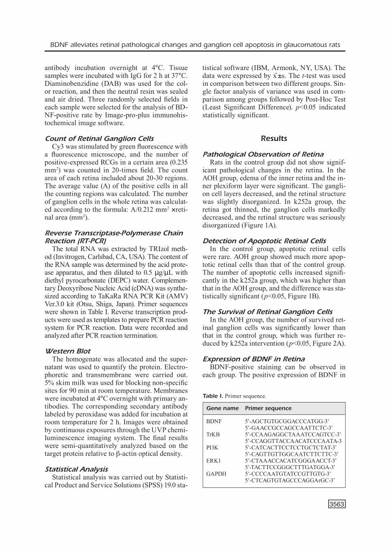
antibody incubation overnight at 4°C. Tissue samples were incubated with IgG for 2 h at 37°C. Diaminobenzidine (DAB) was used for the col-or reaction, and then the neutral resin was sealed and air dried. Three randomly selected fields in each sample were selected for the analysis of BD-NF-positive rate by Image-pro-plus immunohis-tochemical image software.
Count of Retinal Ganglion CellsCy3 was stimulated by green fluorescence with
a fluorescence microscope, and the number of positive-expressed RCGs in a certain area (0.235 mm2) was counted in 20-times field. The count area of each retina included about 20-30 regions. The average value (A) of the positive cells in all the counting regions was calculated. The number of ganglion cells in the whole retina was calculat-ed according to the formula: A/0.212 mm2 ×reti-nal area (mm2).
Reverse Transcriptase-Polymerase Chain Reaction (RT-PCR)
The total RNA was extracted by TRIzol meth-od (Invitrogen, Carlsbad, CA, USA). The content of the RNA sample was determined by the acid prote-ase apparatus, and then diluted to 0.5 μg/μL with diethyl pyrocarbonate (DEPC) water. Complemen-tary Deoxyribose Nucleic Acid (cDNA) was synthe-sized according to TaKaRa RNA PCR Kit (AMV) Ver.3.0 kit (Otsu, Shiga, Japan). Primer sequences were shown in Table I. Reverse transcription prod-ucts were used as templates to prepare PCR reaction system for PCR reaction. Data were recorded and analyzed after PCR reaction termination.
Western BlotThe homogenate was allocated and the super-
natant was used to quantify the protein. Electro-phoretic and transmembrane were carried out. 5% skim milk was used for blocking non-specific sites for 90 min at room temperature. Membranes were incubated at 4°C overnight with primary an-tibodies. The corresponding secondary antibody labeled by peroxidase was added for incubation at room temperature for 2 h. Images were obtained by continuous exposures through the UVP chemi-luminescence imaging system. The final results were semi-quantitatively analyzed based on the target protein relative to β-actin optical density.
Statistical AnalysisStatistical analysis was carried out by Statisti-
cal Product and Service Solutions (SPSS) 19.0 sta-
tistical software (IBM, Armonk, NY, USA). The data were expressed by x ±̅s. The t-test was used in comparison between two different groups. Sin-gle factor analysis of variance was used in com-parison among groups followed by Post-Hoc Test (Least Significant Difference). p<0.05 indicated statistically significant.
Results
Pathological Observation of RetinaRats in the control group did not show signif-
icant pathological changes in the retina. In the AOH group, edema of the inner retina and the in-ner plexiform layer were significant. The gangli-on cell layers decreased, and the retinal structure was slightly disorganized. In k252a group, the retina got thinned, the ganglion cells markedly decreased, and the retinal structure was seriously disorganized (Figure 1A).
Detection of Apoptotic Retinal CellsIn the control group, apoptotic retinal cells
were rare. AOH group showed much more apop-totic retinal cells than that of the control group. The number of apoptotic cells increased signifi-cantly in the k252a group, which was higher than that in the AOH group, and the difference was sta-tistically significant (p<0.05, Figure 1B).
The Survival of Retinal Ganglion CellsIn the AOH group, the number of survived ret-
inal ganglion cells was significantly lower than that in the control group, which was further re-duced by k252a intervention (p<0.05, Figure 2A).
Expression of BDNF in RetinaBDNF-positive staining can be observed in
each group. The positive expression of BDNF in
Table I. Primer sequence.
Gene name Primer sequence
BDNF 5’-AGCTGTGCGGACCCATGG-3’ 5’-GAACCGCCAGCCAATTCTC-3’TrKB 5’-CCAAGAGGCTAAATCCAGTCC-3’ 5’-CCAGGTTACCAACATCCCAATA-3PI3K 5’-CATCACTTCCTCCTGCTCTAT-3’ 5’-CAGTTGTTGGCAATCTTCTTC-3’ERK1 5’-CTAAACCACATCGGGAACCT-3’ 5’-TACTTCCGGGCTTTGATGGA-3’GAPDH 5’-CCCCAATGTATCCGTTGTG-3’ 5’-CTCAGTGTAGCCCAGGArGC-3’

Y. Liang, Y.-H. Yu, H.-J. Yu, L.-S. Ma
3564
the retina of the control group was rare. The num-ber of BDNF-positive cells in AOH group was remarkably higher than that of the control group (p<0.05), which was the highest in the k252a group (p<0.05, Figure 2B).
Transcription Levels of BDNF-TrKB and PI3K/ERK1 in Retina
Compared with the control group, the tran-scriptional levels of BDNF-TrKB and PI3K/ERK1 in the AOH group remarkably increased
(p<0.05). However, transcriptional levels in the k252a group were significantly lower than those in the AOH group (p<0.05, Figure 3A-3D).
Protein Expressions of BDNF-TrKB and PI3K/ERK1 in Retina
Compared with the control group, the ex-pression levels of BDNF-TrKB and PI3K/ERK1 markedly increased (p<0.05). In the k252a group, expression levels of all these proteins decreased compared with those of the AOH group (p<0.05,
Figure 1. HE staining and TUNEL staining in rat retina (magnification 200×). A, HE staining in rat retina of control group, AOH group and k252a group. B, TUNEL staining in rat retina of control group, AOH group and k252a group. Comparison of apoptotic rate in three groups. *: Compared with the control group, the difference was statistically significant (p<0.05); #: Compared with the AOH group, the difference was statistically significant (p<0.05).
Figure 2. Immunohistochemical staining of RGCs and BDNF (magnification 200×). A, RGCs in the control group, AOH group and k252a group. Comparison of survived RGCs in three groups. B, Immunohistochemical staining of BDNF in the control group, AOH group and k252a group. Comparison of number of positive cells in three groups. *: Compared with the control group, the difference was statistically significant (p<0.05); #: Compared with the AOH group, the difference was sta-tistically significant (p<0.05).

BDNF alleviates retinal pathological changes and ganglion cell apoptosis in glaucomatous rats
3565
Figure4A-D). Protein changes of BDNF-TrKB and PI3K/ERK1 in the retina were identical to transcription level changes.
Discussion
Glaucoma is an ophthalmic emergency char-acterized by elevated intraocular pressure. Visu-al dysfunction has resulted from impaired optic nerves and pathways. Lack of timely treatment would lead to irreversible vision loss15,16. The eti-ology of glaucoma is not yet very clear. It is cur-rently believed that the occurrence of glaucoma is closely related to anatomy, genetic suscepti-bility and mental factors17,18. Retinal optic nerve damage, manifesting as characteristic optic at-rophy, is the major pathological performance of glaucoma19. Sands and Barish20 have shown that glaucomatous optic neuropathy is associated with denaturation of trans synaptic neurons in cor-pora geniculatum externum and primary visual
cortex. Histopathological studies showed that the early-stage pathological lesions of glaucoma first occur in the lamina layer. The main manifesta-tions include the loss of the axons, blood vessels and collagen cells, accompanied by the accumu-lation of the temporal and lower pole nerve fibers in the optic disc21. With the further development of the disease course, the optic disk is eventually depressed and could be reversed.
Intraocular pressure (IOP) is the pressure of eyeball contents on the wall of the eyeball. The change of eyeball content inevitably causes IOP. Dynamically balanced aqueous humor circula-tion is the most important factor that maintains IOP stability22. Histopathologically increased IOP leads to the interruption of retinal or optic nerve flow and insufficiency of blood supply. The main pathological changes are the apoptosis of retinal ganglion cells, eventually leading to the irre-versible damage to the optic nerves23. Therefore, protection of ganglion cells, inhibition of further retinal damage and IOP-induced apoptosis have
Figure 3. Transcriptional levels of BDNF-TrKB and PI3K/ERK1. A, The mRNA level of BDNF in three groups. B, The mRNA level of TrKB in three groups. C, The mRNA level of PI3K in three groups. D, The mRNA level of ERK1 in three groups. *: Compared with the control group, the difference was statistically significant (p<0.05); #: Compared with the AOH group, the difference was statistically significant (p<0.05).

Y. Liang, Y.-H. Yu, H.-J. Yu, L.-S. Ma
3566
It is known that BDNF regulates TrKB mainly through the MAPK/PI3K/ERK pathway. The up-stream activator Ras activates the MAPK kinase, thus further stimulating the MAPK/ERK pathway. Subsequently, activated ERK can phosphorylate the cAMP response-element protein Ser133 and the Ser308 site of protein kinase B. Finally, a series of gene expressions and anti-apoptotic pathways are activated31,32.
In this study, the pathological lesions and apop-tosis in retinal tissues were pronounced in the AOH rat model, which were worse after blocking the BDNF-TrKB pathway by k252a treatment. Im-munohistochemical staining showed that BDNF reactivity increased in the AOH group, which was remarkably downregulated in the k252a group. Besides, the expression levels of BDNF, TrKB, PI3K and ERK1 in AOH group were upregulat-ed. However, these expressions were markedly downregulated in the k252a group. It can be seen that RGCs immediately exert protective response after the optic nerve injury by upregulating ex-pressions of BDNF and its receptors. Pease et al13
become the latest research focuses. It is generally believed that effective inhibition of retinal gan-glion cell apoptosis is the key to vision retention. The inhibition of the apoptotic ganglion cells is of great value in the treatment of glaucoma24.
BDNF is a member of the neurotrophin family. It has been widely studied because of its significant inhibitory effect on neuronal apoptosis. BDNF plays an important role in maintaining neuronal growth, differentiation, repair and regeneration in nerve in-jury. Expressions of BDNF and its receptors may be the basis of its local effects. BDNF is capable of inhibiting secondary neuronal apoptosis after spinal cord injury25. It protects central and peripheral nerve damages, and promotes the growth of neuron ax-ons22,23,26. Wordinger et al24 firstly found that BDNF and its receptor are expressed in trabecular cells and trabecular tissues. Also, BDNF participates in au-tocrine and paracrine, and can be expressed in the photoreceptor layer and inner layer of the retina27. BDNF and its main receptor TrKB are widely ex-pressed in the eye. BDNF is found to inhibit RGCs apoptosis after high intraocular pressure injury28-30.
Figure 4. Protein expressions of BDNF-TrKB and PI3K/ERK1. A, Protein expression of BDNF in three groups. B, Protein expression of TrKB in three groups. C, Protein expression of PI3K in three groups. D, Protein expression of ERK1 in three groups. *: Compared with the control group, the difference was statistically significant (p<0.05); #: Compared with the AOH group, the difference was statistically significant (p<0.05).

BDNF alleviates retinal pathological changes and ganglion cell apoptosis in glaucomatous rats
3567
found the interruption of BDNF retrograde trans-port and the accumulation of TrkB receptor of optic nerve papilla in rats with acute and chronic high intraocular pressure and glaucomatous mon-keys, which are the major pathogenesis of RCGs death. However, the specific dose, administration approaches and clinical evaluation of BDNF treat-ment are still needed to be further explored33-35.
Conclusions
We showed that BNDF-TrKB pathway exerts a protective effect on the retina against acute ocular hypertension by reducing retinal cell apoptosis.
Conflict of interestThe authors declare no conflicts of interest.
References
1) Mantravadi av, vadhar n. Glaucoma. Prim Care 2015; 42: 437-449.
2) Gupta d, Chen pp. Glaucoma. Am Fam Physician 2016; 93: 668-674.
3) panG JJ, FrankFort BJ, Gross rL, Wu sM. Elevated intraocular pressure decreases response sensi-tivity of inner retinal neurons in experimental glau-coma mice. Proc Natl Acad Sci U S A 2015; 112: 2593-2598.
4) WeinreB rn, aunG t, Medeiros Fa. The pathophysi-ology and treatment of glaucoma: a review. JAMA 2014; 311: 1901-1911.
5) LuCas dr, neWhouse Jp. The toxic effect of sodium L-glutamate on the inner layers of the retina. AMA Arch Ophthalmol 1957; 58: 193-201.
6) russo r, CavaLiere F, varano Gp, MiLanese M, ador-netto a, nuCCi C, Bonanno G, Morrone La, Co-rasaniti Mt, BaGetta G. Impairment of neuronal glu-tamate uptake and modulation of the glutamate transporter GLT-1 induced by retinal ischemia. PLoS One 2013; 8: e69250.
7) ishikaWa M. Abnormalities in glutamate metabo-lism and excitotoxicity in the retinal diseases. Sci-entifica (Cairo) 2013; 2013: 528940.
8) BaGnis a, izzotti a, CentoFanti M, saCCa sC. Aque-ous humor oxidative stress proteomic levels in pri-mary open angle glaucoma. Exp Eye Res 2012; 103: 55-62.
9) perez Mt, CaMinos e. Expression of brain-derived neurotrophic factor and of its functional receptor in neonatal and adult rat retina. Neurosci Lett 1995; 183: 96-99.
10) rudzinski M, WonG tp, saraGovi hu. Changes in ret-inal expression of neurotrophins and neurotrophin
receptors induced by ocular hypertension. J Neu-robiol 2004; 58: 341-354.
11) iWaBe s, Moreno-Mendoza na, triGo-tavera F, CroWder C, GarCia-sanChez Ga. Retrograde axonal transport obstruction of brain-derived neurotroph-ic factor (BDNF) and its TrkB receptor in the retina and optic nerve of American Cocker Spaniel dogs with spontaneous glaucoma. Vet Ophthalmol 2007; 10 Suppl 1: 12-19.
12) hirsCh s, LaBes M, Bahr M. Changes in BDNF and neurotrophin receptor expression in degenerating and regenerating rat retinal ganglion cells. Restor Neurol Neurosci 2000; 17: 125-134.
13) pease Me, MCkinnon sJ, QuiGLey ha, kerriGan-BauM-rind La, zaCk dJ. Obstructed axonal transport of BDNF and its receptor TrkB in experimental glauco-ma. Invest Ophthalmol Vis Sci 2000; 41: 764-774.
14) Mo X, yokoyaMa a, oshitari t, neGishi h, dezaWa M, Mizota a, adaChi-usaMi e. Rescue of axotomized retinal ganglion cells by BDNF gene electropora-tion in adult rats. Invest Ophthalmol Vis Sci 2002; 43: 2401-2405.
15) sun W, Li yn, ye JF, Guan yQ, Li sJ. MEG3 is in-volved in the development of glaucoma through promoting the autophagy of retinal ganglion cells. Eur Rev Med Pharmacol Sci 2018; 22: 2534-2540.
16) Cook C, Foster p. Epidemiology of glaucoma: what’s new? Can J Ophthalmol 2012; 47: 223-226.
17) WanG r, WiGGs JL. Common and rare genetic risk factors for glaucoma. Cold Spring Harb Perspect Med 2014; 4: a17244.
18) kersey t, CLeMent Ci, BLooM p, Cordeiro MF. New trends in glaucoma risk, diagnosis & manage-ment. Indian J Med Res 2013; 137: 659-668.
19) tathaM aJ, Miki a, WeinreB rn, zanGWiLL LM, Me-deiros Fa. Defects of the lamina cribrosa in eyes with localized retinal nerve fiber layer loss. Oph-thalmology 2014; 121: 110-118.
20) sands sB, Barish Me. A quantitative description of excitatory amino acid neurotransmitter responses on cultured embryonic Xenopus spinal neurons. Brain Res 1989; 502: 375-386.
21) roBerts Md, Grau v, GriMM J, reynaud J, BeLLezza aJ, BurGoyne CF, doWns JC. Remodeling of the connective tissue microarchitecture of the lamina cribrosa in early experimental glaucoma. Invest Ophthalmol Vis Sci 2009; 50: 681-690.
22) MantiLLa CB, Gransee hM, zhan Wz, sieCk GC. Mo-toneuron BDNF/TrkB signaling enhances func-tional recovery after cervical spinal cord injury. Exp Neurol 2013; 247: 101-109.
23) Weishaupt n, Li s, di pardo a, sipione s, Fouad k. Synergistic effects of BDNF and rehabilitative training on recovery after cervical spinal cord in-jury. Behav Brain Res 2013; 239: 31-42.
24) WordinGer rJ, LaMBert W, aGarWaL r, taLati M, CLark aF. Human trabecular meshwork cells se-crete neurotrophins and express neurotrophin re-ceptors (Trk). Invest Ophthalmol Vis Sci 2000; 41: 3833-3841.

Y. Liang, Y.-H. Yu, H.-J. Yu, L.-S. Ma
3568
25) sinGer W, panFord-WaLsh r, knipper M. The function of BDNF in the adult auditory system. Neurophar-macology 2014; 76 Pt C: 719-728.
26) WeBer aJ, harMan Cd. BDNF treatment and ex-tended recovery from optic nerve trauma in the cat. Invest Ophthalmol Vis Sci 2013; 54: 6594-6604.
27) CaLeo M, Menna e, Chierzi s, Cenni MC, MaFFei L. Brain-derived neurotrophic factor is an antero-grade survival factor in the rat visual system. Curr Biol 2000; 10: 1155-1161.
28) BinLey ke, nG Ws, Barde ya, sonG B, MorGan Je. Brain-derived neurotrophic factor prevents den-dritic retraction of adult mouse retinal ganglion cells. Eur J Neurosci 2016; 44: 2028-2039.
29) aBe t, tokita-ishikaWa y, onaMi h, katsukura y, kaJi h, nishizaWa M, naGai n. Intrascleral transplanta-tion of a collagen sheet with cultured brain-de-rived neurotrophic factor expressing cells partially rescues the retina from damage due to acute high intraocular pressure. Adv Exp Med Biol 2014; 801: 837-843.
30) doMeniCi L, oriGLia n, FaLsini B, Cerri e, BarLosCio d, FaBiani C, sanso M, Giovannini L. Rescue of retinal function by BDNF in a mouse model of glaucoma. PLoS One 2014; 9: e115579.
31) arthur Js, FonG aL, dWyer JM, davare M, reese e, oBrietan k, iMpey s. Mitogen- and stress-activat-ed protein kinase 1 mediates cAMP response element-binding protein phosphorylation and activation by neurotrophins. J Neurosci 2004; 24: 4324-4332.
32) ahn Jy, Liu X, Liu z, pereira L, ChenG d, penG J, Wade pa, haMBurGer aW, ye k. Nuclear Akt associates with PKC-phosphorylated Ebp1, preventing DNA fragmentation by inhibition of caspase-activated DNase. EMBO J 2006; 25: 2083-2095.
33) Boye se, Boye sL, LeWin as, hausWirth WW. A com-prehensive review of retinal gene therapy. Mol Ther 2013; 21: 509-519.
34) FenG L, Chen h, yi J, troy JB, zhanG hF, Liu X. Long-term protection of retinal ganglion cells and visual function by brain-derived neurotrophic factor in mice with ocular hypertension. Invest Ophthalmol Vis Sci 2016; 57: 3793-3802.
35) vaLiente-soriano FJ, nadaL-niCoLas FM, saLinas-na-varro M, JiMenez-Lopez M, BernaL-Garro JM, viL-LeGas-perez Mp, aGudoBarriuso M, vidaL-sanz M. BDNF rescues RGCs but not intrinsically pho-tosensitive RGCs in ocular hypertensive albino rat retinas. Invest Ophthalmol Vis Sci 2015; 56: 1924-1936.