Embed Size (px)
Citation preview
Acc
epte
d A
rtic
le
This article has been accepted for publication and undergone full peer review but has not
been through the copyediting, typesetting, pagination and proofreading process, which may
lead to differences between this version and the Version of Record. Please cite this article as
doi: 10.1111/evj.12690
This article is protected by copyright. All rights reserved.
DR. THILO PFAU (Orcid ID : 0000-0002-0702-4289)
Article type : General Article
EVJ-GA-16-131.R3
Effect of a 4-week elastic resistance band training regimen on
back kinematics in horses trotting in-hand and on the lunge.
T. Pfau1,2,*
, V. Simons1, N. Rombach
3, N. Stubbs
4 and R. Weller
1,2
1Department of Clinical Science and Services, Royal Veterinary College, London, UK;
2Structure and Motion Lab, Royal Veterinary College, London, UK;
3 Equinology Inc., California, USA;
4 Department of Equine Sports Medicine, Tierklinik Lűsche, Germany, Samorin, Napoli Slovak
Equestrian Club, Ślovak.
*Corresponding author email: [email protected]
Keywords: horse;
Summary
Reasons for performing study: Training and rehabilitation techniques aiming at improving
core muscle strength may result in increased dynamic stability of the equine vertebral
column. A system of elastic resistance bands is suggested to provide proprioceptive feedback
during motion to encourage recruitment of core abdominal and hindquarter musculature for
improved dynamic stability.

Acc
epte
d A
rtic
le
This article is protected by copyright. All rights reserved.
Objectives: To quantify the effects of a specific resistance band system on back kinematics
during trot in-hand and during lungeing at beginning and end of a 4-week exercise
programme.
Study design: Quantitative analysis of back movement before/after a 4-week exercise
programme.
Methods: Inertial sensor data were collected from seven horses at week 1 and 4 of an
exercise protocol with elastic resistance bands. Translational (dorsoventral, mediolateral) and
rotational (roll, pitch) range of motion of six landmarks from poll to coccygeal region were
quantified during trot in-hand (hard surface) and during lungeing (soft surface, both reins)
with/without elastic exercise bands. A mixed model (p<0.05) evaluated the effects of exercise
bands, time (week) and movement direction (straight, left, right).
Results: The bands reduced roll, pitch and mediolateral displacement in the thoracolumbar
region (all p<0.04). At week 4, independent of band usage, rotational movement (withers,
thoracic) was reduced while dorsoventral movement (thoracic, coccygeal) increased.
Increased back movement was measured in 80% of back movement parameters during
lungeing.
Main limitations: Comparing each horse without and with bands without a control group
does not distinguish whether the differences measured between week 1 and 4 are related to
use of the bands, or only to the exercise regimen.
Conclusion: Results suggest that the elastic resistance bands reduce mediolateral and
rotational movement of the thoracolumbar region (increase dynamic stability) in trot. Further
studies should investigate the underlying mechanism with reference to core abdominal and
hindquarter muscle recruitment and study the long-term effects.

Acc
epte
d A
rtic
le
This article is protected by copyright. All rights reserved.
Introduction
The vertebral column and its associated musculature is fundamental during locomotor
activity to facilitate force transmission from the pelvic limbs through to the thoracic limbs,
neck and head [1]. Due to this interdependency, altered gait patterns due to lameness or other
pain stimuli (e.g. poor saddle fit [2]), can result in asymmetrical loading of the vertebral
column. This can cause altered muscle activation patterns in both the locomotor and postural
trunk muscles, which can then cause functional changes such as muscle spasm [3].
In order to rehabilitate affected muscle groups after veterinary intervention, the use of
physical therapy techniques may be advocated. The evidence base of physical therapy for
rehabilitation and performance development in horses and its relationship to clinical
reasoning has been studied [4]. Protocols are specific to individual cases, but generally
involve initial physical therapy/manipulation techniques, followed by a ground work
programme which can incorporate the use of proprioceptive aids [5]. Recent work has shown
an increased lumbosacral angle and dorsoventral displacement of the horse’s back at trot on
the lunge using the PessoaTM
training aid [6].
The Equiband™,a system (Fig 1) uses resistance band training to promote muscular
rehabilitation and development in horses. The hindquarter band is intended to increase
proprioception through stimulating a neuromuscular response, resulting in greater pelvic limb
muscle activation [7]. The abdominal band fits around the middle third of the abdomen, with
the intention of increasing recruitment of abdominal musculature during locomotion.
Engagement of abdominal and hindquarter musculature is thought to encourage core postural
muscle development and to improve dynamic stability of the back and pelvis, essential for
ridden performance [6]. In people with poor muscular core strength, resistance band training
has been shown to increase muscle activity of the pelvis and lower back [8–12]. In the
presented study we refer to increased ‘dynamic stability’ when a reduction in range of motion
(either translational or rotational) is measured.

Acc
epte
d A
rtic
le
This article is protected by copyright. All rights reserved.
Spinal kinematics can be captured with optical motion capture systems, enabling accurate
measurement of the small movements of the horse’s back [13]. For in-field measurement of
back movement, inertial measurement units (IMUs) are portable, validated [14], can identify
breed-specific back movement patterns [15] and can be positioned under the saddle [16]. In
trot, the range of movement varies between regions of the vertebral column [17,18]. Due to
the vertically orientated articular surfaces and significant transverse vertebral processes in the
lumbar region, there is minimal lateral bending or axial rotation in this region [19,20]. In
comparison, flexion-extension and mediolateral displacement is greatest in the lumbosacral
region [17,18] and may be related to the size and attachment of key muscle groups in this
area. Pitch (or flexion-extension) movement is also maximal in this region due to the large
joint space [19]. Dorsoventral displacement is greatest in the caudal thoracic region and range
of motion is positively correlated with the distance from the body centre of mass (at the level
of T13) [21,22].
We aimed to assess whether the use of a proprioceptive aid provided by an elastic resistance
band resulted in differences in back kinematics in trot. Our objectives were to quantify back
movement parameters indicative of dynamic stability without and with the use of elastic
resistance bands before the start and at the end of a 4-week exercise regimen. We
hypothesised, that a reduced range of motion in the thoracolumbosacral region would be
measurable at the trot with the bands.
Materials and Methods
Horses
Seven privately owned general riding horses in regular (daily) exercise, (5 mares, 2 geldings,
4-22 years of age, 1.52-1.71 m withers height) were included (Supplementary Item 1). Each
horse was considered by their owners as free from overt signs of back pain or lameness.
Horses were training and competing at varying levels mainly for dressage. Data were
collected at each horse’s yard. Handler and site of data collection were consistent between
gait assessments conducted at week 1 and week 4.

Acc
epte
d A
rtic
le
This article is protected by copyright. All rights reserved.
Equipment
Each horse was fitted with its own bridle and a modified saddlepada to which the elastic
hindquarter and abdominal bands were attached using buckle clips. The bands were fitted at
30% tension (see Fig 1). Each handler was requested to check on a weekly basis that the
tension was maintained at 30%. Band tension was checked by the person collecting the data
at week 1 and 4 prior to data collection.
Eight MTxb IMUs were attached to the horse with custom made neoprene pads using double
sided adhesive tape at poll (C1–2), withers (T5), 16th
thoracic dorsal process (T16), lumbar
area (L4-6), os sacrum, right and left tuber coxae and at the tail base (coccygeal area, 2 cm
cranial to the tail head, at the level of Co4–5). These sites were identified by palpation of
skeletal landmarks by the same operator (V.S.) across horses.
The IMUs were placed in the same orientation (sensor x-axis parallel to the sagittal axis of
the horse) and attached to the wireless Xbus transmitterb which was mounted on a lunge
roller. Data were transmitted at a sample rate of 100 Hz per individual channel (tri-axial
acceleration, maximum 18 g, tri-axial rate of turn, maximum 1200 deg/s and tri-axial
magnetic field, maximum 750 mGauss) to a wireless receiver connected to a laptop within
receiving range (up to 100 m) running MT Managerb software.
Exercise and data collection regimen
Week 1: Day 1: Desensitisation of the horse to the resistance bands by gently rubbing them
over the hindquarter and abdominal regions and under the tail. Walk and trot in-hand
and lungeing with the hindquarter band at 10% tension.
Day 2: Walk and trot in-hand and lunge with both abdominal and hindquarter bands at
10% tension.
Day 3: Data collection without and with both bands at 30% tension (Fig 1).
Day 4–7: Use of both bands in-hand/lunge at the start of each workout for 5 minutes.
After removal of bands each horse’s usual exercise regimen was followed.

Acc
epte
d A
rtic
le
This article is protected by copyright. All rights reserved.
Week 2 to 4: Both bands were used during ridden and lunge work at the start of the exercise
session for 10 minutes (week 2, 5 times/week), 20 minutes (week 3, 4 times/week) and
30 minutes (week 4, 3 times/week), with emphasis on transitions in between and within
gaits. On the days of band usage, each session time was shortened by ⅓ (week 3) or ½
(week 4) of the normal work time. The reduction in sessions per week was
implemented to compensate for the increase in exercise duration.
Week 4: Day 7: data collection.
Data Collection Protocol
Inertial sensors were fitted to the horse and a minimum of 25 stride cycles of data were
gathered [23] for each condition. Where the movement condition was not met (subjective
observation of change in gait, accelerating, decelerating or stumbling), data collection was
repeated. Data were obtained in-hand and on the lunge (not during ridden exercise) at trot at
each horse’s favoured speed, on a straight line (hard surface: asphalt or concrete) and on left
and right reins on the lunge on an arena surface (approximately 20 m diameter circle):
1. without bands, straight line
2. with bands, straight line
3. without bands, left rein
4. without bands, right rein
5. with bands, left rein
6. with bands, right rein
7.
Kinematic Analysis
Calculation of kinematic parameters was completed in MATLABc.
Vertebral column 3D kinematics: A right-handed Cartesian coordinate system was used to
calculate translational movement parameters from the inertial sensors with x craniocaudal,
parallel to direction of motion, z dorsoventral, aligned with the gravitational field and y
mediolateral, perpendicular to x and z. Rotational movements of roll (around the sensor x-
axis, the craniocaudal axis of horse or axial rotation) and pitch (around the sensor y-axis, the
mediolateral axis of horse or flexion-extension) were extracted from the sensors. Sensor

Acc
epte
d A
rtic
le
This article is protected by copyright. All rights reserved.
displacements were calculated based on highpass filtering with frequencies of 1.5 Hz for
integration from dorsoventral acceleration to displacement and of 0.75 Hz for integration
from mediolateral acceleration to displacement [14]. After stride segmentation [24], four
range of motion parameters were calculated per sensor and stride (translational: dorsoventral
(DV) and mediolateral (ML) displacement; rotational: roll (R) and pitch (P)) as the difference
between maximum and minimum value over a stride cycle. These parameters were calculated
for the six sensors mounted along the midline of the horse from the poll to the base of the tail
for the initial assessment without and with bands (week 1, day 3) and for the final assessment
without and with bands (week4, day 7).
Movement symmetry measures: Movement symmetry was calculated for the initial
assessment without bands (week 1, day 3) as an indicator of force distribution between
contralateral limbs [25–27]. The symmetry parameters are based on vertical displacement of
poll and pelvis (os sacrum sensor) and specifically were MinD, the difference between
displacement minima during right fore (pelvis: left hind) and left fore (pelvis: right hind)
stance and MaxD, the difference between displacement maxima after right fore (pelvis: left
hind) and left fore (pelvis: right hind) stance [28]. The difference between left and right tuber
coxae upward movement (hip hike difference, HHD) was calculated [29]. All symmetry
parameters were expressed in mm (zero indicating perfect symmetry). For head (pelvic)
movement, positive MinD indicates a higher position of the head during RF stance (of the
pelvis during LH stance) and a positive MaxD indicates a higher position of the head after RF
stance (of the pelvis after LH stance).
Stride time: As part of the stride segmentation procedure, stride time (in ms) was extracted
for each identified stride. Average stride time values for each horse for each exercise
condition were calculated.
Data Analysis: A mixed linear model was implemented in SPSSd, with level of significance
of P<0.05 and translational and rotational range of motion as dependent parameters, horse as
a random factor and band condition (with or without), direction (straight, left rein, right rein)
and time (week1, week4) as fixed factors and stride time as a covariate. The three main

Acc
epte
d A
rtic
le
This article is protected by copyright. All rights reserved.
effects as well as all three possible two-way interactions and the three-way interaction
between band condition, direction and time were assessed. Within each horse, stride time
varied from its subject mean by on average ±5% (±3.8% to ±7% across horses). As a result,
stride time was entered linearly into the model.
Model residual histograms were inspected visually for outliers. Estimated marginal means of
factors with P<0.05 were inspected, and post-hoc tests were carried out (Bonferroni), to
establish pairwise significant differences for factors with more than two categories (i.e.
direction with p-value of 0.05/3).
Results
In total, range of motion data were calculated from 3215 strides of 7 horses assessed at two
time points (week1, week4), for two band conditions (without, with) and three movement
direction (straight, left rein, right rein). Mean values for each horse for each of the 12
conditions were calculated from an average of 38.3 strides (between 25 and 89 strides per
condition). These mean values were used for statistical analysis.
Stride time was on average across all conditions 739 ms (median: 737.5 ms, range: 660 ms to
818 ms). On the straight, average stride time was 724 ms (median: 728.5 ms) compared to
749 ms (744.5 ms) on the left rein and 745 ms (739.5 ms) on the right rein. Average stride
time for assessment without exercise bands was 740 ms (738.5 ms) and with the bands 738
ms (737.5 ms). At week 1, stride time was found to be 732 ms (732 ms) and 746 ms (752 ms)
at week 4.
Movement Symmetry
Movement symmetry parameters for head (MinD, MaxD) and pelvis (MinD, MaxD, HHD)
for the horses during the initial data collection session before application of the exercise
bands are summarised in Figure 2. With the exception of pelvic MinD, interquartile ranges
(boxes) for the symmetry values recorded during in-hand (straight line) trot include zero
(perfect symmetry) with considerable spread seen across the seven horses.
Acc
epte
d A
rtic
le
This article is protected by copyright. All rights reserved.
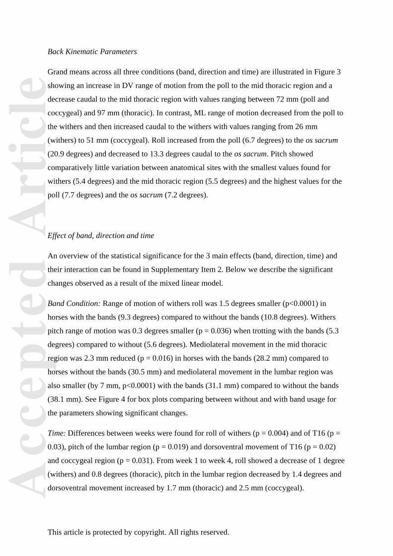
Back Kinematic Parameters
Grand means across all three conditions (band, direction and time) are illustrated in Figure 3
showing an increase in DV range of motion from the poll to the mid thoracic region and a
decrease caudal to the mid thoracic region with values ranging between 72 mm (poll and
coccygeal) and 97 mm (thoracic). In contrast, ML range of motion decreased from the poll to
the withers and then increased caudal to the withers with values ranging from 26 mm
(withers) to 51 mm (coccygeal). Roll increased from the poll (6.7 degrees) to the os sacrum
(20.9 degrees) and decreased to 13.3 degrees caudal to the os sacrum. Pitch showed
comparatively little variation between anatomical sites with the smallest values found for
withers (5.4 degrees) and the mid thoracic region (5.5 degrees) and the highest values for the
poll (7.7 degrees) and the os sacrum (7.2 degrees).
Effect of band, direction and time
An overview of the statistical significance for the 3 main effects (band, direction, time) and
their interaction can be found in Supplementary Item 2. Below we describe the significant
changes observed as a result of the mixed linear model.
Band Condition: Range of motion of withers roll was 1.5 degrees smaller (p<0.0001) in
horses with the bands (9.3 degrees) compared to without the bands (10.8 degrees). Withers
pitch range of motion was 0.3 degrees smaller (p = 0.036) when trotting with the bands (5.3
degrees) compared to without (5.6 degrees). Mediolateral movement in the mid thoracic
region was 2.3 mm reduced (p = 0.016) in horses with the bands (28.2 mm) compared to
horses without the bands (30.5 mm) and mediolateral movement in the lumbar region was
also smaller (by 7 mm, p<0.0001) with the bands (31.1 mm) compared to without the bands
(38.1 mm). See Figure 4 for box plots comparing between without and with band usage for
the parameters showing significant changes.
Time: Differences between weeks were found for roll of withers (p = 0.004) and of T16 (p =
0.03), pitch of the lumbar region (p = 0.019) and dorsoventral movement of T16 (p = 0.02)
and coccygeal region (p = 0.031). From week 1 to week 4, roll showed a decrease of 1 degree
(withers) and 0.8 degrees (thoracic), pitch in the lumbar region decreased by 1.4 degrees and
dorsoventral movement increased by 1.7 mm (thoracic) and 2.5 mm (coccygeal).

Acc
epte
d A
rtic
le
This article is protected by copyright. All rights reserved.
Direction: 79% (19/24) of back kinematic parameters showed a significant effect for
direction (Table 1 and Supplementary Item 2). The majority showed significant differences
between straight line and left rein and between straight line and right rein. Two of the
parameters (mediolateral poll range of motion and coccygeal pitch) additionally showed
differences between left and right rein while three parameters only showed differences
between straight line and one of the reins (dorsoventral withers and pelvis range of motion
and lumbar roll range of motion). All values were greater on the lunge compared to straight
line movement. Average change between straight line and lungeing (average of left and right
rein) of 10% increase was measured for dorsoventral movement (for 6 sensors), 24% increase
for mediolateral movement (for 6 sensors), 16% increase for roll (for 4 sensors) and 23%
increase for pitch (for 3 sensors).
Discussion
We quantified the effects of a specific system of elastic resistance bands (Equiband™) on
back kinematic parameters in seven riding horses over a 4-week period. The resistance bands
significantly reduced withers roll and pitch and thoracic and lumbar mediolateral movement,
providing support for our hypothesis that this proprioceptive aid improves dynamic stability
of the vertebral column in trot in-hand and on the lunge. The effects appeared to be
concentrated on the thoracolumbar area, and no differences were found caudal to the os
sacrum. Whether the changes are related to the stimulation of hindquarter and abdominal
muscle recruitment, resulting in increased activation of the postural core muscles, cannot be
answered by this study. This requires direct measurement of muscle activity of muscles such
as the multifidus and iliopsoas, which are thought to help with limiting energy losses through
decreasing lateral excursion of the vertebral column [30]. It should be acknowledged that
decreased thoracolumbar pitch (flexion-extension) can be seen in older horses and those
exhibiting signs of back pain [19,31]. When asked informally, the riders in this study felt
greater ‘stability of movement’ with the resistance band system. Ridden exercise was part of
the exercise regimen, but no gait analysis data were obtained for this condition. Further
investigation is warranted to quantify the effects of use of resistance bands on back
kinematics during ridden exercise.

Acc
epte
d A
rtic
le
This article is protected by copyright. All rights reserved.
In comparison to the Pessoa training aid (PTA) [6], the resistance bands did not have a direct
effect on lumbosacral flexion (pitch) or overall dorsoventral displacement. Dorsoventral
displacement was increased at week 4 however independent of band usage. Whether or not
this indicates an effect of the band usage over 4 weeks allowing the horses to push off into
the air more efficiently needs to be addressed by future studies. We used a range of horses of
different breed and age. Published in vitro work found that around one third of horses have
anatomical variations in the lumbosacral area which may impact on maximal dorsoventral
displacement [32], however, presence of anatomical variations was not assessed here. In
comparison to attachments of the PTA, the Equiband™ system does not have a direct
connection with the horse’s mouth and hence avoids the oral desensitisation effects seen with
incorrect use of the PTA [33] when using the EquiBandTM
system during lungeing. The
system can of course also be used during ridden exercise.
We assessed horses in-hand and on the lunge. A high proportion of parameters across all
regions showed increased ranges of motion on the lunge compared to straight line trot.
Previous studies on lungeing have mainly focused on movement symmetry and limb angles
of horses on the lunge [34–38], providing little scope for comparison. However, the increased
ranges of motion are likely, independent of band usage, related to the additional production of
centripetal force of locomotion on a curve, resulting in an increase in total force [39] and
increased peak forces measured in the outside front limb [40]. As demonstrated with the PTA
[6] on the lunge, the greater dorsoventral displacement and lumbosacral flexion (pitch) may
be related to increased activation of core postural muscles.
Only 5 differences in movement parameters were measured between weeks. Three of these
were related to rotational range of motion, and each showed a decrease from week 1 to week
4. The two remaining parameters, thoracic and coccygeal, were related to dorsoventral range
of motion, which increased from week 1 to week 4. This is a movement direction that was not
influenced by the resistance bands. The statistical model did not identify an interaction
between use of the exercise bands and time. The study design, comparing each horse without
and with bands, does not distinguish whether the differences between week 1 and 4 are
related to use of the bands, or only to the exercise regimen. This would require a control
group of horses undergoing the same exercises but without the use of the exercise bands. A
Acc
epte
d A
rtic
le
This article is protected by copyright. All rights reserved.
reduction in rotational movement of the thoracolumbar area may be beneficial when
considering the support required to carry a saddle and rider [41], and may also be what the
riders are referring to when subjectively reporting ‘more stability’.
Although not the focus of this study, we assessed movement symmetry of the head and pelvis
at the first data collection. The recorded values are an indicator of symmetry between left and
right fore and hindlimbs with respect to weight bearing and push-off [25]. All horses had
been judged as being ‘fit to perform’ at their respective level of training. In agreement with
studies based on visual assessment [42] or quantitative gait analysis [43,44], based on our
IMU data not all 7 horses would have been classified as within normal limits (± 7.5 mm for
head and ± 4 mm for pelvic movement, thresholds from [45] adapted using the equations
presented in [46]). Without any clinical diagnostics, it is impossible to conclude how many
horses would be classified as lame by a veterinarian. It would also be of interest to evaluate
the effect of elastic resistance bands in the presence of hind limb lameness, since
compensatory force distribution from the hind limbs to the front limbs may be influenced by
proprioceptive feedback from the hindquarters and by increased dynamic stability allowing
more efficient transfer of force from the affected hind limb to the compensatory front limb
[47].
We implemented a field study using privately-owned horses over a period of time. Variability
of rider influence [48,49] during the completion of the 4-week exercise protocol, as well as
protocol compliance could not be controlled. Variables such as the person placing the sensors
and operating the equipment (V.S.), the person handling the horses and the surface used
during gait assessment were kept constant for each horse. It was more challenging to control
circle diameter and speed of motion, which are known to affect movement symmetry and
kinematics [36–38]. Horse height and conformation also influence back movement [19] with
taller horses possessing longer thoracic regions and exhibiting greater lateral bending in the
lumbar region. However, this study design emphasised comparisons within each horse
between exercise with and without use of bands and over time. We chose not to randomise
the order of assessment (always without bands first) for each condition, since it is unknown
whether there is a carry-over effect affecting movement parameters even after removal of the
bands. To minimise the risk of a carry-over effect influencing our results, horses were moved

Acc
epte
d A
rtic
le
This article is protected by copyright. All rights reserved.
in walk after removal of the bands. The existence of a carry-over effect should be
investigated further in future studies with a series of repeat assessments after removal of the
bands.
Conclusion and future work
This study provides quantitative evidence to suggest that use of a specific elastic exercise
band system (Equiband™) as part of an exercise protocol, increases dynamic stability of the
thoracolumbar area in the trotting horse in-hand and on the lunge. The study design did not
allow a judgement of whether the exercise regimen alone (without the band system) would
have similar effects. Further studies should identify whether the effect of the band system is
due to increased activation of the deep core musculature related to dynamic spinal stability.
Authors’ declaration of interests
N.C. Stubbs and N. Rombach developed the Equiband™ system and advised on its correct
use. Neither of them was involved in data collection or processing.
Ethical animal research
This study was authorised by the Royal Veterinary College Ethics and Welfare Committee
(URN 2013 1238). Owners gave informed consent for inclusion of their horses.
Sources of funding
Funding was provided by the Royal Veterinary College in support of completion of V.
Simons’ (V.S.) final year research project.
Acknowledgements
We would like to thank the horse owners for the use of their horses and for participating in
the 4-week training programme.

Acc
epte
d A
rtic
le
This article is protected by copyright. All rights reserved.
Authorship
The study was designed by all authors. V. Simons executed data collection. V. Simons and T.
Pfau performed data processing. All authors were involved in data interpretation, preparation
of the manuscript and gave final approval.
Manufacturers’ addresses
a Equicore Concepts LLC, Grand River Avenue, East Lansing, Michigan, USA.
b Xsens, Enschede, The Netherlands.
c The Mathworks Inc., Natick, Massachusetts, USA.
d SPSS Inc., Chicago, Illinois, USA.
References
1. van Weeren, P.R. and Haussler, K.K. (2010) Science Overview: Development of a
structural and functional understanding of the equine back. Equine Vet. J. 42, 393-400.
2. Greve, L. and Dyson, S. (2015) Saddle fit and management: An investigation of the
association with equine thoracolumbar asymmetries, horse and rider health. Equine
Vet. J. 47, 415-421.
3. Zaneb, H., Kaufmann, V., Stanek, C., Peham, C. and Licka, T.F. (2009) Quantitative
differences in activities of back and pelvic limb muscles during walking and trotting
between chronically lame and nonlame horses. Am. J. Vet. Res. 70, 1129-1134.
4. McGowan, C.M., Stubbs, N.C. and Jull, G.A. (2007) Equine physiotherapy: a
comparative view of the science underlying the profession. Equine Vet. J. 39, 90-94.
5. Paulekas, R. and Haussler, K.K. (2009) Principles and Practice of Therapeutic
Exercise for Horses. J. Equine Vet. Sci. 29, 870-893.
6. Walker, V.A., Dyson, S.J. and Murray, R.C. (2013) Effect of a Pessoa training aid on
temporal, linear and angular variables of the working trot. Vet. J. 198, 404-411.
7. Goff L.S. (2007) Equine Therapy and Rehabilitation. In: Animal Physiotherapy;
Assessment,Treatment and Rehabilitation of Animals, Ed: C.M. McGowan, Blackwell
Publishing Ltd, Oxford. pp 239-250.
8. Andersen, L.L., Saervoll, C.A., Mortensen, O.S., Poulsen, O.M., Hannerz, H. and
Zebis, M.K. (2011) Effectiveness of small daily amounts of progressive resistance
training for frequent neck/shoulder pain: randomised controlled trial. Pain 152, 440-
446.
9. Kell, R.T. and Asmundson, G.J.G. (2009) A comparison of two forms of periodized
exercise rehabilitation programs in the management of chronic nonspecific low-back
pain. J. Strength Cond. Res./Natl. Strength Cond. Assoc. 23, 513-523.
10. Lee, J.H., Ooi, Y. and Nakamura, K. (1995) Measurement of muscle strength of the

Acc
epte
d A
rtic
le
This article is protected by copyright. All rights reserved.
trunk and the lower extremities in subjects with history of low back pain. Spine (Phila.
Pa. 1976). 20, 1994-1996.
11. Macedo, L.G., Maher, C.G., Latimer, J. and McAuley, J.H. (2009) Motor Control
Exercise for Persistent, Nonspecific Low Back Pain: A Systematic Review. Phys.
Ther. 89, 9-25.
12. Sundstrup, E., Jakobsen, M.D., Andersen, C.H., Bandholm, T., Thorborg, K., Zebis,
M.K. and Andersen, L.L. (2014) Evaluation of elastic bands for lower extremity
resistance training in adults with and without musculo-skeletal pain. Scand. J. Med.
Sci. Sport. 24, e353-e359.
13. Faber, M., Schamhardt, H., Weeren, P.R. van and Barneveld, A. (2001) Methodology
and validity of assessing kinematics of the thoracolumbar vertebral column in horses
on the basis of skin-fixated markers. Am. J. Vet. Res. 62, 301-306.
14. Warner, S.M., Koch, T.O. and Pfau, T. (2010) Inertial sensors for assessment of back
movement in horses during locomotion over ground. Equine Vet. J. 42 Suppl. 3, 417-
424.
15. Heim, C., Pfau, T., Gerber, V., Schweizer, C., Doherr, M. and Schüpbach-Regula, G
Witte, S. (2016) Determination of vertebral range of motion using inertial
measurement units in 27 Franches-Montagnes stallions and comparison between
conditions and with a mixed population. Equine Vet. J. doi: 10.1111/evj.12455
16. Martin, P., Cheze, L., Pourcelot, P., Desquilbet, L., Duray, L. and Chateau, H. (in
Press) Effect of the rider position during rising trot on the horse׳s biomechanics (back
and trunk kinematics and pressure under the saddle). J. Biomech. 49, 1027-1033
17. Gómez Alvarez, C.B., Rhodin, M., Byström, A., Back, W. and van Weeren, P.R.
(2009) Back kinematics of healthy trotting horses during treadmill versus over ground
locomotion. Equine Vet. J. 41, 297-300.
18. Faber, M., Johnston, C., Schamhardt, H., van Weeren, R., Roepstorff, L. and
Barneveld, A. (2001) Basic three-dimensional kinematics of the vertebral column of
horses trotting on a treadmill. Am. J. Vet. Res. 62, 757-764.
19. Johnston, C., Holmt, K., Faber, M., Erichsen, C., Eksell, P. and Drevemo, S. (2002)
Effect of conformational aspects on the movement of the equine back. Equine Vet. J.
34, Suppl. 34, 314-318.
20. Jeffcott, L.B. and Dalin, G. (1980) Natural rigidity of the horse’s backbone. Equine
Vet. J. 12, 101-108.
21. Buchner, H.H.F., Obermüller, S. and Scheidl, M. (2000) Body Centre of Mass
Movement in the Sound Horse. Vet. J. 160, 225-234.
22. Townsend, H.G., Leach, D.H. and Fretz, P.B. (1983) Kinematics of the equine
thoracolumbar spine. Equine Vet. J. 15, 117-122.
23. Keegan, K.G., Kramer, J., Yonezawa, Y., Maki, H., Pai, P.F., Dent, E. V, Kellerman,
T.E., Wilson, D.A. and Reed, S.K. (2011) Assessment of repeatability of a wireless
inertial sensor-based lameness evaluation system for horses. Am. J. Vet. Res. 72. 1156-
1163.
24. Starke, S.D., Witte, T.H., May, S.A. and Pfau, T. (2012) Accuracy and precision of
hind limb foot contact timings of horses determined using a pelvis-mounted inertial
measurement unit. J. Biomech. 45, 1522-1528.
25. Pfau, T., Fiske-Jackson, A. and Rhodin, M. (2016) Quantitative assessment of gait
parameters in horses: Useful for aiding clinical decision making? Equine Vet. Educ.
28, 209-215.
26. Bell, R.P., Reed, S.K., Schoonover, M.J., Whitfield, C.T., Yonezawa, Y., Maki, H.,
Pai, P.F. and Keegan, K.G. (2016) Associations of force plate and body-mounted
inertial sensor measurements for identification of hind limb lameness in horses. Am. J.
Acc
epte
d A
rtic
le
This article is protected by copyright. All rights reserved.
Vet. Res. 77, 337-345.
27. Keegan, K.G., Macallister, C.G., Wilson, D.A., Gedon, C.A., Kramer, J., Yonezawa,
Y., Maki, H. and Pai, P.F. (2012) Comparison of an inertial sensor system with a
stationary force plate for evaluation of horses with bilateral forelimb lameness. Am. J.
Vet. Res. 73, 368-374.
28. Kramer, J., Keegan, K.G., Kelmer, G. and Wilson, D.A. (2004) Objective
determination of pelvic movement during hind limb lameness and pelvic height
differences. Am. J. Vet. Res. 65, 741-747.
29. Starke, S.D., Willems, E., May, S.A. and Pfau, T. (2012) Vertical head and trunk
movement adaptations of sound horses trotting in a circle on a hard surface. Vet. J.
193, 73-80.
30. Licka, T.F., Peham, C. and Frey, A. (2004) Electromyographic activity of the
longissimus dorsi muscles in horses during trotting on a treadmill. Am. J. Vet. Res. 65,
155-158.
31. Wennerstrand, J., Johnston, C., Roethlisberger-Holm, K., Erichsen, C., Eksell, P. and
Drevemo, S. (2004) Kinematic evaluation of the back in the sport horse with back
pain. Equine Vet. J. 36, 707-711.
32. Stubbs, N.C., Hodges, P.W., Jeffcott, L.B., Cowin, G., Hodgson, D.R. and McGowan,
C.M. (2006) Functional anatomy of the caudal thoracolumbar and lumbosacral spine in
the horse. Equine Vet. J. 38, Suppl. 36, 393-399.
33. McLean, A.N. and McGreevy, P.D. (2010) Horse-training techniques that may defy
the principles of learning theory and compromise welfare. J. Vet. Behav. Clin. Appl.
Res. 5, 187-195.
34. Pfau, T., Jennings, C., Mitchell, H., Olsen, E., Walker, A., Egenvall, A., Tröster, S.,
Weller, R. and Rhodin, M. (2014) Lungeing on hard and soft surfaces: movement
symmetry of trotting horses considered sound by their owners. Equine Vet. J. 48, 83-
89
35. Rhodin, M., Pfau, T., Roepstorff, L. and Egenvall, A. (2013) Effect of lungeing on
head and pelvic movement asymmetry in horses with induced lameness. Vet. J. 198,
Suppl., e39-e45.
36. Hobbs, S.J., Licka, T. and Polman, R. (2011) The difference in kinematics of horses
walking, trotting and cantering on a flat and banked 10 m circle. Equine Vet. J. 43,
686-694.
37. Clayton, H.M. and Sha, H. (2006) Head and body centre of mass movement in horses
trotting on a circular path. Equine Vet. J. 38, Suppl. 36, 462-467.
38. Pfau, T., Stubbs, N.C., Kaiser, L.J., Brown, L.E.A. and Clayton, H.M. (2012) Effect of
trotting speed and circle radius on movement symmetry in horses during lunging on a
soft surface. Am. J. Vet. Res. 73, 1890-1899.
39. Usherwood, J.R. and Wilson, A.M. (2005) Biomechanics: No force limit on greyhound
sprint speed. Nature 438, 753-754.
40. Chateau, H., Camus, M., Holden-Douilly, L., Falala, S., Ravary, B., Vergari, C.,
Lepley, J., Denoix, J.-M., Pourcelot, P. and Crevier-Denoix, N. (2013) Kinetics of the
forelimb in horses circling on different ground surfaces at the trot. Vet. J. 198, e20-
e26.
41. Cocq, P. de, Weeren, P.R. van and Back, W. (2004) Effects of girth, saddle and weight
on movements of the horse. Equine Vet. J. 36, 758-763.
42. Greve, L. and Dyson, S.J. (2013) The interrelationship of lameness, saddle slip and
back shape in the general sports horse population. Equine Vet. J. 46, 687-694.
43. Rhodin, M., Roepstorff, L., French, A., Keegan, K.G., Pfau, T. and Egenvall, A.
(2015) Head and pelvic movement asymmetry during lungeing in horses with
Acc
epte
d A
rtic
le
This article is protected by copyright. All rights reserved.
symmetrical movement on the straight. Equine Vet. J. 48, 315-320.
44. Pfau, T., Parkes, R.S., Burden, E.R., Bell, N., Fairhurst, H. and Witte, T.H. (2016)
Movement asymmetry in working polo horses. Equine Vet. J. DOI: 10.1111/evj.12467.
45. McCracken, M.J., Kramer, J., Keegan, K.G., Lopes, M., Wilson, D. A., Reed, S.K.,
LaCarrubba, A. and Rasch, M. (2012) Comparison of an inertial sensor system of
lameness quantification with subjective lameness evaluation. Equine Vet. J. 44, 652-
656.
46. Pfau, T., Boultbee, H., Davis, H., Walker, A. and Rhodin, M. (2016) Agreement
between two inertial sensor gait analysis systems for lameness examinations. Equine
Vet. Educ. 28, 203-208.
47. Weishaupt, M.A., Wiestner, T., Hogg, H.P., Jordan, P. and Auer, J.A. (2004)
Compensatory load redistribution of horses with induced weightbearing hindlimb
lameness trotting on a treadmill. Equine Vet. J. 36, 727-733.
48. Licka, T., Kapaun, M. and Peham, C. (2004) Influence of rider on lameness in trotting
horses. Equine Vet. J. 36, 734-736.
49. Lagarde, J., Kelso, J.A.S., Peham, C. and Licka, T. (2005) Coordination dynamics of
the horse-rider system. J. Mot. Behav. 37, 418-424.

Acc
epte
d A
rtic
le
This article is protected by copyright. All rights reserved.
Table
Table 1: Results of the mixed model analysis with regards to trot ‘direction’ comparing
translational (DV: dorsoventral, ML: mediolateral) and rotational (R: roll, P: pitch) ranges of
motion (ROM) between straight line, in-hand trot (S, straight line) and trot on the lunge on
left (L) and right (R) rein from 7 horses. Given are P values (after Bonferroni correction) as
well significant pairwise comparisons with S2L indicating a difference between S and L, S
2R
a difference between S and R and L2R a difference between L and R.
Anatomical
landmark
Kinematic
parameter
P value Posthoc test
result
Poll
DVROM <0.0001 S2L, S2R
MLROM <0.0001 S2L, S2R, L2R RROM <0.0001 S2L, S2R PROM 0.201
Withers
DVROM 0.007 S2R MLROM <0.0001 S2L, S2R RROM 0.179
PROM 0.157
T16
DVROM <0.0001 S2L, S2R MLROM <0.0001 S2L, S2R RROM 0.217 PROM 0.005 S2L, S2R
L4-6
DVROM <0.0001 S2L, S2R
MLROM <0.0001 S2L, S2R RROM 0.029 S2L PROM 0.183
Sacrum
DVROM 0.024 S2L MLROM <0.0001 S2L, S2R RROM <0.0001 S2L, S2R
PROM 0.001 S2L, S2R
Co4-5
DVROM <0.0001 S2L, S2R MLROM <0.0001 S2L, S2R RROM 0.006 S2L, S2R PROM <0.0001 S2L, S2R, L2R

Acc
epte
d A
rtic
le
This article is protected by copyright. All rights reserved.
Figure legends
Fig 1: Picture of one of the horses enrolled in the study with the elastic resistance band
system and the inertial sensor system fitted.
Fig 2: (A) Head and (B) pelvic movement symmetry values of N = 7 horses for trot in-hand
on hard surface (straight) and on the lunge (soft surface) on left and right rein (LR, RR).
Movement symmetry values generally (with the exception of pelvic MinD, the difference
between vertical pelvic displacement minima during left and right hindlimb stance) include
zero (value for perfect symmetry) and show considerable variation between horses.
Median values indicate a lower position of the head during RF stance (negative HDmin) on
the straight line and on the left rein and a lower head position during LF stance (positive
MinDhead) on the right rein. MinDhead indicates a higher position of the head after RF stance
for all three conditions. Median pelvic movement asymmetry shows a higher position of the
pelvis during LH stance (MinDpelvis), most exacerbated on the left rein. MaxDpelvis shows near
zero median values (near symmetrical movement) on the straight and on the right rein and
indicates increased pelvis position after RH stance on the left rein. HHD is positive
throughout indicating increased movement amplitude of the left tuber coxae compared to the
right, most pronounced on the left rein.
Fig 3: Dorsoventral and mediolateral (A) and roll and pitch (B) range of motion of the seven
study horses averaged across all 12 conditions (without/with band, direction [straight, left
rein, right rein] and time [week1/week4]). Presented are grand means extracted from the
mixed model with horse as random factor, movement direction, band usage and time as fixed
factors and stride time as covariate and range of motion parameters as outcome variables.
Fig 4: Box plots illustrating the effect of the band system (the four parameters showing
significant differences without/with band usage in the mixed model) on range of motion of
withers pitch (A) and withers roll (B), of mediolateral range of motion of the mid thoracic
region (C) and the lumbar region (D). Shown are average values for significant changes
between band conditions from N = 7 horses measured across two time points and during

Acc
epte
d A
rtic
le
This article is protected by copyright. All rights reserved.
straight-line trot and while trotting on the lunge (N = 42 values per box). All four significant
changes result in a reduced range of motion (increased dynamic stability) with the use of the
bands.
Supplementary Information
Supplementary Item 1: Horse details.
Supplementary Item 2: Mixed model analysis for range of motion parameters.

Acc
epte
d A
rtic
le
This article is protected by copyright. All rights reserved.

Acc
epte
d A
rtic
le
This article is protected by copyright. All rights reserved.

Acc
epte
d A
rtic
le
This article is protected by copyright. All rights reserved.

Acc
epte
d A
rtic
le
This article is protected by copyright. All rights reserved.